Digital anatomy - the body revealed - Science Photo Library
Digital anatomy - the body revealed - Science Photo Library
Digital anatomy - the body revealed - Science Photo Library

Create successful ePaper yourself
Turn your PDF publications into a flip-book with our unique Google optimized e-Paper software.
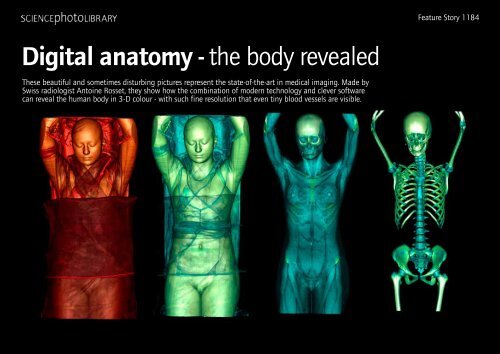
Feature Story 1184<strong>Digital</strong> <strong>anatomy</strong> - <strong>the</strong> <strong>body</strong> <strong>revealed</strong>These beautiful and sometimes disturbing pictures represent <strong>the</strong> state-of-<strong>the</strong>-art in medical imaging. Made bySwiss radiologist Antoine Rosset, <strong>the</strong>y show how <strong>the</strong> combination of modern technology and clever softwarecan reveal <strong>the</strong> human <strong>body</strong> in 3-D colour - with such fine resolution that even tiny blood vessels are visible.
<strong>Science</strong> <strong>Photo</strong> <strong>Library</strong> Feature Stories - <strong>Digital</strong> AnatomySome of Rosset’s subjects arehealthy volunteers. A sequenceof four images begins with afully-clo<strong>the</strong>d woman. Subsequentimages reveal her musculature andvascular system; <strong>the</strong> final imageher skeleton. In ano<strong>the</strong>r image, sheis seen from above, appearing tofly out of <strong>the</strong> frame. O<strong>the</strong>r imagesare of accident victims and surgicalpatients. The head of a severelydeformedchild shows<strong>the</strong> patchwork of facialreconstruction; a skiaccident patient liesswa<strong>the</strong>d in bandages,with <strong>the</strong> outline of <strong>the</strong>respirator and suctiontube that allows himto brea<strong>the</strong>. And o<strong>the</strong>rimages show individualorgans and <strong>body</strong> parts.This is <strong>the</strong> human<strong>body</strong> <strong>revealed</strong> in allits detail. Many of <strong>the</strong> imagesresemble anatomical drawingsfrom earlier times, where cadaverswould be dissected layer by layer.Today, medical science relies ontechnology to strip away <strong>the</strong> layersof <strong>the</strong> <strong>body</strong>, revealing <strong>the</strong> arteriesand muscles that lie just beneath<strong>the</strong> skin; through <strong>the</strong> organs thatlie deeper within <strong>the</strong> <strong>body</strong>, to <strong>the</strong>skeleton itself.Antoine Rosset, M.D., is AssistantProfessor at <strong>the</strong> Radiology Departmentof <strong>the</strong> Geneva University Hospital,Switzerland. He is responsible for<strong>the</strong> Hospital’s two CT scanners.CT - Computed Tomography - hasbecome one of <strong>the</strong> workhorses of today’smedical imaging. Introduced in1990, <strong>the</strong> technology has revolutionisedmedical imaging and diagnosis.Unlike conventional X-ray machines,CT scanners can ‘see’soft tissue, so <strong>the</strong>y arecapable of making detailedvisualisations of<strong>the</strong> internal <strong>anatomy</strong>of <strong>the</strong> <strong>body</strong>.CT scanners use athin X-ray beam tocreate ‘slices’ of <strong>the</strong>patient’s <strong>body</strong>. Theseslices are reconstructedby computer into3-D images. Coloursare assigned according to <strong>the</strong> abilityof <strong>body</strong> parts to block <strong>the</strong> X-ray beam- <strong>the</strong>ir ‘radiodensity’. Fat, for example,is less dense than muscle, which, inturn, is less dense than bone. So fatcan be assigned one colour; muscleano<strong>the</strong>r. This ability to assign coloursto specific <strong>body</strong> structures - blood vessels,for example - makes it possiblefor doctors to highlight areas of interest.It also allows Antoine Rosset to
<strong>Science</strong> <strong>Photo</strong> <strong>Library</strong> Feature Stories - <strong>Digital</strong> Anatomycreate exquisitely coloured images.These images were producedby Rosset, using his ownvisualisation softwarecalled OsiriX. Rosset isa doctor, not a softwaredeveloper.But, he says,modernradiology ishighly computerised,andradiologistshave to be familiarwith both<strong>the</strong> scanners and <strong>the</strong>software:“It’s not like <strong>the</strong> TV seriesER, where <strong>the</strong> doctor looks atan X-ray on a light box,” he says.“All <strong>the</strong> images we work on areon computer.”As well as seeing soft tissue, modernCT scanners produce very highresolution images. Each slice can beas thin as 1 mm, which means thatimages can be incredibly detailed,resolving individual blood vessels. This,says Rosset, gives much better 3-Dimages - but at a price. As <strong>the</strong> scannersare becoming higher in resolution,<strong>the</strong>y are also producing more and moredata, which needs powerful and expensivesoftware to turn into images. Only <strong>the</strong> largerhospitals can afford this software, so Rossetdecided to develop his own:“I was disappointed by <strong>the</strong> commercialsoftware,” he says. “Itwas very expensive. Thebest solution to have <strong>the</strong>perfect tool for my workwas to develop my ownsoftware.”OsiriX is <strong>the</strong> result. It isfreely-available over <strong>the</strong>Internet, is open-source,- so users can modify it<strong>the</strong>mselves - and can takedata from any scanner andturn it into high-resolution colour3-D images. It runs on Applecomputers only - ano<strong>the</strong>r reasonwhy Rosset developed it, sincehe couldn’t find any commercialequivalent for <strong>the</strong> Mac - and alreadyhas several thousand users.The combination of today’s high-resolutionscanners and OsiriX - toge<strong>the</strong>rwith Rosset’s eye and sense of aes<strong>the</strong>tics- have produced <strong>the</strong>se stunning images.They are, quite simply, <strong>the</strong> state-of<strong>the</strong>-artin medical imaging.But, says Rosset, this will not always be <strong>the</strong>case:“The evolution of CT will continue quickly.The next generation of CT scanners will producefaster, higher-resolution images. Medical imagingis rapidly evolving.”ENDS 700 WDS © SCIENCE PHOTO LIBRARY 2007
FULL PICTURE SETFor captions and credits, please refer to <strong>the</strong> captions.txt file<strong>Science</strong> <strong>Photo</strong> <strong>Library</strong> Feature Stories - ????For fur<strong>the</strong>r information, please contact: seymour@sciencephoto.comAll images are copyright, please credit images as stated on <strong>the</strong> captions
FULL PICTURE SETFor captions and credits, please refer to <strong>the</strong> captions.txt file<strong>Science</strong> <strong>Photo</strong> <strong>Library</strong> Feature Stories - ????For fur<strong>the</strong>r information, please contact: seymour@sciencephoto.comAll images are copyright, please credit images as stated on <strong>the</strong> captions