2008-05-07-50_PanLeucogating CD4 counting.pdf - Immunopaedia
2008-05-07-50_PanLeucogating CD4 counting.pdf - Immunopaedia
2008-05-07-50_PanLeucogating CD4 counting.pdf - Immunopaedia

Create successful ePaper yourself
Turn your PDF publications into a flip-book with our unique Google optimized e-Paper software.
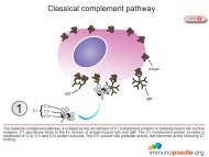
PREVIOUSBlood cells labelled by <strong>CD4</strong>5/<strong>CD4</strong> stainingMonocytesLeucocytesGranulocytesNeutrophilErythrocytesBasophilDendritic cell<strong>CD4</strong>+ HelperT lymphocyteMacrophageCD8+ CytotoxicT lymphocyteLymphocytesEosinophilNaturalKillerCellPlateletsB lymphocyteHIV infection is characterised by a loss of <strong>CD4</strong>+ Helper T lymphocytes. A count of these cells in blood is a useful marker ofdisease state. Flow cytometry is a method used to measure the number of these cells in blood. An alternative strategy toCD3/<strong>CD4</strong> staining to identify these cells uses fluorescent dyes coupled to antibodies against <strong>CD4</strong>5 and <strong>CD4</strong> cell surfacemarkers (panleucogating). <strong>CD4</strong>5 is expressed on all leucocytes while <strong>CD4</strong> is expressed on macrophages and <strong>CD4</strong>+ HelperT lymphocytes. Side scatter is used to distinguish macrophages from Helper T lymphocytes.
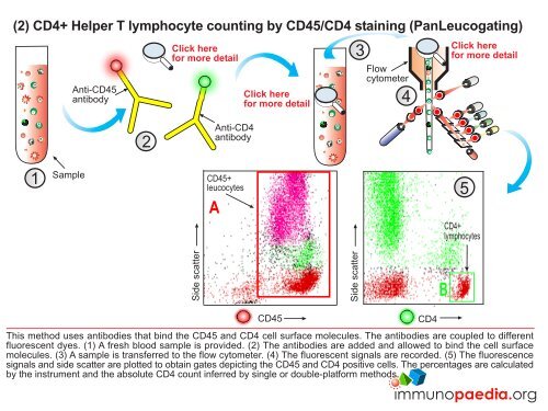
Flow cytometry using fluorescenceBACK TOSTART1Sample2SheathSheathfluid5<strong>CD4</strong>+ CD3+lymphocytes<strong>CD4</strong>FlowchamberPhotonbeam3LensCD3FL1FL24FL3LaserMirrorFluorescentlight detectorsFlow cytometry is also used to differentiate and count cells labelled with fluorescent dyes. (1) Antibodies that bind cell surfacemolecules are added to the sample. Different antibodies are labelled with different colours. (2) A sample containing cells withbound antibody is transferred to a tube that separates the cells from each other. (3) Fluorescent light emitted from individualcells is measured as they pass through the photon beam. (4) Various filters and light detectors record the fluorescence signals.(5) Fluorescence intensity can be plotted to show the subset of cells binding the label.FL4