Out for Blood: Neonatal Hematology Review - FANNP
Out for Blood: Neonatal Hematology Review - FANNP
Out for Blood: Neonatal Hematology Review - FANNP

You also want an ePaper? Increase the reach of your titles
YUMPU automatically turns print PDFs into web optimized ePapers that Google loves.
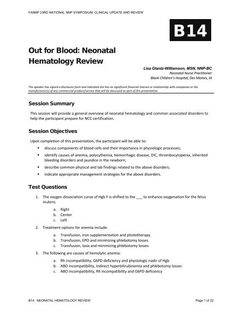
<strong>FANNP</strong> 23RD NATIONAL NNP SYMPOSIUM: CLINICAL UPDATE AND REVIEWB14<strong>Out</strong> <strong>for</strong> <strong>Blood</strong>: <strong>Neonatal</strong><strong>Hematology</strong> <strong>Review</strong>Lisa Glantz-Williamson, MSN, NNP-BC<strong>Neonatal</strong> Nurse PractitionerBlank Children’s Hospital, Des Moines, IAThe speaker has signed a disclosure <strong>for</strong>m and indicated she has no significant financial interest or relationship with companies or themanufacturer(s) of any commercial product/service that will be discussed as part of this presentation.Session SummaryThis session will provide a general overview of neonatal hematology and common associated disorders tohelp the participant prepare <strong>for</strong> NCC certification.Session ObjectivesUpon completion of this presentation, the participant will be able to:• discuss components of blood cells and their importance in physiologic processes;• identify causes of anemia, polycythemia, hemorrhagic disease, DIC, thrombocytopenia, inheritedbleeding disorders and jaundice in the newborn;• describe common physical and lab findings related to the above disorders;• indicate appropriate management strategies <strong>for</strong> the above disorders.Test Questions1. The oxygen dissociation curve of Hgb F is shifted to the ___ to enhance oxygenation <strong>for</strong> the fetusinutero.a. Rightb. Centerc. Left2. Treatment options <strong>for</strong> anemia include:a. Transfusion, iron supplementation and phototherapyb. Transfusion, EPO and minimizing phlebotomy lossesc. Transfusion, lasix and minimizing phlebotomy losses3. The following are causes of hemolytic anemia:a. Rh incompatibility, G6PD deficiency and physiologic nadir of Hgbb. ABO incompatibility, indirect hyperbilirubinemia and phlebotomy lossesc. ABO incompatibility, Rh incompatibility and G6PD deficiencyB14: NEONATAL HEMATOLOGY REVIEW Page 1 of 22
<strong>FANNP</strong> 23RD NATIONAL NNP SYMPOSIUM: CLINICAL UPDATE AND REVIEW4. Thrombocytopenia is defined as:a. A platelet count < 150,000b. A hemoglobin < 15 mg/dLc. A platelet count < 100,0005. A 32 week A positive infant born to an O positive mother who is 36 hours old and has a risingbilirubin > 5 mg/dL/day. The infant is NPO <strong>for</strong> treatment of a PDA with ibuprofen lysine and isreceiving aminophylline. What are you concerned about?a. Nothing, this is physiologic jaundiceb. Unbound bilirubin due to medications competing with albumin binding sites and ABOincompatibilityc. Thrombocytopenia and kernicterusReferencesBagwell, G.A. (2007). Hematologic system. In C. Kenner and J.W. Lott (Eds.), Comprehensive neonatal nursing: A physiologicperspective (4th ed., pp. 221-253). Philadelphia: Saunders.Blackburn, S.T. (2007). Maternal, fetal, & neonatal physiology: A clinical perspective (3rd ed.). St. Louis: Elsevier Saunders.Brodsky, D. & Martin, C. (2003). Neonatology review. Philadelphia: Hanley & Belfus.Cloherty, J.P., Eichenwald, E.C. & Stark, A.R. (2004). Manual of neonatal care (5th ed.). Philadelphia: Lippincott Williams & Wilkins.Diehl-Jones, W. & Fraser Askin, D. (2010). Hematologic disorders. In M.T. Verklan and M. Walden (Eds.). Core curriculum <strong>for</strong>neonatal intensive care nursing (4rd ed., pp. 666-693). St. Louis: Elsevier Saunders.Gleason, C.A. (2005). Hematologic system and disorders of bilirubin metabolism. In H.W. Taeusch, R.A. Ballard & C.A. Gleason(Eds.), Avery’s diseases of the newborn (8th ed., pp. 1135-1256). Philadelphia: Elsevier Saunders.Gomella, T.L. (2009). Neonatology: Management, procedures, on-call problems, diseases, and drugs (6th ed.). New York: Lange.Merenstein, G.B. & Gardner, S.L. (2006). Handbook of neonatal intensive care (6th ed.). St. Louis: Mosby.Sheth, S. (2007). <strong>Hematology</strong>. In R.A. Polin and A.R. Spitzer (Eds.). Fetal and neonatal secrets (2nd ed.). Philadelphia: MosbyElsevier.Watson, R.L. (2004). Gastrointestinal disorders. In M.T. Verklan and M. Walden (Eds.), Core curriculum <strong>for</strong> neonatal intensive carenursing (3rd ed., pp. 685-697). St. Louis: Elsevier Saunders,Zenk, K.E. (2003). <strong>Neonatal</strong> medication and nutrition: A comprehensive guide (3rd ed.). Santa Rosa, CA: NICU INK.Session <strong>Out</strong>lineSee handout on following pages.B14: NEONATAL HEMATOLOGY REVIEW Page 2 of 22
<strong>FANNP</strong> 23RD NATIONAL NNP SYMPOSIUM: CLINICAL UPDATE AND REVIEW<strong>Hematology</strong> in the NeonateLisa Glantz Williamson MSN, RNC-NIC, ARNP, NNP-BC<strong>Neonatal</strong> Nurse PractitionerBlank Children’s HospitalDes Moines, IAHow do blood cells develop?◦ Hematopoiesis• Formation, production, maintenance• Pluripotent Stem Cells◦ All blood cells are made from these• Starts in the yolk sac during 3 rd week ofgestation• Liver vs. Bone◦ Liver• Established by 9 weeks gestation• Peaks at 4-5 months gestation• Regresses as bone marrow production increases◦ Bone• Predominates from 22 weeks gestation <strong>for</strong>ward• Hypoxia, bacterial infection, physiologic stressinfluence the rate of differentiation ofpluripotent cellsHow do blood cells develop?Red <strong>Blood</strong> Cells◦ Erythropoiesis• Production of erythrocytes – aka Red <strong>Blood</strong> Cells• Erythropoietin◦ Hormone◦ Regulates erythropoiesis & hemoglobin synthesis◦ Produced by• Liver prenatally• Kidneys postnatally◦ Increased• Anemia• Low oxygen availability to tissues• Down Syndrome• Intrauterine Growth Restriction (IUGR)• Infants born to women with…diabetes or PIH◦ Decreased• HypertransfusionB14: NEONATAL HEMATOLOGY REVIEW Page 3 of 22
<strong>FANNP</strong> 23RD NATIONAL NNP SYMPOSIUM: CLINICAL UPDATE AND REVIEWThe Nuts and Bolts of <strong>Blood</strong> Cells◦ Hemoglobin (Hgb)• Normal 14-20 g/dL in infants > 34 weeks,slightly lower in preterm infants• Major iron-containing component of RBC• Carries oxygen from the lungs to the tissue cells• HbF – fetal Hgb, begins ~14 days of life◦ RBCs contain 70%-90% HbF at birth◦ Has higher affinity <strong>for</strong> oxygen• HbA – adult Hgb, begins at end of fetal life• Value Factors◦ Gestation◦ Placental transfusion◦ <strong>Blood</strong> sampling site – capillary > centralThe Nuts and Bolts of <strong>Blood</strong> Cells◦ Hematocrit (Hct)• % of RBCs in a unit volume of blood• Value Factors◦ Gestation◦ Placental transfusion◦ <strong>Blood</strong> sampling site – capillary > centralThe Nuts and Bolts of <strong>Blood</strong> Cells◦ Red <strong>Blood</strong> Cells (RBC)• Reticulocyte – immature RBC◦ 1-2 days in the marrow and 1 more dayin the circulation be<strong>for</strong>e full maturation -longer when stress present◦ Retic Count• The ↓ gestation the ↑ the count• Falls to < 2% by 7 days of life• ↑ count indicative of chronic blood loss orhemolysis• Function◦ Oxygen & carbon dioxide transport◦ BufferThe Nuts and Bolts of <strong>Blood</strong> Cells• Value Factors◦ # of circulating mature RBCs/mm 3◦ Count = production vs. destruction/loss• Life Span◦ Adult – 100 to 120 days◦ Term Infant – 60 to 70 days◦ Premature Infant – 35 to 50 days• Nucleated RBC – circulating immature(prereticulocyte) red cells◦ The ↓ gestation the ↑ the #◦ Decline rapidly in the 1 st week of life◦ Indicative of hemolysis, acute blood loss,hypoxemia, congenital heart disease, infectionThe Nuts and Bolts of <strong>Blood</strong> CellsIf you know your hematocrit, yourhemoglobin or your RBC count…then you can figure out the other 2Example:Hgb 15 g%multiply by 3 = Hctdivide by 3 = RBC count◦ White <strong>Blood</strong> Cells (WBC)• Aka leukocytes• Mature in the bone marrow & lymphatictissues• React to <strong>for</strong>eign protein extravascular space• WBC count◦ # of circulating WBC/mm 3◦ Proportional to gestational age – prematurehave 30% to 50% less than term• 3 types of WBC1) Granulocytes2) Lymphocytes3) MonocytesB14: NEONATAL HEMATOLOGY REVIEW Page 4 of 22
<strong>FANNP</strong> 23RD NATIONAL NNP SYMPOSIUM: CLINICAL UPDATE AND REVIEWThe Nuts and Bolts of <strong>Blood</strong> Cells◦ Granulocytes• 3 types1) Basophils◦ Allergic and inflammatory responses◦ Least numerous – 0.5% to 1% of total WBC count2) Eosinophils◦ Similar to neutrophils in function but less effective inresponse◦ Prolonged survival in extravascular space◦ Allergic and anaphylactic responses, parasitic destruction◦ Elevated inversely to gestational age◦ 1-3% of total WBC count3) Neutrophils◦ Phagocytes – ingest & destroy small particles◦ Bacteria, protozoa, cells and their debris, colloids◦ Physiological stress can increase production and bonemarrow release of immature <strong>for</strong>ms◦ Bands, myelocytes, metamyelocytes, promyelocytes◦ May be increased at birth but decrease in the 1 st week oflifeThe Nuts and Bolts of <strong>Blood</strong> Cells◦ Lymphocytes• T-lymphocytes◦ Thymus derived◦ Graft vs. host disease, delayedhypersensitivity reactions• B-lymphocytes◦ Bone marrow derived◦ Production & secretion ofimmunoglobulins & antibodiesThe Nuts and Bolts of <strong>Blood</strong> CellsThe Platelet◦ Monocytes• Immature circulating macrophages• Mature in tissues• “The Cleaners” -clear old blood cells,cellular debris, opsonized bacteria,antigen-antibody complexes, activatedclotting factors from the circulationThe Nuts and Bolts of <strong>Blood</strong> CellsThe RBC “STUFF” at the end of a CBC◦ Platelets• Small, nonnucleated, disk-shaped cells• Hemostasis, coagulation, thrombus <strong>for</strong>mation• Response stimulated by disruption in theendothelium• Derived from megakaryocytes in bone marrow• Circulate in the blood 7-10 days be<strong>for</strong>e beingremoved by the spleen• Hypoactive in the first few days following birth• Platelet Count◦ Normal range 150K – 400K <strong>for</strong> all infants◦ < 150K considered abnormal requiringfurther investigation◦ SGA infants with 20%-25% lower countsCell TypeAnisocytosisMacrocytosisMicrocytosisPoikilocytosisSpherocytosisTarget CellsBurr CellsHowell-JollyBodiesNucleated RBCsClinically SpeakingSevere anemiaVitamin B12 & folic acid deficienciesIron deficiency, spherocytic & hemolytic anemiaSevere anemiaCongenital spherocytosis, hemolytic anemia, aftertransfusion of stored bloodHemoglobinopathies, sickle cell disease, thalassemia,liver diseaseHemolytic anemia, DIC, liver diseaseAsplenia, pernicious anemiaChronic blood loss, significant hemolysis, chronichypoxia, infectionB14: NEONATAL HEMATOLOGY REVIEW Page 5 of 22
<strong>FANNP</strong> 23RD NATIONAL NNP SYMPOSIUM: CLINICAL UPDATE AND REVIEWA Measure of it all…<strong>Blood</strong> VolumeA measure of it all…<strong>Blood</strong> Volume◦ Measured in ml/kg of body weight◦ Volume factors• Gestational age◦ Term: ~80-100 ml/kg◦ Preterm: ~90-105 ml/kg• Placental transfusion◦ Timing of cord clamping, infant positionrelated to placenta (above or below), uterinecontractions, cord compression, onset ofrespirations and decrease in PVR• Maternalfetal or Fetalmaternal transfusion• Twin-twin transfusion• Placenta previa or abruptio placentae• Nuchal cord• Iatrogenic lossA Measure of it all…<strong>Blood</strong> Volume◦ So why is keeping track of blood volumelosses so important?◦ SCENARIO• 2 kg 35 week infant with RDS & pneumonia onthe vent with ABGs every 6 hours & daily labdraws◦ What was the estimated beginning bloodvolume?• 180 ml• On DOL 3 the flow sheet says that the baby isout 15 ml of blood…◦ What % of the beginning blood volume is thebaby out?• 8%...yikes! And that’s if everyone has beenkeeping track accurately…AnemiaWhere did all my volume go?!?Anemia◦ Definition• Central venous Hgb of < 13 g/dL or capillaryHgb of < 14.5 g/dL◦ Oxygen carrying capacity & level ofoxygen available to the tissues reducedby…• Low Hgb concentration• Decreased # of RBCs◦ Etiology• Hemorrhage• Hemolysis• Prematurity• IatrogenicAnemia – Causes of Hemorrhage◦ Twin-to-Twin• Monozygotic, monochorionic (single)placenta• 13%-33% of twin pregnancies• Hgb difference b/w twins > 5 g/dL• Often size discrepancy as well…> 20%difference with chronic hemorrhage◦ Larger Twin: recipient, polyhydramnios;at risk <strong>for</strong> congestive heart disease and/orsystemic/pulmonary hypertension due tovolume overload, hyperviscosity◦ Smaller Twin: donor, oligohydramnios; atrisk <strong>for</strong> anemia with elevated retic count,at risk <strong>for</strong> IUGR and high output cardiacfailure◦ High risk <strong>for</strong> long-term developmentaldelayB14: NEONATAL HEMATOLOGY REVIEW Page 6 of 22
<strong>FANNP</strong> 23RD NATIONAL NNP SYMPOSIUM: CLINICAL UPDATE AND REVIEWAnemia – Causes of Hemorrhage◦ Placental/Cord• Umbilical cord rupture• Cord or placental hematoma• Anomalous cord insertion• Rupture of anomalous vessels of cord orplacenta• Accidental incision of cord or placenta• Placenta previa or abruptio placentae◦ Fetal-Maternal Kleihauer-Betke test• Spontaneous• Traumatic amniocentesis• External cephalic versionAnemia – Causes of Hemorrhage◦ Internal• Intracranial (subdural, subarachnoid,intraventricular), subgaleal• Organ rupture (liver, spleen, adrenal,kidney)• Pulmonary◦ External• Phlebotomy• IatrogenicAnemia – Causes of Hemolysis◦ Rh <strong>Blood</strong> Group Incompatibilities• Aka erythroblastosis fetalis• How does it happen?◦ Rh + fetal cells enter the bloodstream of an Rh –mother resulting in maternal antibodyproduction to the Rh + fetal cells SUBSEQUENT pregnancies will have destructionof fetal RBCs if the fetus is Rh +• Predisposing factors◦ Previous pregnancy or abortion◦ Fetal-maternal hemorrhage during pregnancy◦ Delivery (vaginal, breech, cesarean)◦ Amniocentesis, chorionic villus sampling◦ External version◦ Manual removal of placentaAnemia – Causes of Hemolysis◦ What to look <strong>for</strong>…Rh incompatibility• Anemia ongoing hemolysis• Tissue hypoxia, acidosis decreased oxygencarrying capacity• Congestive heart failure & hydrops fetalis generalized edema due to increased blood volume &cardiac output• Ascites, pleural effusion collection of fluid• Hepatosplenomegaly increased extramedullaryhematopoiesis• Petechiae thrombocytopenia• Hypoglycemia hyperinsulinemia d/t RBCdestruction• Positive direct Coombs test result• Increased retic count ongoing hemolysisAnemia – Causes of Hemolysis◦ ABO <strong>Blood</strong> Group Incompatibilities• Occurs more frequently, but less severethan Rh incompatibility• Most commonly seen with O blood typemother carrying fetus with A or B bloodtype• How does it happen?◦ Can occur with 1 st pregnancy due tomaternal exposure to A & B antigens (food,bacteria, pollen) that results in productionof anti-A & anti-B antibodies◦ Prevents Rh sensitization due to rapiddestruction of fetal A/B cellsPotential ABO Incompatibilities“B” is bad, “A” is awful!Maternal<strong>Blood</strong> GroupIncompatibleFetal <strong>Blood</strong> GroupOA or BBAA or ABB or ABAdapted from Core Curriculum <strong>for</strong> <strong>Neonatal</strong> Intensive Care Nursing, 2004B14: NEONATAL HEMATOLOGY REVIEW Page 7 of 22
<strong>FANNP</strong> 23RD NATIONAL NNP SYMPOSIUM: CLINICAL UPDATE AND REVIEWAnemia – Causes of Hemolysis◦ What to look <strong>for</strong>…ABO incompatibility• Mild hemolysis• Anemia• Reticulocytosist i• HyperbilirubinemiaAnemia – Causes of Hemolysis◦ How to Treat ABO and Rh Incompatibilities• RhoGAM Rh Negative ONLY◦ Prophylactic anti-D immune globulin◦ Blocks maternal antibody production bydestroying fetal red cells in maternal circulation◦ Given at ~28 weeks, then again within 72 hoursfollowing delivery and anytime there may befetal-maternal blood mixing in Rh – pregnantwomen• Phototherapy• Good hydration• IVIG (intravenous immunoglobulin) 1 g/kg over 4hrs• Consider blood or exchange transfusion• Management of multisystem dysfunctionAnemia – Causes of HemolysisNo matter the antibody...Hemolysis will continue until all antibodyinhabited cells are destroyed…that can meanup to months depending on degree ofantibody presence◦ Enzymatic Defect• G6PD (glucose-6-phosphatedehydrogenase) Deficiency• Most common inherited red cell disorder◦ Sex-linked, mainly male offspring,occasional female carriers◦ Most common in American black infants(10% to 15%), also Mediterranean, African,Asian decent• How does it happen?◦ Hemolysis & shortened erythrocyte life dueto deficiency of red cell enzyme & exposureto antioxidant stress (drugs, infection)Anemia – Causes of Hemolysis◦ Infection• Intrauterine◦ Congenital TORCH infections• Toxoplasmosis• Other h (syphilis, hepatitis i B, coxsackievirus,iEpstein-Barr, varicella-zoster Virus, parvovirus)• Rubella• Cytomegalovirus• Herpes Simplex Virus• Postnatal◦ Bacterial infections• Both may cause…◦ Hemolysis, anemia, thrombocytopenia, DICAnemia of Prematurity◦ Considered physiologic◦ How does it happen?• Erythropoietin falls to minimal level d/t improvedrelative oxygenation after birth• Hgb falls by 1 g/dL/week in preterm infants,starting at~2 weeks of age to an average nadir of7-9 g/dL at 6 to 8 weeks of life◦ Smaller & more immature infants will reachlower nadir at an earlier age d/t shortened RBClife span◦ Transfusions result in a greater fall in Hgb d/tpresence of HbA, will still undergo nadir◦ Premature infants have persistent hepaticpathway◦ The ensuing anemia triggers a hypoxic stimulus,thus increasing the presence erythropoietin andultimately RBC productionB14: NEONATAL HEMATOLOGY REVIEW Page 8 of 22
<strong>FANNP</strong> 23RD NATIONAL NNP SYMPOSIUM: CLINICAL UPDATE AND REVIEWAnemia of Prematurity◦ What to look <strong>for</strong>…• Symptoms of hypoxia◦ Poor feeding, poor weight gain,dyspnea, tachypnea, tachycardia,diminished activity, pallor, increasedapnea/bradycardia events◦ Retic count◦ How to treat…• Minimizing blood losses• Iron supplementation• Transfusion• Recombinant Human Erythropoietin (EPO)Iatrogenic AnemiaCaused by need <strong>for</strong> frequent bloodsampling of critically illinfants…removal of > 20% of bloodvolume over 24-48 hours canproduce anemiaWhat to look <strong>for</strong>…Anemia◦ Acute <strong>Blood</strong> Loss• Pallor followed by cyanosis & desaturation• Shallow, rapid, irregular respirations• Tachycardia• Weak or absent peripheral pulses• Low or absent blood pressure• Acidosis• Hgb may be normal initially, then fall over 4-12 hoursd/t hemodilution◦ Chronic <strong>Blood</strong> Loss• Pallor w/o signs of acute distress• Normal blood pressure• Low Hgb level• Possible s/s of congestive heart failure w/hepatomegaly◦ On exam…• Jaundice, cephalohematoma, abdominal distention ormass, petechiae, purpura, murmur, gallop rhythm,hydropic changesReticCountNormalor↓The Lab Low Down <strong>for</strong> AnemiaBilirubinLevelCoombs’TestRBCMorphologyDiagnosticPossibilitiesNormal Negative Normal Physiologic anemia;Congenital hypoplasticanemia; Other causes ofdecreased productionNormal Negative Normal; Acute hemorrhage;HypochromicChronic hemorrhagemicrocytesNormalor↑↑ ↑ Positive Spherocytes Immune hemolysisNormalor↑↑ Negative Variety ofabnormalAdapted from Manual of <strong>Neonatal</strong> Care 5th edition, 2004Hereditaryspherocytosis;Thalassemia; Pyruvatekinasedeficiency; DIC;G6PD; Infection;Enclosed hemorrhagePolycythemia…The Ins and <strong>Out</strong>sPolycythemiaToo much of a good thing!◦ Definition• Hemoglobin > 22 g/dL or Hematocrit > 65% in the1 st week of life• Hyperviscosity◦ <strong>Blood</strong> viscosity increases with hematocrits > 60%◦ Leads to a reduction in organ blood flow◦ Causes• Intrauterine hypoxia, placental insufficiency◦ Hypoxia stimulates erythropoiesis increased RBCproduction◦ Preeclampsia, eclampsia, placenta previa,postmaturity syndrome, IUGR• Maternal-fetal, twin-to-twin transfusion• Placental hypertransfusion• Maternal diabetes• Congenital adrenal hyperplasia, Beckwith-Wiedemann syndromeB14: NEONATAL HEMATOLOGY REVIEW Page 9 of 22
<strong>FANNP</strong> 23RD NATIONAL NNP SYMPOSIUM: CLINICAL UPDATE AND REVIEWWhat to look <strong>for</strong>…Polycythemia◦ On exam• Often asymptomatic• Plethora - common• Cyanosis• CNS abnormalities◦ Lethargy, jitteriness, seizures• Respiratory distress◦ Tachypnea, pulmonary edema, pulmonaryhemorrhage• Tachycardia, congestive heart failure, PPHN• Hypo…glycemia, calcemia, magnesemia• Poor feeding behaviors◦ Poor nippling, emesisHow to treat…Polycythemia◦ The Labs• Hemoglobin…>22 g/dL• Hematocrit…> 65%• Bilirubin…often elevated• Retic count…may be elevated• Platelet count…may be low◦ Things to do…• Good hydration◦ May need normal saline boluses◦ Total fluids usually 100-120 ml/kg/day• Monitor CBC, bilirubin, retic & platelet counts• Partial exchange transfusion◦ Controversial when asymptomatic◦ Goal is to reduce hematocrit to < 60%◦ Can cause GI complicationsWhere it all begins…BilirubinHyperbilirubinemiaThey call me mellow yellow…Shedding some light on jaundice◦ Produced from the breakdown ofheme-containing proteins• 75% from erythrocyte Hgb breakdown• 25% from breakdown of other proteins◦ Excreted in bile, gives stool color◦ Found in amniotic fluid 12-37 weeks• Increased amounts concerning <strong>for</strong>hemolytic disease or intestinal obstructionbelow bile ducts◦ Potent antioxidant◦ Protection from oxygen free radicals?What is Indirect Bilirubin?◦ Unconjugated bilirubin◦ Fat soluble attracted to fatty tissues likesubcutaneous tissue and brain tissue◦ Binds to albumin reversible• Metabolic Issues…hypoxia, acidosis• Hypothermia• Infection• Drugs…salicylates, sulfonamides, sodiumbenzoate, indomethacin, ampicillin• Free Fatty Acids…intralipids, starvation,hypothermia, hypoglycemia, anoxia◦ Bound bilirubin typically nontoxic and doesnot cross blood/brain barrier◦ Unbound bilirubin can cross blood/brainbarrier kernicterusFast Facts on Indirect Hyperbilirubinemia◦ 50-60% of term and up to 80% of preterminfants will develop visible jaundice◦ Progresses from head to toe as levels rise• Important to document level of jaundice on dailyexam◦ Visibly apparent when bilirubin levels > 5-7mg/dL◦ High levels during the first 24 hrs of life areNOT normal!• Must assess <strong>for</strong>…◦ Hemolytic Disease Rh and ABO◦ Congenital Infection◦ Polycythemia• AAP Guidelines◦ http://aappolicy.aappublications.org/cgi/content/full/pediatrics;114/1/297B14: NEONATAL HEMATOLOGY REVIEW Page 10 of 22
<strong>FANNP</strong> 23RD NATIONAL NNP SYMPOSIUM: CLINICAL UPDATE AND REVIEWRisk Factors <strong>for</strong> Indirect Hyperbilirubinemia◦ Ethnicity• Chinese, Japanese, Korean, Native American, Greek,Hispanic◦ Perinatal Events• ↑ Delayed cord clamping• ↑ Breech, vacuum or <strong>for</strong>ceps• ↑ Oxytocin or epidural bupivacaine• ↑ Asphyxia• ↑ Maternal diabetes• ↓ Early feeding◦ Other Factors• Sibling with history of jaundice• Male• Exclusive breast feeding• Late preterm infant (< 37 weeks)• Maternal age > 25 years• Early discharge from hospital (36-48 hrs of age)Controversial TerminologyPhysiologicvs.PathologicJaundiceIndirect HyperbilirubinemiaThe 2 Old School TypesPhysiologic◦ Normal in 1 st week oflife◦ Multifactorial• ↑ bilirubin load to liver• ↓ hepac uptake• Defective conjugation• ↓ excreon of bilirubin◦ Peaks DOL 3 in termand DOL 5-6 in pretermPathologic◦ Defined by any of thefollowing…• Appears in 1 st 24 hrs of life• ↑ level > 5 mg/dL/day• Level > 12.9 mg/dL in term or> 15 mg/dL in preterm• Lasting > 1 week in term or >2 weeks in preterm◦ Etiology• Hemolysis• Extravasation of blood• Swallowed blood• ↑ enterohepatic circulation• ↓ hepatic function/perfusion• Hypothyroidism• Hypopituiarism• Inborn errors of metabolismWhen to see the light…Treatment of Indirect HyperbilirubinemiaTreatment based upon…Age gestation and hours of ageMethod of feeding EBM vs. <strong>for</strong>mulaEthnicity be on the watch <strong>for</strong> G6PDHydration ESSENTIALRate of riseAAP GuidelinesOptions <strong>for</strong> TreatmentIndirect HyperbilirubinemiaTreatment of choice based uponetiology and AAP recommendations◦ Observation and monitoring ofserial bilirubin levelse ◦ Early initiation of feeds◦ Phototherapy◦ Exchange transfusion◦ IVIG administration◦ Early follow-up after discharge• The rule of 2’s – 2 days, 2 weeksAAP Guidelines <strong>for</strong> PhototherapyB14: NEONATAL HEMATOLOGY REVIEW Page 11 of 22
<strong>FANNP</strong> 23RD NATIONAL NNP SYMPOSIUM: CLINICAL UPDATE AND REVIEWBreast Milk vs. Breast Feeding JaundiceTreatment of Breast Milk/Feeding JaundiceAcceptable Options from the AAPBreast Milk◦ Late, DOL 4-7◦ Incidence 10-30%from 2-6 weeks of life◦ Levels 12-20 mg% <strong>for</strong>up to 8 weeks◦ Caused by ingredientsin breast milkBreast Feeding◦ Early, DOL 2-4◦ Caused by inadequatefrequency and/orintake of milk decreased fluid/caloricintake, dehydration◦ Prevention withfrequent breastfeeding 8-12times/day until milksupply establishedInterruption of breast feeding notencouraged in HEALTHY infants…◦ Observe◦ Breast feed with phototherapy◦ Bottle feeding supplementation with breastfeeding, phototherapy optional◦ Suspend breast feeding and bottle feed◦ Suspend breast feeding, bottle feed andphototherapyLevels should decrease by 72 hours…if not must look <strong>for</strong> other causesCore Curriculum <strong>for</strong> <strong>Neonatal</strong> Intensive Care Nursing (3rd ed.). St. Louis, 2004.Bilirubin Gone Bad…Kernicterus◦ Rare acute encephalopathy• Bilirubin staining of neurons and neuronalinjury, especially in basal ganglia, causedby unbound bilirubin crossing blood/brainbarrier◦ Increased risk with decreasedalbumin/bilirubin binding and/or alteredintegrity of blood/brain barrier• Hypoproteinemia• Drugs that compete with albumin binding• Sepsis, acidosis, hypoxiaBilirubin Gone Bad…Kernicterus◦ Treatment is prevention• Early follow-up is key…The rule of 2’s• See AAP Guidelines• Litigation Landmine…Almost alwayspreventable!◦ The Late Preterm Infant• The Greatest Risk…Why?◦ Increased risk of feeding difficulties◦ Decreased blood/brain barrier◦ Discharged “early” at 36-48 hours of agevs. further monitoring in hospitalBilirubin Gone Bad…KernicterusClinical Findings◦ Initial Phase• Slight stupor/lethargy, hypotonia, paucity ofmovement, poor suck, high pitched cry◦ Intermediate Phase• Moderate stupor, irritability, hypertonia, reverseC-shaped arching (retrocollis or opisthotonos),fever◦ Advanced Phase• Deep stupor to coma, pronounced retrocollis oropisthotonos, no feeding◦ Chronic Findings• Hearing loss, cerebral palsy, gazeabnormalities, intellectual deficitsCore Curriculum <strong>for</strong> <strong>Neonatal</strong> Intensive Care Nursing (3rd ed.). St. Louis, 2004.Kernicterus…MRI Findingshttp://commons.wikimedia.org/wiki/Image:Kernicterus.jpgB14: NEONATAL HEMATOLOGY REVIEW Page 12 of 22
<strong>FANNP</strong> 23RD NATIONAL NNP SYMPOSIUM: CLINICAL UPDATE AND REVIEWWhat is direct hyperbilirubinemia?Always pathologic!◦ Direct component > 1-2 mg/dL◦ Causes• Liver Cell Injury◦ TPN cholestasis, infection (viral, bacterial,parasitic), hepatitis, drugs• Bile Flow Obstruction◦ Biliary atresia (clay colored stools), extrahepaticobstruction (choledochal cyst, Trisomy 13/18,polysplenia), intrahepatic obstruction(choledochal cyst, bile duct stenosis, bile ductrupture, tumors, CF)• Excessive Bilirubin Load• Maternal/Fetal <strong>Blood</strong> Group Incompatibility◦ ABO, RhDirect Hyperbilirubinemia◦ Exam hepatosplenomegaly, petechiae,chorioretinitis, microcephaly, IUGR◦ Labs/Studies AST, ALT, GGT, alpha-1-antitrypsine, hepatitis panel, PT, PTT,serum albumin level, type and screen,viral and TORCH studies, septic workup,abdominal ultrasound◦ Treatment and prognosis based uponcause…consider Pediatric GI consult• Time to heal, getting off of TPN and ontogoal feeds (may need medium chain TG<strong>for</strong>mula), drugs typically started when > 2mg/dL (ADEK, phenobarbital, ursodiol),surgery including liver biopsySticking together…CoagulationSticking together…A Closer Look at the Coagulation Process◦ Deficiencies in Clotting• Transient decrease in platelet function◦ Drugs and bugs• Transient decrease in clotting factors◦ II, VII, IX, X, XI, XII◦ Causes – immaturity of hepatic enzymes,transient deficiency of vitamin K (needed <strong>for</strong>II, VII, IX, X)◦ Concentration proportional to gestational age◦ Hemostasis• Reactions produced…◦ Vascular – vessel contraction◦ Intravascular – platelet plug <strong>for</strong>mation◦ Extravascular – compression by surroundingtissue, release of thromboplastinSticking together…Coagulation◦ The Process• Cascade of cellular & plasmareactions whose product is a fibrinbasedclot◦Calcium, iron, & phospholipids arekey components◦Endothelial and tissue injurycascades result in the activation ofFactor X ultimately leading to the<strong>for</strong>mation of a stable fibrin clot• Clotting is balanced by concurrentfibrinolysisThe Coagulation Processwww.rnceus.com/coag/images/factor_test.gifB14: NEONATAL HEMATOLOGY REVIEW Page 13 of 22
<strong>FANNP</strong> 23RD NATIONAL NNP SYMPOSIUM: CLINICAL UPDATE AND REVIEWSticking together…Coagulation◦ The Labs• Platelet count – platelet #• Prothrombin time (PT) – extrinsic &common portions of the coagulationcascade• Partial thromboplastin time (PTT) –intrinsic & common portions of thecoagulation cascade• Fibrinogen – circulating level of thisprotein substrate, required <strong>for</strong> clot<strong>for</strong>mation• FDP/FSP – fibrinolytic activity• D-Dimer – fibrin degradation productHemorrhagic DiseaseHemorrhagic tendency caused byvitamin K deficiency AND decreasedactivity of factors II, VII, IX, XHemorrhagic Disease…Etiology◦ Primary vitamin K deficiency◦ Vitamin K is required <strong>for</strong>…• Activation of clotting factors II, VII, IX, X• Activation of proteins C & S after liver synthesiss◦ Vitamin K is affected by…• Lack of bacterial presence◦ Intestinal flora is needed <strong>for</strong> vitamin K synthesis◦ Intestinal tract is virtually free of bacteria untilfeedings initiated◦ Antibiotics can alter intestinal colonization◦ May result in intraventricular/intracranialhemorrhage3 Types Early, Classic, Late◦ Early• Least common• Bleeding within 1 st 24 hrs of life• Typically associated with maternalanticonvulsant therapy• Cannot be prevented withadministration of vitamin K• Therapy = treatment of mother withlarge doses of vitamin K >/= 10 daysprior to delivery3 Types Early, Classic, Late◦ Classic• 2-5 days of life• Generalized and occasional dramaticbleeding (GI, umbilicus, circ site, skin,internal organs)• Typically breast fed infant who has notreceived prophylactic vitamin K and isnot taking adequate amounts of EBM• At Risk late preterm, near term, postc/section3 Types Early, Classic, Late◦ Late• After 7 days• More devastating d/t increasedincidence of intracranial hemorrhage(~60%), permanent sequalae (~25%),mortality rate (~15%)• Associated with chronic diseases thatinterfere with fat absorption orper<strong>for</strong>mance of intestinal floraB14: NEONATAL HEMATOLOGY REVIEW Page 14 of 22
<strong>FANNP</strong> 23RD NATIONAL NNP SYMPOSIUM: CLINICAL UPDATE AND REVIEWHemorrhagic Disease◦ What to look <strong>for</strong>…• Bleeding◦ Begins 24-72 hours after delivery◦ Localized or diffuse◦ Rarely life threatening◦ Late onset possible at 2-3 weeks of age• Oozing◦ Localized typically to the GI system◦ May also be noted from umbilical cord,circumcision, puncture sites• On exam…◦ Diffuse ecchymosis, petechiae, abdominaldistention, jaundice• The labs…◦ Prolonged PT/PTT, low vitamin K dependentclotting factorsHemorrhagic Disease◦ How to treat…• Prophylactic vitamin K administrationimmediately following delivery◦ 05 0.5 – 1 mg IM, may be given IV in ELBW◦ Has virtually eliminated the disease• May need transfusion in presence ofsignificant bleeding• Preterm Infants◦ May have persistent bleeding• FFP to replace clotting factors• Repeat doses of Vitamin KDisseminatedIntravascularCoagulationAn acquired hemorrhagic disorderassociated with an underlying diseasemanifested as uncontrolled activationof coagulation & fibrinolysisHeading down the road to DIC…Precipitating Factors◦ Maternal• Preeclampsia, eclampsia, placental abruption• Placental abnormalities◦ Intrapartal• Fetal distress with hypoxia & acidosis• Dead twin fetus• Traumatic delivery◦ <strong>Neonatal</strong>• Infection of any type• Conditions causing hypoxia, acidosis, shock• Severe Rh incompatibility• Thrombocytopenia• Tissue injury (birth trauma, breech crushinjury)The DIC CascadeWhat to look <strong>for</strong>…DIC◦ The warning signs…• Hemorrhage, Ischemia, Anemia (Hi-Ya)◦ Depleted clotting factors & platelets◦ <strong>Blood</strong> loss & red cell fragmentation◦ Microvascular thrombi lead to ischemia &necrosis of any organ…kidneys are a favorite◦ On exam…• Prolonged oozing from puncture sites orumbilicus• Petechiae, purpura, ecchymosis• Pulmonary, cerebral, GI hemorrhage• Localized necrosis & gangrene• SHOCKB14: NEONATAL HEMATOLOGY REVIEW Page 15 of 22
<strong>FANNP</strong> 23RD NATIONAL NNP SYMPOSIUM: CLINICAL UPDATE AND REVIEWHow to treat…DIC◦ The Labs• Platelet count…low• PT/PTT…prolonged• Fibrinogen level…low• D-dimer…sensitive, detects mild DIC• Factors VIII & II, proteins C & S, antithrombinIII…decreased• Peripheral blood smear◦ Things to do…• Aggressively treat underlying disease• Transfusion of blood, platelets, FFP,cryoprecipitate, Antithrombin III• Heparin therapy…controversial• Exchange transfusion…poor tolerance, more ofa last ditch therapy d/t complication risksThrombocytopeniaA platelet count of < 150KThe most common newborn bleeding disorderDestruction of PlateletsWhere did all my platelets go?!?Destruction vs. Impaired Production◦ Autoimmune – 80%• Maternal autoantibody condition◦ Idiopathic thrombocytopenic purpura,systemic lupus erythematosus◦ Seek & Destroy…maternal t IgG antibodiescross the placenta & destroy fetal platelets• Nadir occurs on DOL 2• Counts depressed as long as antibodies arepresent, typically as long as 4 months◦ Maternal platelet count LOW◦ Treat supportively◦ No evidence of severe IVH◦ Mortality rate 1% - 10%Destruction of Platelets◦ Alloimmune – 20%• Analogous to Rh incompatibility◦ Affects 33% - 50% of 1 st pregnancies◦ 1 in 2000 – 5000 live births◦ Maternal production of antibodies to fetalplatelets in maternal circulation resulting indestruction of fetal platelets…paternalinheritance• Nadir occurs in 1 st few days, normal by 1month of age◦ Maternal platelet count NORMAL◦ Treat with transfusion of maternal platelets◦ 15% - 25% have intracranial hemorrhagewith ~10% - 15% occurring in utero, mostb/w 30 – 35 weeks gestation◦ Mortality rate 10% - 15% due to moresevere bleedingDestruction of Platelets◦ Infection• Bacterial, TORCH• Can cause DIC d/t increased consumption• Increased platelet sequestration• May <strong>for</strong>m antigen/antibody relationship with the bug◦ Thrombotic Disorders• Large-vessel disease…renal vein thrombosis• Microvascular disease…NEC, RDS, PPHN• DIC…platelet consumption◦ Birth Depression – Apgar score < 7• Fetal megakaryocytes with increased sensitivity tohypoxic injury◦ Giant Hemangiomas• Consumption, mechanical destruction & sequestration◦ Exchange Transfusion• Shortened platelet survivalB14: NEONATAL HEMATOLOGY REVIEW Page 16 of 22
<strong>FANNP</strong> 23RD NATIONAL NNP SYMPOSIUM: CLINICAL UPDATE AND REVIEWImpaired Production of PlateletsTAR Syndrome◦ RARE (< 5%)◦ Associated with congenital mal<strong>for</strong>mations• Trisomy 13, 18◦ Bone marrow hypoplasia can cause decreasedmegakaryocyte production• TAR syndrome◦ Thrombocytopenia w/ Absent Radii◦ Megakaryocyte progenitor cell defective◦ Presents at birth, improvement follows◦ Anomalies of the radius only, thumb okay• Fanconi anemia◦ Rarely presents in the neonatal period, worsens w/time◦ Thumb, skeletal, renal, CNS anomalies w/ café-aulaitspots• Rare syndromes◦ Unusually small or large plateletsPlatelet Interference…Last but not Least◦ Caused by maternal drug ingestion• Interferes with platelet aggregation• Associated drugs◦ Demerol (meperidine)◦ Phenergan (promethazine)• Maternal history of hyperemesis◦ Acetylsalicylic acid◦ Sulfonamides◦ Quinidine◦ Quinine◦ ThiazidesWhat to look <strong>for</strong>…Thrombocytopenia◦ Signs of bleeding r/t low platelets• Petechiae, purpura, epistaxis• Ecchymosis over presenting part• Cephalohematoma• Bleeding from mucous membranes, GI tract, GUsystem, umbilical cord, puncture sites, superficialcuts, abrasions◦ <strong>Review</strong> the history• Family history of bleeding• Maternal factors• Hypoxia at birth• Infection risk◦ On exam• Signs of bleeding, jaundice, IUGR, microcephaly,hepatosplenomegaly with infectious etiology,anomalies associated with syndromesHow to treat…Thrombocytopenia◦ The Labs• Platelet count…low• PT/PTT…normal• Bleeding time…prolonged• Peripheral blood smear• Maternal studies◦ Platelet count◦ HPA (Human Platelet Antigen) type◦ Platelet specific antibody• Severe cases – platelet typing of parents& infantHow to treat…Thrombocytopenia◦ Things to do…• Supportive care• Treatment of underlying disease◦ Most often secondary to other diseases• Platelet transfusion◦ Donor or maternal• Screening cranial ultrasound• IVIG• Exchange transfusion◦ Life threatening hemorrhage orhyperbilirubinemia only• SteroidsB14: NEONATAL HEMATOLOGY REVIEW Page 17 of 22
<strong>FANNP</strong> 23RD NATIONAL NNP SYMPOSIUM: CLINICAL UPDATE AND REVIEWThrombosis…Clotting Gone Wrong<strong>Neonatal</strong> ThrombosisClotting Gone Wrong◦ Neonates at greater risk d/t diminishedfibrinolysis r/t decreased plasminogenlevels◦ Increased risk associated with…• Umbilical vessel catheters (most significant)• Asphyxia• Sepsis• Polycythemia or Hyperviscosity◦ Dehydration, IDM, IUGR, congenital heart disease• Shock• Deficiences in protein C, S, ATIII, factor V Leiden◦ Treatment• Antithrombolytic therapy typically reserved <strong>for</strong>massive and/or life threatening occurences• tPA, heparin, LMW heparinThrombosis…What to look <strong>for</strong> and How to diagnose◦ Clinical Presentations• Renal Vein Thrombosis (most common, often LGA)◦ Symptoms flank mass/enlarged kidney, hematuria,hypertension, renal failure, proteinuria, thrombocytopenia,depletion of coagulation factors◦ More likely with hypercoaguable states◦ Often indwelling umbilical venous catheter (> 80%)• Renal Artery Thrombosis◦ Symptoms flank mass/enlarged kidney, hematuria, renal failure;may have hypertension◦ Often indwelling umbilical arterial catheter (> 90%)• Stroke◦ Symptoms seizures, thrombocytopenia• Also found in inferior vena cava, aorta, portal vein, hepaticveins, adrenal veins◦ Diagnosis• Renal ultrasound with doppler studies• Abdominal ultrasound with doppler studies• Echocardiogram• MRI of brain◦ CongenitalThrombosis…Inherited Thrombophilias• Family history, early age of onset, recurrentdisease, unusual/multiple locations ofthrombosis• Deficiencies of protein C, protein S,antithrombin, activated protein C resistance(factor V Leiden and prothrombin G20210Amutations), MTHFR deficiency◦ Acquired• Coagulation factor deficiency r/t placentaltransfer of maternal anti-phospholipidantibodies (example: maternal lupus)◦ Both may present with purpura fulminansYour brain…Inherited Bleeding DisordersThe Rare, The Random, The SevereB14: NEONATAL HEMATOLOGY REVIEW Page 18 of 22
<strong>FANNP</strong> 23RD NATIONAL NNP SYMPOSIUM: CLINICAL UPDATE AND REVIEWInherited Bleeding Disorders◦ Hemophilia• 90%◦ Classic hemophilia ~70% (hemophilia A, factorVIII deficiency, ~10% present in neonatal periodwith bleeding r/t blood draws, circumcision, ICH)◦ Christmas disease ~30% (hemophilia B, factor IXdeficiency, similar to Classic)• Treatment replacement of missing factor• Symptoms directly r/t plasma factor levels• X-linked recessive inheritance◦ Gene on X chromosome• Females are carriers only due to XX• Males are affected due to XY – no normal X counterbalance◦ 25% chance of occurrence with each pregnancywith 50% of male offspring affected◦ 75% with family history of male bleeding disordersInherited Bleeding Disorders◦ von Willebrand disease• Component of factor VIII functioning as ligandb/w platelet and vessel• Autosomal dominant or recessive inheritance◦ Males and females equally affected◦ Each pregnancy with 50% chance of occurrence◦ Vertical transmission – seen in successivegenerations◦ Family history may be unremarkable• Rarely presents in neonatal period mucousmembrane bleeding• Diagnose with ristocetin factor• Treatment◦ Cryoprecipitate; factor VIII with vWF,desmopressin acetateInherited Bleeding Disorders◦ Factor XIII deficiency• Autosomal recessive inheritance◦ Parents phenotypically normal carriers◦ Males and females equally affected◦ With each pregnancy• 25% of offspring will be affected• 50% will be carriers• 25% will be normal◦ Horizontal expression• Deficiency seen in siblings• Skips a generation• Symptoms prolonged bleeding fromumbilical stump or several days aftercircumcision, ICH, wound dehiscence• Treatment◦ Cryoprecipitate, factor XIII concentrateWhat to look <strong>for</strong>…Inherited Bleeding Disorders◦ On exam• Rarely presents in newborns◦ Except <strong>for</strong> factor XIII deficiency• Well infant with prolonged bleeding◦ Umbilical cord (> 80% with factor XIII)◦ Circumcision oozing• Rare intracranial hemorrhage◦ Family History…it may be the keyHow to treat…Inherited Bleeding Disorders◦ The Labs• PT/PTT…typically normal• Platelet count…typically normal• Fibrinogen level…typically normal• Factor assays◦ Factor VIII…should be = to adult levels◦ Factor IX…< 2% activity◦ Things to do…• Transfuse with FFP◦ Contains all clotting factors but factor VIII• Transfuse with cryoprecipitate◦ Contains factor VIII, use if FFP not effective• Diagnosis & replacement of specific factorA sticky mess…Quickly Assessing the Bleeding InfantWell BabyPlatelets PT PTT Fibrinogen FactorVIIIThrombocytopenia ↓ N N N NVitamin KN ↑ ↑ N NDeficiencyClassic Hemophilia N N ↑ N ↓↓Sick InfantDIC ↓ ↑ ↑ ↓↓ ↓Liver Disease N - ↓ ↑ ↑ Slightly ↓ N - ↓Infection N - ↓ N N - ↑ ↑ ↑B14: NEONATAL HEMATOLOGY REVIEW Page 19 of 22
<strong>FANNP</strong> 23RD NATIONAL NNP SYMPOSIUM: CLINICAL UPDATE AND REVIEWGet ready to rumble!An Overview of the NeutrophilThe Neutrophil…The most sensitive indicator of sepsis◦ Significant variance in count in 1 st few days oflife…lower in preterm infants◦ Mature and Immature <strong>for</strong>ms• Mature = polymorphonuclear (PMNs)• Immature = promyelocytes, myelocytes,metamyelocytes and bands◦ Absolute Neutrophil Count (ANC)• WBC x (% immature neutrophils + % matureneutrophils)• Example◦ WBC count 5.25 (use 5250 <strong>for</strong> calculation)◦ 23% neutrophils, 2% bands, 1% metamyelocytes, 3%myelocytes (use .29 <strong>for</strong> calculation)◦ ANC = 1523◦ Immature/Total Neutrophil (I/T) Ratio• Increase in I/T ratio is a “Left Shift”• Ratio > 0.20 is suggestive of infection, sensitivity 90%• % immature <strong>for</strong>ms / % mature + % immature <strong>for</strong>msSpotlight on the NeutrophilNeutropenia◦ Definition ANC < 1500◦ Most accuratepredictor of infection◦ Clinical Findings• Maternal hypertension• Periventricularhemorrhage• Severe asphyxia• Reticulocytosis after 14days of ageNeutrophilia◦ Less predictive, but stillassociated with infection◦ Normal at birth (up to26K) r/t birth stress,increased production/rateof release◦ Clinical Findings• Hemolytic disease• Asymptomatic hypoglycemia• Trisomy 21• Oxytocin during labor• Maternal fever• Perinatal stress• Exogenous steroids• Pneumothorax• Meconium aspiration• Seizures• Stress – including prolongedcrying and surgeryReplenishing what you’ve lost…The Ins and <strong>Out</strong>s of TransfusionsandA Closer Look at Other Treatment OptionsIn<strong>for</strong>med Consent – Right of ChoiceBe proactive…Discuss be<strong>for</strong>e you mustIn<strong>for</strong>med Consent – The Risks◦ Infection• <strong>Blood</strong> products screened <strong>for</strong> HIV, HBV, HCV, HTLV,syphilis…occasionally CMV• NICU donors maybe more thoroughlyscreened…repeatedly test negative over time◦ Transfusion reactions• Fever – most common• Allergic reactions – rare in the neonate• Hemolytic reactions – type, screen, crossmatch, use ofdonor blood vs. parental blood◦ Graft versus host disease• Occurs after transfusion…within 100 days• Foreign lymphocytes not rejected d/t immatureimmune function infection & neutropenia• Rash, diarrhea, hepatic dysfunction, pancytopenia d/tbone marrow suppression• Irradiation of blood productsB14: NEONATAL HEMATOLOGY REVIEW Page 20 of 22
<strong>FANNP</strong> 23RD NATIONAL NNP SYMPOSIUM: CLINICAL UPDATE AND REVIEWIn<strong>for</strong>med Consent – The Benefits◦ Whole <strong>Blood</strong> - Hematocrit ~35%• Replacement of blood volume• Massive hemorrhage, exchange transfusion◦ PRBC - Hematocrit ~60% - 90%• Oxygenation – carrying capacity, tissues• Symptomatic anemia, active bleeding, hemolyticdisease, ECMO therapy• Less volume <strong>for</strong> benefit than whole blood◦ Platelets• Improved coagulation• Hemorrhage d/t thrombocytopenia or plateletdysfunction◦ FFP• Replacement of clotting factor deficiency◦ Albumin• Volume expansion, improved oncotic pressure• Hypovolemia, 3rd space lossesIn<strong>for</strong>med Consent – The Alternatives◦ Family & Friend Donations• Donate with compatible blood type• <strong>Blood</strong> must be irradiated to prevent graft versushost disease• No evidence of increased safety◦ Parental Donation• Maternal◦ Plasma not usable due to risk <strong>for</strong> antibodypresence◦ Platelets & RBC can be used if washed• Paternal◦ Risky due to risk of paternal antigen inheritance◦ Recombinant Human Erythropoietin (EPO)• Prophylaxis & treatment of anemia• Variable results – investigating dosage & intervalTurning up the volume…◦ Must use irradiated, leukocyte depletedproducts – CMV negative ideal, but notalways a reality anymore◦ Single donor unit divided id d <strong>for</strong> multiplelPRBC transfusions <strong>for</strong> an individualpatient – typically good <strong>for</strong> ~30 days◦ Check your institution <strong>for</strong> consentguidelinesTurning up the volume…◦ PRBC• Increments of 5 – 15 ml/kg to preventvolume overload◦ Partial Exchange• Normal saline – polycythemia◦ ↓ hematocrit w/o ↓ blood volume◦ <strong>Blood</strong> volume x (Measured Hct – DesiredHct) / Measured Hct• PRBC – hydrops fetalis◦ Correct anemia w/o ↑ blood volume◦ <strong>Blood</strong> volume x (Desired Hct – MeasuredHct) / PRBC Hct – Measured HctTurning up the volume…◦ Complete Exchange• Removes 70% - 85% of infant blood• Use blood < 5 days old to decrease risk ofhyperkalemia• Indications - hyperbilirubinemia, DIC,autoimmune thrombocytopenia• May cause hypo…glycemia, calcemia,magnesemia◦ Platelets• 1 unit = ~40 ml 10 – 20 ml/kg• Count increase dependent on age of platelets◦ Typical rise 75K – 100K◦ Lack of rise indicative of destruction• Concentrating volume not indicatedTurning up the volume…◦ FFP• Increments of 10 ml/kg to prevent volumeoverload• Plasma obtained from whole blood & frozenwithin 6 hours of collection◦ Cryoprecipitate• Usually 1 unit/kg (1 unit = ~15 ml)• Fibrinogen, factor VIII, factor XIII◦ Albumin• Volume expansion 5% in increments of 10 ml/kg• Oncotic pressure 25% in increments of 4 ml/kgB14: NEONATAL HEMATOLOGY REVIEW Page 21 of 22
<strong>FANNP</strong> 23RD NATIONAL NNP SYMPOSIUM: CLINICAL UPDATE AND REVIEWPharmacologic Treatment Options…◦ Erythropoetin• Used <strong>for</strong> treatment of anemia ofprematurity• Dose◦ 500-1400 units/kg/week given single ordivided, SC or IV• Mechanism of Action◦ Colony-stimulating factor◦ Stimulates RBC production and induces therelease of reticulocytes from the bone marrow• Need to be on iron supplement to supportincreased erythropoiesis• Minimize phlebotomy losses and restricttransfusionsPharmacologic Treatment Options…◦ Ferrous Sulfate• Prophylaxis <strong>for</strong> and/or treatment of anemia ofprematurity• May also be used in older infants with significantanemia• Dose◦ 2-5 mg/kg/day elemental iron given daily with afeeding <strong>for</strong> total of 4-6 mg/kg/day◦ Iron <strong>for</strong>tified <strong>for</strong>mula can provide up to 2 mg/kg/day• Mechanism of Action◦ Essential mineral◦ ↑ iron stores• Preterm infants need iron supplement until 12months of age• Overdose (serum > 300 mg/dL)◦ Induce emesis, NaHCO3 lavage and deferoxaminechelationPharmacologic Treatment Options…◦ Heparin Sodium• Uses◦ Anticoagulant typically used <strong>for</strong> heparin locks and to maintainpatency of central catheters; controversial use <strong>for</strong> treatment of DIC;during treatment with ECMO; treatment of thrombosis• Dose◦ Heparin Lock 1-2 ml of 10 unit/ml solution every 4-6 hrs and PRN◦ Continuous Infusion <strong>for</strong> Central Line 0.5-1 unit/ml in infusion fluid◦ Continuous Infusion <strong>for</strong> Thrombosis dedicated line if possible,initial bolus 75 units/kg followed by continuous infusion 28units/kg/hour, titrate down <strong>for</strong> lower gestations• Laboratory Monitoring Heparin Activity Level (anti-factor Xalevel) 0.3-0.7 units/ml and/or PTT, CBC to assess <strong>for</strong>thrombocytopenia• Antidote◦ Termination of therapy◦ Protamine Sulfate IV dose dependent on heparin dose• Mechanism of Action◦ Inhibits the intrinsic clotting cascade and prevents fibrin <strong>for</strong>mation• Precautions◦ Platelet count < 50K, suspected ICH, GI bleeding, shock, severehypotension, uncontrolled bleedingPharmacologic Treatment Options…◦ Enoxaparin (Lovenox)• Low molecular weight heparin• Advantages over standard heparin◦ Predictable pharmacokinetics, decreased need <strong>for</strong>laboratory monitoring, subcutaneous dosing,reduced risk of thrombocytopenia, possiblereduced risk of bleeding• Dose◦ 0.5-1.5 mg/kg/dose SQ BID◦ Laboratory Monitoring target anti-factor Xalevel 0.5-1 unit/ml obtained 4-6 hrs afterinjection, CBC to assess <strong>for</strong> thrombocytopenia• Antidote◦ Termination of injections◦ Protamine Sulfate 1 mg/1 mg LMW heparin givenin last injectionPharmacologic Treatment Options…◦ Tissue Plasminogen Activator (tPA)• Minimal data <strong>for</strong> safety and efficacy• Agent of choice due to…◦ Allergic reactions associated with streptokinase◦ Availability of urokinase◦ Shortest half-life◦ Less stimulation of systemic prolytic state d/t poor binding ofcirculating plasminogen and maximal impact on fibrin boundplasminogen• Indications◦ Arterial thrombosis, massive thrombosis with evidence of organdysfunction or compromised limb viability, life threatening thrombosis• Dosing◦ 0.1-0.5 mg/kg/hr <strong>for</strong> 6-12 hrs lysis continues after infusion stops• Laboratory Monitoring/Imaging◦ Prior to Therapy CBC, PT, PTT, fibrinogen; consider evaluation <strong>for</strong>ICH◦ PT, PTT, fibrinogen every 4 hrs initially then every 12-24 hrs◦ CBC every 12-24 hrs◦ Imaging of thrombosis every 6-24 hrs• Management◦ Maintain fibrinogen > 100 mg/dL and platelet count 50K-100KMarathon…a : anendurancecontestb : something(as an event,activity, orsession)characterizedby great lengthor concentratedef<strong>for</strong>tB14: NEONATAL HEMATOLOGY REVIEW Page 22 of 22