79262_Geistlich_Bio Gide_Paro_Broschüre_20 ... - Paro Aachen
79262_Geistlich_Bio Gide_Paro_Broschüre_20 ... - Paro Aachen
79262_Geistlich_Bio Gide_Paro_Broschüre_20 ... - Paro Aachen

Create successful ePaper yourself
Turn your PDF publications into a flip-book with our unique Google optimized e-Paper software.
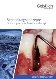
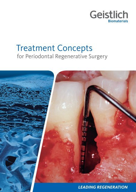
Treatment Conceptsfor Periodontal Regenerative Surgery
2ContentContentWhy periodontal regeneration? 3Regenerative therapy: getting to the root of the problem 4Suggested treatment concept for periodontally compromised teeth 5Defect morphology influences outcome of regenerative therapy 6Scientific and clinical evidence for the surgical preservative phase 7Case 1: Dr. Frank Bröseler | Intrabony 2-wall defect: interproximal crater 8Case 2: Dr. Diego Capri | 3-wall defect: rapid progression of lesion 9Case 3: Prof. Dr. Michael Christgau | Extended 2-wall defect 10Case 4: Dr. Pierpaolo Cortellini | Periodontal regenerative surgery 11Case 5: Dr. Daniel Etienne | Treatment of infrabony 1-wall defect 12Case 6: Prof. Dr. Markus Hürzeler | Combination defect 13Case 7: Dr. Syed Mahnaz | Regenerative surgery 11 – perio-endo 14Case 8: Prof. Dr. Giulio Rasperini | 2-wall defect in the non-aesthetic region 15Case 9: Prof. Dr. Anton Sculean | Deep intrabony 2-wall defect 16Case 10: Dr. Beat Wallkamm | 2-wall defect in the aesthetic zone 17Case 11: Prof. Dr. Giovanni Zucchelli | 2-wall wide intrabony defect 18References 19Product Range for periodontal treatment <strong>20</strong>
Why periodontal regeneration?Helping patients affected by periodontitis to create and The present treatment concept serves to summarise provenGuided Bone Regeneration (GBR) and Guided Tissuemaintain good oral health, function, and aesthetics is thegoal of every dentist. To accomplish this, various therapeuticapproaches have been developed in response to the ment of common periodontal defects.Regeneration (GTR) techniques for the successful treat-grades of severity of periodontitis. The role of biomaterials It provides scientific evidence and presents step-by-stepin treating periodontal disease has gained in significance clinical cases, demonstrating stable favorable outcomes.and is now an integral part of many protocols. Carefully This guide is intended for the clinician and highlights reliabletreatment options with the highest quality biomateri-selected biomaterials used with proven treatment protocolsmay not only stop progression of periodontal disease, als. It aims to present techniques and tools used for oralbut effectively regenerate both hard and soft tissue. 1,2 tissue regeneration to offer optimised therapy, leading togreater patient long-term satisfaction. 2Introduction 3Table 1. Prognosis of periodontally affected teeth: For classification at least one of the parameters (respectively two for hopeless teeth) has to be met. 6-8Good Questionable Hopeless> teeth with < 50% bone loss > teeth with 50-75% bone loss or> 6–8 mm PD or> class 2 furcation or> angular defect> teeth with > 75% bone loss or> more than 8mm PD or> Class 3 furcation or> Class 3 mobility or> teeth with at least 2 characteristicsof questionable categoryTooth preservation or implant?Teeth will last for life, unless they are affected by oral diseasesor service interventions. Many retained teeth thereforemay be an indicator of positive oral health behaviourthroughout the life course. Tooth longevity is largely dependenton the health status of the periodontium, the pulpor periapical region and the extent of reconstructions. 3Multiple risks lead to a critical appraisal of the value of atooth. Choosing between periodontal regeneration to supporttooth preservation and tooth extraction has beencalled one of the most complex and debatable decisions adentist is confronted with in daily clinical practice. 4Assigning a questionable prognosis – where the tooth requiresadvanced treatment to maybe preserve it –or a hopeless prognosis, where the tooth needs to be extractedas soon as possible, is often a delicate situation.This decision significantly impacts both treatment planingand patient lifestyle. Accordingly, it has been argued thatperiodontally compromised teeth should be treated for aslong as possible, and only being extracted when periodontaland endodontic treatment is no longer possible. 4, 5Regardless of whether the tooth is preserved or extracted,biomaterials are often required to reach the individualtherapeutic goals. Some criteria to categorise the prognosisof periodontally affected teeth are summarised inTable 1. 6-8
4Regenerative therapy:getting to the root of the problemRegenerative TherapyGood – Questionable – Hopeless … now what?Moreover, periodontally compromised but treated teethIn advance of any regenerative therapy, an initial nonsurgicalhygienic phase is crucial. This may include patient of implants in well-maintained patients. 12are known to have survival rates equal to the survival rateseducation on oral hygiene, scaling and root planing, antibacterialtherapy, and removal of plaque retentive fac-regeneration can result in long-term retention of teethA growing amount of evidence indicates that periodontaltors – all aimed to yield a good tissue response by eliminatinginfection and alleviating inflammation. When these intra-bony defects. 12-15 A randomised, long-term clinicaloriginally presenting with deep pockets associated withmethods fail to prevent bone loss, surgical or even regenerativetherapy for periodontally compromised teeth is with extraction and prosthetic replacement of hopelesstrial in 50 patients comparing periodontal regenerationthe recommended next-line therapy (Figure 2). 9-11teeth showed that regenerative therapy enabled retentionof 92% “hopeless” teeth scheduled for extraction. 7In questionable cases, regenerative therapy may be favoredover tooth extraction. This because extracting periometers,comfort and function for the follow-up period ofThe retained teeth had clinically stable periodontal paradontitis-affectedteeth will not resolve the underlying host 5-years (Figure 1). 12response-related problems contributing to the disease.Aims of Regenerative Treatment> Restoration of the complete tooth attachment apparatus with bone, cementum, and ligament> Prevention of long junctional epithelial down growth as a risk factor for recurrence of periodontitis> Long-term tooth retention> Aesthetic appearanceNumber of observations262524232221<strong>20</strong>Tooth preservation (test, n=25)Extraction / implantation (control, n=24)Baseline 1 2 3 4 5YearsFigure 1. Survival analysis. Comparison between hopeless teeth (test group) treated with periodontal regeneration and implant supported teeth at extractionsites of hopeless teeth (control group). Survival at 5 years was 100% in the control group versus 92% in the test group. 12
Suggested treatment concept forperiodontally compromised teethThe following treatment plan outlines a possible clinical methodologY:Treatment Concept 5DIAGNOSISnonsurgicalphasePhase I Initial TherapyPlaque control and patient educationScaling / deep scaling (root planing)nonsurgicalphaseREEVALUATIONtreatment decisionphasePhase II Consolidation TherapyControl of clinical parameters: bleeding on probing(BOP), clinical attachment level (CAL), pocketdepth (PD). Decision on further treatmenttreatment decisionphasesurgical preservativephasePHASE IIIa Tooth preservationPeriodontal surgery, with GBR/GTR* oropen flap depridement (OFD)surgical restorativephasePHASE IIIb Tooth extractionProsthetic restoration or implant replacement oftoothno surgicaltreatmentneededmaintenancephasePhase IV Maintenance TherapyPlaque control with or without antibioticaltreatment. Periodic control of clinical parameters:BOP, CAL, PD and bone loss (peri-implantitis)in case of implant placementmaintenancephaseFigure 2. Suggested Treatment Concept (Adapted from Newman, Lindhe, Rateitschak 9-11 )* the present Treatment Concept presents only cases with GBR/GTR
6Defect morphology influencesoutcome of regenerative therapyDefect MorphologyThere is a wide range of general factors that are known or The present Treatment Concept shows different cases thatassumed to influence periodontal healing (e.g., age, smoking,concomitant medication, postsurgical care, periodon-the remaining walls and the vertical dimension of the bonyhave been appointed to a classification system combiningtal maintenance, oral hygiene, nutrition, stress).defect (Figure 4).Furthermore, defect morphology is a key factor for thetherapy outcome. 16 Each periodontal osseous lesion presentsa unique anatomy. A first level of classification dif-Osseous defectsferentiates between horizontal, infrabony, and furcationHorizontal Defectsdefects as represented in Figure 3. 17Horizontal defects are defined when the base of the pocketis located coronal to the alveolar crest whereas infra-Infrabony Defectsbony defects are apical (vertical defects).Intrabony DefectsRegenerative therapy (GBR, GTR) is indicated in bony defectswith three, two or at least one remaining walls. Tosome extend also Class II furcation defects can be treatedwith GTR. 18 There is evidence, that 2- and 3 wall intrabonydefects respond better to GTR therapy than 1-wall defects.However, the deeper the infrabony defect, the more attachmentgain and bony fill may be expected. 16 Other defectcharacteristics influencing outcomes of regenerativetherapy are presented in Table 2:1 Wall2 Walls3 WallsCombinationsCratersFurcation DefectsTable 2: Positive and negative defect characteristics 16Positive INFLUENCE> Deep infrabony component(> 3 mm)> Narrow radiographicdefect angle> Deep baseline pocketdepthNegative INFLUENCE> Shallow infrabonycomponent (≤ 3 mm)> Wide radiographic defectangle> Tooth motilityClass I: Horizontal loss up to 3 mmClass II: Horizontal loss > 3 mm; not totalClass III: Total loss of tissue in furcationFigure 3. Classification of periodontal osseous defects (modified fromPapapanou et al. <strong>20</strong>00) 171 wall defect 2 wall defect 3 wall defect Interproximal craterFigure 4. Infrabony defects (modified from Papapanou et al. <strong>20</strong>00) 17
Scientific and clinical evidencefor the surgical preservative phaseUpon decision to preserve the tooth, the next step is todecide for a surgical therapy: Leading treatment methodsoften utilise a combination of a slowly resorbing osteoconductivebone substitute and a membrane. 19Guided Tissue RegenerationSome evidence shows, that Guided Tissue Regeneration(GTR) is superior to Open Flap Debridement (OFD) forthe treatment of periodontal intrabony and furcationdefects. <strong>20</strong>-22 Overall, GTR is consistently more effectivethan OFD in reducing:> open horizontal furcation depths,> horizontal and vertical attachment levels, and> pocket depths for mandibular or maxillary class IIfurcation defects.BONBBONBLNCFigure 6. The histologic assessment demonstrates the presence of newperiodontal ligament, cementum, and bone. The newly formed wovenbone can be observed maturating into bone trabeculae completelysurrounding <strong>Geistlich</strong> <strong>Bio</strong>-Oss particles. BO=<strong>Bio</strong>-Oss; NB=new boneL=ligament; NC=new cementum; OC=old cementum; D=dentin 19D<strong>20</strong>0 µmScientific & Clinical Evidence 7With the use of <strong>Geistlich</strong> <strong>Bio</strong>-Oss ® orthodontic movementis possible in patients after GTR therapy. 23,24 Moreover, resorbablemembranes have proven superior to non-resorbablemembranes in generating vertical bone fill. 15mmmm76543210765432CAL gainCAL gainp < 0.01 p < 0.011.8 4.0 1.4 3.7After 1 yearAfter 5 yearsPD reductionPD reductionp ≤ 0.05 p ≤ 0.05<strong>Geistlich</strong> <strong>Bio</strong>-Oss ® (Collagen) and<strong>Geistlich</strong> <strong>Bio</strong>-<strong>Gide</strong> ® (Perio)Combined filling of periodontal defects with the graft material<strong>Geistlich</strong> <strong>Bio</strong>-Oss ® Collagen or <strong>Geistlich</strong> <strong>Bio</strong>-Oss ®followed by <strong>Geistlich</strong> <strong>Bio</strong>-<strong>Gide</strong> ® membrane coverage has ahistory of proven effectiveness in regenerative periodontaltherapy. 25-31Treatment of intra-bony defects with <strong>Geistlich</strong> <strong>Bio</strong>-Oss ®and <strong>Geistlich</strong> <strong>Bio</strong>-<strong>Gide</strong> ® Perio resulted in sustained higherclinical attachment level gain as compared to treatmentwith OFD alone after 5 years (Figure 5). 2First clinical and histological results of treatment of endodontic-periodonticlesion with endodontic therapyfollowed by Guided Tissue Regeneration with <strong>Geistlich</strong><strong>Bio</strong>-Oss ® and <strong>Geistlich</strong> <strong>Bio</strong>-<strong>Gide</strong> ® demonstrated that thecombined approach can promote the formation of new cementum,periodontal ligament, and bone around the apex,as well as the complete bone regeneration of the buccalbone plate (Figure 6). 19104.0 5.4 3.3 4.8After 1 yearAfter 5 yearsopen flap debridement + <strong>Geistlich</strong> <strong>Bio</strong>-Oss ® and <strong>Geistlich</strong> <strong>Bio</strong>-<strong>Gide</strong> ® Perio (n=10)open flap debridement (n=9)Figure 5. The gain in clinical attachment level (CAL) and the reduction in pocket depth (PD) are significantly larger in the test group than in the controlgroup respectively, (p=0.01 and ≤ 0.05 respectively) both after one year and after 5 years. 2
8Case 1: Dr. Frank BröselerIntrabony 2-wall defect: interproximal craterSURGERY by Dr. Frank Bröseler, <strong>Aachen</strong> (DE)AIM Functional and esthetic reconstruction in chronic periodontitis with deep intrabony defects.Tooth # CAL (mm) PD (mm) Depth of bonydefect (mm)Defect morphology11 mesial 10 mesial 10 10 interproximal crater21 buccal 6 mesial 10 buccal 5 mesial 10 9<strong>Bio</strong>materialsSuture materialTechniquePeriodontal treatment> <strong>Geistlich</strong> <strong>Bio</strong>-Oss ® Collagen and <strong>Geistlich</strong> <strong>Bio</strong>-<strong>Gide</strong> ® Perio.> 4-0 classic and 6-0 monofilament with cutting needle> Full thickness flap, split released, papilla preservation> Patient instruction and plaque control for at least 8 weeks.01 Initial situation after anti-infective therapy.Radiographically, the intrabony defect cannot berepresented in toto due to palatal bone plate.02 Intrasurgical situation after preparation ofthe mucoperiostal full-thickness flap revealsdeep osseous defect.03 Palatinal view of the defect after applicationof <strong>Geistlich</strong> <strong>Bio</strong>-Oss ® Collagen.04 The grafted site is covered with<strong>Geistlich</strong> <strong>Bio</strong>-<strong>Gide</strong> ® Perio.05 The flap is repositioned and sutured torelieve flap tension and obtain primaryclosure of the interdental space.06 Postoperative x-ray control afterregenerative procedure using <strong>Geistlich</strong><strong>Bio</strong>-Oss ® Collagen.07 Clinical situation at 3 years follow-up 08 4.5 years post-op radiograph showingsustained defect fill from <strong>Geistlich</strong> <strong>Bio</strong>-Oss ®Collagen.09 Clinical situation at 7 years follow-up; notethe naturally reformed papilla between thecentral incisors, and no gingival recession.CONCLUSION After controlling the periodontal disease, this guided tissue regeneration technique leads to a long-term stablebony situation with pleasant soft-tissue appearance.
3-wall defect: rapid progression of lesionSURGERY by Dr. Diego Capri, bologna (IT)AIM Regeneration of a 2 to 3 wall defect caused by a cemental tear.Tooth # CAL (mm) PD (mm) Depth of bonydefect (mm)Defect morphology35 distal 12 distal 7 5 3 wall defectwithout furcationCase 2: Dr. Diego Capri 9<strong>Bio</strong>materials > <strong>Geistlich</strong> <strong>Bio</strong>-Oss ® , autogeneous bone, <strong>Geistlich</strong> <strong>Bio</strong>-<strong>Gide</strong> ®Suture material > Gore-Tex ® Suture CV7Technique> Periodontal regeneration of the defect by means of GTRPeriodontal treatment > Periodontal defect debridement with hand and ultrasonic instrumentation.01 Clinical preoperative view of the affectedarea showing the lesion.02 DIAGNOSIS: Cemental tear – likely causedby a parafunctional habit overlapped to partialedentulism and malocclusion in the area.03 After reflection of a mucoperiosteal flap theperiodontal defect is de-granulated and thefractured portion of the cementum is visible.04 The root surface is thoroughly scaled andplaned.05 The defect is filled with a mixture ofautogenous bone and <strong>Geistlich</strong> <strong>Bio</strong>-Oss ® .06 A trimmed <strong>Geistlich</strong> <strong>Bio</strong>-<strong>Gide</strong> ® collagenmembrane is positioned on the augmentedarea.07 Primary wound closure is achieved, afterproper releasing of the flap with internalmattress and single interrupted Gore sutures.08 4 months after periodontal regenerativesurgery a probing depth of 3 mm anda clinical attachment loss of 6 mm wasmeasured distally.09 Intraoral radiographic aspect of the siteshowing the healing of the defect.CONCLUSION The rapid progression of the lesion was arrested and the bone at the defect side successfullyregenerated.
10Case 3: Prof. Dr. Michael ChristgauExtended 2-wall defectSURGERY by Prof. Dr. MichaEl Christgau, Düsseldorf (DE)AIM Defect resolution of an extended 2-wall defect with regenerative periodontal surgery.Tooth # CAL (mm) PD (mm) Depth of bonydefect (mm)Defect morphology32 mesial 14 distal 4 mesial 11 distal 2 ca. 10 2 wall defectbuccal 4 oral 4 buccal 1 oral 2<strong>Bio</strong>materials> <strong>Geistlich</strong> <strong>Bio</strong>-Oss ® Collagen, <strong>Geistlich</strong> <strong>Bio</strong>-<strong>Gide</strong> ® Perio, autogenous boneSuture material > Seralene ® 5-0 and 6-0Technique> Papilla-Preservation technique, sulcular incision Regio 41–33 without vertical releasing incisionsPeriodontal treatment > Semipermanent adhesive tooth splinting with composite material and non-surgical periodontal therapywith additional systemic antibiotic therapy ( 3 x 400 mg metronidazol, 7 days)01 Preoperative clinical and radiological situationshowing an inflammation-free gingivaand the bone defect.02 Intraoperative view of the extended 2-walldefect.03 Basal defect is filled with autogenous bonechips after debridement and root planing.04 Autogeneous bone covered and defect filledcompletely with <strong>Geistlich</strong> <strong>Bio</strong>-Oss ® Collagen.05 Coverage with a trimmed <strong>Geistlich</strong> <strong>Bio</strong>-<strong>Gide</strong> ®Perio membrane without further fixation.06 Coronal flap repositioning and wound closurewith horizontal mattress and single sutures.07 Clinical and radiological situation after6 months with clinical attachment gainof 7 mm mesial and vast defect fill.08 Clinical and radiological situation at12 months with clinical attachment gainof 8 mm mesial and considerable defect fill.09 Clinical and radiological situation 6 yearsafter surgery showing stable long-termsituation.CONCLUSION Regenerative periodontal surgery with <strong>Geistlich</strong> <strong>Bio</strong>-Oss ® Collagen and <strong>Geistlich</strong> <strong>Bio</strong>-<strong>Gide</strong> ® Perio results in longtermdefect resolution.
Periodontal regenerative surgerySURGERY by Dr. Pierpaolo CORTELLINI, Firenze (IT)AIM Resolution of deep pockets associated with deep intrabony defects and preservation of aesthetics on upper incisors.Tooth # CAL (mm) PD (mm) Depth of bonydefect (mm)Defect morphology21 (22) mesial 7 (4) distal 2 (7) mesial 6 (2) distal 2 (6) max. 10 (8) 2 wall defectbuccal 4 (4) lingual 3 (4) buccal 4 (2) lingual 3 (3) without furcationCase 4: Prof. Dr. Pierpaolo Cortellini 11<strong>Bio</strong>materials > <strong>Geistlich</strong> <strong>Bio</strong>-Oss ®Suture material > Gore-Tex ® Suture 6-0Technique > Modified minimally invasive surgical procedure (M-MIST) with a Microblade USM 6900Periodontal treatment > Root planing was performed before surgery.01 Preoperative probing at tooth 21 showingprobing depth of 6 mm.02 Preoperative probing at tooth 22 withprobing depth of 6 mm.03 Preoperative radiograph showing the intrabonydefects mesial to tooth 21 and distal totooth 22 .04 Buccal incision design. 05 Intraoperative probing at tooth 21. Notethe absence of the interdental bone peakbetween teeth 11 and 21 and the severebuccal dehiscence. <strong>Geistlich</strong> <strong>Bio</strong>-Oss ® wasused to prevent the postoperative shrinkageof the soft tissues.06 <strong>Geistlich</strong> <strong>Bio</strong>-Oss ® is positioned to fill theintrabony components of the defects.In larger and/or less contained defects, theadditional use of a collagen membrane, suchas <strong>Geistlich</strong> <strong>Bio</strong>-<strong>Gide</strong> ® , is recommended.07 The flap is sealed over <strong>Geistlich</strong> <strong>Bio</strong>-Oss ®with internal modified mattresssutures.08 1 year clinical situation showing healthycondition and a minimal gingival recessionrelative to baseline.09 1 year radiographs showing the resolution ofthe intrabony components of the defects.CONCLUSION The combination of the modified minimally invasive surgical technique with <strong>Geistlich</strong> <strong>Bio</strong>-Oss ® was effective intreating multiple intrabony defects associated with deep pockets in the upper incisors.ReferencesCortellini P, Tonetti MS. Improved wound stability with a modified minimally invasivesurgical technique in the regenerative treatment of isolated interdental intrabonydefects. J Clin Periodontol <strong>20</strong>09: 36: 157–163.Cortellini P, Tonetti MS. Clinical and radiographic outcomes of the modified minimallyinvasive surgical technique with and without regenerative materials: a randomized- controlledtrial in intra-bony defects. J Clin Peridontol <strong>20</strong>11: 38: 365–373.
12Case 5: Dr. Daniel EtienneTreatment of infrabony 1-wall defectSURGERY by Dr. Daniel Etienne, Paris (FR)Non Surgical Periodontal Therapy: Dr. Sofia Aroca, Saint-Germain en Laye (FR)AIM 1 wall periodontal defect treatment before orthodontic tooth intrusion and diastema closure.Tooth # CAL (mm) PD (mm) Depth of bonydefect (mm)Defect morphology11 buccal mesial 6 distal 5 mesial 6 distal 5 6 1-wall defect11 lingual mesial 6 distal 3 mesial 6 distal 3 without furcation<strong>Bio</strong>materialsSuture materialTechniquePeriodontal treatment> <strong>Geistlich</strong> <strong>Bio</strong>-Oss ® small granules, <strong>Geistlich</strong> <strong>Bio</strong>-<strong>Gide</strong> ® 25x25mm, Emdogain> 6-0 Ethicon PDS-II> Remote palatal papilla incision and Guided Tissue Regeneration (GTR)> 1. Plaque control 2. GTR 3. Orthodontic treatment by Dr. Catherine Galletti (Paris)01 Preoperative clinical and radiological situationshowing an angular bony defect at themesial aspect of tooth 11. No inflammationof the soft tissue is observed. Presence of adiastema and a small papilla collapse mesialof 11.02 1 wall defect of 6 mm CAL on mesio-buccaland mesio-lingual of 11.03 After debridement and root planing, root oftooth 11 is covered with Emdogain. Defectfill with Emdogain and <strong>Geistlich</strong> <strong>Bio</strong>-Oss ®granules. The augmented site is covered witha <strong>Geistlich</strong> <strong>Bio</strong>-<strong>Gide</strong> ® membrane.04 Repositioning and suturing of the flap using6-0 Ethicon PD-S II sutures.05 Clinical situation 1 week after surgery andsuture removal. No inflammation is observed.06 Clinical situation and x-ray of the augmentedsite just before start of orthodontic treatment10 months after surgery.07 Clinical and radiological situation afterorthodontic treatment (intrusion of 11 anddiastema closure) and 3 years after surgery.08 Clinical and radiological images with stabletissue conditions 4 years after surgery.09 X-ray 5 years after surgery showing a slightand stable crestal bone remodelling in themesial aspect of tooth 11.CONCLUSION Slight crestal bone remodelling on the mesial aspect of tooth 11 was observed after orthodontic treatment,with 5 mm probing after papilla remodeling. Clinical attachment stability is observed during maintenance.
Combination defectSURGERY by prof. DR. Markus Hürzeler, München (DE)AIM Periodontal regeneration of two teeth severely compromised by attachment loss at the apex.Tooth # CAL (mm) PD (mm) Depth of bonydefect (mm)21, 11, 12 mesial 6,10, 11 distal 6, 10, 7 mesial 6, 10, 11 distal 6, 10, 7 max 10 -buccal 5, 8, 9 lingual 5, 6, 7 buccal 5, 8, 9 lingual 5, 6, 7Defect morphologyCase 6: Prof. Dr. Markus Hürzeler 13<strong>Bio</strong>materials> <strong>Geistlich</strong> <strong>Bio</strong>-Oss ® , <strong>Geistlich</strong> <strong>Bio</strong>-<strong>Gide</strong> ® , AmelogeninSuture material > Seralene ® suture, DS 12, 15 / 7.0Technique> Microsurgical access flap with modified papilla incision techniquePeriodontal treatment > Anti-infectious therapy, Doxycyclin (Ligosan ® Heraeus), DH (24 hours scaling), reevaluation,11 + 21 Ca(OH) 2and root canal filling 21, recall.01 Pre-operative radiological view of theextended bone loss.02 Clinical situation preoperatively afterDoxycyclin antibiotic treatment.03 Surgical site after debridement and rootplaning.04 Defect fill with <strong>Geistlich</strong> <strong>Bio</strong>-Oss ® aftertreatment with amelogenin derivativematrix.05 Coverage with <strong>Geistlich</strong> <strong>Bio</strong>-<strong>Gide</strong> ® tostabilise the augmented area.06 Situtation after wound closure.07 1 month after surgery an improvement of thebony situation is visible.08 Clinical situation after 5 months beforeclosing the inter-approximate defect withcomposite.09 Final restoration 10 months after surgery.CONCLUSION Successful preservation of two “hopeless” teeth with periodontal regenerative therapy.
14Case 7: Dr. Syed MahnazRegenerative surgery 11 – perio-endoSURGERY by Dr. Syed Mahnaz, Perth (AUS)AIM Retention of the central incisor and improvement of its mobility.Tooth # CAL (mm) PD (mm) Depth of bonydefect (mm)Defect morphology11 mesial 9 distal 5 mesial 7 distal 4 4 2 wall defectbuccal 5 lingual 5 buccal 3 lingual 3<strong>Bio</strong>materials > <strong>Geistlich</strong> <strong>Bio</strong>-Oss ® , <strong>Geistlich</strong> <strong>Bio</strong>-<strong>Gide</strong> ®Suture material > Vicryl 5.0 suture materialsTechnique> Endodontic treatment followed by non-surgical debridement and a modified papilla preservation technique.Periodontal treatment > Nonsurgical periodontal debridement therapy under local anaesthesia with endodontic treatment wasundertaken.01 Non-responding residual pocket associatedwith a perio-endo involved tooth 11.02 Radiograph of infrabony angular defecton tooth 11 with subsequent endodontictreatment.03 Elevation of flap with papilla preservationto access the infrabony pocket.04 <strong>Geistlich</strong> <strong>Bio</strong>-Oss ® granules in the defect. 05 <strong>Geistlich</strong> <strong>Bio</strong>-<strong>Gide</strong> ® membrane trimmed and 06 Immediate post-op passive closure andplaced in the interproximal region .coronal repositioning of the mucosa.07 Improved pocketing and mobility 8 monthsafter surgery and additional composite bondingto improve the aesthetics.08 <strong>Geistlich</strong> <strong>Bio</strong>-Oss ® mesial of tooth 11 is wellintegrated after 8 months.08 Follow up 2 years post surgery showing goodbone stability and improved clinical status ofthis tooth.CONCLUSION Predictable treatment outcomes were achieved to help retain teeth in situations where perio-endo problemsexist. Regenerative surgery offers sustainable options for treatment of advanced periodontal disease.
2-wall defect in the non-aesthetic regionSURGERY by Prof. Dr. Giulio Rasperini, Milan (IT)AIM Periodontal regeneration to reduce probing depth by increasing bone and periodontal attachment with a minimalgingival recession, to change the prognosis of the tooth # 46 and preserve its function.Tooth # CAL (mm) PD (mm) Depth of bonydefect (mm)Defect morphology46 mesial 14 distal 3 mesial 14 distal 3 max 10 2 wall defectwithout furcationCase 8: Prof. Dr. Giulio Rasperini 15<strong>Bio</strong>materials > <strong>Geistlich</strong> <strong>Bio</strong>-Oss ® , <strong>Geistlich</strong> <strong>Bio</strong>-<strong>Gide</strong> ®Suture material > Gore-Tex ® Suture 5-0Technique> Periodontal regeneration procedure with preservation of the interdental tissue and mesial releasing incision.Periodontal treatment > Cause related periodontal therapy, including motivation and instructions for home care; professionalsupra-gingival debridment and sub-gingival root planing. Re-evaluation for potential additional therapy.01 Baseline situation showing the 14 mm pocketdepth mesial to tooth 46.02 Baseline radiograph showing the presence ofan angular bony defect involving the mesialsite of tooth 46.03 Elevation of a full-thickness buccal andlingual flap with papilla preservation.The 10 mm deep, 2-wall intrabony defectmesial to tooth 46 is evident after carefuldebridement.04 The <strong>Geistlich</strong> <strong>Bio</strong>-Oss ® fills the defectand is protected by a <strong>Geistlich</strong> <strong>Bio</strong>-<strong>Gide</strong> ®membrane. After flap release, the woundis closed without tension.05 Re-evaluation at 1 year. A residual 5 mmprobing depth is present with a 9 mmprobing depth loss as compared to baselinemeasurements.06 Nearly complete bone fill of the angulardefect at 1 year.CONCLUSION 2 months after conclusion of presurgical, cause-related therapy, the patient reported the complete resolution ofinflammation, resulting in a decrease of the full mouth plaque and bleeding scores. 1 year after the surgery, the soft-tissuewas well preserved and represented with a sufficient width of keratinised gingiva. Radiographs after 1 year show a stablesituation with an almost complete bone fill.
16Case 9: Prof. Dr. Anton SculeanDeep intrabony 2-wall defectSURGERY by Prof. Dr. Anton Sculean, Bern (CH)AIM Treatment of intrabony defect with a complicated, noncontained morphology using a combination of collagen barriermembrane and a natural bone mineral.Tooth # CAL (mm) PD (mm) Depth of bonydefect (mm)Defect morphology36 distal 11 distal 11 5 2 wall, large non-containeddefect<strong>Bio</strong>materials > <strong>Geistlich</strong> <strong>Bio</strong>-<strong>Gide</strong> ® Perio, <strong>Geistlich</strong> <strong>Bio</strong>-Oss ®Suture material > 4-0 silkTechnique> Periodontal regeneration of a large non-contained defect through GTR with the use of grafting material.Periodontal treatment > Hygienic phase 3 months before regenerative surgery consisting of patient instruction for oral hygiene,and full-mouth scaling and root planing in conjunction with systemically administered antibiotic therapy(3 x 375 mg Amoxicillin and 3 x 250 mg Metronidazol) for one week.01 Preoperative probing indicating the presenceof a deep pocket distal to the mandibular leftmolar.02 Preoperative radiograph demonstrating theextent of bone loss.03 Intraoperative view revealing a deep noncontainedintrabony defect.04 Following removal of granulation tissue androot planing, the defect is filled with <strong>Geistlich</strong><strong>Bio</strong>-Oss ® .05 The grafting material and the surroundingalveolar bone are covered with a <strong>Geistlich</strong><strong>Bio</strong>-<strong>Gide</strong> ® Perio.06 Minimal recession of the soft tissues andattachment gain and reduced PD measured6mm and 7 mm respectivelyat 1 year.07 Postoperative radiograph at 1 year reveals analmost complete fill of the intrabony defect.CONCLUSION Good appearance of soft tissue and sufficient bone fill at 1 year after regeneration of a deep non-contained bonydefect.
2-wall defect in the aesthetic zoneSURGERY by Dr. BEAT WALLKAMM, LANgenthal (CH)AIM Periodontal regeneration with a minimally invasive surgical technique in combination with <strong>Geistlich</strong> <strong>Bio</strong>-Oss ® Collagenand <strong>Geistlich</strong> <strong>Bio</strong>-<strong>Gide</strong> ® Perio.Tooth # CAL (mm) PD (mm) Depth of bonydefect (mm)Defect morphology11 mesial 11 distal 4 mesial 8 distal 3 5 2 wall defectbuccal 4 lingual 4 buccal 2 lingual 3Case 10: Dr. Beat Wallkamm 17<strong>Bio</strong>materials> <strong>Geistlich</strong> <strong>Bio</strong>-Oss ® Collagen, <strong>Geistlich</strong> <strong>Bio</strong>-<strong>Gide</strong> ® PerioSuture material > Seralene ® 7/0 (PVDF, Serag Wiessner)Technique > Minimal invasive surgical technique (MIST) (Cortellini <strong>20</strong>09)Periodontal treatment > Initial periodontal treatment (4hrs), 3-months recall01 Tooth 11 presents with a pocket depth of8 mm and a clinical attachment level of 11mm with some loss of papillary tissue.02 Baseline radiograph shows the bone lossmesially to the first right incisor reaching theapical third of the root.03 After elevation of a tiny buccal flap andpositioning of the interdental papilla slightlyto the palatal side, the defect is debrided.04 A trimmed <strong>Geistlich</strong> <strong>Bio</strong>-<strong>Gide</strong> ® Perio isinserted lingually and <strong>Geistlich</strong> <strong>Bio</strong>-Oss ®Collagen is applied into the defect.05 The <strong>Geistlich</strong> <strong>Bio</strong>-<strong>Gide</strong> ® Perio is folded overthe augmented site and inserted under thebuccal full thickness flap.06 Primary closure of the wide interdentalpapilla is obtained with an internal mattresssuture with an external loop and two obliquehang-up mattress sutures.07 6 weeks after surgery the inter-dental softtissues are well healed.08 Clinical situation after 2 years with a probingpocket depth of 3 mm and a clinical attachmentlevel gain of 5 mm.09 The 2 year radiograph shows a horizontalgain of 3 mm bone in the treated area.CONCLUSION The minimally invasive surgical technique in combination with <strong>Geistlich</strong> <strong>Bio</strong>-Oss ® Collagen and <strong>Geistlich</strong><strong>Bio</strong>-<strong>Gide</strong> ® Perio resulted in markedly improved clinical and radiographic outcome.ReferenceCortellini P, Tonetti MS. Improved wound stability with a modified minimally invasive surgical technique in the regenerative treatment of isolated interdental intrabony defects.J Clin Periodontol <strong>20</strong>09: 36: 157–163.
18Case 11: Prof. Dr. Giovanni Zucchelli2-wall wide intrabony defectSURGERY by Prof. Dr. Giovanni Zucchelli, Bologna (IT)AIM Regenerative surgery of a severely compromised tooth in aesthetic area.Tooth # CAL (mm) PD (mm) Depth of bonydefect (mm)Defect morphology21 mesial 3 distal 13 mesial 3 distal 11 13 combined intrabony defect<strong>Bio</strong>materialsSuture materialTechniquePeriodontal treatmentbuccal 11 lingual 3 buccal 11 lingual 3> <strong>Geistlich</strong> <strong>Bio</strong>-Oss ® , <strong>Geistlich</strong> <strong>Bio</strong>-<strong>Gide</strong> ® , Amelogenin> PGA 7.0 in the papilla / PGA 6.0 in the flap> Regenerative surgery with CAF combined with simplified papilla preservation> Ultrasonic periodontal therapy before the surgery01 Pre-operative view of the affected upper leftincisor.02 Radiographic situation before treatment.The wide defect reaches the apex of thetooth.03 The defect after degranulation.04 EDTA and an amelogenin derivative matrixare applied to condition the root surface .05 <strong>Geistlich</strong> <strong>Bio</strong>-Oss ® fills the wide defect and<strong>Geistlich</strong> <strong>Bio</strong>-<strong>Gide</strong> ® prevents tissue collapsewhile stabilizing the site.06 Post-op view of suturing: note the primaryintention closure of the interdental papillaabove the defect.07 Clinical situation at 12 month follow-up.A regrowth of the interdental papilla couldbe achieved.08 Follow-up radiograph at 12 months revealscomplete bone fill.CONCLUSION Healthy hard- and soft-tissue situation with regrowth of the interdental papilla after 1 year.
References:1Wang HL et al., J Periodontol. <strong>20</strong>05 Sep; 76(9):1601-16222Sculean A et al., J Clin Periodontol. <strong>20</strong>07 Jan;34(1):72-773Holm-Pederson et al, Clin. Oral Impl. Res. 18 (Suppl. 3), <strong>20</strong>07 / 15–194Donos N et al., Periodontol <strong>20</strong>00. <strong>20</strong>12 Jun;59(1):89-1105Zitzmann NU et al., Int Endod J. <strong>20</strong>09 Sep;42(9):757-7746Checchi L et al., J Clin Periodontol. <strong>20</strong>02 Jul; 29(7): 651–6567Samet N et al., Quintessence Int. <strong>20</strong>09 May; 40(5):377–3878Becker W et al., J Periodontol. 1984 Sep; 55(9):505–5099Newman, Takei, Klokkevold, Carranza. CARRANZA’S CLINICAL PERIODONTOLOGY. ISBN 13 978-1-4160-2400-2.10Lindhe, Karring, Lang. Clinical Periodontology and Implant Dentistry. BlackwellMunksgaard. ISBN 1-4051-0236-5.11Rateitschak, Wolf. Farbatlanten der Zahnmedizin 1. <strong>Paro</strong>dontologie. Thieme. ISBN 3-13-655601-1.12Cortellini P. et al., J Clin Periodontol. <strong>20</strong>11 Oct;38(10):915-92413Cortellini P., Tonetti MS., J Periodontol. <strong>20</strong>04 May;75(5):672-67814Sculean A. et al., J Clin Periodontol. <strong>20</strong>08 Sep;35(9):817-82415Kinaia BM. et al., J Periodontol. <strong>20</strong>11 Mar; 82 (3):413-42816Sculean Anton. Periodontal Regenerative Therapy. Quintessence Publishing. ISBN-13: 978185097158017Papapanou PN., Tonetti MS., Periodontol <strong>20</strong>00. <strong>20</strong>00 Feb;22:8-2118Reddy KP et al., J Contemp Dent Pract. <strong>20</strong>06 Feb 15;7(1):60-7019Ghezzi et al Clin. Oral Impl. Res. 18 (Suppl. 3), <strong>20</strong>07 / 15–19<strong>20</strong>Murphy KG et Gunsolley JC, Ann Periodontol, Dec, <strong>20</strong>03, Vol 8. Number 1, 266-30221Houser BE et al., Int J Periodontics Restorative Dent., <strong>20</strong>01 Apr, 21 (2): 161-16922Paolantonio M et al., J Periodontol. <strong>20</strong>10 Nov;81(11):1587-159523Da Silva VC et al., J Clin Periodontol. <strong>20</strong>06 Jun;33(6):440-44824Cardaropoli D et al., Int J Periodontics Restorative Dent. <strong>20</strong>06 Dec;26(6):553-55925Cosyn J et al., J Clin Periodontol. <strong>20</strong>12; Oct;39(10):979-98626Camelo Int J Periodontics Restorative Dent. 1998 Aug;18(4):321-33127Lundgren D, Slotte C, J Clin Periodontol. 1999 Jan;26(1):56-6228Camargo PM et al., J Clin Periodontol. <strong>20</strong>00 Dec;27(12):889-89629Sculean A et al., J Clin Periodontol. <strong>20</strong>03 Jan;30(1):73-8030Tonetti MS et al., J Clin Periodontol. <strong>20</strong>04 Sep;31(9):770-77631Liñares M et al., J Clin Periodontol. <strong>20</strong>06 May;33(5):351-358References 19Adding convenience to periodontal treatments!Simple to handle, simple to shapePerio-System Combi-Pack<strong>Geistlich</strong> <strong>Bio</strong>-Oss ®Collagen 100 mg<strong>Geistlich</strong> <strong>Bio</strong>-<strong>Gide</strong> ® Periowith sterile templates16 x 22 mm
Product Range for periodontal treatment *<strong>Geistlich</strong> <strong>Bio</strong>-<strong>Gide</strong> ® PerioResorbable bilayer membranewith sterile templatesAvailable sizes:16 mm x 22 mm<strong>Geistlich</strong> <strong>Bio</strong>-<strong>Gide</strong> ®Resorbable bilayer membraneAvailable sizes:25 mm x 25 mm30 mm x 40 mm<strong>Geistlich</strong> <strong>Bio</strong>-Oss ® CollagenSpongious bone substitutePreformed block with CollagenAvailable sizes:100 mg250 mg500 mg<strong>Geistlich</strong> Combi-Kit Collagen<strong>Geistlich</strong> <strong>Bio</strong>-Oss ®Collagen 100 mg<strong>Geistlich</strong> <strong>Bio</strong>-<strong>Gide</strong> ®16 x 22 mm<strong>Geistlich</strong> <strong>Bio</strong>-Oss ®Spongious bone substituteSmall granules 0.25 mm – 1 mmAvailable sizes:0.25 g ≈ 0.5 cc0.5 g ≈ 1 cc2 g ≈ 4 cc<strong>Geistlich</strong> <strong>Bio</strong>-Oss ®Spongious bone substituteLarge granules 1 mm – 2 mmAvailable sizes:0.5 g ≈ 1.5 cc2 g ≈ 6 cc* Product availability may vary from country to country.Subsidiary Great Britain, Ireland<strong>Geistlich</strong> <strong>Bio</strong>materials<strong>Geistlich</strong> Sons Limited1 st Floor, Thorley HouseBailey Lane, Manchester AirportGB-Manchester M90 4ABPhone +44 1 614 902 038Fax +44 1 614 986 988www.geistlich.co.ukManufacturer©<strong>Geistlich</strong> Pharma AGBusiness Unit <strong>Bio</strong>materialsBahnhofstrasse 40CH-6110 WolhusenPhone +41-41-4925 630Fax +41-41-4925 639www.geistlich-pharma.comMore details about our distribution partners:www.geistlich-pharma.com/mycontact31601.1/1303/en