Exercise 3: Internal Anatomy of the Lubber Grasshopper, Romalea
Exercise 3: Internal Anatomy of the Lubber Grasshopper, Romalea
Exercise 3: Internal Anatomy of the Lubber Grasshopper, Romalea

Create successful ePaper yourself
Turn your PDF publications into a flip-book with our unique Google optimized e-Paper software.
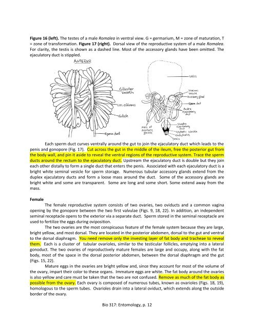
Figure 16 (left). The testes <strong>of</strong> a male <strong>Romalea</strong> in ventral view. G = germarium, M = zone <strong>of</strong> maturation, T<br />
= zone <strong>of</strong> transformation. Figure 17 (right). Dorsal view <strong>of</strong> <strong>the</strong> reproductive system <strong>of</strong> a male <strong>Romalea</strong>.<br />
For clarity, <strong>the</strong> testis is shown as a dashed line. Most <strong>of</strong> <strong>the</strong> accessory glands have been omitted. The<br />
ejaculatory duct is stippled.<br />
Each sperm duct curves ventrally around <strong>the</strong> gut to join <strong>the</strong> ejaculatory duct which leads to <strong>the</strong><br />
penis and gonopore (Fig. 17). Cut across <strong>the</strong> gut in <strong>the</strong> middle <strong>of</strong> <strong>the</strong> ileum, free <strong>the</strong> posterior gut from<br />
<strong>the</strong> body wall, and pin it aside to reveal <strong>the</strong> ventral regions <strong>of</strong> <strong>the</strong> reproductive system. Trace <strong>the</strong> sperm<br />
ducts around <strong>the</strong> rectum to <strong>the</strong> ejaculatory duct. Upstream <strong>the</strong> ejaculatory duct is double but <strong>the</strong>y join<br />
each o<strong>the</strong>r distally to form a single duct that enters <strong>the</strong> penis. Associated with each ejaculatory duct is a<br />
bright white seminal vesicle for sperm storage. Numerous tubular accessory glands extend from <strong>the</strong><br />
duplex ejaculatory ducts and form a loose mass around <strong>the</strong> duct. Some <strong>of</strong> <strong>the</strong> accessory glands are<br />
bright white and some are transparent. Some are long and some short. Some extend away from <strong>the</strong><br />
mass.<br />
Female<br />
The female reproductive system consists <strong>of</strong> two ovaries, two oviducts and a common vagina<br />
opening by <strong>the</strong> gonopore between <strong>the</strong> two first valvulae (Figs. 9, 18, 22). In addition, an independent<br />
seminal receptacle opens to <strong>the</strong> exterior via a separate duct. Sperm stored in <strong>the</strong> seminal receptacle are<br />
used to fertilize <strong>the</strong> eggs during oviposition.<br />
The two ovaries are <strong>the</strong> most conspicuous feature <strong>of</strong> <strong>the</strong> female system because <strong>the</strong>y are large,<br />
bright yellow, and most dorsal. They are located in <strong>the</strong> posterior abdomen, dorsal to <strong>the</strong> gut and ventral<br />
to <strong>the</strong> dorsal diaphragm. You need remove only <strong>the</strong> investing layer <strong>of</strong> fat body and tracheae to reveal<br />
<strong>the</strong>m. Each is a cluster <strong>of</strong> tubular ovarioles, similar to <strong>the</strong> testicular follicles, emptying into a lateral<br />
gonoduct. The two ovaries <strong>of</strong> reproductively mature females are large and occupy, along with <strong>the</strong> fat<br />
body, most <strong>of</strong> <strong>the</strong> space in <strong>the</strong> dorsal posterior abdomen, between <strong>the</strong> dorsal diaphragm and <strong>the</strong> gut<br />
(Figs. 15, 22).<br />
Mature eggs in <strong>the</strong> ovaries are bright yellow and, since <strong>the</strong>y account for most <strong>of</strong> <strong>the</strong> volume <strong>of</strong><br />
<strong>the</strong> ovary, impart <strong>the</strong>ir color to <strong>the</strong>se organs. Immature eggs are white. The fat body around <strong>the</strong> ovaries<br />
is also yellow and care must be taken that <strong>the</strong> two are not confused. Remove as much <strong>of</strong> <strong>the</strong> fat body as<br />
possible from <strong>the</strong> ovary. Each ovary is composed <strong>of</strong> numerous tubes, known as ovarioles (Figs. 18, 19),<br />
homologous to <strong>the</strong> sperm tubes. Ovarioles drain into a lateral oviduct, which extends along <strong>the</strong> outside<br />
border <strong>of</strong> <strong>the</strong> ovary.<br />
Bio 317: Entomology, p. 12