Remodeling of the Vertebral Body in Hereditary Lordoscoliotic ...
Remodeling of the Vertebral Body in Hereditary Lordoscoliotic ...
Remodeling of the Vertebral Body in Hereditary Lordoscoliotic ...

You also want an ePaper? Increase the reach of your titles
YUMPU automatically turns print PDFs into web optimized ePapers that Google loves.
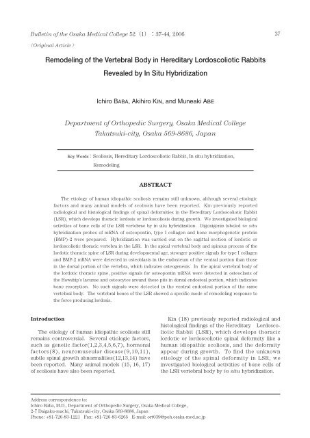
Bullet<strong>in</strong> <strong>of</strong> <strong>the</strong> Osaka Medical College 52137-44, 2006 37Orig<strong>in</strong>al Article<strong>Remodel<strong>in</strong>g</strong> <strong>of</strong> <strong>the</strong> <strong>Vertebral</strong> <strong>Body</strong> <strong>in</strong> <strong>Hereditary</strong> <strong>Lordoscoliotic</strong> RabbitsRevealed by In Situ HybridizationIchiro BABA, Akihiro KIN, and Muneaki ABEDepartment <strong>of</strong> Orthopedic Surgery, Osaka Medical CollegeTakatsuki-city, Osaka 569-8686, JapanKey WordsScoliosis, <strong>Hereditary</strong> <strong>Lordoscoliotic</strong> Rabbit, In situ hybridization,<strong>Remodel<strong>in</strong>g</strong>ABSTRACTThe etiology <strong>of</strong> human idiopathic scoliosis rema<strong>in</strong>s still unknown, although several etiologicfactors and many animal models <strong>of</strong> scoliosis have been reported. K<strong>in</strong> previously reportedradiological and histological f<strong>in</strong>d<strong>in</strong>gs <strong>of</strong> sp<strong>in</strong>al deformities <strong>in</strong> <strong>the</strong> <strong>Hereditary</strong> <strong>Lordoscoliotic</strong> Rabbit(LSR), which develops thoracic lordosis or lordoscoliosis dur<strong>in</strong>g growth. We <strong>in</strong>vestigated biologicalactivities <strong>of</strong> bone cells <strong>of</strong> <strong>the</strong> LSR vertebrae by <strong>in</strong> situ hybridization. Digoxigen<strong>in</strong> labeled <strong>in</strong> situhybridization probes <strong>of</strong> mRNA <strong>of</strong> osteopont<strong>in</strong>, type I collagen and bone morphogenetic prote<strong>in</strong>(BMP)-2 were prepared. Hybridization was carried out on <strong>the</strong> sagittal section <strong>of</strong> lordotic orlordoscoliotic thoracic vertebra <strong>in</strong> <strong>the</strong> LSR. In <strong>the</strong> apical vertebral body and sp<strong>in</strong>ous process <strong>of</strong> <strong>the</strong>lordotic thoracic sp<strong>in</strong>e <strong>of</strong> LSR dur<strong>in</strong>g developmental age, stronger positive signals for type I collagenand BMP-2 mRNA were detected <strong>in</strong> osteoblasts <strong>in</strong> <strong>the</strong> endosteum <strong>of</strong> <strong>the</strong> ventral portion than those<strong>in</strong> <strong>the</strong> dorsal portion <strong>of</strong> <strong>the</strong> vertebra, which <strong>in</strong>dicates osteogenesis. In <strong>the</strong> apical vertebral body <strong>of</strong><strong>the</strong> lordotic thoracic sp<strong>in</strong>e, positive signals for osteopont<strong>in</strong> mRNA were detected <strong>in</strong> osteoclasts <strong>of</strong><strong>the</strong> Howship’s lacunae and osteocytes around <strong>the</strong>se pits <strong>in</strong> dorsal endosteal portion, which <strong>in</strong>dicatesbone resorption. No such signals were detected <strong>in</strong> <strong>the</strong> ventral endosteal portion <strong>of</strong> <strong>the</strong> samevertebral body. The vertebral bones <strong>of</strong> <strong>the</strong> LSR showed a specific mode <strong>of</strong> remodel<strong>in</strong>g response to<strong>the</strong> force produc<strong>in</strong>g lordosis.IntroductionThe etiology <strong>of</strong> human idiopathic scoliosis stillrema<strong>in</strong>s controversial. Several etiologic factors,such as genetic factor(1,2,3,4,5,6,7), hormonalfactors(8), neuromuscular disease(9,10,11),subtle sp<strong>in</strong>al growth abnormalities(12,13,14) havebeen reported. Many animal models (15, 16, 17)<strong>of</strong> scoliosis have also been reported.K<strong>in</strong> (18) previously reported radiological andhistological f<strong>in</strong>d<strong>in</strong>gs <strong>of</strong> <strong>the</strong> <strong>Hereditary</strong> <strong>Lordoscoliotic</strong>Rabbit (LSR), which develops thoraciclordotic or lordoscoliotic sp<strong>in</strong>al deformity like ahuman idiopathic scoliosis, and <strong>the</strong> deformityappear dur<strong>in</strong>g growth. To f<strong>in</strong>d <strong>the</strong> unknownetiology <strong>of</strong> <strong>the</strong> sp<strong>in</strong>al deformity <strong>in</strong> LSR, we<strong>in</strong>vestigated biological activities <strong>of</strong> bone cells <strong>of</strong><strong>the</strong> LSR vertebral body by <strong>in</strong> situ hybridization.Address correspondence to:Ichiro Baba, M.D., Department <strong>of</strong> Orthopedic Surgery, Osaka Medical College,2-7 Daigaku-machi, Takatsuki-city, Osaka 569-8686, JapanPhone: +81-726-83-1221 Fax: +81-726-83-6265 E-mail: ort039@poh.osaka-med.ac.jp
38Ichiro BABA, Akihiro KIN, and Muneaki ABEMaterials and MethodsLSRLSR is a type <strong>of</strong> Japanese White Rabbit whichwas <strong>in</strong>cidentally found by a breeder <strong>of</strong> RabbitonInstitute(Hyogo, Japan) <strong>in</strong> 1982(Fig.1). S<strong>in</strong>cethat time, crossbreed<strong>in</strong>g has been cont<strong>in</strong>ued. InLSR, sp<strong>in</strong>al deformities are not recognized atbirth, but appear at 4-6 weeks <strong>of</strong> age and developuntil 16 weeks. Most <strong>of</strong> <strong>the</strong> rabbits demonstratedlordosis, which was seen at <strong>the</strong> T7-10 levels (44degrees measured by Cobb’s method(19) atmaximum), and was <strong>of</strong>ten accompanied by slightscoliosis(40 degrees at maximum)(Fig.2). Nosp<strong>in</strong>al anomalies were observed but severedeformities sometimes impaired <strong>the</strong>ir gait andability to eat. The <strong>in</strong>cidence <strong>of</strong> <strong>the</strong> deformitiesranged from 50 to 60%, and no gender differenceswere observed.Tissue PreparationLSR were sacrificed at 6-14 weeks <strong>of</strong> age under<strong>in</strong>tra venous general anes<strong>the</strong>sia and <strong>the</strong> thoracicFig.1Fig.2aFig.2Typical example <strong>of</strong> LSR.Fig.2bRadiographs show<strong>in</strong>g scoliosis (a) andlordosis(b) <strong>of</strong> <strong>the</strong> thoracic sp<strong>in</strong>e <strong>in</strong> LSR.sp<strong>in</strong>es were harvested with surround<strong>in</strong>g s<strong>of</strong>ttissue and fixed for 5 days at 4 <strong>in</strong> 4% freshlymade paraformaldehyde(PFA) <strong>in</strong> 0.1M phosphatebuffer. Then specimens were dehydrated <strong>in</strong>ethanol, cleared <strong>in</strong> chlor<strong>of</strong>orm, and decalcifiedwith Morse’s solution(10% sodium citrate and22.5% formic acid) until <strong>the</strong> specimen becames<strong>of</strong>t(it took 3 to 4 weeks). Decalcified specimenswere dehydrate and embedded <strong>in</strong> paraff<strong>in</strong>.Sagittal sections <strong>of</strong> 6m thick were cut andmounted on 3-am<strong>in</strong>opropyl- triethoxysilanecoatedslides(20).Probe PreparationBone samples <strong>of</strong> <strong>the</strong> skull, sp<strong>in</strong>e, ribs andfemurs dissected from <strong>the</strong> fetus <strong>of</strong> Japanese WhiteRabbits 1 week before birth under <strong>in</strong>tra venousgeneral anes<strong>the</strong>sia were frozen <strong>in</strong> liquid nitrogen.Messenger RNA was extracted from <strong>the</strong> specimenus<strong>in</strong>g <strong>the</strong> Micro-Fast Track mRNA isolationkit(Invitrogen Corp., Sandiego, CA), accord<strong>in</strong>g to<strong>the</strong> manufacture’s <strong>in</strong>structions. ComplementaryDNA(cDNA) was made so that mRNA was reversetranscribed <strong>in</strong> 20l reaction mixture conta<strong>in</strong><strong>in</strong>greverse transcriptase and random primer.Thereafter, 1l <strong>of</strong> cDNA was amplified <strong>in</strong> 25lpolymerase cha<strong>in</strong> reaction(PCR) mixtureconta<strong>in</strong><strong>in</strong>g 0.125U Taq DNA polymerase and12.5pmol <strong>of</strong> each primer for type I collagen,osteopont<strong>in</strong>(Osp) and bone morphogeneticprote<strong>in</strong>-2(BMP-2) by PCR. Oligonucleotides forPCR were as follows: type I collagen, 5’-CAAGGGAGAACGTGGTTACC-3’ (5’sense;312-331) and 5’-TTCTTCCGGGA GCCTTCAGG-3’(3’antisense;934-963); and Osp, 5’-CACCGCAGAATGCTATGTCC-3’(5’ sense;251-270) and 5’-GGCTCGATGGCTAGCTTGTC-3’(3’antisense;922-941); and BMP-2, 5’-ATGCCAAGTCCTGCTAGGAG-3’(5’sense;395-414)and 5’-GATCGGCTAATCCTGACA TG-3’(3’antisense;1110-1129). PCR conditions were asfollows a total <strong>of</strong> 30 cycles was performed with<strong>the</strong> Astec DNA <strong>the</strong>rmal cycler(Astec, Fukuoka,Japan) at 94 for 0.5 m<strong>in</strong>utes, 55 for 1 m<strong>in</strong>ute,72 for 1 m<strong>in</strong>ute, and <strong>the</strong>n at 72 for 5 m<strong>in</strong>utesat <strong>the</strong> end <strong>of</strong> <strong>the</strong> procedure. PCR products weresubcloned <strong>in</strong>to multiclon<strong>in</strong>g sites <strong>of</strong> <strong>the</strong> plasmid,and analyzed with <strong>the</strong> ABI Model 373A DNASequencer us<strong>in</strong>g <strong>the</strong> PRISM Ready ReactionDyeDeoxy Term<strong>in</strong>ator Cycle Sequenc<strong>in</strong>gKit(Perk<strong>in</strong>-Elmer Corporation) accord<strong>in</strong>g to <strong>the</strong>manufacture’s <strong>in</strong>structions. The sequence resultswere identical to those <strong>of</strong> <strong>the</strong> rabbit type Icollagen (GenBank accession no.D49399), BMP-2Bullet<strong>in</strong> <strong>of</strong> <strong>the</strong> Osaka Medical College 52137-44, 2006
Etiology <strong>of</strong> Sp<strong>in</strong>al deformity <strong>in</strong> LSR 39(GenBank accession no.D30751, X56848) andOsp(GenBank accession no.D11411) previouslyreported(21). Digoxigen<strong>in</strong>(DIG)-labeled s<strong>in</strong>glestrandantisense and sense complementary RNAprobes were prepared us<strong>in</strong>g <strong>the</strong> DIG RNA label<strong>in</strong>gkit(Boehr<strong>in</strong>ger Mannheim GmbH, Biochemica,Manheim, Germany) accord<strong>in</strong>g to <strong>the</strong>manufacturer’s <strong>in</strong>structions(20).In situ hybridizationIn situ hybridization was carried out accord<strong>in</strong>gto <strong>the</strong> manufacturer’s protocol(Boehr<strong>in</strong>gerMannheim Yamanouchi, Tokyo, Japan) with m<strong>in</strong>ormodifications. Sections were deparaff<strong>in</strong>ized,fixed <strong>in</strong> 4%PFA <strong>in</strong> 0.1M PB (pH7.4), pretreatedwith HCl to <strong>in</strong>activate endogenous alkal<strong>in</strong>e phosphatase,and acetylated with acetic anhydrate.Hybridization was carried out at 50 for 16hus<strong>in</strong>g DIG labeled probes at a concentration <strong>of</strong>approximately 0.5g/ml <strong>in</strong> hybridization bufferand was washed with Rnase A (10g/ml) at 37for 30 m<strong>in</strong>utes. Hybridized probes were detectedus<strong>in</strong>g a nucleic acid detection kit(Boehr<strong>in</strong>gerMannheim GmbH, Biochemica, Mannheim,Germany) accord<strong>in</strong>g to <strong>the</strong> manufacture’s<strong>in</strong>structions.Controls hybridized with <strong>the</strong> sense probe didnot show positive signals.ResultsHistological f<strong>in</strong>d<strong>in</strong>gsThe thoracic sp<strong>in</strong>e <strong>of</strong> 4-week-old LSR has nodeformity (Fig.3). In <strong>the</strong> apical vertebra and<strong>in</strong>tervertebral disc <strong>in</strong> <strong>the</strong> lordotic thoracic sp<strong>in</strong>e <strong>of</strong>a 8-week-old LSR, <strong>the</strong> ma<strong>in</strong> histological f<strong>in</strong>d<strong>in</strong>gswere a deviation <strong>of</strong> <strong>the</strong> nucleus pulposus andextension <strong>in</strong> ventral and compression <strong>in</strong> <strong>the</strong> dorsalportion <strong>of</strong> <strong>the</strong> annulus fibrosus(Fig.4). No markedFig.3Sagittal section <strong>of</strong> <strong>the</strong> normal thoracicvertebra <strong>of</strong> LSR <strong>of</strong> 4 weeks <strong>of</strong> age. H.E.sta<strong>in</strong> (x 3) vb:vertebral body sc:sp<strong>in</strong>alcordFig.4a Fig.4b Fig.4cFig.4(a)Sagittal section <strong>of</strong> <strong>the</strong> normal thoracic vertebra <strong>of</strong> LSR <strong>of</strong> 10 weeks <strong>of</strong> age.(b)(c)Sagittal section <strong>of</strong> <strong>the</strong> apical thoracic lordotic vertebra <strong>of</strong> LSR <strong>of</strong> 10 weeks <strong>of</strong> age. There isdeviation <strong>of</strong> <strong>the</strong> nucleus pulposus and extension <strong>in</strong> ventral and compression <strong>in</strong> <strong>the</strong> dorsal portion<strong>of</strong> <strong>the</strong> annulus fibrosus. No marked abnormality is observed <strong>in</strong> <strong>the</strong> epiphysis or <strong>in</strong> <strong>the</strong> growthplate <strong>of</strong> <strong>the</strong> vertebral body. H.E. sta<strong>in</strong> (x 3)vb:vertebral body sc:sp<strong>in</strong>al cord np:nuculeus pulposusBullet<strong>in</strong> <strong>of</strong> <strong>the</strong> Osaka Medical College 52137-44, 2006
40Ichiro BABA, Akihiro KIN, and Muneaki ABEabnormality was observed <strong>in</strong> <strong>the</strong> epiphysis or <strong>in</strong><strong>the</strong> growth plate <strong>of</strong> <strong>the</strong> vertebral body.In <strong>the</strong> apical vertebra <strong>of</strong> a 12-week-old LSR,proliferation <strong>of</strong> columnar cartilage composed <strong>of</strong> avertebral growth plate was found on <strong>the</strong> ventralside. On <strong>the</strong> dorsal side, narrow<strong>in</strong>g <strong>of</strong> <strong>the</strong>vertebral growth plate and proliferation <strong>of</strong>marg<strong>in</strong>al epiphysial cartilage cells were found.In <strong>the</strong> apical vertebra <strong>of</strong> a 24-week-old LSR, awedg<strong>in</strong>g deformity <strong>in</strong> <strong>the</strong> epiphysis was found.The <strong>in</strong>tervertebral disc was degenerated and <strong>the</strong>nucleus pulposus was destroyed.Fig.5aFig.5b Fig.5c Fig.5d Fig.5eFig.5Type I collagen <strong>in</strong> situ hybridization.Sagittal section <strong>of</strong> <strong>the</strong> apical thoracic lordotic vertebra <strong>of</strong> LSR <strong>of</strong> 10 weeks <strong>of</strong> age.(a)Arrows <strong>in</strong>dicate markedly strong positive signals for type I collagen mRNA. (x 3)(b)Arrows <strong>in</strong>dicate markedly strong positive signals for type I collagen mRNA <strong>in</strong> osteoblasts <strong>in</strong><strong>the</strong> endosteum at <strong>the</strong> ventral side <strong>of</strong> <strong>the</strong> vertebral body. (x 50)(c)Arrows <strong>in</strong>dicate positive signals for type I collagen mRNA <strong>in</strong> osteoblasts <strong>in</strong> <strong>the</strong> extraosteum at<strong>the</strong> dorsal side <strong>of</strong> vertebral body. (x 50)Sagittal section <strong>of</strong> <strong>the</strong> normal thoracic vertebra <strong>of</strong> LSR <strong>of</strong> 10 weeks <strong>of</strong> age.(d) Arrows <strong>in</strong>dicates slightly positive signals for type I collagen mRNA <strong>in</strong> osteoblasts <strong>in</strong> <strong>the</strong>endosteum at <strong>the</strong> ventral side <strong>of</strong> <strong>the</strong> vertebral body. (x 50)(e) Positive signals for type I collagen mRNA are not seen at <strong>the</strong> dorsal portion <strong>of</strong> <strong>the</strong> vertebralbody. (x 50)v:ventral side d:dorsal side cr:cranial side ca:caudal side sc:sp<strong>in</strong>al canalBullet<strong>in</strong> <strong>of</strong> <strong>the</strong> Osaka Medical College 52137-44, 2006
Etiology <strong>of</strong> Sp<strong>in</strong>al deformity <strong>in</strong> LSR 41In situ hybridizationIn <strong>the</strong> apical vertebral body and sp<strong>in</strong>ousprocess <strong>in</strong> <strong>the</strong> lordotic thoracic sp<strong>in</strong>e <strong>of</strong> LSRdur<strong>in</strong>g grow<strong>in</strong>g age, stronger positive signals fortype I collagen mRNA were detected <strong>in</strong>osteoblasts <strong>in</strong> <strong>the</strong> endosteum <strong>of</strong> <strong>the</strong> ventral thanthose <strong>in</strong> <strong>the</strong> dorsal portion <strong>of</strong> <strong>the</strong> vertebra(Fig.5).In <strong>the</strong> vertebra without deformity, no differences<strong>of</strong> positive signals between <strong>the</strong> ventral and dorsalportion were observed. Positive signals for BMP-2mRNA showed a similar pattern as that observed<strong>in</strong> type I collagen mRNA pattern(Fig.6).In <strong>the</strong> apical vertebral body <strong>of</strong> <strong>the</strong> lordoticthoracic sp<strong>in</strong>e, positive signals for Osp mRNAwere detected <strong>in</strong> osteoclasts <strong>in</strong> Howship’s lacunaeand osteocytes around <strong>the</strong>se pits <strong>in</strong> dorsalendosteal portion. No such signals were detected<strong>in</strong> <strong>the</strong> ventral endosteal portion <strong>of</strong> <strong>the</strong> samevertebral body(Fig.7). In <strong>the</strong> vertebral bodywithout deformity, <strong>the</strong>re were no differences <strong>in</strong><strong>the</strong> Osp mRNA expression pattern betweenventral and dorsal portion.DiscussionTo <strong>in</strong>vestigate <strong>the</strong> etiology <strong>of</strong> idiopathicscoliosis, several scoliotic animal models havebeen developed, <strong>in</strong>clud<strong>in</strong>g paravertebral muscleexcision(22), division <strong>of</strong> <strong>the</strong> <strong>in</strong>tercostalnerves(22), resection <strong>of</strong> <strong>the</strong> ribs(23), resection <strong>of</strong><strong>the</strong> costotransvers jo<strong>in</strong>ts(24), rib elongation(25),rib shorten<strong>in</strong>g(26). Recent report showed that<strong>the</strong> scoliosis which develops <strong>in</strong> chickens afterp<strong>in</strong>ealectomy was similar to human idiopathicscoliosis, and thus may be a useful model <strong>of</strong>idiopathic scoliosis(17). Moreover surgicalte<strong>the</strong>r<strong>in</strong>g <strong>of</strong> <strong>the</strong> sp<strong>in</strong>ous apophysis and transverseapophysis on <strong>the</strong> same side <strong>of</strong> <strong>the</strong> sp<strong>in</strong>e producedscoliosis with characteristics similar to those <strong>of</strong>human idiopathic scoliosis(27). However <strong>the</strong>sescoliotic animal models can not show deformitywithout surgery, <strong>the</strong>refore <strong>the</strong>se are models <strong>of</strong>secondary sp<strong>in</strong>al deformity. On <strong>the</strong> o<strong>the</strong>r hand<strong>the</strong> sp<strong>in</strong>al deformities observed <strong>in</strong> LSR weresimilar to those <strong>of</strong> human idiopathic scoliosis; nosp<strong>in</strong>al anomaly, no deformity at birth, <strong>the</strong>deformities <strong>of</strong> <strong>the</strong> sp<strong>in</strong>e appears at 4-6weeks <strong>of</strong>age and progresses without surgery, anddeformities <strong>in</strong>clude lordosis or lordoscoliosis withFig.6aFig.6bFig.6BMP mRNA <strong>in</strong> situ hybridization.Sagittal section <strong>of</strong> <strong>the</strong> apical thoracic lordotic vertebra <strong>of</strong> LSR <strong>of</strong> 10 weeks <strong>of</strong> age.(a)Arrows <strong>in</strong>dicate positive signals for BMP mRNA <strong>in</strong> osteoblasts <strong>in</strong> <strong>the</strong> endosteum <strong>in</strong> <strong>the</strong> ventralside <strong>of</strong> <strong>the</strong> vertebral body. (x 50)(b)Positive signals for BMP mRNA are not seen <strong>in</strong> <strong>the</strong> endosteum at <strong>the</strong> dorsal portion dorsalside <strong>of</strong> <strong>the</strong> vertebral body. (x 50)v:ventral side d:dorsal side cr:cranial side ca:caudal sideBullet<strong>in</strong> <strong>of</strong> <strong>the</strong> Osaka Medical College 52137-44, 2006
42Ichiro BABA, Akihiro KIN, and Muneaki ABEFig.7a Fig.7b Fig.7cFig.7Osp mRNA <strong>in</strong> situ hybridization.Sagittal section <strong>of</strong> <strong>the</strong> apical thoracic lordotic vertebra <strong>of</strong> LSR at 10 weeks <strong>of</strong> age.(a) Asterisk <strong>in</strong>dicates positive signals for Osp mRNA <strong>in</strong> osteoclasts at Howship’s lacunae andosteocytes around <strong>the</strong>se pits <strong>in</strong> extraosteum at <strong>the</strong> ventral side <strong>of</strong> <strong>the</strong> vertebral body. (x 50)(b) Asterisks <strong>in</strong>dicate positive signals for Osp mRNA <strong>in</strong> osteoclasts <strong>in</strong> Howship’s lacunae andosteocytes around <strong>the</strong>se pits <strong>in</strong> endosteum at <strong>the</strong> dorsal side <strong>of</strong> <strong>the</strong> vertebral body. (x 50)Sagittal section <strong>of</strong> <strong>the</strong> normal thoracic vertebra <strong>of</strong> LSR at 10 weeks <strong>of</strong> age.(c) Positive signals for Osp mRNA are not seen <strong>in</strong> <strong>the</strong> endosteum at <strong>the</strong> dorsal side <strong>of</strong> <strong>the</strong>vertebral body. (x 50)v:ventral side d:dorsal side cr:cranial side ca:caudal siderotation from middle to lower thoracic levels(18).In <strong>the</strong> LSR with severe lordosis, rabbits cannot eator reproduce, so <strong>the</strong>y have to rotate <strong>the</strong>ir sp<strong>in</strong>e.This posture may enforce <strong>the</strong>ir lordosis <strong>in</strong>toscoliosis. Therefore LSR is an useful animalmodel for study<strong>in</strong>g etiology <strong>of</strong> human idiopathicscoliosis.Concern<strong>in</strong>g histological f<strong>in</strong>d<strong>in</strong>gs observed <strong>in</strong>present study, <strong>in</strong> <strong>the</strong> early stage <strong>of</strong> <strong>the</strong> deformity,ma<strong>in</strong> changes <strong>in</strong>cluded a deviation <strong>in</strong> <strong>the</strong> nucleuspulposus <strong>of</strong> <strong>the</strong> <strong>in</strong>tervertebral disc and extension<strong>in</strong> <strong>the</strong> ventral portion and compression <strong>in</strong> <strong>the</strong>dorsal region <strong>of</strong> <strong>the</strong> annulus fibrosus. In <strong>the</strong> latestage, proliferation <strong>of</strong> columnar cartilagecomposed <strong>of</strong> <strong>the</strong> vertebral growth plate was foundon <strong>the</strong> ventral side. On <strong>the</strong> dorsal side, narrow<strong>in</strong>g<strong>of</strong> <strong>the</strong> vertebral growth plate and proliferation <strong>of</strong>marg<strong>in</strong>al epiphyseal cartilage cells were found.F<strong>in</strong>ally, wedg<strong>in</strong>g deformity <strong>in</strong> <strong>the</strong> epiphysis wasfound, <strong>the</strong> <strong>in</strong>tervertebral disc was degeneratedand <strong>the</strong> nucleus pulposus was destroyed. Thesef<strong>in</strong>d<strong>in</strong>gs were considered to be secondary changesresult<strong>in</strong>g from progressive local lordosis <strong>of</strong> <strong>the</strong>thoracic sp<strong>in</strong>e.Dubousset et al.(28) found that scoliosisrout<strong>in</strong>ely developed <strong>in</strong> p<strong>in</strong>ealectomized chickens,and scoliosis was caused from decreas<strong>in</strong>gmelaton<strong>in</strong> production. Sobajima et al. (29)reported that serum melaton<strong>in</strong> levels <strong>in</strong> LSR weresignificant higher than those <strong>of</strong> Japanese whiterabbit. And <strong>the</strong>y suggest that causes <strong>of</strong> sp<strong>in</strong>aldeformities <strong>in</strong> <strong>the</strong> LSR may be <strong>the</strong> result <strong>of</strong> <strong>the</strong>contribution <strong>of</strong> melaton<strong>in</strong> receptors as well as that<strong>of</strong> altered serum melaton<strong>in</strong> levels <strong>in</strong> <strong>the</strong> LSR.Although fur<strong>the</strong>r studies will be required to<strong>in</strong>vestigate <strong>the</strong> expression <strong>of</strong> melaton<strong>in</strong> receptor<strong>in</strong> o<strong>the</strong>r tissues <strong>of</strong> <strong>the</strong> LSR as well as to del<strong>in</strong>eate<strong>the</strong> role <strong>of</strong> melaton<strong>in</strong> <strong>in</strong> <strong>the</strong> pathogenesis <strong>of</strong>idiopathic scoliosis.Type I collagen is a ma<strong>in</strong> component <strong>of</strong>connective tissues <strong>of</strong> <strong>the</strong> sk<strong>in</strong>, tendon and bone.In present study, strong positive signals for type Icollagen mRNA were detected <strong>in</strong> osteoblasts <strong>of</strong><strong>the</strong> endosteum <strong>of</strong> <strong>the</strong> ventral portion <strong>of</strong> <strong>the</strong>Bullet<strong>in</strong> <strong>of</strong> <strong>the</strong> Osaka Medical College 52137-44, 2006
Etiology <strong>of</strong> Sp<strong>in</strong>al deformity <strong>in</strong> LSR 43lordotic thoracic vertebral body and sp<strong>in</strong>ousprocess, <strong>in</strong>dicat<strong>in</strong>g osteogenesis. BMP-2 is one <strong>of</strong><strong>the</strong> most potent prote<strong>in</strong> <strong>of</strong> osteogenesis and hasbeen shown to <strong>in</strong>duce bone formation successfullyat heterotopic locations. Aga<strong>in</strong> <strong>in</strong> present studyBMP-2 mRNA showed a similar pattern to type Icollagen mRNA, <strong>in</strong>dicat<strong>in</strong>g osteogenesis. OspmRNA-positive osteoclasts <strong>in</strong> Howship’s lacunaeand osteocytes around <strong>the</strong> pits were detected <strong>in</strong><strong>the</strong> dorsal endosteal portion <strong>of</strong> <strong>the</strong> same vertebralbody, <strong>in</strong>dicat<strong>in</strong>g bone resorption(30). These bonecells activity <strong>in</strong>dicates <strong>the</strong> remodel<strong>in</strong>g fordistraction force to <strong>the</strong> anterior side andcompression force to <strong>the</strong> posterior side <strong>in</strong> <strong>the</strong>thoracic lordotic vertebral body. The vertebralbones showed a specific mode <strong>of</strong> remodel<strong>in</strong>gresponse to <strong>the</strong> force produc<strong>in</strong>g lordosis.Asymmetrical load<strong>in</strong>g <strong>of</strong> <strong>the</strong> sp<strong>in</strong>e is onepossible etiology <strong>of</strong> idiopathic scoliosis.Carp<strong>in</strong>tero et al.(27) demonstrated thatlordoscoliosis could be <strong>in</strong>duced <strong>in</strong> a rabbit bysurgical te<strong>the</strong>r<strong>in</strong>g <strong>of</strong> <strong>the</strong> sp<strong>in</strong>ous apopyhsis andtransverse apophysis. Our results suggest thatgrowth imbalance which may exist between <strong>the</strong>anterior component and <strong>the</strong> posterior componentwith muscle and ligament, enforced develop<strong>in</strong>glordosis. This f<strong>in</strong>d<strong>in</strong>gs suggest that asymmetricalload<strong>in</strong>g <strong>of</strong> <strong>the</strong> sp<strong>in</strong>e can be an etiology <strong>of</strong>idiopathic scoliosis. Whe<strong>the</strong>r asymmetricalload<strong>in</strong>g is <strong>the</strong> cause <strong>of</strong> idiopathic scoliosis or <strong>the</strong>result <strong>of</strong> sp<strong>in</strong>al deformity, is still unclear. Because<strong>the</strong> etiology <strong>of</strong> sp<strong>in</strong>al deformity <strong>in</strong> LSR may bemultifactorial same as human idiopathic scoliosis,fur<strong>the</strong>r studies is necessary to know <strong>the</strong> etiology<strong>of</strong> sp<strong>in</strong>al deformity <strong>of</strong> LSR.Acknowledgments The authors thank Sh<strong>in</strong>taroNomura, PhD (Department <strong>of</strong> Pathology, MedicalSchool <strong>of</strong> Osaka University), Akira Tsutsumi,MD(Department <strong>of</strong> Surgical Pathology, OsakaMedical College) for technical assistance andadvice.References1. De George FV, Fisher RL: Idiopathic scoliosis:genetic and enviromental aspects.J MedGenet. 1967;4:251-257.2. Wynne-Davis R: Familial(idiopathic)scoliosis:A family survey. J Bone Jo<strong>in</strong>tSurg(Br). 1968;50:24-30.3. Risenborough Ej, Wynne-Davies R: A genericsurvey <strong>of</strong> idiopathic scoliosis <strong>in</strong> Boston,Massachusetts. J Bone Jo<strong>in</strong>t Surg (Am).1973;55:974-982.4. Wynne-Davis R: Genetic aspects <strong>of</strong> idiopathicscoliosis. Dev. Med Child Neurol.1973;15:809-811.5. Rob<strong>in</strong> GC, Cohen T: Familial scoliosis:A cl<strong>in</strong>icalReport. J Bone Jo<strong>in</strong>t Surg (Br). 1975;57:146-148.6. Miller NH: Cause and natural history <strong>of</strong>adolescent idiopathic scoliosis. Orthop Cl<strong>in</strong>iNorth Am. 1999;30:343-352.7. Hadley MN: Sp<strong>in</strong>e update:Genetics <strong>of</strong> familialidiopathic scoliosis. Sp<strong>in</strong>e.2000;25:2416-2418.8. Machida M, Dubousset J, imamura Y, MiyashitaY, Yamada T, Kimura J: Melaton<strong>in</strong>: A possiblerole <strong>in</strong> pathogenesis <strong>of</strong> adolescent idiopathicscoliosis. Sp<strong>in</strong>e.1996;21:1147-1152.9. Yamada K, Yamamoto H, Nakagawa Y,TezukaA, Tamura T, Kawata S:Etiology <strong>of</strong> idiopathicscoliosis. Cl<strong>in</strong>i Orthop.1980;152:232-236.10. Zadeh HG, Sakka SA, Powell MP, Mehta MH:Absent superficial abdom<strong>in</strong>al reflexes <strong>in</strong>children with scoliosis: An early <strong>in</strong>dicator <strong>of</strong>syr<strong>in</strong>gomyelia. J Bone Jo<strong>in</strong>t Surg (Br).1995;77:762-767.11. Samuelsson L, L<strong>in</strong>dell D: Scoliosis as <strong>the</strong> firstsign <strong>of</strong> a cystic sp<strong>in</strong>al cord. Eur Sp<strong>in</strong>e J.1995;4:284-290.12. Perdriolle R, Becchetti S, Vidal J, Lopez P:Mechanical process and growth cartilages:Essentioal factors <strong>in</strong> <strong>the</strong> progression <strong>of</strong>scoliosis.Sp<strong>in</strong>e.1993;18:343-349.13. Stokes Ia, Spence H, Aronsson Dd, Kilmer N:Mechanical modulation <strong>of</strong> vertbral bodygrowth:Implication for scoliosis progression.Sp<strong>in</strong>e.1996;21:1162-1167.14. Porter Rw:Idiopathic scoliosis:The relationbetween <strong>the</strong> vertebral canal and <strong>the</strong> bodies.Sp<strong>in</strong>e. 2000;25:1360-1366.15. Langenskiöd A, Michelsson E: Experimentalprogressive scoliosis <strong>in</strong> <strong>the</strong> rabbit.J Bone Jo<strong>in</strong>tSurg (Br). 1961;43:116-12016. Machida M, Dubousset J, Imamura Y, ImamuraY, Iwaya T, Yamada T, Kimura J: Aexperimental study <strong>in</strong> chickens <strong>of</strong> <strong>the</strong>pathogenesis <strong>of</strong> idiopathic scoliosis.Sp<strong>in</strong>e.1993;18:1609-1615.17. Kanemura T, Kawakami N, Deguchi M,Mimatsu K, Iwata H: Natural course <strong>of</strong>exterimental scoliosis <strong>in</strong> p<strong>in</strong>ealectomizedchickens. Sp<strong>in</strong>e.1997;22:1563-1567.18. K<strong>in</strong> A: Radiological and histological studies on<strong>the</strong> sp<strong>in</strong>al deformity <strong>in</strong> hereditary lordoscolioticrabbits J. Jpn. Orthop. Assoc. 1994;68:458-469.(<strong>in</strong> Japanese)Bullet<strong>in</strong> <strong>of</strong> <strong>the</strong> Osaka Medical College 52137-44, 2006
44Ichiro BABA, Akihiro KIN, and Muneaki ABE19. Cobb JR: American Academy <strong>of</strong> OrthopaedicSurgeons Instructional Course Lectures.1948;5:261.20. Nomura S, Hirakawa K, Nagoshi J: Method fordetect<strong>in</strong>g <strong>the</strong> expression on bone matrixprote<strong>in</strong> by <strong>in</strong> situ hybridization us<strong>in</strong>gdecalcified m<strong>in</strong>eralized tissue. ActaHistochem.Cytochem. 1993; 26, (4): 303-309.21. GenomeNet. http://www.genome.jp/Japanese/22. Bisgard Jd: Experimental thoracogenicscoliosis. J Thoracic Surg .1935;4,435.23. Langenskiöd A, Michelsson JE: Thepathogenesis <strong>of</strong> experimental progressivescoliosis. Acta Orthop Scand. Suppl. 1962;59,3-26.24. Karaharju EO: Deformation <strong>of</strong> vertebrae <strong>in</strong>experimental scoliosis. Acta Orthop Scand.Suppl. 1967;105:7-7925. Sevastik J, Agadir M, Sevastik Befits <strong>of</strong> ribelongation on <strong>the</strong> sp<strong>in</strong>e. I. Distortion <strong>of</strong> <strong>the</strong>vertebral alignment <strong>in</strong> <strong>the</strong> rabbit.Sp<strong>in</strong>e.1990;15:822-82526. Sevatikglou JA, Aaro S, L<strong>in</strong>dholm TS,Dahlborn MD: Experimental scoliosis <strong>in</strong>grow<strong>in</strong>g rabbits by operations on <strong>the</strong> rib cage.Cl<strong>in</strong> Orthop.1978;136:282-28627. Carp<strong>in</strong>tero P, Mesa M, Garcia J, Carp<strong>in</strong>tero P:Scoliosis <strong>in</strong>duced by asymmetric lordosis androtation. Sp<strong>in</strong>e.1997;22,(19):2202-2206.28. Dubousset J, Queneau P, Thillard MJ:Experimental scoliosis <strong>in</strong>duced by p<strong>in</strong>eal anddiencephalic lesions <strong>in</strong> young chickens. Itsrelation between with cl<strong>in</strong>ical f<strong>in</strong>d<strong>in</strong>gs <strong>in</strong>idiopathic scoliosis. Orthop. Trans. 1983;7:7.29. Sobajima S, K<strong>in</strong> A, Baba I, Kanbara K, SemotoY, Abe M: Implication for melaton<strong>in</strong> and itsreceptor <strong>in</strong> <strong>the</strong> sp<strong>in</strong>al deformity <strong>of</strong> hereditary<strong>Lordoscoliotic</strong> Rabbit. Sp<strong>in</strong>e. 2003;28(6):554-558.30. Ikeda T, Yamaguchi A, Yokose S, Nagai Y,Yamato H, Nakamura T, Tsurukami H,Tanizawa T, Yoshiki S: Changes <strong>in</strong> biologicalactivity <strong>of</strong> bone cells <strong>in</strong> ovariectomized ratsrevealed by <strong>in</strong> situ hybridization. J. Bone andM<strong>in</strong>eral Research.1996;11,(6): 780-788.Received December 14, 2005Accepted January 5, 2006Bullet<strong>in</strong> <strong>of</strong> <strong>the</strong> Osaka Medical College 52137-44, 2006