Hemepath Case 21: 9-Year-Old Boy
Hemepath Case 21: 9-Year-Old Boy
Hemepath Case 21: 9-Year-Old Boy

You also want an ePaper? Increase the reach of your titles
YUMPU automatically turns print PDFs into web optimized ePapers that Google loves.
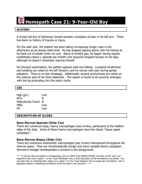
HISTORY<strong>Hemepath</strong> <strong>Case</strong> <strong>21</strong>: 9-<strong>Year</strong>-<strong>Old</strong> <strong>Boy</strong>A 9-year-old boy of Ashkenazi Jewish ancestry complains of pain in his left arm. Therehas been no history of trauma or injury.For the past year, the patient has been taking increasingly longer naps in theafternoons as he always feels tired. He has stopped playing sports with his friends ashe feels out of breath when he runs. About 8 months ago, he began having regularnosebleeds (about 1 episode per month) and acquires frequent bruises on his legs,although he doesn’t remember injuring himself.On physical examination, the patient appears pale and listless. Localized tendernessand swelling are noted on his left forearm, and he winces with pain during gentlepalpation. There is no skin breakage. Additionally, several ecchymoses are noted onthe anterior part of his shins bilaterally. The spleen is found to be severely enlarged,with the tip protruding into the pelvic cavity.CBCHgb (g/L) LowMCVNReticulocyte Count NWBCLowPltLowDESCRIPTION OF SLIDESBone Marrow Aspirate (Slide <strong>21</strong>a)There are numerous large, foamy macrophages (see circles), particularly at the featheredge of the slide. Some of these foamy macrophages have the classic “tissue papercytoplasm”.Bone Marrow Biopsy (Slide <strong>21</strong>b)There are numerous eosinophilic macrophages (see circles) interspersed throughout themarrow space. They are morphologically benign and show variable foamy cytoplasm.Normal tri-lineage hematopoiesis is present in the background.*** To see the slide annotations in Imagescope, click on VIEW, then ANNOTATIONS, and then on the "eye" iconadjacent to the word "Layers". In the “Layer Attributes” box, a brief description of the annotations is provided. Youmay also click on individual layer region (e.g. region 1) in the “Layer Regions” box to locate each annotation – this isespecially helpful in identifying annotations when the slide is not zoomed in. ***
MORPHOLOGICAL DIAGNOSISGaucher’s diseaseDISCUSSIONGaucher’s disease is a rare autosomal recessive condition in which deficient function ofthe lysosomal hydrolase, β-glucocerebrosidase, results in buildup of glucocerebroside incells of the monocyte-macrophage system. It is most frequently found in patients ofAshkenazi Jewish descent, and can be further classified based on clinical symptoms andsigns. Type I is the most common form of this condition and can present with anemia,thrombocytopenia, splenomegaly, and skeletal disease, as is evident in our patient.Histologically, Gaucher’s disease is characterized by lipid-laden macrophages with acrumpled cytoplasmic appearance and a displaced nucleus (Gaucher cells). Glycolipidscan also accumulate in the bone marrow (resulting in pancytopenia and giving rise tobony infarcts and pathologic fractures) and in other organs, such as the liver and thespleen (producing hepatosplenomegaly).