intradural chondroma: a case report and literature review
intradural chondroma: a case report and literature review
intradural chondroma: a case report and literature review

You also want an ePaper? Increase the reach of your titles
YUMPU automatically turns print PDFs into web optimized ePapers that Google loves.
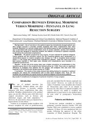
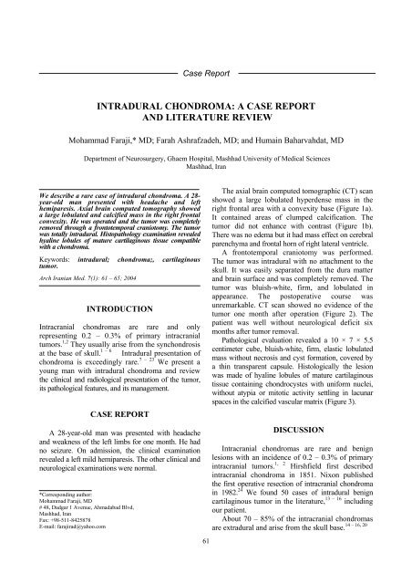
Case ReportINTRADURAL CHONDROMA: A CASE REPORTAND LITERATURE REVIEWMohammad Faraji,* MD; Farah Ashrafzadeh, MD; <strong>and</strong> Humain Baharvahdat, MDDepartment of Neurosurgery, Ghaem Hospital, Mashhad University of Medical SciencesMashhad, IranWe describe a rare <strong>case</strong> of <strong>intradural</strong> <strong>chondroma</strong>. A 28-year-old man presented with headache <strong>and</strong> lefthemiparesis. Axial brain computed tomography showeda large lobulated <strong>and</strong> calcified mass in the right frontalconvexity. He was operated <strong>and</strong> the tumor was completelyremoved through a frontotemporal craniotomy. The tumorwas totally <strong>intradural</strong>. Histopathology examination revealedhyaline lobules of mature cartilaginous tissue compatiblewith a <strong>chondroma</strong>.Keywords: <strong>intradural</strong>; <strong>chondroma</strong>;, cartilaginoustumor.Arch Iranian Med. 7(1): 61 – 65; 2004INTRODUCTIONIntracranial <strong>chondroma</strong>s are rare <strong>and</strong> onlyrepresenting 0.2 – 0.3% of primary intracranialtumors. 1,2 They usually arise from the synchondrosisat the base of skull. 1 – 6 Intradural presentation of<strong>chondroma</strong> is exceedingly rare. 7 – 23 We present ayoung man with <strong>intradural</strong> <strong>chondroma</strong> <strong>and</strong> <strong>review</strong>the clinical <strong>and</strong> radiological presentation of the tumor,its pathological features, <strong>and</strong> its management.CASE REPORTA 28-year-old man was presented with headache<strong>and</strong> weakness of the left limbs for one month. He hadno seizure. On admission, the clinical examinationrevealed a left mild hemiparesis. The other clinical <strong>and</strong>neurological examinations were normal.*Corresponding author:Mohammad Faraji, MD# 48, Dadgar 1 Avenue, Ahmadabad Blvd,Mashhad, IranFax: +98-511-8425878E-mail: farajirad@yahoo.com61The axial brain computed tomographic (CT) scanshowed a large lobulated hyperdense mass in theright frontal area with a convexity base (Figure 1a).It contained areas of clumped calcification. Thetumor did not enhance with contrast (Figure 1b).There was no edema but it had mass effect on cerebralparenchyma <strong>and</strong> frontal horn of right lateral ventricle.A frontotemporal craniotomy was performed.The tumor was <strong>intradural</strong> with no attachment to theskull. It was easily separated from the dura matter<strong>and</strong> brain surface <strong>and</strong> was completely removed. Thetumor was bluish-white, firm, <strong>and</strong> lobulated inappearance. The postoperative course wasunremarkable. CT scan showed no evidence of thetumor one month after operation (Figure 2). Thepatient was well without neurological deficit sixmonths after tumor removal.Pathological evaluation revealed a 10 × 7 × 5.5centimeter cube, bluish-white, firm, elastic lobulatedmass without necrosis <strong>and</strong> cyst formation, covered bya thin transparent capsule. Histologically the lesionwas made of hyaline lobules of mature cartilaginoustissue containing chondrocystes with uniform nuclei,without atypia or mitotic activity settling in lacunarspaces in the calcified vascular matrix (Figure 3).DISCUSSIONIntracranial <strong>chondroma</strong>s are rare <strong>and</strong> benignlesions with an incidence of 0.2 – 0.3% of primaryintracranial tumors. 1, 2 Hirshfield first describedintracranial <strong>chondroma</strong> in 1851. Nixon publishedthe first operative resection of intracranial <strong>chondroma</strong>in 1982. 24 We found 50 <strong>case</strong>s of <strong>intradural</strong> benigncartilaginous tumor in the <strong>literature</strong>, 13 – 16 includingour patient.About 70 – 85% of the intracranial <strong>chondroma</strong>s14 – 16, 20are extradural <strong>and</strong> arise from the skull base.
Intradural <strong>chondroma</strong>Figure 1. The axial brain CT scan showed a large lobulated,calcified, hyperdense mass lesion in the right frontal area (a)which was not enhanced with contrast medium (b). No edemawas visible in either of the CT scans.Skull base <strong>chondroma</strong>s were <strong>report</strong>ed inassociation with Ollier’s disease 25 – 27 <strong>and</strong> Maffuci’ssyndrome. 24 Malignant change was <strong>report</strong>ed in anintracranial <strong>chondroma</strong> in a patient with Maffuci’ssyndrome. 28 Approximately 15 – 30% of intracranial<strong>chondroma</strong>s do not arise from the skull base <strong>and</strong> are13, 15, 16, 20<strong>intradural</strong>. They were <strong>report</strong>ed in thechoroids plexus/intraventricular, 29, 30 sellar <strong>and</strong>parasellar, 31,32 intracerebral (pons), 33 <strong>and</strong> attached to11, 16, 18, 20,the dura matter (convexity or falx) (Table 1).23Attachment to dura, particularly overcerebral62
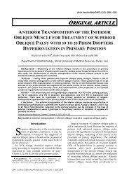
AIM, 7(1), 2004Figure 3. a) Histologically, the photomicrograph revealed tumor capsule with lobules of mature cartilaginous tissue (H <strong>and</strong> E ×10). b) The connective tissue without meningeal cells between lobules of cartilaginous tissue (H <strong>and</strong> E × 10). c). The maturecartilaginous tissue containing chondrocytes without atypia <strong>and</strong> mitosis settled in the lacunae (H <strong>and</strong> E × 20).Figure 2. There is no evidence of tumor in CT scan taken onemonth after operation.convexities included 70% of <strong>intradural</strong> <strong>chondroma</strong><strong>and</strong> 15% of intracranial <strong>chondroma</strong>. 15The origin of intracranial <strong>chondroma</strong> is notknown for sure. Many theories have been suggested.The skull base <strong>chondroma</strong>s have been believed tooriginate from embryonic rests of chondrogeniccells along baseline synchondrosis. 1,4,6,15,26,34 It isthought that <strong>intradural</strong> <strong>chondroma</strong>s develop fromheterotropic chondrocystes or metaplasia of othernormal tissue, including meningeal fibroblasts orperivascular mesenchymal tissue. 13 – 15 Intracranial<strong>chondroma</strong>s are usually seen in females in the 2 nd to5 th decades of life. 1, 35 In <strong>intradural</strong> <strong>chondroma</strong>, themean age is 29 years at presentation, with slightmale predominance (63.5% versus 37.5%) (Tablemay show evidence of hyperostosis of the internaltable of the skull, increased intracranial pressure,<strong>and</strong> areas of calcification. 16,20 The convexity<strong>chondroma</strong>s may have stippled, flocculent, <strong>and</strong> ringcalcification. 14 However, falcine <strong>chondroma</strong>s do notusually show calcification. 20 According to Lacerte etal, 15 the <strong>intradural</strong> <strong>chondroma</strong>s have two distinct CTscan presentations. The type 1, named classical, ismore common <strong>and</strong> reveals mixed density withminimal or moderate enhancement, whereas type 2is less frequent <strong>and</strong> has a central hypodense area,which is composed of a cystic degeneration 11, 14 orof a very loose-texture connective tissue withoutnecrosis in pathological evaluation. 15 Tanohota et alstress that enhancement of <strong>chondroma</strong>s increasesafter 30 minutes of contrast injection. 20The MRI features have been <strong>report</strong>ed in a few<strong>case</strong>s of intracranial <strong>chondroma</strong>s. The tumor showsheterogenous signal intensity with more hypodenseon T1 spin echo scan <strong>and</strong> iso- to hyper-intense on2). Because of the noninvasive <strong>and</strong> slow-growingnature of <strong>intradural</strong> <strong>chondroma</strong>s the patients oftenpresent with a long-st<strong>and</strong>ing history of headache<strong>and</strong> symptoms of increased intracranial pressure.Patients may have signs <strong>and</strong> symptoms related tocompression of adjacent structure, like seizure,personality changes, <strong>and</strong> hemiparesis. 14, 15, 20 Despitepaucity of symptoms, the intracranial <strong>chondroma</strong>are usually very large (mean diameter, 6 cm <strong>and</strong>mean weight, 170 grams) at diagnosis which may beexplained by their slow-growing nature <strong>and</strong> theircommon location in frontoparietal area, 15 thereforeas part of a work-up for other reasons. 14The radiological appearance of <strong>intradural</strong><strong>chondroma</strong>s is fairly typical. On skull X-ray,<strong>chondroma</strong>s, particularly over cerebral convexities,T2 spin echo scan. The tumor enhances minimallyto moderately following administration of contrast. 15In type 2, the T2 spin echo scan shows a peripheralheterogenous hypointense area <strong>and</strong> a welldemarcatedhyperintense central area. 14, 15 Theformer may enhance with contrast <strong>and</strong> show ringTable 1. Reported location of <strong>intradural</strong> <strong>chondroma</strong>.*Location No. of <strong>case</strong>s (%)Menings 37 (74 %)Convexity 22 (44 %)1. Frontoparietal 20 (40 %)2. Others 2 (4 %)Falx 15 (30 %)Intracerebral 7 (14 %)Choroid plexus / intraventricular 6 (12 %)Total 50 (100 %)* Based on references 16, 19, <strong>and</strong> 21.enhancement on T1 spin echo scan. 9, 14, 15, 18 Thelatter did not enhance with contrast 13 <strong>and</strong> its signalintensity in T2 spin echo scan may be explained63
AIM, 7(1), 200413. Holthouse DJ, Robbins PD, knuckey NK. Solitary <strong>intradural</strong>fibro<strong>chondroma</strong> in a 16-year-old boy. J Clin Neuroscie. 1999;6: 355 – 7.14. Khosrovi H, Sadrolhefazi A, el-Kadi H, Bloomfield SM,Scochet SS. Intradural convexity <strong>chondroma</strong>: a <strong>case</strong> <strong>report</strong> <strong>and</strong><strong>review</strong> of diagnostic features. W V Med J. 2000; 96: 612 – 6.15. Lacerte D, Gagne F, Copty M. Intracranial <strong>chondroma</strong>:<strong>report</strong> of two <strong>case</strong>s <strong>and</strong> <strong>review</strong> of the <strong>literature</strong>. Can J NeurtolSci. 1996; 23: 132 – 7.16. Mapstone TB, Wongmongkolrit T, Roessman U, RatchesonRA. Intradural <strong>chondroma</strong>: a <strong>case</strong> <strong>report</strong> <strong>and</strong> <strong>review</strong> of the<strong>literature</strong>. Neurosurgery. 1983; 12: 111 – 4.17. Matz S, Israeli Y, Shlit MN, Cohen ML. Computedtomography in intracranial supratentorial osteo<strong>chondroma</strong>. JComput Assist Tomogr. 1981; 5: 109 – 15.18. Nakazawa T, Inoue T, Suzuki F, Nakasu S, H<strong>and</strong>a J.Solitary intracranial <strong>chondroma</strong> of the convexity dura: <strong>case</strong><strong>report</strong>. Surg Neurol. 1993; 40: 495 – 8.19. Ozgen T, Pamir MN, Akalan N, Bertan V, Onol B.Intracranial solitary <strong>chondroma</strong>. Case <strong>report</strong>. J Neurosurg.1984; 61: 399 – 401.20. Sebbag M, Schmidt V, Leboucq N, Bitoun J, Castan PH,Frerebeau PH. Chondrome dure-mérien; a propos d’un cas etrevue de la <strong>literature</strong> [in French]. J Radiol. 1990. 71: 495 – 8.21. Siris JH, Angrist A. Chondroblastic meningioma. Am JSurg. 1942; 57: 162 – 7.22. Wu WQ, Lapi A. Primary nonskeletal intracranialcartilaginous neoplasms: <strong>report</strong> of <strong>chondroma</strong> <strong>and</strong> amesenchymal chondrosarcoma. J Neurol Neurosurg Psychiatry.1970; 33: 469 – 75.23. Yang PJ, Seeger JF, Carmody RF, Fleischer AS.Chondroma of falx: CT findings. J Comput Assist Tomogr.1986; 10: 1075 – 6.24. Chakrabortty S, Tamaki N, Kondoh T, Kojima N,Kamikawa H, Matsumoto S. Maffucci’s syndrome associatedwith intracranial en<strong>chondroma</strong> <strong>and</strong> aneurysm: <strong>case</strong> <strong>report</strong>. SurgNeurol. 1991; 36: 216 – 20.25. Dany A, Vidal J, Dumas M, Ravon R, Bokor J.Cerebellopontine angle <strong>chondroma</strong>. A <strong>report</strong> of one personal<strong>case</strong> (author's transl) [in French]. Neurochirurgie. 1980; 26:355 – 7.26. Dutton J. Intracranial solitary <strong>chondroma</strong>. Case <strong>report</strong>. JNeurosurg. 1978; 49: 460 – 3.27. Traflet RF, Babaria AR, Barolat G, Doan HT, Gonzalez C,Mishkin MM. Intracranial <strong>chondroma</strong> in a patient with Ollier’sdisease. J Neurosurg. 1989; 70: 274 – 6.28. Bushe KA, Naumann M, Warmuth-Metz MM,Meixensberger J, Muller J. Maffucci’s syndrome with bilateralcartilaginous tumors of cerebellopontine angle. Neurosurgery.1990; 27: 625 – 8.29. Salazar J, Vaquero J, Ar<strong>and</strong>a IF, Menendez J, Jimenez MD,Bravo G. Choroid plexus papilloma with <strong>chondroma</strong>: <strong>case</strong><strong>report</strong>. Neurosurgry. 1986; 18: 781 – 3.30. Valdueza JM, Freckmann N, Herrmann HD.Chondromatosis of the choroid plexus: <strong>case</strong> <strong>report</strong>.Neurosurgery. 1990; 27: 291 – 4.31. Furui T, Iwata K, Yamamoto H, Murakami A. A <strong>case</strong> ofintracranial <strong>chondroma</strong> presenting with hemorrhage [inJapanese]. No Shinkei Geka. 1990; 18: 543 – 6.65
Intradural <strong>chondroma</strong>32. Munemitsu H, Matsuda M, Hirai O, Fukumitsu T,Kanamura J. Intrasellar <strong>chondroma</strong>. Neurol Med Chir (Tokyo).1981; 21: 775 – 80.33. Ishii R, Sato S, Ueki K, Oyake Y. Myxoosteo<strong>chondroma</strong> inthe pons. Case <strong>report</strong>. J Neurosurg. 1974; 41: 240 – 3.34. List CF. Osteo<strong>chondroma</strong> arising from the base of the skull.Surg Gynecol Obstet. 1943; 76: 480 – 92.35. Doran SE, Gebarski SS, Hoff JT. Tumors of skull. In:Youmans JR, ed. Neurological Surgery: A comprehensiveReference Guide to the Diagnosis <strong>and</strong> Management ofNeurosurgical Problems. 4th ed. Vol 3. Philadelphia: WBSaunders; 1996: 2998 – 3023.36. Ahyai A, Spoerri O. Intracerebral <strong>chondroma</strong>. Surg Neurol.1979; 11: 431 – 3.37. Kretzschmar HA, Eggert HR, Beck U, Furmaier R.Intracranial <strong>chondroma</strong>: <strong>case</strong> <strong>report</strong>. Surg Neurol. 1989; 32:121 – 5.38. Osburn AG. Diagnostic Neuroradiology. St Louis: Mosby;1994.39. McKeiver PE, Blairas M, Nelson JS. Tumors: application oflight microscoic methods. In: Gracia JH, ed. Neuropatholgy,the Diagnostic Approach. St Louis: Mosby; 1997: 97 – 192.■■66