Hashimoto Encephalopathy and Down Syndrome - AMA Publishing ...
Hashimoto Encephalopathy and Down Syndrome - AMA Publishing ...
Hashimoto Encephalopathy and Down Syndrome - AMA Publishing ...
You also want an ePaper? Increase the reach of your titles
YUMPU automatically turns print PDFs into web optimized ePapers that Google loves.
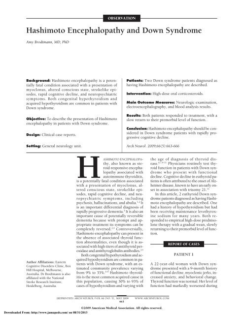
OBSERVATION<strong>Hashimoto</strong> <strong>Encephalopathy</strong> <strong>and</strong> <strong>Down</strong> <strong>Syndrome</strong>Amy Brodtmann, MD, PhDBackground: <strong>Hashimoto</strong> encephalopathy is a potentiallyfatal condition associated with a presentation ofmyoclonus, altered conscious state, strokelike episodes,rapid cognitive decline, <strong>and</strong> neuropsychiatricsymptoms. Both congenital hypothyroidism <strong>and</strong>acquired hypothyroidism are common in patients with<strong>Down</strong> syndrome.Objective: To describe the presentation of <strong>Hashimoto</strong>encephalopathy in patients with <strong>Down</strong> syndrome.Design: Clinical case reports.Setting: General neurology unit.Patients: Two <strong>Down</strong> syndrome patients diagnosed ashaving <strong>Hashimoto</strong> encephalopathy are described.Intervention: High-dose oral corticosteroids.Main Outcome Measures: Neurologic examination,electroencephalography, <strong>and</strong> blood analysis results.Results: Both patients responded to treatment, with aslow return to their premorbid level of function.Conclusion: <strong>Hashimoto</strong> encephalopathy should be consideredin <strong>Down</strong> syndrome patients with rapidly progressivecognitive decline.Arch Neurol. 2009;66(5):663-666Author Affiliations: EasternCognitive Disorders Clinic, BoxHill Hospital, Melbourne,Australia. Dr Brodtmann is alsoaffiliated with the NationalStroke Research Institute,Heidelberg, Australia.HASHIMOTO ENCEPHALOPAthy,also known as steroid-responsiveencephalopathyassociated withautoimmune thyroiditis,is a potentially fatal condition associatedwith a presentation of myoclonus, alteredconscious state, strokelike episodes,rapid cognitive decline, <strong>and</strong> neuropsychiatricsymptoms, includingpsychosis, hallucinations, <strong>and</strong> abulia. 1,2 Itis an important differential diagnosis ofrapidly progressive dementia. 3 It is also animportant cause of potentially reversibledementia because with prompt <strong>and</strong> appropriatetreatment its symptoms can becompletely reversed. 2,4 Controversially,<strong>Hashimoto</strong> encephalopathy can present inthe absence of associated thyroid functionabnormalities, even though it is associatedwith high titers of antithyroid peroxidase<strong>and</strong> antithyroglobulin antibodies. 2,5Both congenital hypothyroidism <strong>and</strong> acquiredhypothyroidism are common in patientswith <strong>Down</strong> syndrome, with an estimatedcommunity prevalence varyingfrom 9% to 35%. 6-9 <strong>Hashimoto</strong> thyroiditisis the most common acquired cause inthis population, causing 30% to 93% ofcases of hypothyroidism <strong>and</strong> varying withthe age of diagnosis of thyroid disease.6,7,9-11 Physicians routinely test thyroidfunction in patients with <strong>Down</strong> syndromewho present with functionaldecline. Cognitive decline in euthyroid patientsis often attributed to the onset of Alzheimerdisease, known to have an early onsetin association with trisomy 21. 12In this article, 2 euthyroid <strong>Down</strong> syndromepatients diagnosed as having <strong>Hashimoto</strong>encephalopathy are described. Onehad a history of hypothyroidism but hadbeen receiving maintenance levothyroxinesodium for many years. Both respondedto empirical high-dose prednisolonetherapy with a gradual wean, slowlyreturning to their premorbid level of function.REPORT OF CASESPATIENT 1A 22-year-old woman with <strong>Down</strong> syndromepresented with a 9-month historyof functional decline, myoclonic jerks, increasedanxiety, <strong>and</strong> behavioral change.Thyroid function was normal. Her level offunction had markedly worsened during(REPRINTED) ARCH NEUROL / VOL 66 (NO. 5), MAY 2009663WWW.ARCHNEUROL.COM©2009 American Medical Association. All rights reserved.<strong>Down</strong>loaded From: http://www.jamapeds.com/ on 08/31/2013
On Presentation 10 Months Following Diagnosis 1 Year Following DiagnosisFigure 1. Serial electroencephalograms from patient 1. A double banana montage is shown; the same filter settings were used for each for comparison. Note theimprovement in background rhythm from a diffuse encephalopathic state to normal rhythm.ABPatient 1 Patient 2Figure 2. Brain images of patients 1 <strong>and</strong> 2. T1 (A) <strong>and</strong> T2 (B) images areshown. Patient 1 underwent axial magnetic resonance imaging at the level ofthe basal ganglia. There was evidence of brachycephaly but no atrophy.Patient 2 underwent noncontrast axial computed tomography at the level ofthe basal ganglia (top). Two sections (bottom) shown demonstratewidespread atrophy. Note the incidental basal ganglia calcification.the preceding 2 months, with increasing weakness, reducedverbal output, increased somnolence, possible visualhallucinations, <strong>and</strong> leg jerks. During the 4 weeks beforehospital admission she had required assistance withall of her activities of daily living. She had presented tothe emergency department 1 month before admission withaltered thinking <strong>and</strong> behavioral change. A possible underlyingpsychosis was considered. Risperidone, 2 mg/d,was administered <strong>and</strong> later venlafaxine hydrochloride,150 mg, was added in view of a possible diagnosis of depression.Despite these therapies, she presented 1 monthlater for investigation of worsening level of function <strong>and</strong>increasing jerks.Vital signs were normal. Neurologic examination revealedmarked bradyphrenia <strong>and</strong> abulia, with a positiveglabellar tap <strong>and</strong> palmomental reflexes but no grasp reflex.The patient was poorly cooperative, <strong>and</strong> no highermental function testing could be performed. The resultsof cranial nerve examination were normal. There wasevidence of mild hypertonia <strong>and</strong> hyperreflexia in herlower limbs. Limb power, sensation, <strong>and</strong> coordinationwere intact. Frequent bilateral arm <strong>and</strong>, to a lesser extent,leg myoclonic jerks were noted throughout theexamination.Admission electroencephalography (EEG) revealed abackground of admixed polymorphic <strong>and</strong> , consistentwith a diffuse encephalopathic process (Figure 1).Magnetic resonance imaging of the brain revealed brachycephalybut no significant cortical or white matter lesions(Figure 2). The results of basic blood analyses werenormal, including a normal vitamin B 12 level (590 pg/mL; reference range, 187-1059 pg/mL [to convert to picomolesper liter, multiply by 0.7378]), <strong>and</strong> she was euthyroid(thyrotropin, 2.90 mIU/mL; free thyroxine, 1.0ng/dL [to convert to picomoles per liter, multiply by12.871]; free triiodothyronine, 240 pg/dL [to convert topicomoles per liter, multiply by 0.0154]; all within thereference range for our laboratory). Lumbar puncture revealeda bl<strong>and</strong> cerebrospinal fluid: lymphocytes, 1; redblood cells, 0; protein, 0.02 g/dL; <strong>and</strong> glucose, 59 mg/dL. The results of the cerebrospinal fluid herpes simplexvirus polymerase chain reaction were negative. Test-(REPRINTED) ARCH NEUROL / VOL 66 (NO. 5), MAY 2009664WWW.ARCHNEUROL.COM©2009 American Medical Association. All rights reserved.<strong>Down</strong>loaded From: http://www.jamapeds.com/ on 08/31/2013
ing for autoantibodies revealed an antinuclear antibodytiter of 1:640. A diagnosis of <strong>Hashimoto</strong> encephalopathywas considered: her antithyroid peroxidase antibodylevel was 132 IU/mL (reference range, 35 IU/mL), <strong>and</strong> her antithyroglobulin antibody level was 976IU/mL (reference range, 40 IU/mL). A diagnosis of<strong>Hashimoto</strong> encephalopathy was made, <strong>and</strong> treatmentcommenced.She was administered oral prednisolone, 75 mg/d (1.5mg/kg), without prior intravenous dosing. Her myoclonusimproved during the next 24 hours, <strong>and</strong> staff <strong>and</strong>family noted increased alertness <strong>and</strong> cooperation duringthe 72 hours after treatment. Rapid functional gainswere made on the ward, <strong>and</strong> 14 days after commencingoral prednisolone therapy she was discharged home witha slowly weaning prednisolone regimen. Slow incrementalgains in her cognition <strong>and</strong> function were made duringthe next 7 months. Her EEG normalized during a periodof 12 months (Figure 1) as did her autoantibody levels(antithyroid peroxidase antibody, 10 IU/mL; antithyroglobulinantibody, 31 IU/mL). Low-dose maintenanceprednisolone therapy had been given during thistime.On review 8 months after initiation of corticosteroidtherapy, she was fully oriented. Results of her neurologicexamination were normal, with no evidence of hypertonia,hyperreflexia, or myoclonus. She had returnedto her normal level of function, including a returnto her workplace 5 days a week.PATIENT 2A 46-year-old woman with <strong>Down</strong> syndrome was admittedwith a 6-week history of increasing drowsiness, behavioralchange, <strong>and</strong> twitching. Her history included hypothyroidism<strong>and</strong> probable early-onset Alzheimer disease,diagnosed on a background of 1 to 2 years of gradual functional<strong>and</strong> cognitive decline that necessitated transfer toa nursing home. Before her recent illness she was able totransfer from bed to chair <strong>and</strong> chair to walking frame <strong>and</strong>walk short distances with a gutter-frame with assistance.She was incontinent of urine. Her medications werevalproate sodium, 200 mg 3 times daily, <strong>and</strong> thyroxine,100 µg/d.During the preceding 6 weeks, her mobility had markedlydeclined. She became dependent in all of her activitiesof daily living, incontinent of feces <strong>and</strong> urine, <strong>and</strong>wheelchair bound. She had ceased to verbalize or evenattempt to communicate with staff. Her family reportedthat she always seemed confused. In the 2 weeks beforehospital admission, increasing drowsiness <strong>and</strong> wholebodytwitching had developed, which occurred for mostof her waking hours. She developed some right-sidedweakness <strong>and</strong> difficulty with swallowing <strong>and</strong> was notedto choke on her foods. Admission was arranged for treatmentof nonconvulsive status epilepticus <strong>and</strong> considerationfor insertion of a percutaneous endoscopic gastrostomytube. Nasogastric tube feeding was commenced onthe ward.On admission, her blood pressure was 120/60 mm Hg<strong>and</strong> her heart rate was 90/min. She was afebrile, with aGlasgow coma scale score of 9. The patient was intermittentlydrowsy <strong>and</strong> uncommunicative. She was unableto follow simple comm<strong>and</strong>s. She appeared to be movingher left side more than the right. Higher mentalfunction testing was not performed. The results of cranialnerve examination were within normal limits. Generalizedlimb hypertonia <strong>and</strong> hyperreflexia were noted.Her plantar reflexes were extensor, <strong>and</strong> intermittent myoclonuswas seen in all limbs.The results of basic blood analyses were normal, includinga C-reactive protein level less than 0.21 mg/L (toconvert to nanomoles per liter, multiply by 9.524) <strong>and</strong> avitamin B 12 level of 689 pg/mL. The results of thyroid functiontesting were consistent with adequate thyroxine replacement(thyrotropin, 3.39 mIU/L; thyroxine, 1.3 µg/dL[to convert to nanomoles per liter, multiply by 12.871]).Her EEG revealed a background of high-amplitude polymorphic with high-amplitude triphasic waves. Frequentmyoclonic jerks in the absence of associated corticalactivity were noted. Computed tomography of thebrain revealed generalized atrophy consistent with herearlier diagnosis of Alzheimer disease <strong>and</strong> bilateral basalganglia calcification (Figure 2). Autoantibody testing revealedan antithyroid peroxidase antibody titer of 87IU/mL <strong>and</strong> a normal antithyroglobulin antibody level(20 IU/mL; reference range, 40 IU/mL). A diagnosisof probable <strong>Hashimoto</strong> encephalopathy was made, <strong>and</strong>no further serologic or cerebrospinal fluid studies wereperformed.Intravenous methylprednisolone, 500 mg, was givenfor 3 days. Within 24 hours of treatment initiation herjerking stopped, with only some small twitches observed.Increased alertness was noted 72 hours after treatment.Five days after commencement of corticosteroid,her nasogastric tube was removed <strong>and</strong> oral intake commenced,with rapid return to her premorbid diet. Her familymembers commented on increased interaction <strong>and</strong> cooperation.She was started on a trial of reconditioningphysiotherapy but was still transferring with the 2-personassist or hoist transfer. Eleven days after commencementof treatment, she was discharged back to her nursinghome with a prescription for 50 mg/d of oralprednisolone with a plan to gradually wean this dose. Fecalcontinence was regained.On review 3 months after discharge, she had returnedto her normal level of communication <strong>and</strong> oralintake. She was alert but not obeying simple verbal comm<strong>and</strong>s.Neurologic examination was limited but revealedno evidence of myoclonus, hypertonia, or hyperreflexia.Plantar responses were flexor. Her thyroidantibody level had returned to the reference range.COMMENTThe association of <strong>Hashimoto</strong> encephalopathy with <strong>Down</strong>syndrome has not been previously described. Two patientswith <strong>Down</strong> syndrome are described, both with aclinical presentation of <strong>Hashimoto</strong> encephalopathy diagnosedon the basis of a history of myoclonus, rapid cognitivedecline, abnormal EEG, <strong>and</strong> positive antithyroidperoxidase antibody titers. Both patients were euthyroid<strong>and</strong> had a rapid response to corticosteroid therapy,(REPRINTED) ARCH NEUROL / VOL 66 (NO. 5), MAY 2009665WWW.ARCHNEUROL.COM©2009 American Medical Association. All rights reserved.<strong>Down</strong>loaded From: http://www.jamapeds.com/ on 08/31/2013
with early resolution of myoclonus <strong>and</strong> slow return totheir premorbid level of function. In the patient followedup with serial EEGs, this was associated with normalizationof the recording from a background of <strong>and</strong> to normal reactive rhythm.Alzheimer disease is often considered in <strong>Down</strong> syndromepatients who present with functional decline, withpatients presenting in their fourth or fifth decades of life.High levels of -amyloid deposition by the fourth decadein this patient population are well described. 13,14 A numberof factors influence the age at onset of cognitive decline,including susceptibility genotypes, atypical karyotypes,<strong>and</strong> estrogen deficiency. 14 However, alternativediagnoses, such as thyroid dysfunction, should be consideredin younger patients <strong>and</strong> those who present with a rapidonset of functional decline. Alzheimer disease usually hasan insidious onset <strong>and</strong> gradual progression <strong>and</strong> is not usuallyassociated with altered conscious state, early seizures,focal neurologic abnormalities, <strong>and</strong> myoclonus.Although rare, the association of <strong>Hashimoto</strong> encephalopathyin patients with <strong>Down</strong> syndrome is not surprisinggiven the high incidence of autoimmune thyroid diseasein this population. Even in the setting of normalthyroid function, thyroid antibody levels should be measured<strong>and</strong> the diagnosis of <strong>Hashimoto</strong> encephalopathyconsidered in patients with <strong>Down</strong> syndrome who presentwith rapid cognitive decline, particularly in associationwith myoclonus <strong>and</strong> an abnormal EEG result.Accepted for Publication: July 16, 2008.Correspondence: Amy Brodtmann, MD, PhD, NationalStroke Research Institute, Level 1, Neurosciences Building,Austin Health, Heidelberg 3084, Australia (amyb@alphalink.com.au).Financial Disclosure: None reported.Funding/Support: Dr Brodtmann is a current holder ofa National Health <strong>and</strong> Medical Research Council AustralianResearch Training Fellowship (part-time).REFERENCES1. Brain L, Jellinek EH, Ball K. <strong>Hashimoto</strong>’s disease <strong>and</strong> encephalopathy. Lancet.1966;2(7462):512-514.2. Castillo P, Woodruff B, Caselli R, et al. Steroid-responsive encephalopathy associatedwith autoimmune thyroiditis. Arch Neurol. 2006;63(2):197-202.3. Ferracci F, Carnevale A. The neurological disorder associated with thyroidautoimmunity. J Neurol. 2006;253(8):975-984.4. Marshall GA, Doyle JJ. Long-term treatment of <strong>Hashimoto</strong>’s encephalopathy. J NeuropsychiatryClin Neurosci. 2006;18(1):14-20.5. Ferracci F, Bertiato G, Moretto G. <strong>Hashimoto</strong>’s encephalopathy: epidemiologicdata <strong>and</strong> pathogenetic considerations. J Neurol Sci. 2004;217(2):165-168.6. Loudon MM, Day RE, Duke EM. Thyroid dysfunction in <strong>Down</strong>’s syndrome. ArchDis Child. 1985;60(12):1149-1151.7. Lobo H, Khan M, Tew J. Community study of hypothyroidism in <strong>Down</strong>’s syndrome.Br Med J. 1980;280(6226):1253.8. Tüysüz B, Beker DB. Thyroid dysfunction in children with <strong>Down</strong>’s syndrome. ActaPaediatr. 2001;90(12):1389-1393.9. Karlsson B, Gustafsson J, Hedov G, Ivarsson SA, Anneren G. Thyroid dysfunctionin <strong>Down</strong>’s syndrome: relation to age <strong>and</strong> thyroid autoimmunity. Arch DisChild. 1998;79(3):242-245.10. Noble SE, Leyl<strong>and</strong> K, Findlay CA, et al. School based screening for hypothyroidismin <strong>Down</strong>’s syndrome by dried blood spot TSH measurement. Arch Dis Child.2000;82(1):27-31.11. Pueschel SM, Jackson IM, Giesswein P, Dean MK, Pezzullo JC. Thyroid functionin <strong>Down</strong> syndrome. Res Dev Disabil. 1991;12(3):287-296.12. Coppus A, Evenhuis H, Verberne GJ, et al. Dementia <strong>and</strong> mortality in personswith <strong>Down</strong>’s syndrome. J Intellect Disabil Res. 2006;50(pt 10):768-777.13. Devenny DA, Wegiel J, Schupf N, et al. Dementia of the Alzheimer’s type <strong>and</strong>accelerated aging in <strong>Down</strong> syndrome. Sci Aging Knowledge Environ. 2005;2005(14):dn1.14. Schupf N, Sergievsky GH. Genetic <strong>and</strong> host factors for dementia in <strong>Down</strong>’ssyndrome. Br J Psychiatry. 2002;180:405-410.AnnouncementVisit www.archneurol.com. As an individual subscriber,you may elect to be contacted when a specificarticle is cited. Receive an e-mail alert when the articleyou are viewing is cited by any of the journals hosted byHighWire. You will be asked to enter the volume, issue,<strong>and</strong> page number of the article you wish to track. Youre-mail address will be shared with other journals in thisfeature; other journals’ privacy policies may differ fromJ<strong>AMA</strong> & Archives Journals. You may also sign up to receivean e-mail alert when articles on particular topicsare published.(REPRINTED) ARCH NEUROL / VOL 66 (NO. 5), MAY 2009666WWW.ARCHNEUROL.COM©2009 American Medical Association. All rights reserved.<strong>Down</strong>loaded From: http://www.jamapeds.com/ on 08/31/2013


