Allyl type radical formation in X-irradiated glutarimide crystals ...
Allyl type radical formation in X-irradiated glutarimide crystals ...
Allyl type radical formation in X-irradiated glutarimide crystals ...
Create successful ePaper yourself
Turn your PDF publications into a flip-book with our unique Google optimized e-Paper software.
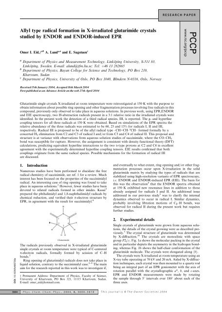
RESEARCH PAPER<strong>Allyl</strong> <strong>type</strong> <strong>radical</strong> <strong>formation</strong> <strong>in</strong> X-<strong>irradiated</strong> <strong>glutarimide</strong> <strong>crystals</strong>studied by ENDOR and ENDOR-<strong>in</strong>duced EPROmer I. Eid,y ab A. Lund* a and E. Sagstuen cPCCPwww.rsc.org/pccpa Department of Physics and Measurement Technology, L<strong>in</strong>köp<strong>in</strong>g University, S-511 83,L<strong>in</strong>köp<strong>in</strong>g, Sweden. E-mail: alund@ifm.liu.se; Tel: þ46 13 282665b Department of Physics, Bayan College for Science and Technology, PO Box 210,Khartoum, Sudanc Department of Physics, University of Oslo, PO Box 1048, Bl<strong>in</strong>dern N-0316, Oslo, NorwayReceived15th January 2004, Accepted18th March 2004F|rst published as an Advance Article on the web 27th April 2004Glutarimide s<strong>in</strong>gle <strong>crystals</strong> X-<strong>irradiated</strong> at room temperature were re<strong>in</strong>vestigated at 150 K with the purpose toobta<strong>in</strong> <strong>in</strong><strong>formation</strong> about possible r<strong>in</strong>g open<strong>in</strong>g and other fragmentation processes <strong>in</strong>volv<strong>in</strong>g free <strong>radical</strong>s <strong>in</strong> thiscompound, previously only observed to take place <strong>in</strong> aqueous solutions. In previous work, us<strong>in</strong>g EPR,ENDORand EIE spectroscopy, two H-abstraction <strong>radical</strong>s present <strong>in</strong> a 3:1 relative ratio <strong>in</strong> the <strong>irradiated</strong> <strong>crystals</strong> wereidentified. In the present work the detection of a third <strong>radical</strong> species, III, is reported. The g- and hyperf<strong>in</strong>ecoupl<strong>in</strong>g tensors for all three <strong>radical</strong>s at 150 K were obta<strong>in</strong>ed. Based on simulations of the EPR spectra therelative abundance of the three <strong>radical</strong>s was estimated to be 60, 25 and 15% for <strong>radical</strong>s I, II and III,respectively. Radical III is proposed to be of the allyl <strong>radical</strong> <strong>type</strong> –CH=CH– CH– formed formally by aconcerted H 2 elim<strong>in</strong>ation from C2 and C3 of <strong>radical</strong> I and/or from C3 and C4 of <strong>radical</strong> II. This proposal andstructure is at variance with observations from aqueous solution studies of succ<strong>in</strong>imide, where the CO–CH 2bond was susceptible for rupture. However, the assignment is consistent with density functional theory (DFT)calculations, predict<strong>in</strong>g equivalent hyperf<strong>in</strong>e <strong>in</strong>teractions to the two a-<strong>type</strong> protons at C2 and C4 <strong>in</strong> excellentagreement with the experimentally determ<strong>in</strong>ed hyperf<strong>in</strong>e coupl<strong>in</strong>g tensors. EIE results confirmed that bothcoupl<strong>in</strong>gs orig<strong>in</strong>ate from the same <strong>radical</strong> species. Possible mechanisms for the <strong>formation</strong> of <strong>radical</strong> IIIare discussed.DOI: 10.1039/b400730a1. IntroductionNumerous studies have been performed to elucidate the free<strong>radical</strong> chemistry of succ<strong>in</strong>imide, see ref. 1 for a review. Much<strong>in</strong>terest has been focused on the properties of the succ<strong>in</strong>imidyl<strong>radical</strong>. An <strong>in</strong>terest<strong>in</strong>g case of r<strong>in</strong>g open<strong>in</strong>g was found to takeplace <strong>in</strong> aqueous solutions. 2 However, fewer studies have beendevoted to related <strong>radical</strong>s formed <strong>in</strong> other imides. Kasai 3prepared the phthalimidyl and 1,8-naphthalimidyl <strong>radical</strong>s bychemical reduction, and verified their p-electron structure byEPR, <strong>in</strong> agreement with the result for succ<strong>in</strong>imidyl. 4The <strong>radical</strong>s previously observed <strong>in</strong> X-<strong>irradiated</strong> <strong>glutarimide</strong>s<strong>in</strong>gle <strong>crystals</strong> at room temperature were typical of C-centeredp-electron <strong>radical</strong>s, formally formed by scission of C–Hbonds. 5R<strong>in</strong>g open<strong>in</strong>g of glutarimidyl <strong>radical</strong>s does not take place <strong>in</strong>liquid solution, contrary to the succ<strong>in</strong>imidyl case. 6–8 The ma<strong>in</strong>aim for the research reported <strong>in</strong> this work was to <strong>in</strong>vestigate if,y Permanent Address: Department of Physics, Faculty of Science,University of Khartoum, PO Box 321, 11115 Khartoum, Sudan.E-mail: omer_eid@hotmail.com.and eventually to what extent, r<strong>in</strong>g open<strong>in</strong>g and/or other fragmentationprocesses occur upon X-irradiation <strong>in</strong> the solid<strong>glutarimide</strong> matrix by study<strong>in</strong>g the <strong>type</strong>s of <strong>radical</strong>s that arestabilized us<strong>in</strong>g high-resolution variants of EPR spectroscopy,i.e. ENDOR and ENDOR-<strong>in</strong>duced EPR (EIE). The basis forthis was the observation 9 that the ENDOR spectra obta<strong>in</strong>edat 150 K exhibited new resonance l<strong>in</strong>es <strong>in</strong> addition to thosealready assigned for <strong>radical</strong>s I and II. An additional issueaddressed <strong>in</strong> our previous study 5 was to clarify the <strong>in</strong>ternaldynamics observed to occur <strong>in</strong> <strong>radical</strong> I. Similar dynamics,probably <strong>in</strong>volv<strong>in</strong>g libration motions of C b –H bonds, wasobserved for <strong>radical</strong> II dur<strong>in</strong>g the present work but requiresfurther studies.2. Experimental detailsS<strong>in</strong>gle <strong>crystals</strong> of <strong>glutarimide</strong> were grown from aqueous solutions,the details of the crystal grow<strong>in</strong>g were as described previously.5 The crystal structure of <strong>glutarimide</strong> was determ<strong>in</strong>edby X-diffraction. 10 The <strong>crystals</strong> are monocl<strong>in</strong>ic with spacegroup P2 l /c. Fig. 1a shows the molecular pack<strong>in</strong>g <strong>in</strong> the crystaland <strong>in</strong> particular depicts the asymmetry <strong>in</strong> the hydrogen bond<strong>in</strong>g,whereas Fig. 1b shows the half-chair con<strong>formation</strong> of the<strong>glutarimide</strong> molecule. The <strong>crystals</strong> were elongated along hbi.The <strong>crystals</strong> were X-<strong>irradiated</strong> at room temperature us<strong>in</strong>g anX-ray tube operat<strong>in</strong>g at 70 kV and 20 mA. Aided by X-diffractiontechniques, each crystal was then mounted to a quartz rodbe<strong>in</strong>g an <strong>in</strong>tegral part of an EPR goniometer with the axis ofrotation parallel with the crystallographic a*, b, and c-axes.EPR and ENDOR measurements were made by rotat<strong>in</strong>gthe sample through 5 <strong>in</strong>tervals over 180 about each of thethree axes.3604 Phys. Chem. Chem. Phys., 2004, 6, 3604–3610 This journal is Q The Owner Societies 2004
e determ<strong>in</strong>ed. 13 The fit was done simultaneously for j ¼ 1to3to obta<strong>in</strong> the tensor. Spectrum simulations were made us<strong>in</strong>gthe program KVASEC, as described previously. 14 Crystallographiccoord<strong>in</strong>ates were calculated us<strong>in</strong>g a modified versionof the crystallographic data program ORFEE, 15 complementedby locally developed programs.The DFT calculations were performed us<strong>in</strong>g the GAUS-SIAN98 program package. 16 The s<strong>in</strong>gle po<strong>in</strong>t calculationsused the B3LYP hybrid functional and the 6-311þG(2df,p)basis set based on an optimized structure evaluated us<strong>in</strong>gthe 6-31þG(d) basis set us<strong>in</strong>g a start<strong>in</strong>g geometry obta<strong>in</strong>edfrom the X-diffraction data. 10Fig. 1 (a) The <strong>glutarimide</strong> structure viewed down hci. Broken l<strong>in</strong>es<strong>in</strong>dicate hydrogen bonds. Only C3=O1 participates <strong>in</strong> hydrogen bond<strong>in</strong>g,to N1–H of a neighbor<strong>in</strong>g molecule around a centre of symmetry.(b) A view of the <strong>glutarimide</strong> molecule, depict<strong>in</strong>g the half chair con<strong>formation</strong>around the C3 atom.The EPR, ENDOR and ENDOR-<strong>in</strong>duced EPR (EIE) spectrawere recorded us<strong>in</strong>g an X-band Bruker ESP300E spectrometerequipped with a microwave frequency counter andNMR gaussmeter. The temperature was controlled us<strong>in</strong>g aBRUKER ER 4112 variable temperature control unit. ForENDOR experiments, BRUKER’s DICE technology for generationof the rf-field <strong>in</strong> comb<strong>in</strong>ation with the BRUKERENDOR cavity (TM 110 ) was employed together with a 200W ENI rf-amplifier. The system was set to generate a rf-fieldsquare-wave-like frequency modulation of the rf-field at12.5 kHz with a modulation depth of typically 140 kHz.The ENDOR signal <strong>in</strong>tensity was first <strong>in</strong>vestigated at differenttemperatures and it was noted that for the present systemthe ENDOR enhancement showed a maximum at 150 K.The measur<strong>in</strong>g temperature was hence chosen to be 150 K.The ENDOR frequency n M depends on the orientation ofthe magnetic field l ¼ B/B and is given by 11n 2 M ¼ l M Mg gA n N1g Ag nN1l ð1ÞIt conta<strong>in</strong>s contributions from the hyperf<strong>in</strong>e tensor A and fromthe nuclear Zeeman term n N ¼ g N m N B/h, both given <strong>in</strong> frequencyunits. 1 is the unit tensor, and M ¼ 1 2is the electronicquantum number. A QuickBasic program allow<strong>in</strong>g the evaluationof g- and A-tensors from EPR s<strong>in</strong>gle crystal measurementsby non-l<strong>in</strong>ear or l<strong>in</strong>ear least squares fits to eqns. (1) or (2), wasused to derive the proton hfc tensors from the ENDOR data.In general, a non-l<strong>in</strong>ear least squares fit has to be employed toextract the tensor elements of A, assumed to be symmetric, butdue to the small g-anisotropy a l<strong>in</strong>ear procedure was sufficient.The hyperf<strong>in</strong>e tensor of each coupl<strong>in</strong>g was accord<strong>in</strong>gly obta<strong>in</strong>ableby the method of Schonland, 12 adopt<strong>in</strong>g the expression:n 2 M ¼ a j þ b j cos 2y þ c j s<strong>in</strong> 2yThe measured ENDOR frequencies depend<strong>in</strong>g on magneticfield orientation y <strong>in</strong> the three different planes ( j) wereemployed to calculate the values of a j , b j and c j that are l<strong>in</strong>earfunctions of the elements of the hyperf<strong>in</strong>e coupl<strong>in</strong>g tensor A toð2Þ3. ENDOR resultsThe EPR spectra obta<strong>in</strong>ed after irradiation at 295 K andrecorded at 150 K were difficult to <strong>in</strong>terpret and analyze dueto a large number of overlapp<strong>in</strong>g resonance l<strong>in</strong>es from severaldifferent <strong>radical</strong> species. In contrast, the ENDOR spectraobta<strong>in</strong>ed at 150 K revealed a number of well-resolved resonancel<strong>in</strong>es with different <strong>in</strong>tensities. This is illustrated <strong>in</strong>Fig. 2, where the ENDOR l<strong>in</strong>es account<strong>in</strong>g for the previouslyreported <strong>radical</strong>s I and II are clearly shown (a I , b I1 , b I2 anda II , b II1 , b II2 ).z This ENDOR spectrum is recorded with theexternal magnetic field parallel to ha*i. New resonance l<strong>in</strong>es,not previously detected at room temperature were clearlyobservable <strong>in</strong> the spectrum at 150 K. Based on the experimentalresults presented below it has been concluded that thesel<strong>in</strong>es are due to a new <strong>radical</strong> species denoted III, and the resonancel<strong>in</strong>es are given the symbols a III1 and a III2 . Additionall<strong>in</strong>es were observed <strong>in</strong> the 15–22 MHz range. They were difficultto follow, and could not be unambiguously attributed to aparticular species. The significant l<strong>in</strong>e broaden<strong>in</strong>g observed forthe two b-protons of <strong>radical</strong> II may be due to the dynamics<strong>in</strong>volved <strong>in</strong> this system. Different dynamical models were discussed<strong>in</strong> our previous paper. 5 The dynamics is not discussed<strong>in</strong> the present work.Figs. 3a–cz show the dependence of the ENDOR resonancefrequencies measured at 150 K with the angle of rotation of theFig. 2 ENDOR spectrum of a <strong>glutarimide</strong> s<strong>in</strong>gle crystal X-<strong>irradiated</strong>at 295 K and recorded at 150 K with the static magnetic field parallelto ha*i. L<strong>in</strong>es a I , b I1 , b I2 belong to <strong>radical</strong> I; l<strong>in</strong>es a II , b II1 , b II2 are dueto <strong>radical</strong> II; l<strong>in</strong>es a III1 , a III1 have been assigned to <strong>radical</strong> III. L<strong>in</strong>emarked by arrow could not be analyzed.z An ENDOR l<strong>in</strong>e is superscripted with þ if it the transition orig<strong>in</strong>sfrom the M ¼ 1/2 manifold of states, and with if it orig<strong>in</strong>s fromthe M ¼ 1/2 manifold.This journal is Q The Owner Societies 2004 Phys. Chem. Chem. Phys., 2004, 6, 3604–3610 3605
Fig. 3 Angular variation of the ENDOR l<strong>in</strong>e positions for radial I(a), II(b) and III(c) from measurements <strong>in</strong> the a*b, bc and ca* planes of the<strong>glutarimide</strong> s<strong>in</strong>gle crystal <strong>irradiated</strong> at 295 K and recorded at 150 K. a I , b I1 , b I2 l<strong>in</strong>es are due to <strong>radical</strong> I, a II , b II1 , b II2 l<strong>in</strong>es are due to <strong>radical</strong> II,and l<strong>in</strong>es marked a III1 , a III2 due to <strong>radical</strong> III.crystal for each of the three orthogonal planes of rotation. Thel<strong>in</strong>es belong<strong>in</strong>g to <strong>radical</strong>s I and II were easily followed <strong>in</strong> thethree planes a*b, bc, and ca*, as <strong>in</strong>dicated. The hyperf<strong>in</strong>e coupl<strong>in</strong>gsof the two <strong>radical</strong>s I and II at 295 K were reported <strong>in</strong> aprevious paper, 5 but due to the different temperature of measurement,the coupl<strong>in</strong>gs are significantly different <strong>in</strong> the presentcase and were re-analyzed to obta<strong>in</strong> sufficiently precise data forEPR spectrum simulations at this temperature. In addition,new l<strong>in</strong>es orig<strong>in</strong>at<strong>in</strong>g from <strong>radical</strong> III were easily detected, asdemonstrated <strong>in</strong> Fig. 3c. The six ENDOR transitions labeleda I , b I1 , b I2 , a II , b II1 , b II2 , a III1 and a III2 , could be fairly wellfollowed <strong>in</strong> all three planes. The a-label <strong>in</strong>dicates resonancel<strong>in</strong>es exhibit<strong>in</strong>g a large angular anisotropy, characteristic forthe <strong>in</strong>teraction with a proton <strong>in</strong> a-position to the site of majorunpaired sp<strong>in</strong> density. The b-label <strong>in</strong>dicates a large hyperf<strong>in</strong>e<strong>in</strong>teraction with small hyperf<strong>in</strong>e anisotropy, typical for hyperf<strong>in</strong>e<strong>in</strong>teraction with a proton <strong>in</strong> b-position to the major site ofunpaired sp<strong>in</strong> density.The hyperf<strong>in</strong>e tensor of each coupl<strong>in</strong>g was obta<strong>in</strong>ed asdescribed above, and the pr<strong>in</strong>cipal values and correspond<strong>in</strong>geigenvectors are given <strong>in</strong> Table 1.4. EIE resultsIn addition to the practice of us<strong>in</strong>g the EIE technique <strong>in</strong> differentiat<strong>in</strong>gbetween different <strong>radical</strong>s contribut<strong>in</strong>g to a complexEPR resonance pattern, it can also be used to obta<strong>in</strong> theg-tensors for the various <strong>radical</strong>s. Calculations of the g-valueof an EPR pattern require knowledge of the value of the staticmagnetic field at the center of the resonance (as well as themicrowave frequency of the experiment). From the ENDORspectrum only the hyperf<strong>in</strong>e coupl<strong>in</strong>g parameters may beobta<strong>in</strong>ed. In EIE, first the ENDOR spectrum for an EPR l<strong>in</strong>eis recorded. Next, the <strong>in</strong>tensity of one of these l<strong>in</strong>es is observedby lock<strong>in</strong>g the rf oscillator to a particular ENDOR frequencywhile sweep<strong>in</strong>g the magnetic field. For each ENDOR l<strong>in</strong>e monitored,an EIE spectrum is observed. For I ¼ 1/2, the sameEIE spectrum occurs for all monitored ENDOR l<strong>in</strong>es belong<strong>in</strong>gto the same <strong>radical</strong>. Alternatively, if the ENDOR l<strong>in</strong>es donot belong to the same <strong>radical</strong>, different EIE spectra will result.Thus, measur<strong>in</strong>g the EIE at a sufficient number of ENDORfrequencies at a sufficient number of orientations will enableextract<strong>in</strong>g the g-tensors for the <strong>radical</strong>s contribut<strong>in</strong>g to thecomplex, overlapp<strong>in</strong>g EPR pattern. This procedure wasrecently discussed <strong>in</strong> detail by Nelson et al. 17Fig. 4 shows the EIE spectra obta<strong>in</strong>ed at 150 K with theexternal magnetic field parallel to hbi. The spectra (I, II, II)were recorded upon lock<strong>in</strong>g the rf to the a I , a II , and a III resonancel<strong>in</strong>es respectively. The EIE spectra obta<strong>in</strong>ed from thea III1 and a III2 l<strong>in</strong>es were almost identical and very differentfrom the EIE spectra recorded when irradiat<strong>in</strong>g at the a I anda II positions. This supports our hypothesis that a different<strong>radical</strong> species, III, causes these ENDOR transitions.In the present work, a number of EIE spectra were obta<strong>in</strong>ed asdescribed above for the three different <strong>radical</strong>s at different crystalorientations relative to the magnetic field. These data enabledthe calculation of the g-tensors for each <strong>radical</strong> us<strong>in</strong>g basicallythe same procedure as that described for hyperf<strong>in</strong>e coupl<strong>in</strong>gs. 7The g-tensors for the three <strong>radical</strong>s are listed <strong>in</strong> Table 2.From the ENDOR and EIE experiments it cannot be concludedif the substructure (see Fig. 4) is caused by hyperf<strong>in</strong>e<strong>in</strong>teraction with H, N, or both. L<strong>in</strong>es from weakly coupledprotons did appear <strong>in</strong> the ENDOR experiments but couldnot be unambiguously assigned to <strong>radical</strong> III species.5. Radical identificationThe X-<strong>irradiated</strong> <strong>glutarimide</strong> s<strong>in</strong>gle crystal studied at 150 Kyields three different <strong>radical</strong>s. Radicals I and II were identifiedpreviously to have the structure shown <strong>in</strong> the scheme below. 5The new <strong>radical</strong>, III, exhibit<strong>in</strong>g anisotropic hyperf<strong>in</strong>e coupl<strong>in</strong>gto two protons is the ma<strong>in</strong> issue of the present study.3606 Phys. Chem. Chem. Phys., 2004, 6, 3604–3610 This journal is Q The Owner Societies 2004
Table 1 The hyperf<strong>in</strong>e coupl<strong>in</strong>g tensors (<strong>in</strong> MHz) obta<strong>in</strong>ed from theENDOR angular variation data for the X-<strong>irradiated</strong> <strong>glutarimide</strong> s<strong>in</strong>gle<strong>crystals</strong> measured at 150 KTensorIsotropicvaluesPr<strong>in</strong>cipalvaluesEigenvectors aa* b ca I 55.73 25.54 0.3696 0.9167 0.152155.34 0.4693 0.0429 0.882086.30 0.8020 0.3973 0.4460b I1 132.24 127.00 0.1196 0.9928 0.0098130.12 0.6827 0.1088 0.4938139.60 0.3913 0.0506 0.8695b I2 37.98 33.62 0.1491 0.4273 0.893434.44 0.4878 0.8174 0.306345.88 0.8601 0.3902 0.3286a II 54.40 24.42 0.9460 0.1628 0.280254.58 0.2941 0.0682 0.953384.20 0.1361 0.9843 0.1124b II1 59.64 56.18 0.9873 0.0000 0.158857.46 0.1588 0.0000 0.987365.28 0.0000 1.0000 0.0000b II2 110.37 104.68 0.7289 0.3785 0.5705110.98 0.6675 0.2077 0.7151115.46 0.1522 0.9020 0.4040a III1 33.59 15.15 0.9472 0.1133 0.300134.02 0.2990 0.0263 0.953951.60 0.1160 0.9932 0.0089a III2 34.61 16.07 0.3917 0.9093 0.140234.78 0.3233 0.0066 0.946352.98 0.8614 0.4160 0.2914Crystallographic directions:C4–C5 bond direction 0.6156 0.7762 0.1359N–C1 bond direction 0.3585 0.9075 0.2187N–C5 bond direction 0.9162 0.2186 0.3358Perpendicular to the N–C5–C4 plane 0.3212 0.1000 0.9417a Eigenvectors are given for one magnetic site <strong>in</strong> the unit cell, revers<strong>in</strong>gthe sign of the b-component yields the other site.The two protons belong<strong>in</strong>g to <strong>radical</strong> III show relativelylarge anisotropic hyperf<strong>in</strong>e coupl<strong>in</strong>g behaviour with an angulardependency similar to those of a I and a II . These are spectralcharacteristics of protons directly bonded to carbon atom(s)carry<strong>in</strong>g the ma<strong>in</strong> unpaired sp<strong>in</strong> density, that is, a-protons.The eigenvectors associated with the <strong>in</strong>termediate pr<strong>in</strong>cipalcomponents of the coupl<strong>in</strong>g tensors a III1 , and a III2 are with<strong>in</strong>1.8 of be<strong>in</strong>g parallel to each other, see Table 1. Similarly,the angle between the two eigenvectors associated with them<strong>in</strong>imum pr<strong>in</strong>cipal components of the two a-proton coupl<strong>in</strong>gtensors of <strong>radical</strong> III is 121.1 . The very similar isotropic coupl<strong>in</strong>gvalues both <strong>in</strong>dicate a 2pp-sp<strong>in</strong> density of about 0.48based on the McConnell relation 18 us<strong>in</strong>g a Q-value of 70MHz. The dipolar coupl<strong>in</strong>g elements likewise <strong>in</strong>dicate a p-sp<strong>in</strong>density of 0.48 (ref. 18) and hence the available experimentaldata suggests a planar p-electron structure with trigonal hybridizationfor the a-carbon(s) to which both protons are bonded.Accord<strong>in</strong>g to the crystal structure data (see Table 1), theangle between the C4–C5 bond direction and the direction correspond<strong>in</strong>gto the sum of the eigenvectors for the m<strong>in</strong>imumpr<strong>in</strong>cipal tensor component of a III1 , and a III2 ( 0.5646,0.8091, 0.1625) is 3.6 . The angles between the normal to theN–C5–C4 plane and the <strong>in</strong>termediate pr<strong>in</strong>cipal componentsof the two a tensors were at most 5.4 i.e. almost parallel.Furthermore, the eigenvectors for the m<strong>in</strong>imum pr<strong>in</strong>cipal tensorcomponents of a III1 , and a III2 make angles of 6.6 and 4.9 with the N–C5 and N–C1 bond directions, respectively. Onemay also note that the smallest g-factor component of <strong>radical</strong>III is near the free electron value and oriented approximatelyFig. 4 ENDOR-<strong>in</strong>duced EPR spectra obta<strong>in</strong>ed at 150 K for anX-<strong>irradiated</strong> s<strong>in</strong>gle crystal of <strong>glutarimide</strong>. The static magnetic field isparallel to hbi. Spectra (I), (II), and (III) are obta<strong>in</strong>ed upon lock<strong>in</strong>gthe external magnetic field to the ENDOR l<strong>in</strong>es a I , a II and a III l<strong>in</strong>es,respectively.along the N–C1–C5 normal (deviation 15.3 ), as expected fora p-<strong>radical</strong>. Similarly, the eigenvector for the maximum g-value component is close to the N–H bond direction (deviation17.8 ).The experimental results reported above are <strong>in</strong> agreementwith three possible <strong>radical</strong> candidates, III, IVa and IVb below,as well as various tautomeric forms of these.Table 2 ENDOR-<strong>in</strong>duced EPR determ<strong>in</strong>ed g-tensors of the three<strong>radical</strong>s I–III formed <strong>in</strong> X-<strong>irradiated</strong> <strong>glutarimide</strong> s<strong>in</strong>gle <strong>crystals</strong>recorded at 150 KTensorPr<strong>in</strong>cipal valuesEigenvectors aa* b cg I 2.0010 0.3820 0.1865 0.90522.0038 0.7420 0.6458 0.18012.0058 0.5509 0.7404 0.3851g II 2.0008 0.5088 0.4477 0.73532.0032 0.6183 0.4043 0.67392.0055 0.5990 0.7976 0.0711g III 2.0013 0.2612 0.3548 0.89772.0046 0.5099 0.7389 0.44042.0050 0.8196 0.5728 0.0121a Eigenvectors are given for one magnetic site <strong>in</strong> the unit cell, revers<strong>in</strong>gthe sign of the b-component yields the other site.This journal is Q The Owner Societies 2004 Phys. Chem. Chem. Phys., 2004, 6, 3604–3610 3607
Structure III is the allylic <strong>radical</strong> 19 characterized by the twosimilar a-proton coupl<strong>in</strong>gs, each associated with a little lessthan 50% sp<strong>in</strong> density. The central carbon (C3), which isnow situated <strong>in</strong> the ma<strong>in</strong> molecular plane, is expected to carrya small, negative sp<strong>in</strong> density yield<strong>in</strong>g a third a-<strong>type</strong> coupl<strong>in</strong>gwith eigenvectors <strong>in</strong> directions somewhat different from thoseof an ord<strong>in</strong>ary a-proton coupl<strong>in</strong>g. 13,19 The g-tensor will bevery dependent upon the tautomeric nature of the <strong>radical</strong>,whether it is protonated at one or both of the oxygens, eventually<strong>in</strong> comb<strong>in</strong>ation with deprotonation of the nitrogen.The actual structure depicted above is expected to exhibit amaximum g-value <strong>in</strong> the C1–C5 direction, apparently <strong>in</strong> contradictionwith observations. However, the dist<strong>in</strong>ction betweenthe maximum and <strong>in</strong>termediate pr<strong>in</strong>cipal g-values is small andprobably with<strong>in</strong> experimental uncerta<strong>in</strong>ties. Except for the failureof clearly observ<strong>in</strong>g the third a-proton <strong>in</strong>teraction <strong>in</strong> theENDOR spectra (see also Fig. 3), all other reported spectralcharacteristics are compatible with this basic <strong>type</strong> of <strong>radical</strong>structure.The possibility for structures IVa (b) to be compatible withthe available data has to be considered. These a–CH 2 structureswith a C=O group attached to the methylene group arevery similar to those observed for <strong>radical</strong>s of the <strong>type</strong>CH 2 COO stabilised, e.g., <strong>in</strong> <strong>irradiated</strong> malonic acid 20,21and z<strong>in</strong>c acetate. 22,23 The pr<strong>in</strong>cipal values of the proton tensorsa III1 and a III2 <strong>in</strong> Table 1 are, however, numerically much smallerthan those for the CH 2 COO <strong>radical</strong>, e.g. ( 15.15 34.0251.60) MHz as compared to ( 29.6, 58.0 92.4) MHz <strong>in</strong>z<strong>in</strong>c acetate 23 and similar values <strong>in</strong> malonic acid. 21 Unlessthe actual sp<strong>in</strong> distribution will be much dependent on the tautomericnature of the <strong>radical</strong> as well as the nature of the fragmentR (be<strong>in</strong>g –CH=CH 2 if only a pla<strong>in</strong> r<strong>in</strong>g open<strong>in</strong>g betweenC4(C2) and C3(C1) takes place) the assignment to <strong>radical</strong> IVappears less probable. The DFT calculations reported belowdid not provide evidence for such dependency. From this itis concluded that the a–CH 2 structures are not compatible withthe experimental data, and that a r<strong>in</strong>g open<strong>in</strong>g of the <strong>glutarimide</strong>r<strong>in</strong>g is not likely to occur <strong>in</strong> the solid state, <strong>in</strong> agreementwith observations <strong>in</strong> liquid solution. 6–8A good test for the correctness of the analysis is us<strong>in</strong>g EPRspectrum simulations. Fig. 6 shows the result of add<strong>in</strong>g thesimulated spectra based on the recorded g- and hyperf<strong>in</strong>e coupl<strong>in</strong>gtensors from the three <strong>radical</strong>s <strong>in</strong> different amounts.Good agreement with the experimental spectrum was achievedwith the relative weights of the added spectra be<strong>in</strong>g 60, 25, and15% for <strong>radical</strong>s I, II, and III, respectively. One also notes thatupon ignor<strong>in</strong>g <strong>radical</strong> III and only consider<strong>in</strong>g I and II withthe ratio 3:1 the simulated spectrum does not accurately fitwith the experimental one, yield<strong>in</strong>g <strong>in</strong> particular a clear discrepancy<strong>in</strong> the centre of the spectrum.6. DFT calculationsThe experimental data referred to the above <strong>in</strong>dicate that the<strong>radical</strong> center is planar, delocalised with a sp<strong>in</strong> density eitheron an a–CH 2 as <strong>in</strong> structures IVa and b of about 0.5, or moreprobably <strong>in</strong> an allyl <strong>type</strong> <strong>radical</strong> III. This issue was furtheraddressed us<strong>in</strong>g quantum chemical calculations <strong>in</strong> the hopeof obta<strong>in</strong><strong>in</strong>g <strong>in</strong><strong>formation</strong> about the nature and degree ofsp<strong>in</strong> delocalisation at adjacent atoms. Gaussian98/DFTcalculations were performed for this purpose, as described <strong>in</strong>Section 2.a–CH 2 structures IVThe start<strong>in</strong>g po<strong>in</strong>t for this part of the calculations was the conclusiondrawn experimentally that the a–CH 2 fragment had tobe at the C2 or C4 position. This conclusion was based on thecomparison of crystallographic coord<strong>in</strong>ates for the moleculeFig. 5 (a) The fully optimized structure of the O1-protonated versionof <strong>radical</strong> structure IVa. (b) The fully optimized <strong>radical</strong> structure III.and ENDOR and EIE data for the <strong>radical</strong>. This seems difficultto accommodate <strong>in</strong> an <strong>in</strong>tact <strong>glutarimide</strong> structure, and a r<strong>in</strong>gopen<strong>in</strong>g to yield structure IV was therefore considered. If ther<strong>in</strong>g open<strong>in</strong>g is to proceed from an imidyl precursor <strong>radical</strong>,then a b-(isocyanatocarbonyl) alkyl <strong>radical</strong> denoted PI <strong>in</strong> thework by L<strong>in</strong>d et al. has been <strong>in</strong>ferred to form. 1,2 S<strong>in</strong>ce no largehyperf<strong>in</strong>e coupl<strong>in</strong>gs attributable to b–H at carbon positionsadjacent to the a–CH 2 are observed, this structure seemsexcluded and was not considered <strong>in</strong> the calculations. A structurelike IVa,b above is more <strong>in</strong> l<strong>in</strong>e with the experimentalobservations and was used as a start<strong>in</strong>g po<strong>in</strong>t for the calculations.The structure of the term<strong>in</strong>at<strong>in</strong>g R group could not bededuced from the experiments, and was <strong>in</strong>itially assumed tobe v<strong>in</strong>yl, –CH=CH 2 . Leav<strong>in</strong>g all bond lengths and bond<strong>in</strong>gangles free for optimization, the results shown <strong>in</strong> Table 3 wereobta<strong>in</strong>ed. The calculated isotropic coupl<strong>in</strong>gs of the two a-protons,about 52 MHz, are smaller <strong>in</strong> magnitude than thoseexpected for a p-electron planar structure with unit sp<strong>in</strong> densityand corresponds to a calculated sp<strong>in</strong> density of ca. 0.89at C2, the rema<strong>in</strong><strong>in</strong>g part resid<strong>in</strong>g ma<strong>in</strong>ly on the O4 oxygen(0.25). The magnitude of the coupl<strong>in</strong>gs is larger by a factorof 1.5 than the experimental ones of ca. 34 MHz. Also thecalculated dipolar a-proton coupl<strong>in</strong>gs <strong>in</strong> the pr<strong>in</strong>cipal axes systemare larger <strong>in</strong> magnitude than the experimental ones byapproximately the same factor. On the other hand, these calculatedresults are quite similar to the experimental data for theCH 2 COO commented on above.Different protonation states of the carbonyl oxygens and thenitrogen atom of structure IVa might <strong>in</strong>fluence the sp<strong>in</strong> densitydistribution. However, DFT calculations on a variety of differentlyprotonated/deprotonated structures did not yield anyresults comparable to those obta<strong>in</strong>ed experimentally. Likewise,the possibility of a different sp<strong>in</strong> localization depend<strong>in</strong>g on thenature of the term<strong>in</strong>at<strong>in</strong>g group R was also <strong>in</strong>vestigated.3608 Phys. Chem. Chem. Phys., 2004, 6, 3604–3610 This journal is Q The Owner Societies 2004
Table 3 Hyperf<strong>in</strong>e coupl<strong>in</strong>gs (<strong>in</strong> MHz) for <strong>radical</strong> structures III andIVa calculated us<strong>in</strong>g DFT methods, as compared with experimentaldataIII a IVa a ExperimentalAtomisotr. dipolar isotr. dipolar isotr. dipolarH a (C 2 ) 32.09 19.20 51.97 31.64 34.61 18.371.09 0.38 0.1720.29 34.40 18.54H b (C2) 32.09 19.20 51.74 31.64 33.59 18.011.09 1.81 0.4320.29 33.45 18.44N 3.14 2.26 1.69 0.84b0.98 0.351.28 1.19H(NH) 0.18 2.15 1.95 2.10b0.13 1.542.28 3.64H(C3) 9.75 4.11 — —b1.35 —5.45 —a Calculated us<strong>in</strong>g the completely optimised structures b Barelyresolved substructure shown <strong>in</strong> Fig. 4 <strong>in</strong> EIE spectrum of Radical III.Assum<strong>in</strong>g R to be methyl, CH 3 , the calculated sp<strong>in</strong> densitiesand hyperf<strong>in</strong>e coupl<strong>in</strong>g constants were, however, similar tothe ones obta<strong>in</strong>ed with v<strong>in</strong>yl as a term<strong>in</strong>at<strong>in</strong>g group. It seemstherefore that the term<strong>in</strong>at<strong>in</strong>g group has little <strong>in</strong>fluence onthe local structure at the <strong>radical</strong> centre.These f<strong>in</strong>d<strong>in</strong>gs <strong>in</strong>dicate that the assignment to an a–CH 2<strong>radical</strong> is not compatible with the calculations, and that a r<strong>in</strong>gopen<strong>in</strong>g of the <strong>glutarimide</strong> r<strong>in</strong>g appears less likely also from atheoretical po<strong>in</strong>t of view.<strong>Allyl</strong>-<strong>type</strong> <strong>radical</strong> IIITable 3 also shows the results from the DFT calculation onstructure III. It appears that the a-proton hyperf<strong>in</strong>e coupl<strong>in</strong>gconstants are <strong>in</strong> excellent agreement with those experimentallyobserved, with regard to the isotropic as well as to the anisotropiccomponents. There is also a very good agreement withthe allyl-<strong>radical</strong> parameters determ<strong>in</strong>ed us<strong>in</strong>g EPR spectroscopyof X-<strong>irradiated</strong> glutaconic acid <strong>crystals</strong> by Heller andCole. 19 The major argument aga<strong>in</strong>st this structure is the lackof a clear ENDOR l<strong>in</strong>e due to the proton at C3. As commentedabove, there are l<strong>in</strong>es <strong>in</strong> the 15–22 MHz range that weredifficult to follow (see, e.g., l<strong>in</strong>e marked with arrow <strong>in</strong> Fig.2). One might envisage several reasons for this and an importanttask for future experiments will also be a more detailedexam<strong>in</strong>ation of the ENDOR spectra <strong>in</strong> the weakly coupl<strong>in</strong>gregion. However, the EIE spectrum (III) <strong>in</strong> Fig. 4 shows clearfeatures that could, <strong>in</strong> part, be due to additional small coupl<strong>in</strong>gs(other possible sources are satellite l<strong>in</strong>es and forbiddentransitions). This spectrum is recorded with the externalmagnetic field along hbi, which is also <strong>in</strong> a direction wherethe C3–H coupl<strong>in</strong>g should be fairly large as the (numerically)m<strong>in</strong>imum value will occur perpendicular to the molecularplane. The maximum value is predicted from the DFT calculation<strong>in</strong> Table 3 to be about 15 MHz or more than 0.5 mT.7. Radical <strong>formation</strong>The <strong>formation</strong> of an allylic <strong>radical</strong> from the <strong>in</strong>itially saturated<strong>glutarimide</strong> molecule represents a mechanistically complexalthough structurally simple situation. The lack of knowledgeabout precursors for the <strong>radical</strong>s observed after X-irradiationat 295 K leaves only speculations to be made at this stage.Fig. 6 EPR spectra at 150 K from a s<strong>in</strong>gle crystal of <strong>glutarimide</strong> withthe static magnetic field parallel to hci. Top: Simulated spectrumobta<strong>in</strong>ed us<strong>in</strong>g the parameters <strong>in</strong> Tables 1 and 2 and add<strong>in</strong>g the three<strong>radical</strong>s I, II, and III <strong>in</strong> relative amounts 60, 25 and 15%, respectively.Middle: Experimental spectrum. Bottom: Simulated spectrumobta<strong>in</strong>ed by add<strong>in</strong>g <strong>radical</strong>s I and II only <strong>in</strong> relative amounts 75 and25%, respectively. Note the discrepancy <strong>in</strong> the centre of the spectrum.One might envisage that <strong>radical</strong>s I or II are formed either byremov<strong>in</strong>g the axial or the equatorial hydrogens from C2 orC4, respectively (see Fig. 1b). If one of the axial hydrogens isabstracted, it appears that only small rearrangements wouldbe required for obta<strong>in</strong><strong>in</strong>g the stable structures I or II. However,if one of the equatorial hydrogens is abstracted, significantelectronic and geometric rearrangements would follow<strong>in</strong> order to atta<strong>in</strong> the most stable structure. It is possible <strong>in</strong> thissituation that it is energetically more favourable for the reactionrather to proceed by a concerted H 2 elim<strong>in</strong>ation towardsthe stable allylic-<strong>type</strong> <strong>radical</strong> as endpo<strong>in</strong>t. This might well bean overall exothermic process consider<strong>in</strong>g the extensive delocalizationof the unpaired electron over the entire molecular skeletonand the associated ga<strong>in</strong> <strong>in</strong> resonance stabilization energy.8. ConclusionThe study of the <strong>glutarimide</strong> s<strong>in</strong>gle <strong>crystals</strong> at 150 K revealedthe existence of a new <strong>radical</strong> (III) <strong>in</strong> addition to those (I andII) already reported formed by X-irradiation at 295 K. Thepresent results did not provide evidence for r<strong>in</strong>g open<strong>in</strong>g processesto occur <strong>in</strong> <strong>glutarimide</strong> <strong>in</strong> the solid state. Radical III,most probably an allylic <strong>type</strong> <strong>radical</strong>, provides <strong>in</strong>direct evidencethat radiation <strong>in</strong>duced reactions <strong>in</strong>volv<strong>in</strong>g hydrogenelim<strong>in</strong>ation can take place <strong>in</strong> solid phase <strong>glutarimide</strong>. Theseresults suggest strongly that further studies should bemade of <strong>glutarimide</strong> and also other simple cyclic imidesThis journal is Q The Owner Societies 2004 Phys. Chem. Chem. Phys., 2004, 6, 3604–3610 3609
(e.g., succ<strong>in</strong>imide, oxazolid<strong>in</strong>one 24 ) <strong>in</strong> the solid state toelucidate the pathways for secondary <strong>radical</strong> reactions <strong>in</strong> thesesystems.AcknowledgementsGrants from IPPS, University of Uppsala and from IFM,University of L<strong>in</strong>köp<strong>in</strong>g to OIE are gratefully acknowledged.References1 P. S. Skell and J. C. Day, Acc. Chem. Res., 1978, 11, 381.2 J. L<strong>in</strong>d, M. Jonsson, T. E. Eriksen and G. Merenyi, J. Phys.Chem., 1993, 97, 1610.3 P. Kasai, J. Am. Chem. Soc., 1992, 114, 2875.4 A. Lund, P-O. Samskog, L. Eberson and S. Lunell, J. Phys.Chem., 1982, 86, 2458.5 N. A. Salih, O. I. Eid, N. P. Benetis, M. L<strong>in</strong>dgren, A. Lund andE. Sagstuen, Chem. Phys., 1996, 212, 409.6 U. Lün<strong>in</strong>g, S. Seshadri and P. S. Skell, J. Org. Chem., 1986, 51,2071.7 U. Lün<strong>in</strong>g, D. S. McBa<strong>in</strong> and P. S. Skell, J. Org. Chem., 1986, 51,2077.8 J. C. Day, N. Gov<strong>in</strong>darai, D. S. McBa<strong>in</strong> and P. S. Skell, J. Org.Chem., 1986, 51, 4959.9 O. I. Eid, Glutarimide, sodium hydrogen oxalate s<strong>in</strong>gle <strong>crystals</strong>: AnESR/ENDOR study. PhD Thesis, University of Khartoum,Sudan, 1999.10 C. S. Petersen, Acta Chem. Scand., 1971, 25, 379.11 O. Claesson, A. Lund, P. Jørgensen and E. Sagstuen, J. Magn.Reson., 1980, 41, 229.12 D. S. Schonland, Proc. Phys. Soc., 1958, 73, 788.13 A. Carr<strong>in</strong>gton and D. L. McLachlan, Introduction to MagneticResonance with Applications to Chemistry and Chemical Physics,New York, Harper and Row, London, 1967.14 A. Lund and R. Erickson, Acta Chem. Scand., 1998, 52, 261.15 W. R. Bus<strong>in</strong>g, K. O. Mart<strong>in</strong> and H. A. Levy, ORNL-TM-306 OakRidge National Laboratories, Oak Ridge, TN 37830, 1964.16 M. J. Frisch, G. W. Trucks, H. B. Schlegel, G. E. Scuseria,J. R. C. M. A. Robb, V. G. Zakrzewski, J. A. Montgomery, Jr.,J. C. B. R. E. Stratmann, S. Dapprich, J. M. Millam,K. N. K. A. D. Daniels, M. C. Stra<strong>in</strong>, O. Farkas, J. Tomasi,M. C. V. Barone, R. Cammi, B. Mennucci, C. Pomelli, C. Adamo,J. O. S. Clifford, G. A. Petersson, P. Y. Ayala, Q. Cui,D. K. M. K. Morokuma, A. D. Rabuck, K. Raghavachari,J. C. J. B. Foresman, J. V. Ortiz, A. G. Baboul, G. L. B. B.Stefanov, A. Liashenko, P. Piskorz, I. Komaromi, R. L. M. R.Gomperts, D. J. Fox, T. Keith, M. A. Al-Laham, A. N. C. Y.Peng, M. Challacombe, P. M. W. Gill, W. C. B. Johnson,M. W. Wong, J. L. Andres, C. Gonzalez, E. S. R. M. Head-Gordon and J. A. Pople, Gaussian 98, Revision A.9, GaussianInc., Pittsburgh, PA, 1998.17 J. Kang, S. Tokdemir, J. Shao and W. H. Nelson, J. Magn.Reson., 2003, 165, 128.18 W. A. Bernhard, J. Chem. Phys., 1982, 81, 5928.19 C. Heller and T. Cole, J. Chem. Phys., 1962, 37, 324.20 A. Horsfield, J. R. Morton and D. H. Whiffen, Mol. Phys., 1961,4, 327.21 R. C. McCalley and A. L. Kwiram, J. Phys. Chem., 1993, 97,2888.22 W. M. Tolles, L. P. Crawford and J. L. Valenti, J. Chem. Phys.,1968, 49, 4745.23 M. Brustolon, A. L. Maniero and U. Segre, Mol. Phys., 1988,65, 447.24 A. Hosse<strong>in</strong>i, A. Lund and E. Sagstuen, Phys. Chem. Chem. Phys.,2002, 4, 6086.3610 Phys. Chem. Chem. Phys., 2004, 6, 3604–3610 This journal is Q The Owner Societies 2004


