A high-resolution MRI study of linear growth of the human fetal skull ...
A high-resolution MRI study of linear growth of the human fetal skull ...
A high-resolution MRI study of linear growth of the human fetal skull ...

You also want an ePaper? Increase the reach of your titles
YUMPU automatically turns print PDFs into web optimized ePapers that Google loves.
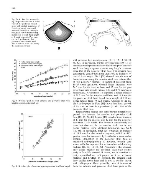
364Fig. 7a,b. Sketches summarisingtemporal variations. a Axialview <strong>of</strong> <strong>the</strong> posterior cranialfossa with shaded increments <strong>of</strong>2 weeks showing that widthincreases in relation to length.b Sagittal view demonstratingincrements <strong>of</strong> <strong>skull</strong> base lengthat 2-week intervals. Plus signsare used to illustrate that<strong>growth</strong> <strong>of</strong> <strong>the</strong> anterior <strong>skull</strong>base is almost twice that along<strong>the</strong> posterior portionFig. 8. Bivariate plot <strong>of</strong> total, anterior and posterior <strong>skull</strong> baselengths against gestational ageFig. 9. Bivariate plot <strong>of</strong> anterior against posterior <strong>skull</strong> base lengthwith previous key investigations [10, 11, 12, 13, 38, 39,40, 52]. In particular, Burdi’s investigations [10, 13] <strong>of</strong>hemisectioned specimens show that <strong>the</strong> slope <strong>of</strong> anterior<strong>skull</strong> base length against crown-rump length is almosttwice that <strong>of</strong> <strong>the</strong> posterior <strong>skull</strong> base. The anterior baseconsistently contributes more than 50% to increases <strong>of</strong>overall base length. Birch [38] showed that <strong>the</strong> rate <strong>of</strong><strong>linear</strong> increase along <strong>the</strong> anterior <strong>skull</strong> base is twice that<strong>of</strong> <strong>the</strong> posterior segment in sectioned material from10–27 weeks gestation. Overall <strong>linear</strong> increases were24.5 mm for <strong>the</strong> anterior base and 12 mm for <strong>the</strong> posteriorbase with <strong>growth</strong> rates <strong>of</strong> 1.44 and 0.71 mm/week,respectively. Kvinnsland [14] reported a <strong>linear</strong> increase<strong>of</strong> 21.7 mm for <strong>the</strong> anterior <strong>skull</strong> base and 11.5 mm for<strong>the</strong> posterior <strong>skull</strong> base based on a sample <strong>of</strong> 159 sectionedfetuses from 10–32.5 weeks. Analysis <strong>of</strong> <strong>the</strong> Table4 in <strong>the</strong> paper by Ford [11] shows that <strong>linear</strong> <strong>growth</strong><strong>of</strong> <strong>the</strong> anterior base is approximately twice that <strong>of</strong> <strong>the</strong>posterior <strong>skull</strong> base.Radiographic studies also demonstrate differences <strong>of</strong><strong>growth</strong> rate between <strong>the</strong> anterior and posterior <strong>skull</strong>base [12, 17, 39, 40]. Levihn [12] noted a <strong>linear</strong> increase<strong>of</strong> 17 mm for <strong>the</strong> anterior and 12 mm for <strong>the</strong> posteriorbase from 12–24 weeks. The former is considerably lessthan that observed for <strong>the</strong> anterior <strong>skull</strong> base in sectionedmaterial using identical landmarks, i.e., Na-S[10, 38]. In particular, Birch [38] observed an increase<strong>of</strong> 24.5 mm for <strong>the</strong> anterior segment, which is 44%greater than that measured by Levihn for a comparablesample. Elongation <strong>of</strong> <strong>the</strong> posterior <strong>skull</strong> base asmeasured radiographically is, however, broadly consistentwith that reported for sectioned material and myfindings [10, 12, 14, 38, 39]. Presumably, this discrepancyarises because <strong>the</strong> posterior <strong>skull</strong> base ossifiesearly in <strong>fetal</strong> life, around 11 weeks, and is <strong>the</strong>refore aseasily quantified on radiographs as it on sectionedmaterial and hr<strong>MRI</strong>. In contrast, <strong>the</strong> anterior <strong>skull</strong>base ossifies much later and is not so readily shownwith radiographs. More generally, rates <strong>of</strong> increase <strong>of</strong>both <strong>the</strong> anterior and posterior <strong>skull</strong> base given in <strong>the</strong>