Platelet-rich plasma influence on human ... - Clauberth Oliveira
Platelet-rich plasma influence on human ... - Clauberth Oliveira
Platelet-rich plasma influence on human ... - Clauberth Oliveira

You also want an ePaper? Increase the reach of your titles
YUMPU automatically turns print PDFs into web optimized ePapers that Google loves.
Cimara Fortes Ferreira<br />
Márcia Cristina Carriel<br />
Gomes<br />
José Scarso Filho<br />
José Mauro Granjeiro<br />
Cláudia Maria <strong>Oliveira</strong><br />
Simões<br />
Ricardo de Souza Magini<br />
Authors’ affiliati<strong>on</strong>s:<br />
Cimara Fortes Ferreira, Department of Dental<br />
Implantology, University of Santa Catarina,<br />
Florianópolis, Brazil<br />
Márcia Cristina Carriel Gomes, Department of<br />
Biotechnology, University of Santa Catarina,<br />
Florianópolis, Brazil<br />
José Scarso Filho, Department of Oral Surgery,<br />
University of Araraquara, Araraquara, Brazil<br />
José Mauro Granjeiro, Department of Biochemistry,<br />
School of Dentistry, Bauru, Brazil<br />
Cláudia Maria <strong>Oliveira</strong> Simões, Department of<br />
Pharmaceutical Sciences, University of Santa<br />
Catarina, Florianópolis, Brazil<br />
Ricardo de Souza Magini, Department of<br />
Period<strong>on</strong>tology, University of Santa Catarina,<br />
Florianópolis, Brazil<br />
Corresp<strong>on</strong>dence:<br />
Dr Ricardo de Souza Magini<br />
Universidade Federal de Santa Catarina – Centro de<br />
Ensino e Pesquisa em Implantes Dentários<br />
(UFSC-CEPID)<br />
Centro de Ciências da Saúde–Campus<br />
Universitário Trindade<br />
88040-970<br />
Florianópolis, SC<br />
Brazil<br />
Tel.: þ 55 48 331 9077<br />
Fax: þ 55 48 234 1788<br />
e-mail: cimarafortes@hotmail.com<br />
Date:<br />
Accepted 18 October 2004<br />
To cite this article:<br />
Ferreira CF, Gomes MCC, Filho JS, Granjeiro JM,<br />
Simões CMO, Magini RS. <str<strong>on</strong>g>Platelet</str<strong>on</strong>g>-<str<strong>on</strong>g>rich</str<strong>on</strong>g> <str<strong>on</strong>g>plasma</str<strong>on</strong>g><br />
<str<strong>on</strong>g>influence</str<strong>on</strong>g> <strong>on</strong> <strong>human</strong> osteoblasts growth.<br />
Clin. Oral Impl. Res. 16, 2005; 456–460<br />
doi: 10.1111/j.1600-0501.2005.01145.x<br />
Copyright r Blackwell Munksgaard 2005<br />
456<br />
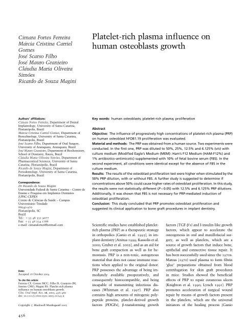
<str<strong>on</strong>g>Platelet</str<strong>on</strong>g>-<str<strong>on</strong>g>rich</str<strong>on</strong>g> <str<strong>on</strong>g>plasma</str<strong>on</strong>g> <str<strong>on</strong>g>influence</str<strong>on</strong>g> <strong>on</strong><br />
<strong>human</strong> osteoblasts growth<br />
Key words: <strong>human</strong> osteoblasts; platelet-<str<strong>on</strong>g>rich</str<strong>on</strong>g> <str<strong>on</strong>g>plasma</str<strong>on</strong>g>; proliferati<strong>on</strong><br />
Abstract<br />
Objective: The <str<strong>on</strong>g>influence</str<strong>on</strong>g> of progressively high c<strong>on</strong>centrati<strong>on</strong>s of platelet-<str<strong>on</strong>g>rich</str<strong>on</strong>g> <str<strong>on</strong>g>plasma</str<strong>on</strong>g> (PRP)<br />
<strong>on</strong> <strong>human</strong> osteoblast hFOB1.19 proliferati<strong>on</strong> was evaluated.<br />
Material and methods: The PRP was obtained from a <strong>human</strong> source. Two experiments were<br />
c<strong>on</strong>ducted. In the first <strong>on</strong>e, PRP was diluted to 50%, 25%, 12.5% and 6.125% (v/v) with<br />
culture medium (Modified Eagle’s Medium (MEM) : Ham’s F12 Medium (HAM-F12%) and<br />
1% antibiotics–antimicotic) supplemented with 10% of fetal bovine serum (FBS). In the<br />
sec<strong>on</strong>d experiment, all c<strong>on</strong>diti<strong>on</strong>s were identical except for the absence of FBS in the<br />
culture medium.<br />
Results: The results of the osteoblast proliferati<strong>on</strong> test were higher when stimulated by the<br />
50% PRP diluti<strong>on</strong>, with or without FBS. A further study is suggested to determine if<br />
c<strong>on</strong>centrati<strong>on</strong>s above 50% could cause higher rates of osteoblast proliferati<strong>on</strong>. In this study,<br />
the results were not statistically different (Po0.05) with 12.5% and 6.125% PRP diluti<strong>on</strong>s.<br />
Additi<strong>on</strong>ally, it was shown that FBS is not necessary for PRP-mediated inducti<strong>on</strong> of<br />
osteoblast proliferati<strong>on</strong>.<br />
C<strong>on</strong>clusi<strong>on</strong>: This study c<strong>on</strong>cluded that PRP promotes osteoblast proliferati<strong>on</strong> and<br />
suggested its clinical applicati<strong>on</strong> to b<strong>on</strong>e graft procedures in implant dentistry.<br />
Scientific studies have established platelet<str<strong>on</strong>g>rich</str<strong>on</strong>g><br />
<str<strong>on</strong>g>plasma</str<strong>on</strong>g> (PRP) as a therapeutic strategy<br />
in orthopedics (Ganio et al. 1993), in implant<br />
dentistry (Anitua 1999; Kassolis et al.<br />
2000; Gruber et al. 2002), and as an aid for<br />
b<strong>on</strong>e graft compacti<strong>on</strong> as well as for hemostasis.<br />
PRP is a n<strong>on</strong>-toxic, autogenous<br />
material that does not cause immune reacti<strong>on</strong>s<br />
when applied to the original d<strong>on</strong>or.<br />
PRP possesses the advantage of being immediately<br />
available preoperatively, and<br />
c<strong>on</strong>sequently histocompatible, and being<br />
incapable of transmitting infectious diseases<br />
(Whitman et al. 1997). PRP also<br />
c<strong>on</strong>tains high amounts of mitogenic polypeptide<br />
proteins, platelet-derived growth<br />
factors (PDGFs), b-transforming growth<br />
factors (TGF-b’s) and I-insulin-like growth<br />
factors, which appear to accelerate the<br />
osteogenesis in oral and maxillofacial surgery,<br />
as well as platelets, which are a<br />
source of growth factors that induce b<strong>on</strong>e,<br />
epithelial and c<strong>on</strong>nective tissue repair. It<br />
has been successfully used since the 1970s.<br />
Matras (1970) used <str<strong>on</strong>g>plasma</str<strong>on</strong>g> to form fibrin<br />
‘glue’ preparati<strong>on</strong>s obtained from blood<br />
centrifugati<strong>on</strong> for skin graft procedures<br />
in mice. Studies showed the beneficial<br />
effects of PRP to repair cutaneous ulcers<br />
(Knight<strong>on</strong> et al. 1990; Lynch 1991). PRP<br />
promotes accelerati<strong>on</strong> of surgical wound<br />
repair by means of growth factors present<br />
in the platelets, which are the universal<br />
initiators of the healing process (Ganio
et al. 1993; Tayap<strong>on</strong>gsak et al. 1994; Anitua<br />
1999). Its benefits were also c<strong>on</strong>firmed<br />
in oral and maxillofacial surgeries, (Ganio<br />
et al. 1993; Kassolis et al. 2000). Lucarelli<br />
et al. (2003) investigated mesenchymal<br />
stem cell proliferati<strong>on</strong> in culture medium<br />
supplemented with PRP and verified that<br />
the use of 10% PRP was sufficient to<br />
accelerate mineralizati<strong>on</strong> in vitro. Clinicians<br />
and researchers have investigated<br />
the use of PRP in dentistry as a form of<br />
accelerating the natural healing process.<br />
Carls<strong>on</strong> & Roach (2002) showed that PRP<br />
and its growth factors are promising for<br />
surgical wound healing. The use of PRP,<br />
in associati<strong>on</strong> with autogenous b<strong>on</strong>e grafts<br />
in implant dentistry, was shown to optimize<br />
the quality and quantity of newly<br />
formed b<strong>on</strong>e (Scarso Filho 2002).<br />
A systematic review of maxillary sinus<br />
augmentati<strong>on</strong> procedures <strong>on</strong> the survival<br />
of endosseous dental implants was d<strong>on</strong>e<br />
when the efficacy of the sinus augmentati<strong>on</strong><br />
procedure was compared with various<br />
surgical grafting materials, including the<br />
use of PRP associated with b<strong>on</strong>e graft<br />
material. In spite of the favorable resp<strong>on</strong>ses<br />
shown in the literature in using<br />
PRP as an adjuvant for b<strong>on</strong>e-grafting<br />
procedures, the authors of this systematic<br />
review c<strong>on</strong>cluded that there are insufficient<br />
data to recommend the use of PRP<br />
in sinus graft surgery (Wallace & Froum<br />
2003).<br />
Another study <strong>on</strong> the use of PRP associated<br />
with autologous b<strong>on</strong>e as filling<br />
material for sinus lift procedures shows<br />
increased b<strong>on</strong>e maturati<strong>on</strong> rate and improved<br />
b<strong>on</strong>e density when PRP, or its<br />
recombinant growth factors, are added to<br />
small b<strong>on</strong>y defects or to larger defects that<br />
use autogenous b<strong>on</strong>e as the grafting material.<br />
Histomorphometric analysis indicated<br />
that the additi<strong>on</strong> of PRP to the grafts did<br />
not show significant difference either in<br />
vital b<strong>on</strong>e producti<strong>on</strong> or in interfacial<br />
b<strong>on</strong>e c<strong>on</strong>tact <strong>on</strong> implants placed in these<br />
sites (Froum et al. 2002). Other studies<br />
using animal models showed that PRP<br />
associated with autogenous b<strong>on</strong>e did not<br />
enhance b<strong>on</strong>e formati<strong>on</strong> (Roldan et al.<br />
2004; Schlegel et al. 2004; Wiltfang et al.<br />
2004).<br />
The effect of PRP <strong>on</strong> b<strong>on</strong>e regenerati<strong>on</strong>,<br />
when associated to autogenous b<strong>on</strong>e grafts<br />
was evaluated in vitro in a canine model<br />
(Choi et al. 2004). The findings suggested<br />
that the additi<strong>on</strong> of PRP does not appear to<br />
enhance new b<strong>on</strong>e formati<strong>on</strong>.<br />
The purpose of the present study was to<br />
evaluate progressively high c<strong>on</strong>centrati<strong>on</strong>s of<br />
PRP <strong>on</strong> proliferati<strong>on</strong> of <strong>human</strong> osteoblasts.<br />
Material and methods<br />
Cell culture<br />
Human osteoblasts (hFOB1.19, no. CRL<br />
11,372) were provided from the American<br />
Type Culture Collecti<strong>on</strong>. Those used in the<br />
experiment were derived from passages 5<br />
and 6. The cell m<strong>on</strong>olayers were cultured<br />
in Modified Eagle’s medium (MEM, Gibco,<br />
Invitrogen Corporati<strong>on</strong>, Carlsbad, CA,<br />
USA) and Ham’s F12 medium (HAM-<br />
F12) (Cultilab, Campinas, SP, Brazil) (1 : 1)<br />
supplemented with 10% of fetal bovine<br />
serum (FBS, Gibco BRL, Campinas, Brazil),<br />
and 1% of antibiotics–antimycotic (penicillin<br />
G sodium – 100 U/ml), streptomycin –<br />
100 mg/ml and amphotericin B – 0.025 mg/<br />
ml; Gibco BRL). The cells were maintained<br />
in a humidified incubator with 5% CO2 at<br />
371C. When cells reached c<strong>on</strong>fluency they<br />
were subcultivated using 0.1% of trypsin<br />
(Sigma, St. Louis, MO, USA; 5 mg/ml) and<br />
0.1% of calcium- and magnesium-free Dulbecco’s<br />
phosphate-buffer saline soluti<strong>on</strong><br />
(Gibco, Invitrogen Corporati<strong>on</strong>).<br />
<str<strong>on</strong>g>Platelet</str<strong>on</strong>g> collecti<strong>on</strong><br />
Plasma was obtained from the venous<br />
blood of a healthy male volunteer. Blood<br />
was drawn into a 5 ml Vacutainer s (BD,<br />
Curitiba, Paraná, Brazil) c<strong>on</strong>taining 500 ml<br />
of the anticoagulant sodium citrate. PRP<br />
was obtained following the protocol developed<br />
by Macedo et al. (2003). Briefly, blood<br />
was centrifuged twice at 120 g in an<br />
ALC centrifuge (Centrifugette 4206 ALC,<br />
ACE Surgical Supply Company Inc.,<br />
Brockt<strong>on</strong>, MA, USA) for 10 min at 201C<br />
to remove red blood cells (first time) to<br />
obtain the PRP (sec<strong>on</strong>d time). Three hours<br />
later, the PRP was diluted in the cell<br />
culture medium for proliferative assessment<br />
of the osteoblasts. A previous study<br />
revealed that the activity of growth factors<br />
present in PRP decreased 4 h after blood<br />
was drawn (Ledent et al. 1995). C<strong>on</strong>sequently,<br />
the PRP used here was obtained<br />
approximately 3 h after blood collecti<strong>on</strong>.<br />
This procedure was repeated three times,<br />
in a weekly interval, to obtain mean va-<br />
Ferreira et al . Human osteoblasts growth<br />
lues. Therefore, the blood was drawn three<br />
times from the same male volunteer.<br />
Evaluati<strong>on</strong> of <strong>human</strong> osteoblast<br />
proliferati<strong>on</strong> by 3-(4,5-dimethylthiazol-2yl)-2,5-diphenyl<br />
tetrazolium bromide<br />
(MTT) assay<br />
hFOB1.19 cell cultures (3 10 4 cells/well)<br />
were prepared in 96-well tissue culture<br />
plates (Corning, NY, USA). After a 24 h<br />
period of incubati<strong>on</strong>, the cell culture medium<br />
was replaced by <strong>on</strong>e c<strong>on</strong>taining 50%,<br />
25%, 12.5% and 6.125% of diluted PRP<br />
without or with 10% FBS.<br />
Next, platelet gel was obtained up<strong>on</strong><br />
additi<strong>on</strong> of 5% calcium chloride to the<br />
medium. Four days after replacing original<br />
culture medium with another <strong>on</strong>e c<strong>on</strong>taining<br />
PRP, the MTT method (Mossman 1983)<br />
was applied with modificati<strong>on</strong>s (Andrighetti-Fröhner<br />
et al. 2003). After 4 days, at<br />
371C inhumidified5%CO2atmosphere, 50 ml of MTT (Sigma, 1 mg/ml) soluti<strong>on</strong><br />
prepared in MEM : HAM-F12 (1 : 1) cell<br />
medium was added to each well and the<br />
plates were incubated for 4 h at 371C. Next,<br />
the medium was not removed with sucti<strong>on</strong>,<br />
and 100 ml of dimethyl sulfoxide (Merck<br />
Biosciences, Darmstadt, Germany) was<br />
added to each well to dissolve formazan<br />
crystals. After gently shaking the plates for<br />
5 min, whereby crystals were completely<br />
dissolved, the absorbance was read <strong>on</strong> a<br />
multiwell spectrophotometer (Bio-Tek, Elx<br />
800, Winooski, VT, USA) at 540 nm. The<br />
original MTT technique (Mossman 1983)<br />
was modified, the cell culture medium was<br />
not removed, for a gel clot was formed inside<br />
the wells, and, by means of light microscopy,<br />
the cells had migrated to the clot.<br />
Afterwards, the <str<strong>on</strong>g>influence</str<strong>on</strong>g> of the PRP was<br />
assessed by a scanning spectrophotometer<br />
(Ultrospec 3000-Pharmacia Bio-tek, Winooski,<br />
VT, USA) in the four c<strong>on</strong>centrati<strong>on</strong>s<br />
used in this research. This evaluati<strong>on</strong><br />
showed no <str<strong>on</strong>g>influence</str<strong>on</strong>g> of the PRP in the cell<br />
culture medium when read at 540 nm.<br />
Therefore, PRP did not <str<strong>on</strong>g>influence</str<strong>on</strong>g> the results<br />
obtained from the spectrophotometer (Bio-<br />
Tek, Elx 800) at 540 nm. The percentage of<br />
cell growth was calculated as [(A B)/<br />
A 100], where A and B are the absorbances<br />
of c<strong>on</strong>trol and treated cells, respectively.<br />
In this calculati<strong>on</strong>, B was subtracted<br />
from A and divided by the absorbance of the<br />
treated group multiplied by 100, resulting in<br />
the percentage of cell growth.<br />
457 | Clin. Oral Impl. Res. 16, 2005 / 456–460
Ferreira et al . Human osteoblasts growth<br />
The percentages of cell growth by the<br />
PRP in relati<strong>on</strong> to hFOB1.19 osteoblasts<br />
represent mean standard error of the<br />
mean values of three different experiments.<br />
Variance analysis and the Tukey test<br />
(Po0.05) were carried out as appropriate.<br />
Results<br />
Evaluati<strong>on</strong> of <strong>human</strong> osteoblast<br />
proliferati<strong>on</strong> by MTT assay<br />
The results of the <str<strong>on</strong>g>influence</str<strong>on</strong>g> of different<br />
c<strong>on</strong>centrati<strong>on</strong>s of PRP <strong>on</strong> hFOB1.19 osteoblast<br />
proliferati<strong>on</strong> in MEM : HAM-F12<br />
(1 : 1) without and with 10% FBS are expressed<br />
as percentages of cell growth that<br />
indicate mean values of three independent<br />
experiments. The results were evaluated<br />
statistically using analysis of variance and<br />
Tukey analysis (Fig. 1).<br />
A comparis<strong>on</strong> of the results of hFOB1.19<br />
osteoblast proliferati<strong>on</strong> by PRP with and<br />
without 10% FBS showed that cell growth<br />
increases as a functi<strong>on</strong> of the c<strong>on</strong>centrati<strong>on</strong><br />
of PRP. Cell growth is always higher in the<br />
groups receiving 10% FBS, showing the<br />
highest <str<strong>on</strong>g>influence</str<strong>on</strong>g> <strong>on</strong> cell growth between<br />
675.7% and 824% with 695.7% as the<br />
median value for the groups receiving 50%<br />
PRP. Figure 1 shows the results of the<br />
variability c<strong>on</strong>sidered in this research for<br />
the groups receiving 10% FBS and those<br />
not receiving FBS.<br />
Discussi<strong>on</strong><br />
Past investigati<strong>on</strong>s have established the<br />
<str<strong>on</strong>g>influence</str<strong>on</strong>g> of growth factors <strong>on</strong> cell prolif-<br />
Mean cell growth<br />
900<br />
800<br />
700<br />
600<br />
500<br />
400<br />
300<br />
200<br />
100<br />
0<br />
10%<br />
0%<br />
256.9 ± 26.9<br />
148.3 ± 20.7<br />
191.2 ± 20.6<br />
116.1 ± 10.4<br />
erati<strong>on</strong> (Gr<strong>on</strong>thos & Simm<strong>on</strong>s 1995; Kuznetov<br />
et al. 1997; Lind 1998; Tian et al.<br />
1999). PRP, which c<strong>on</strong>tains many growth<br />
factors, has been shown to stimulate cell<br />
proliferati<strong>on</strong> in vitro (Slater et al. 1995;<br />
Nakanishi et al. 1997; Weib<str<strong>on</strong>g>rich</str<strong>on</strong>g> et al.<br />
2002; Kawasa et al. 2003; Lucarelli et al.<br />
2003; Okuda et al. 2003). Whitman et al.<br />
(1997) described the preparati<strong>on</strong> of platelet<br />
gel showing its advantages in b<strong>on</strong>e graft<br />
procedures to accelerate the healing process.<br />
Literature <strong>on</strong> b<strong>on</strong>e cell physiology<br />
shows many instances where PRP was in<br />
associati<strong>on</strong> with b<strong>on</strong>e graft procedures. In<br />
these cases, PRP has dem<strong>on</strong>strated an accelerati<strong>on</strong><br />
of the formati<strong>on</strong> of mature b<strong>on</strong>e.<br />
In additi<strong>on</strong>, a greater formati<strong>on</strong> of cancellous<br />
b<strong>on</strong>e was noted when compared<br />
with b<strong>on</strong>e grafts that did not receive<br />
PRP when evaluated 4–6 m<strong>on</strong>ths after the<br />
procedure (Marx & Garg 2003). In vitro<br />
investigati<strong>on</strong>s have identified that the<br />
PDGF, a subcomp<strong>on</strong>ent of the PRP, has<br />
a significant effect <strong>on</strong> cell proliferati<strong>on</strong><br />
(Nakanishi et al. 1997; Weib<str<strong>on</strong>g>rich</str<strong>on</strong>g> et al.<br />
2002; Kawase et al. 2003; Okuda et al.<br />
2003).<br />
However, questi<strong>on</strong>s are raised about the<br />
efficacy of PRP in dental applicati<strong>on</strong>s.<br />
There are clinical studies where PRP was<br />
used for grafting the floor of the maxillary<br />
sinus in associati<strong>on</strong> with b<strong>on</strong>e graft material<br />
for increasing alveolar b<strong>on</strong>e height prior<br />
to the placement of endosseous dental<br />
implants in the posterior maxilla. To<br />
determine the efficacy of the sinus<br />
augmentati<strong>on</strong> procedure compared with<br />
the results achieved with various surgical<br />
techniques, grafting materials and im-<br />
444.1 ± 23.1<br />
433.5 ± 31.9<br />
731.8 ± 46.5<br />
666.4 ± 20.4<br />
6.125 12.5 25 50<br />
PRP c<strong>on</strong>centrati<strong>on</strong> (%)<br />
Fig. 1. The <str<strong>on</strong>g>influence</str<strong>on</strong>g> of different c<strong>on</strong>centrati<strong>on</strong>s of platelet-<str<strong>on</strong>g>rich</str<strong>on</strong>g> <str<strong>on</strong>g>plasma</str<strong>on</strong>g> (PRP) <strong>on</strong> hFOB1.19 osteoblast<br />
proliferati<strong>on</strong> in MEM : HAM-F12 (1 : 1) medium without (0%) and with 10% of fetal bovine serum, expressed<br />
as cell growth percentages. The mean values are presented <strong>on</strong> top of the bars with the standard error value.<br />
458 | Clin. Oral Impl. Res. 16, 2005 / 456–460<br />
plants, a systematic review of clinical studies<br />
was c<strong>on</strong>ducted by Wallace & Froum<br />
(2003). In this investigati<strong>on</strong>, the authors<br />
verified the effect <strong>on</strong> implant survival of<br />
maxillary sinus augmentati<strong>on</strong> vs. implant<br />
placement in the n<strong>on</strong>-grafted posterior<br />
maxilla. The authors c<strong>on</strong>cluded that there<br />
were insufficient data to recommend the<br />
use of PRP in sinus graft surgery, a very<br />
comm<strong>on</strong> procedure used in implant<br />
dentistry.<br />
In another investigati<strong>on</strong>, the effect of<br />
PRP <strong>on</strong> b<strong>on</strong>e regenerati<strong>on</strong> associated with<br />
autogenous b<strong>on</strong>e graft was evaluated in<br />
animal models where b<strong>on</strong>e defects received<br />
autogenous b<strong>on</strong>e in associati<strong>on</strong> with PRP,<br />
and without PRP. Results of these studies<br />
showed that the additi<strong>on</strong> of PRP does not<br />
appear to enhance new b<strong>on</strong>e formati<strong>on</strong> in<br />
autogenous b<strong>on</strong>e grafts (Schlegel et al.<br />
2003, 2004; Choi et al. 2004; Wiltfang<br />
et al. 2004).<br />
Further, PRP was studied <strong>on</strong> b<strong>on</strong>e augmentati<strong>on</strong><br />
procedures in vitro and was<br />
compared with b<strong>on</strong>e morphogenetic protein<br />
when added to autologous b<strong>on</strong>e grafts.<br />
The authors c<strong>on</strong>cluded that, by means of<br />
a histomorphometric study, PRP failed<br />
to enhance b<strong>on</strong>e formati<strong>on</strong> (Roldan et al.<br />
2004).<br />
Froum et al. (2002) studied the efficacy<br />
of PRP <strong>on</strong> b<strong>on</strong>e growth and osseointegrati<strong>on</strong><br />
in <strong>human</strong> maxillary sinus grafts when<br />
associated with grafts of anorganic bovine<br />
b<strong>on</strong>e that c<strong>on</strong>tained minimal or no autogenous<br />
b<strong>on</strong>e. The results of their studies<br />
showed that the effect of PRP did not show<br />
a significant difference either in vital b<strong>on</strong>e<br />
producti<strong>on</strong> or in interfacial b<strong>on</strong>e c<strong>on</strong>tact <strong>on</strong><br />
the test implants.<br />
The studies enumerated above suggest<br />
uncertainty in recommending the use of<br />
PRP in b<strong>on</strong>e-regenerati<strong>on</strong> procedures.<br />
However, the present investigati<strong>on</strong> shows<br />
in vitro that PRP has a positive effect <strong>on</strong><br />
osteoblast proliferati<strong>on</strong>.<br />
The results of this investigati<strong>on</strong> are in<br />
accordance with the results obtained by<br />
Nakanishi et al. (1997) and Kawase et al.<br />
(2003) showing that PRP stimulates cell<br />
proliferati<strong>on</strong> when added to culture medium<br />
c<strong>on</strong>taining osteoblasts. In this study,<br />
we observed a positive correlati<strong>on</strong> between<br />
the increased c<strong>on</strong>centrati<strong>on</strong> of PRP in culture<br />
medium and the rate of osteoblasts<br />
proliferati<strong>on</strong>, c<strong>on</strong>sistent with the results<br />
published by Lucarelli et al. (2003), which
showed that 10% of PRP added to osteoblast<br />
culture medium was sufficient to<br />
induce evident cell proliferati<strong>on</strong>. The present<br />
study showed that 50% PRP caused<br />
the best proliferative results in osteoblast<br />
culture. Further studies are needed to establish<br />
if progressively higher c<strong>on</strong>centrati<strong>on</strong>s<br />
of PRP would have a corresp<strong>on</strong>ding<br />
<str<strong>on</strong>g>influence</str<strong>on</strong>g> <strong>on</strong> proliferati<strong>on</strong>. In additi<strong>on</strong>, our<br />
results showed that FBS is not necessary for<br />
PRP-mediated inducti<strong>on</strong> of hFOB1.19 osteoblast<br />
proliferati<strong>on</strong> as shown by Lucarelli<br />
et al. (2003).<br />
The c<strong>on</strong>tent of PDGF and TGF-b present<br />
in platelet gel was studied during the preparati<strong>on</strong><br />
and storage of PRP. The c<strong>on</strong>tent of<br />
PDGF and TGF-b decreases according to<br />
higher storage periods, showing less cell<br />
growth promoti<strong>on</strong> 4 h to 3 days after blood<br />
is drawn. In this study, the experimental<br />
use of blood to supplement the cell growth<br />
media did not exceed 3 h after blood was<br />
drawn (Ledent et al. 1995).<br />
Currently, cultivati<strong>on</strong> of <strong>human</strong> osteoblasts<br />
in vitro is challenging (Lind 1998),<br />
and PRP has dem<strong>on</strong>strated a significant<br />
<str<strong>on</strong>g>influence</str<strong>on</strong>g> <strong>on</strong> osteoblast reproducti<strong>on</strong>.<br />
The positive results of in vitro<br />
(Nakanishi et al. 1997; Weib<str<strong>on</strong>g>rich</str<strong>on</strong>g> et al.<br />
2002; Lucarelli et al. 2003) studies encourage<br />
research into further refinement<br />
of methods for osteoblast cultivati<strong>on</strong>.<br />
Methods c<strong>on</strong>ducive to high yields of<br />
osteoblast proliferati<strong>on</strong> may c<strong>on</strong>tribute<br />
ultimately to the development of processes<br />
that promote growth or replacement<br />
of substitute b<strong>on</strong>e material. They also<br />
could potentially have far-reaching benefits<br />
in orthopedics, maxillofacial surgery as<br />
well as implant dentistry by eliminating<br />
the need for surgically complex b<strong>on</strong>e harvesting<br />
as well as reducing associated<br />
pain and costs. However, the effects of<br />
PRP associated with autogenous b<strong>on</strong>e<br />
grafts in clinical situati<strong>on</strong>s require further<br />
investigati<strong>on</strong>.<br />
The purpose of the present study was<br />
to evaluate the <str<strong>on</strong>g>influence</str<strong>on</strong>g> of progressively<br />
higher c<strong>on</strong>centrati<strong>on</strong>s of PRP <strong>on</strong> cell<br />
growth medium. The maximum amount<br />
of PRP added to the growth medium was<br />
50%, showing the best proliferative results<br />
in <strong>human</strong> osteoblast culture. Therefore,<br />
further studies are suggested to answer<br />
if higher c<strong>on</strong>centrati<strong>on</strong>s of PRP would<br />
alter the proliferative rate of <strong>human</strong><br />
osteoblasts.<br />
Acknowledgements: The authors are<br />
grateful to Prof. Dr Zenilda Laurita<br />
Bouz<strong>on</strong> from the Department of Cell<br />
Biology, Embryology and Genetics of<br />
the University of Santa Catarina for her<br />
assistance during the experimental part<br />
of the research; to Esther Takamori,<br />
MSc (Faculty of Dentistry, Bauru, SP)<br />
for her assistance during laboratory<br />
training and to Kenneth Faulkner<br />
Weaver for his editorial assistance.<br />
Résumé<br />
Le but de cette étudeaété d’évaluer l’<str<strong>on</strong>g>influence</str<strong>on</strong>g> de<br />
c<strong>on</strong>centrati<strong>on</strong>s progressivement plus importantes de<br />
<str<strong>on</strong>g>plasma</str<strong>on</strong>g> <str<strong>on</strong>g>rich</str<strong>on</strong>g>e en plaquettes (PRP) sur la proliférati<strong>on</strong><br />
des ostéoblastes hFOB1.19 humain. Le PRP a été<br />
obtenu de source humaine. Deux expériences <strong>on</strong>t été<br />
c<strong>on</strong>duites. Dans la première, le PRP a été dilué à 50,<br />
25, 12,5 et 6,125 (v/v) avec un milieu de culture<br />
(MEM : HAM-F12 et 1% d’antibiotiques-antimicotique)<br />
avec 10% de sérum bovin foetal (FBS). Dans la<br />
sec<strong>on</strong>de expérience, toutes ces c<strong>on</strong>diti<strong>on</strong>s étaient<br />
identiques sauf qu’il n’y avait pas de FBS dans le<br />
milieu de culture. Les résultats du test de la proliférati<strong>on</strong><br />
des ostéoblastes étaient plus élevés quand ils<br />
étaient stimulés par une diluti<strong>on</strong> PRP de 50% avec<br />
ou sans FBS. Une étude supplémentaireaété suggérée<br />
pour déterminer si les c<strong>on</strong>centrati<strong>on</strong>s supérieures<br />
à 50% pouvaient apporter des taux plus élevés<br />
de proliférati<strong>on</strong> des ostéoblastes. Dans l’étude présente,<br />
les résultats n’étaient pas différents (Po0,05)<br />
avec des diluti<strong>on</strong>s de PRP à 12,5 et 6,125%. De plus,<br />
il était c<strong>on</strong>staté que le FBS n’était pas nécessaire<br />
pour l’inducti<strong>on</strong> de la proliférati<strong>on</strong> des ostéoblastes<br />
par PRP. Le PRP améliore d<strong>on</strong>c la proliférati<strong>on</strong> des<br />
ostéoblastes et propose s<strong>on</strong> applicati<strong>on</strong> clinique dans<br />
les processus de greffe osseuse en chirurgie buccale<br />
implantaire.<br />
Zusammenfassung<br />
Der Einfluss v<strong>on</strong> plättchenreichem Plasma auf das<br />
Wachstum v<strong>on</strong> menschlichen Osteoblasten<br />
Ziel: Es sollte der Einfluss v<strong>on</strong> progressiv erhöhten<br />
K<strong>on</strong>zentrati<strong>on</strong>en v<strong>on</strong> plättchenreichem Plasma<br />
(PRP) auf die Proliferati<strong>on</strong> v<strong>on</strong> menschlichen Osteoblsaten<br />
hfOB1.19 untersucht werden.<br />
Material und Methoden: Das PRP wurde bei<br />
Menschen gew<strong>on</strong>nen. Zwei Experimente wurden<br />
durchgeführt. Beim ersten wurde das PRP mit Kulturmedium<br />
(MEM:HAM-F12 und 1% Antibiotika-<br />
Antimykotika) auf 50, 25, 12.5 und 6.125 (v/v)<br />
verdünnt und mit 10% fetalem bovinem Serum<br />
(FBS) ergänzt. Beim zweiten Experiment waren alle<br />
Bedingungen identisch, nur das FBS fehlte im Kulturmedium.<br />
Resultate: Die Proliferati<strong>on</strong> der Osteoblasten war<br />
höher, wenn sie durch eine 50% PRP Verdünnung<br />
mit oder ohne FBS stimuliert wurde. Es wird eine<br />
weitere Studie vorgeschlagen, um zu bestimmen, ob<br />
K<strong>on</strong>zentrati<strong>on</strong>en über 50% höhere Proliferati<strong>on</strong>sraten<br />
der Osteoblasten zu Folge haben. In dieser<br />
Untersuchung waren die Resultate mit 12.5 und<br />
6.125% Verdünnungen nicht statistisch signifikant<br />
verschieden (Po0.05). Zusätzlich k<strong>on</strong>nte gezeigt<br />
werden, dass FBS für die PRP gesteuerte Indukti<strong>on</strong><br />
der Osteoblastenproliferati<strong>on</strong> nicht nötig ist.<br />
Schlussfolgerung: Diese Studie führt zur Schlussfolgerung,<br />
dass PRP die Osteoblastenproliferati<strong>on</strong><br />
fördert. Es wird die klinische Anwendung v<strong>on</strong> PRP<br />
bei Knochentransplantati<strong>on</strong>en in Zusammenhang<br />
mit dentalen Implantaten vorgeschlagen.<br />
Resumen<br />
Ferreira et al . Human osteoblasts growth<br />
Objetivo: Evaluar la influencia de altas c<strong>on</strong>centraci<strong>on</strong>es<br />
progresivamente de <str<strong>on</strong>g>plasma</str<strong>on</strong>g> rico en plaquetas<br />
(PRP) en la proliferación de osteoblastos <strong>human</strong>os<br />
hFOB1.19.<br />
Material y métodos: El PRP se obtuvo de fuentes<br />
<strong>human</strong>as. Se llevar<strong>on</strong> a cabo dos experimentos. En el<br />
primero, el PRP se diluyó a 50, 25, 12.5 y 6.125 (v/v)<br />
c<strong>on</strong> medio de cultivo (MEM : HAM-F12 y 1% antibióticos-antimicóticos)<br />
suplementado c<strong>on</strong> 10% de<br />
suero fetal bovino (FBS). En el segundo experimento,<br />
todas las c<strong>on</strong>dici<strong>on</strong>es fuer<strong>on</strong> idénticas salvo por la<br />
ausencia de FBS en el medio de cultivo.<br />
Resultados: Losresultadosdelapruebadeproliferación<br />
de los osteoblastos fuer<strong>on</strong> mayores cuando se<br />
estimular<strong>on</strong> c<strong>on</strong> la dilución de PRP al 50% c<strong>on</strong> o sin<br />
FBS. Se sugiere un estudio ulterior para determinar si<br />
c<strong>on</strong>centraci<strong>on</strong>es por encima del 50% pueden causar<br />
mayores índices de proliferación de osteoblastos. En<br />
este estudio, los resultados no fuer<strong>on</strong> estadísticamente<br />
diferentes (Po0.005) c<strong>on</strong> diluci<strong>on</strong>es de PRP<br />
de 12.5 y 6.125. Adici<strong>on</strong>almente, se mostró que el<br />
FBS no es necesario para la inducción mediada por<br />
PRPdelaproliferación de osteoblastos.<br />
C<strong>on</strong>clusión: Este estudio c<strong>on</strong>cluye que el PRP promueve<br />
la proliferación de osteoblastos y sugirió la su<br />
aplicación clínica para procedimientos de injerto<br />
óseo en od<strong>on</strong>tología deimplantes.<br />
459 | Clin. Oral Impl. Res. 16, 2005 / 456–460
Ferreira et al . Human osteoblasts growth<br />
References<br />
Andrighetti-Fröhner, C.R., Ant<strong>on</strong>io, R.V., Creczynski-Pasa,<br />
T.B., Barardi, C.R.M. & Simões,<br />
C.M.O. (2003) Cytotoxicity and potential antiviral<br />
evaluati<strong>on</strong> of violacein produced by Chromobacterium<br />
violaceum. Memórias do Instituto<br />
Oswaldo Cruz 98: 843–848.<br />
Anitua, E. (1999) Plasma <str<strong>on</strong>g>rich</str<strong>on</strong>g> in growth factors:<br />
preliminary results of use in the preparati<strong>on</strong> of<br />
future sites for implants. Internati<strong>on</strong>al Journal<br />
Oral & Maxillofacial Implants 14: 529–535.<br />
Carls<strong>on</strong>, N.E. & Roach, R.B. Jr (2002) <str<strong>on</strong>g>Platelet</str<strong>on</strong>g>-<str<strong>on</strong>g>rich</str<strong>on</strong>g><str<strong>on</strong>g>plasma</str<strong>on</strong>g>:<br />
clinical applicati<strong>on</strong>s in dentistry. Journal<br />
of the American Dental Associati<strong>on</strong> 133: 1383–<br />
1386.<br />
Choi, B.H., Im, C.J., Huh, J.Y., Suh, J.J. & Lee, S.H.<br />
(2004) Effect of platelet-<str<strong>on</strong>g>rich</str<strong>on</strong>g> <str<strong>on</strong>g>plasma</str<strong>on</strong>g> <strong>on</strong> b<strong>on</strong>e<br />
regenerati<strong>on</strong> in autogenous b<strong>on</strong>e graft. Internati<strong>on</strong>al<br />
Journal of Oral and Maxillofacial Surgery<br />
33: 56–59.<br />
Froum, S.J., Wallace, S.S., Tarnow, D.P. & Cho,<br />
S.C. (2002) Effect of platelet-<str<strong>on</strong>g>rich</str<strong>on</strong>g> <str<strong>on</strong>g>plasma</str<strong>on</strong>g> <strong>on</strong> b<strong>on</strong>e<br />
growth and osseointegrati<strong>on</strong> in <strong>human</strong> maxillary<br />
sinus grafts: three bilateral case reports. Internati<strong>on</strong>al<br />
Journal of Period<strong>on</strong>tics and Restorative<br />
Dentistry 22: 45–53.<br />
Ganio, C., Tenewitz, F.E., Wils<strong>on</strong>, R.C. & Moyles,<br />
B.G. (1993) The treatment of chr<strong>on</strong>ic n<strong>on</strong>leaking<br />
wounds using autologo platelet-derived growth<br />
factors. Journal of Foot and Ankle Surgery 32:<br />
263–267.<br />
Gr<strong>on</strong>thos, S. & Simm<strong>on</strong>s, P.J. (1995) The growth<br />
factor requirements of STRO-1-positive <strong>human</strong><br />
b<strong>on</strong>e marrow stromal precursors under serumdeprived<br />
c<strong>on</strong>diti<strong>on</strong>s in vitro. Blood 85: 929–940.<br />
Gruber, R., Varga, F., Fischer, M.B. & Watzek, G.<br />
(2002) <str<strong>on</strong>g>Platelet</str<strong>on</strong>g>s stimulate proliferati<strong>on</strong> of b<strong>on</strong>e<br />
cells: involvement of platelet-derived growth factor,<br />
microparticles and membranes. Clinical Oral<br />
Implants Research 13: 529–535.<br />
Kassolis, J.D., Rosen, P.S. & Reynolds, S.A. (2000)<br />
Alveolar ridge and sinus augmentati<strong>on</strong> utilizing<br />
platelet-<str<strong>on</strong>g>rich</str<strong>on</strong>g> <str<strong>on</strong>g>plasma</str<strong>on</strong>g> in combinati<strong>on</strong> with freezedried<br />
b<strong>on</strong>e allograft: case series. Journal of Period<strong>on</strong>tology<br />
71: 1654–1661.<br />
Kawase, T., Okuda, K., Wolff, L.F. & Yoshie, H.<br />
(2003) <str<strong>on</strong>g>Platelet</str<strong>on</strong>g>-<str<strong>on</strong>g>rich</str<strong>on</strong>g> <str<strong>on</strong>g>plasma</str<strong>on</strong>g>-derived fibrin clot<br />
formati<strong>on</strong> stimulates collagen synthesis in period<strong>on</strong>tal<br />
ligament and osteoblastic cells in vitro.<br />
Journal of Period<strong>on</strong>tology 74: 856–874.<br />
Knight<strong>on</strong>, D.R., Ciresi, K., Fiegel, V.D., Schumerth,<br />
S., Butler, E. & Cerra, F. (1990) Stimulati<strong>on</strong> of<br />
repair in cutaneous ulcers using platelet-derived<br />
wound healing formula. Surgery, Gynecology &<br />
Obstetrics 170: 56–60.<br />
460 | Clin. Oral Impl. Res. 16, 2005 / 456–460<br />
Kuznetov, S.A., Friedenstein, K. & Robey, P.G.<br />
(1997) Factors required for b<strong>on</strong>e marrow stromal<br />
fibroblasts col<strong>on</strong>y formati<strong>on</strong> in vitro. British Journal<br />
of Haematology 97: 561–570.<br />
Ledent, E., Wastes<strong>on</strong>, A. & Berlin, G. (1995)<br />
Growth factor release during preparati<strong>on</strong> and storage<br />
of platelet c<strong>on</strong>centrates. Vox Sanguinis 64:<br />
205–209.<br />
Lind, M. (1998) Growth factor stimulati<strong>on</strong> of b<strong>on</strong>e<br />
healing. Effects <strong>on</strong> osteoblasts, osteomies, and<br />
implants fixati<strong>on</strong>. Acta Orthopaedica Scandinavica<br />
283 (Suppl.): 2–32.<br />
Lucarelli, E., Beccher<strong>on</strong>i, A., D<strong>on</strong>ati, D., Sangiorgi,<br />
L., Cenacchi, A., Del Vento, A.M., Meotti, C.,<br />
Bertoja, A.Z., Giardino, R., Fornasari, P.M., Mercuri,<br />
M. & Picci, P. (2003) <str<strong>on</strong>g>Platelet</str<strong>on</strong>g>-derived<br />
growth factors enhance proliferati<strong>on</strong> of <strong>human</strong><br />
stromal stem cells. Biomaterials 24: 3095–3100.<br />
Lynch, S.E. (1991) Interacti<strong>on</strong>s of growth factors in<br />
tissue repair. Progress in Clinical and Biological<br />
Research 365: 341–357.<br />
Macedo, A., Ferreira, C.F., Souza, D.C., Aldecoa,<br />
E.A., Coura, G.S., Castro, K.N.O., P<strong>on</strong>tual,<br />
M.A., Magini, R.S. & Magnani, O. (2003) Protocolo<br />
de obtenção e aplicações clínicas do PRP. In:<br />
P<strong>on</strong>tual, M.A.B. & Magini, R.S., eds. Plasma rico<br />
em plaquetas e fatores de crescimento. 1stediti<strong>on</strong>,<br />
189–230. São Paulo: Editora Santos.<br />
Marx, R.E. & Garg, A.K. (2003) Applicati<strong>on</strong>s of PRP<br />
in oral and maxillofacial surgery. Available in:<br />
http://perfusi<strong>on</strong>partners.com/platelegel/geljournal1.html<br />
Access in: Nov 2nd 2003.<br />
Matras, H. (1970) Effect of various fibrin preparati<strong>on</strong>s<br />
<strong>on</strong> reimplantati<strong>on</strong>s in the rat skin. Osterreichische<br />
Zeitschrift fur Stomatologie 67: 338–359.<br />
Mossman, T. (1983) Rapid colorimetric assay for<br />
cellular growth and survival: applicati<strong>on</strong> to proliferati<strong>on</strong><br />
and cytotoxicity assays. Journal of Immunological<br />
Methods 65: 55–63.<br />
Nakanishi,H.,Yamanouchi,K.,Gotoh,Y.&Nagayama,<br />
M. (1997) The associati<strong>on</strong> of platelet-derived<br />
growth factor (PDGF) receptor tyrosine<br />
phosphorylati<strong>on</strong> to mitogenic resp<strong>on</strong>se of <strong>human</strong><br />
osteoblastic cells in vitro. Oral Diseases 3:236–242.<br />
Okuda, K., Kawase, T., Momose, M., Murata, M.,<br />
Saito, Y., Suzuki, H., Wolfe, L.F. & Yoshie, H.<br />
(2003) <str<strong>on</strong>g>Platelet</str<strong>on</strong>g>-<str<strong>on</strong>g>rich</str<strong>on</strong>g>-<str<strong>on</strong>g>plasma</str<strong>on</strong>g> c<strong>on</strong>tains high levels of<br />
PDGF e TGF-b and modulates da proliferati<strong>on</strong> of<br />
period<strong>on</strong>tally related cells in vitro. Journal of<br />
Period<strong>on</strong>tology 74: 849–857.<br />
Roldan, J.C., Jepsen, S., Miller, J., Freitag, S., Rueger,<br />
D.C., Acil, Y. & Terheyden, H. (2004) B<strong>on</strong>e<br />
formati<strong>on</strong> in the presence of platelet-<str<strong>on</strong>g>rich</str<strong>on</strong>g> <str<strong>on</strong>g>plasma</str<strong>on</strong>g><br />
vs. b<strong>on</strong>e morphogenetic protein-7. B<strong>on</strong>e 34: 80–90.<br />
Scarso Filho, J. (2002) Avaliação do <str<strong>on</strong>g>plasma</str<strong>on</strong>g> rico em<br />
plaquetas na proliferação celular–Estudo ‘‘in vitro’’.<br />
75f. Thesis (Doctorate in Dentistry, Opti<strong>on</strong><br />
Implant Dentistry)–Graduate Programm in Dentistry,<br />
Federal University of Santa Catarina, Florianópolis.<br />
Schlegel, K.A., Kloss, F.R., Schultze-Mosgau, S.,<br />
Nenkam, F.W. & Wiltfang, J. (2003) Osseous<br />
defect regenerati<strong>on</strong> using autogenous b<strong>on</strong>e al<strong>on</strong>e<br />
or combined with Biogran or Algipore with and<br />
without added thrombocytes. A microradiologic<br />
evaluati<strong>on</strong>. Mund Kiefer Gesichtschir. 7:<br />
112–118.<br />
Schlegel, K.A., D<strong>on</strong>ath, K., Rupprecht, S., Falk, S.,<br />
Zimmermann, R., Felszeghy, E. & Wiltfang, J.<br />
(2004) De novo b<strong>on</strong>e formati<strong>on</strong> using bovine<br />
collagen and platelet-<str<strong>on</strong>g>rich</str<strong>on</strong>g> <str<strong>on</strong>g>plasma</str<strong>on</strong>g>. Biomaterials<br />
25: 5387–5393.<br />
Slater, M., Patava, J., Kingham, K. & Mas<strong>on</strong>, R.S.<br />
(1995) Involvement of platelets in stimulating<br />
osteogenic activity. Journal of Orthopaedic Research<br />
13: 655–663.<br />
Tayap<strong>on</strong>gsak, P., O’Brien, D.A., M<strong>on</strong>teiro, C.B. &<br />
Arceo-Diaz, L.Y. (1994) Autologous fibrin adhesive<br />
in mandibular rec<strong>on</strong>structi<strong>on</strong> with particulate<br />
cancellous b<strong>on</strong>e and marrow. Journal of Oral and<br />
Maxillofacial Surgery 52: 161–165.<br />
Tian, W., Gao, X., Wang, D. & Chen, W. (1999)<br />
The effects of combined use of multiple growth<br />
factors <strong>on</strong> proliferati<strong>on</strong> and differentiati<strong>on</strong> of <strong>human</strong><br />
osteoblast-like cells. Hua Xi Yi Ke Da Xue<br />
Xue Bao 30: 271–273.<br />
Wallace, S.S. & Froum, S.J. (2003) Effect of maxillary<br />
sinus augmentati<strong>on</strong> <strong>on</strong> the survival of endosseous<br />
dental implants. A systematic review.<br />
Annals of Period<strong>on</strong>tology 8: 328–343.<br />
Weib<str<strong>on</strong>g>rich</str<strong>on</strong>g>, G., Gnoth, S.H., Otto, M., Reichert, T.E.<br />
& Wagner, W. (2002) Growth stimulati<strong>on</strong> of <strong>human</strong><br />
osteoblast-like cells by thrombocyte c<strong>on</strong>centrate<br />
in vitro. Mund Kiefer Gesichtschir 6:<br />
168–174.<br />
Whitman, D.H., Berry, R.L. & Green, D.M. (1997)<br />
<str<strong>on</strong>g>Platelet</str<strong>on</strong>g> gel: an autologous alternative to fibrin<br />
glue with applicati<strong>on</strong>s in oral and maxillofacial<br />
surgery. Journal of Oral and Maxillofacial Surgery<br />
55: 1294–1299.<br />
Wiltfang, J., Kloss, F.R., Kessler, P., Nkenke, E.,<br />
Schultze-Mosgau, S., Zimmermann, R. & Schlegel,<br />
K.A. (2004) Effects of platelet-<str<strong>on</strong>g>rich</str<strong>on</strong>g> <str<strong>on</strong>g>plasma</str<strong>on</strong>g> <strong>on</strong><br />
b<strong>on</strong>e healing in combinati<strong>on</strong> with autogenous<br />
b<strong>on</strong>e and b<strong>on</strong>e substitutes in critical-size defects.<br />
An animal experiment. Clinical Oral Implants<br />
Research 15: 187–193.