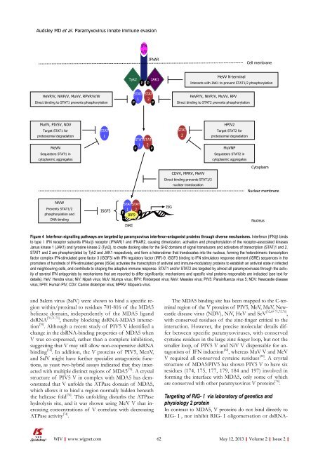
Audsley MD et al . <strong>Paramyxovirus</strong> <strong>innate</strong> immune <strong>evasion</strong>IFNα/βIFNARCell membraneTyk2PJAK1PMeVV N-terminalInteracts with JAK1 to prevent STAT1/2 phosphorylationHeVP/V, NiVP/V, MuVV, RPVP/V/WDirect binding to STAT1 prevents phosphorylationSTAT1STAT2PHeVP/V, NiVP/V, MuVV, RPVDirect binding to STAT2 prevents phosphorylationMuVV, PIV5V, NDVTarget STAT1 <strong>for</strong>proteosomal degradationMeVNSequesters STAT1 incytoplasmic aggregatesSTAT1PSTAT1PSTAT2PSTAT2HPIV2Target STAT2 <strong>for</strong>proteosomal degradationMuVNPSequesters STAT2 incytoplasmic aggregatesCytoplasmCDVV, MPRV, MeVVDirect binding prevents STAT1/2nuclear translocationNuclear membraneNiVWPrevents STAT1/2phosphorylation andDNA-bindingISGF3STAT STAT1 2P PIRF9ISREISGNucleusFigure 4 Interferon signalling pathways are targeted by paramyxovirus interferon-antagonist proteins through diverse mechanisms. Interferon (IFN)β bindsto type Ⅰ IFN receptor subunits IFNα/β receptor (IFNAR)1 and IFNAR2, causing dimerization, activation and phosphorylation <strong>of</strong> the receptor-associated kinasesJanus kinase 1 (JAK1) and tyrosine kinase 2 (Tyk2), to create docking sites <strong>for</strong> the SH2 domains <strong>of</strong> signal transducers and activators <strong>of</strong> transcription (STAT)1 and 2.STAT1 and 2 are phosphorylated by Tyk2 and JAK1 respectively, and <strong>for</strong>m a heterodimer that translocates into the nucleus, <strong>for</strong>ming the heterotrimeric transcriptionfactor complex IFN-stimulated gene factor 3 (ISGF3) with IFN regulatory factor (IRF)-9. ISGF3 binding to IFN stimulatory response element (ISRE) sequences in thepromoters <strong>of</strong> hundreds <strong>of</strong> IFN-stimulated genes (ISGs) activates the transcription <strong>of</strong> antiviral and immune-modulatory proteins to establish an antiviral state in infectedand neighbouring cells, and contribute to shaping the adaptive immune response. STAT1 and/or STAT2 are targeted by almost all paramyxoviruses through the activity<strong>of</strong> several IFN antagonists by mechanisms that are reported to differ significantly; mechanisms and specific viral proteins responsible are indicated (see text <strong>for</strong>details). HeV: Hendra virus; NiV: Nipah virus; MuV: Mumps virus; RPV: Rinderpest virus; MeV: Measles virus; PIV5: Parainfluenza virus 5; NDV: Newcastle diseasevirus; hPIV: Human PIV; CDV: Canine distemper virus; MPRV: Mapuera virus.and Salem virus (SalV) were shown to bind a specific regionwithin/proximal to residues 701-816 <strong>of</strong> the MDA5helicase domain, independently <strong>of</strong> the MDA5 liganddsRNA [70,71,73] , thereby blocking dsRNA-MDA5 interaction[70] . Although a recent study <strong>of</strong> PIV5 V identified achange in the dsRNA-binding properties <strong>of</strong> MDA5 whenV was co-expressed, rather than a complete inhibition,suggesting that V may still allow non-cooperative dsRNAbinding [74] . In addition, the V proteins <strong>of</strong> PIV5, MenV,and SalV might have further specialist antagonistic functions,as yeast two-hybrid assays indicated that they interactedwith multiple distinct regions <strong>of</strong> MDA5 [70] . A crystalstructure <strong>of</strong> PIV5 V in complex with MDA5 has demonstratedthat V unfolds the ATPase domain <strong>of</strong> MDA5,which allows it to bind a region normally hidden beneaththe helicase fold [74] . This unfolding disturbs the ATPasehydrolysis site, and it was shown using MeV V that increasingconcentrations <strong>of</strong> V correlate with decreasingATPase activity [74] .The MDA5 binding site has been mapped to the C-terminalregion <strong>of</strong> the V proteins <strong>of</strong> PIV5, MeV, MuV, Newcastledisease virus (NDV), NiV, HeV and SeV [32,69-71,75,76] ,with conserved residues <strong>of</strong> the zinc-finger critical to theinteraction. However, the precise molecular details differbetween specific paramyxoviruses, with conservedcysteine residues in the large zinc finger loop, but not thesmaller loop, <strong>of</strong> PIV5 V and NiV V dispensable <strong>for</strong> antagonism<strong>of</strong> IFN induction [32] , whereas MuV V and MeVV required all conserved cysteine residues [32] . A crystalstructure <strong>of</strong> MDA5:PIV5 has shown PIV5 V to have sixresidues (174, 175, 177, 179, 184 and 197) involved in<strong>for</strong>ming the interface with MDA5, only some <strong>of</strong> whichare conserved with other paramyxovirus V proteins [74] .Targeting <strong>of</strong> RIG-Ⅰ via laboratory <strong>of</strong> genetics andphysiology 2 proteinIn contrast to MDA5, V proteins do not bind directly toRIG-Ⅰ, nor inhibit RIG-Ⅰ oligomersation or dsRNA-WJV|www.wjgnet.com62 May 12, 2013|Volume 2|Issue 2|
Audsley MD et al . <strong>Paramyxovirus</strong> <strong>innate</strong> immune <strong>evasion</strong>binding [70] , which has been assumed to indicate that theyhave no direct role in inhibiting RIG-Ⅰ activation, butrather target downstream signalling components such asIRF-3 (see below). However, recent data has indicated thatV proteins can inhibit RIG-Ⅰ by interaction with anothercellular helicase, the laboratory <strong>of</strong> genetics and physiology2 (LGP2) [73] , via a region <strong>of</strong> LGP2 homologous to the Vprotein binding region in MDA5 [71,73] . The interaction appearsto be dependent on the unique C-terminal domain<strong>of</strong> V protein, as PIV5 P protein did not bind to LGP2,but the C-terminal domains <strong>of</strong> MeV and MuV V proteinswere necessary and sufficient <strong>for</strong> the interaction [71,73] . Importantly,V proteins were able to inhibit RIG-Ⅰ signallingonly in cells where LGP2 was coexpressed [73] , and RIG-Ⅰ-LGP2 interaction was detected only in cells expressing Vprotein, suggesting that V facilitates or mediates this interactionto shutdown RIG-Ⅰ activation [73] . Because LGP2 ishomologous to RIG-Ⅰ and MDA5, but lacks the CARDdomain to activate downstream signalling, it is thoughtto be a negative regulator <strong>of</strong> IFN induction, consistentwith the inhibitory effects <strong>of</strong> V protein expression. However,there is evidence that LGP2 can positively regulateIFN induction under some conditions [77-80] , so the precisemechanisms <strong>of</strong> V protein/LGP2 antagonism <strong>of</strong> RIG-Ⅰremain to be determined.Inhibition <strong>of</strong> IRF-3 activationIn addition to inhibition <strong>of</strong> PRRs, paramyxoviruses targetdownstream signalling components to prevent activation<strong>of</strong> IRF-3, potentially as a mechanism to inhibit signallingby both RLRs and TLRs (Figure 3). Rubulavirusesincluding MuV, hPIV2, and PIV5 use V protein as a decoysubstrate <strong>for</strong> the IRF-3 kinases TANK-binding kinase 1(TBK-1) and inhibitor <strong>of</strong> NF-κB kinase (IKK)ε (Figure 3),both inhibiting phosphorylation <strong>of</strong> IRF-3 and facilitatingIKKε/TBK-1 polyubiquitination and degradation to preventfurther signalling [81] .Henipavirus V proteins do not cause IKKε/TBK-1degradation [81] or block TLR-3/IRF-3 dependent signalling[76,81] . For henipaviruses, this appears to be a function<strong>of</strong> the W protein, as NiV W, although having no effecton MDA5 signalling, inhibited TLR-3-dependent phosphorylation<strong>of</strong> IRF-3 [82] . It is possible this is due to bindingand sequestration <strong>of</strong> inactive IRF-3 in the nucleuswhere NiV W localises, to prevent interaction withcytoplasmic IKKε/TBK-1 [82] . This model is consistentwith the reported importance <strong>of</strong> NiV W protein nuclearlocalisation to its inhibition <strong>of</strong> TLR3-dependent IFNinduction [82] . MeV C protein also inhibits IFN induction,correlating with its nuclear localisation [83] , although MeVC does not affect IRF-3 directly, and appears to have anundetermined nuclear target [83] . By contrast, cytoplasmicNDV and SeV V protein bind directly to IRF-3, therebypreventing its nuclear translocation [76] . Thus, paramyxovirustargeting <strong>of</strong> IRF-3-mediated signalling involvesmechanisms that appear to differ significantly betweenspecies.Targeting <strong>of</strong> STATs by rubulaviruses: degradation andmis-localisationAlmost all rubulavirus V proteins target STAT1 or STAT2<strong>for</strong> degradation by the host-cell proteosomal pathways [84-87]through assembly <strong>of</strong> a V-degradation complex (VDC)containing V protein, STAT1, STAT2, and components<strong>of</strong> an E3 ubiquitin ligase complex, specifically the UVdamage-specific DNA binding protein 1 (DDB1), andCul4A [88-92] , which likely mediate the STAT1/2 polyubiquitination[93] . In vitro studies/crystallographic analysis<strong>of</strong> the PIV5 V-DBB1 complex have indicated that boththe N-terminal and unique C-terminal regions <strong>of</strong> PIV5V are required <strong>for</strong> VDC assembly and STAT1 degradation[33,88,93,94] . Intriguingly, although some rubulavirusestarget only STAT1 or STAT2 <strong>for</strong> degradation (see Figure 4<strong>for</strong> details) [95,96] , both STATs are required, with the nondegradedSTAT acting as a “co-factor” [97,98] .The MuV V protein VDC polyubiquitinates and degradesnot only STAT1, but also STAT3 [84,99] , such thatMuV V protein can inhibit STAT3-dependent transcriptionalactivation by IL-6 and v-Src [99] . MuV targeting <strong>of</strong>STAT3 is independent <strong>of</strong> STAT1 targeting, as a pointmutation abrogating targeting <strong>of</strong> STAT3 did not affectSTAT1 [100] , and STAT3 degradation does not require theSTAT2 “c<strong>of</strong>actor” [99] . STAT3 targeting by the V protein<strong>of</strong> MuV is also highly specific to this species, as the Vproteins <strong>of</strong> the rubulaviruses MPRV, hPIV2 and hPIV4do not reduce cellular levels <strong>of</strong> STAT3 [87,101,102] .Intriguingly, the V proteins <strong>of</strong> hPIV4a and hPIV4bdo not degrade STATs or measurably affect their localisationor phosphorylation, but still bind to STAT1, STAT2and other VDC components [101] . While these virusesappear to lack the ability to antagonise STAT signalling,the specific binding capacity <strong>of</strong> the proteins is suggestive<strong>of</strong> a previous role in STAT antagonism, which may havebeen lost due to changes in selective pressures [101] .MPRV V protein, by contrast with those <strong>of</strong> other rubulaviruses,binds to STAT1 and STAT2 to prevent theirnuclear translocation without inducing degradation [102] .This is similar to reports <strong>for</strong> the V proteins <strong>of</strong> the henipavirusesand morbilliviruses (see below), except in thatMPRV V does not inhibit STAT1 phosphorylation andcan bind to STAT1 and STAT2 independently [102] . Asimilar mechanism may be used by the MuV NP protein,which co-localises with STAT2 in punctate aggregates inthe cytoplasm <strong>of</strong> infected cells [99] , indicating that NP protein,like P protein, can mediate both replication and IFNantagonist functions.Targeting <strong>of</strong> STATs by avulavirusesIn common with rubulaviruses such as PIV5, the avulavirusNDV targets STAT1, but not STAT2, <strong>for</strong> degradation.Deletion <strong>of</strong> the C-terminal region <strong>of</strong> V protein, ordeletion <strong>of</strong> both V and W C-termini by disruption <strong>of</strong>the RNA editing site, prevented STAT1 degradation byrecombinant NDV [95] . As little difference was observedbetween virus deleted <strong>for</strong> both V and W, and virus deletedWJV|www.wjgnet.com63 May 12, 2013|Volume 2|Issue 2|