Understanding Feline Cardiomyopathy (pdf)
Understanding Feline Cardiomyopathy (pdf)
Understanding Feline Cardiomyopathy (pdf)

You also want an ePaper? Increase the reach of your titles
YUMPU automatically turns print PDFs into web optimized ePapers that Google loves.
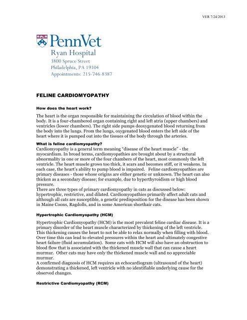
VER 7/24/2013Ryan Hospital3800 Spruce StreetPhiladelphia, PA 19104Appointments: 215-746-8387FELINE CARDIOMYOPATHYHow does the heart work?The heart is the organ responsible for maintaining the circulation of blood within thebody. It is a four-chambered organ containing right and left atria (upper chambers) andventricles (lower chambers). The right side pumps deoxygenated blood returning fromthe body into the lungs. From the lungs, oxygenated blood enters the left side of theheart where it is pumped out into the tissues of the body through the arteries.What is feline cardiomyopathy?<strong>Cardiomyopathy</strong> is a general term meaning “disease of the heart muscle” - themyocardium. In broad terms, cardiomyopathies are brought about by a structuralabnormality in one or more of the four chambers of the heart, most commonly the leftventricle. The heart muscle grows too thick, it scars and becomes stiff, or it weakens. Ineach case, the heart’s ability to pump blood is impaired. <strong>Feline</strong> cardiomyopathies areprimary diseases - those whose origins are either genetic or unknown. The heart can alsothicken as a secondary disease; for example, due to hyperthyroidism or high bloodpressure.There are three types of primary cardiomyopathy in cats as discussed below:hypertrophic, restrictive, and dilated. Cardiomyopathies primarily affect adult cats andalthough all cats are susceptible, a genetic predisposition for the disease has been shownin Maine Coons, Ragdolls, and in some American shorthair cats.Hypertrophic <strong>Cardiomyopathy</strong> (HCM)Hypertrophic <strong>Cardiomyopathy</strong> (HCM) is the most prevalent feline cardiac disease. It is aprimary disorder of the heart muscle characterized by thickening of the left ventricle.This thickening causes the heart to not be able to relax normally when filling with blood.Over time this can lead to elevated pressures within the heart and ultimately congestiveheart failure (fluid accumulation). Some cats with HCM will also have an obstruction toblood flow that is associated with the thickened muscle wall that can cause a heartmurmur. Other cats may have only the thickened muscle wall and no appreciablemurmur.A confirmed diagnosis of HCM requires an echocardiogram (ultrasound of the heart)demonstrating a thickened, left ventricle with no identifiable underlying cause for theobserved changes.Restrictive <strong>Cardiomyopathy</strong> (RCM)
VER 7/24/2013Restrictive cardiomyopathy is caused by excessive buildup of scar tissue (fibrosis) on theinner lining of the ventricle. This prevents the ventricle from adequately relaxing, filling,and emptying with each heart beat. The clinical signs are similar to HCM and aconfirmed diagnosis also requires an echocardiogram.Dilated <strong>Cardiomyopathy</strong> (DCM)Dilated cardiomyopathy is rarely seen in cats today. Historically, it was linked to adietary deficiency in taurine, which has been corrected by most cat food manufacturers.DCM is characterized by a poorly contracting dilated left ventricle and oftentimesenlarged atria. Cats with DCM usually progress to congestive heart failure.Potential OutcomesAlthough there is variable progression with feline cardiomyopathy with many catsremaining asymptomatic for years, many will progress to developing clinical signsassociated with their disease at some point.A common outcome with cardiomyopathy is congestive heart failure (CHF; fluidaccumulation). The most common location for fluid accumulation in cats is within thelungs (pulmonary edema) or around the lungs (pleural effusion). This results indifficulty breathing and is a medical emergency.Cats with significant heart disease are predisposed to the formation of blood clots. If aclot forms within the heart and enters the circulation, small pieces may lodge within thearteries. The most common location for this to happen is within the terminal aorta,causing an occlusion of flow to the hind legs. This process is accompanied by severe pain,lameness and/or paralysis of one or both hind legs.What are the goals of treatment?Currently, there is no cure cardiomyopathy in cats and no drugs have been shown to slowthe progression. In a small subset of cats with a rapid heart rate or significantobstruction to blood flow within the heart, a drug called a beta blocker may be prescribedprior to development of clinical signs, in order to slow the heart rate and decrease theworkload on the heart. A beta blocker should not be prescribed prior to anechocardiogram. In most other cases, treatment is not initiated until the development ofCHF, and is aimed at alleviating fluid accumulation, improving heart function,suppressing arrhythmias, and reducing the risk of blood clot formation. Please see thebrochure on <strong>Understanding</strong> Canine and <strong>Feline</strong> Congestive Heart Failure for moreinformation on treatment.What should I monitor at home?Breathing rate: In an effort to catch CHF early, is important to become familiar withyour cat’s normal resting breathing rate and effort at home. When your cat is at rest,watch his/her sides rise and fall as he/she breathes normally. One rise and fall cycle isequal to one breath. Count the number of breaths he/she takes in 6 seconds, thenmultiply this number by 10 to get total breaths per minute. For example, if you count 3breaths in 6 seconds, that is equal to 30 (3x10) breaths per minute. A normal cat at restshould have a respiratory rate less than 40. If you notice this number increasingconsistently, or notice an increase in the effort it takes to breathe, please contact a
VER 7/24/2013veterinarian. It may be helpful to keep a daily log of your pet’s breathing rate so thatyou will notice increases or changes from normal breathing.Use of limbs: Cats with heart disease are at a higher risk for forming blood clots. Themost common way this manifests is through loss of use of one or more limbs, mostcommonly the hind legs. Monitor your cat for any dragging or inability to use the limbs.General: Monitor for any lethargy, collapse, exercise intolerance, coughing or decreasedappetite. Contact a veterinarian if you notice any of these changes.Thank you for visiting the cardiology service at the Ryan Veterinary Hospital. If you haveany further questions, please do not hesitate to contact us