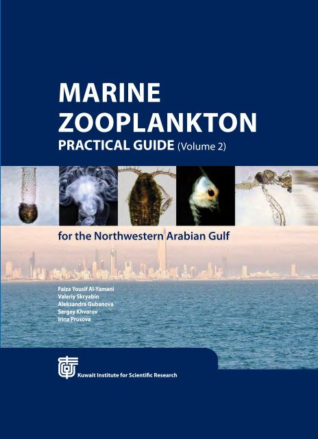
marine zooplankton practical guide - KISR
marine zooplankton practical guide - KISR
marine zooplankton practical guide - KISR

You also want an ePaper? Increase the reach of your titles
YUMPU automatically turns print PDFs into web optimized ePapers that Google loves.
Family CENTROPAGIDAE 42Genus Centropages 42Centropages furcatus 44Centropages orsinii 46Centropages tenuiremis 46Family PSEUDODIAPTOMIDAE 48Genus Pseudodiaptomus 48Pseudodiaptomus arabicus 49Pseudodiaptomus ardjuna 50Family TEMORIDAE 51Genus Temora 51Temora discaudata 52Temora turbinata 54Family CANDACIIDAE 56Genus Candacia 56Candacia bradyi 57Family PONTELLIDAE 58Genus Calanopia 58Calanopia elliptica 58Calanopia minor 60Genus Labidocera 62Labidocera acuta 62Labidocera bengalensis 66Labidocera kroyeri 68Labidocera minuta 70Labidocera sp. 72Genus Pontella 74Pontella danae 74Pontella investigatoris 76Genus Pontellopsis 78Pontellopsis herdmani 78Family ACARTIIDAE 80Genus Acartia 80Acartia (Acanthacartia) fossae 82Acartia (Odontacartia) amboinensis 84Acartia (Odontacartia) ohtsukai 86Genus Acartiella 88Acartiella faoensis 88MARINE ZOOPLANKTON PRACTICAL GUIDE for the Northwestern Arabian Gulf
Family TORTANIDAE 90Genus Tortanus 90Tortanus barbatus 90Tortanus forcipatus 92Order CYCLOPOIDA 94Family OITHONIDAE 94Genus Oithona 94Oithona attenuata 96Oithona brevicornis 98Oithona nana 98Oithona plumifera 100Oithona sp. 102Family ONCAEIDAE 104Genus Oncaea 104Oncaea clevei 106Family SAPPHIRINIDAE 106Genus Copilia 108Copilia mirabilis 108Genus Sapphirina 110Sapphirina nigromaculata 110Family CORYCAEIDAE 112Genus Corycaeus 112Subgenus Dithrichocorycaeus 113Corycaeus (Dithrichocorycaeus) andrewsi 113Corycaeus (Dithrichocorycaeus) dahli 114Corycaeus (Dithrichocorycaeus) lubbocki 116Subgenus Onychocorycaeus 116Corycaeus (Onychocorycaeus) agilis 117Corycaeus (Onychocorycaeus) pacificus 118Order HARPACTICOIDA 120Family ECTINOSOMATIDAE 120Genus Microsetella 120Microsetella sp. 120Family MIRACIIDAE 122Genus Macrosetella 122Macrosetella gracilis 122Kuwait Institute for Scientific Research
Family EUTERPINIDAE 124Genus Euterpina 124Euterpina acutifrons 124Family CLYTEMNESTRIDAE 124Genus Clytemnestra 125Clytemnestra scutellata 1252.14. Mysida 126Rhopalophthalmus sp. 1262.15. Decapoda 128Metapenaeus ensis, protozoea II 128Parapenaeopsis stylifera, protozoea III 129Penaeus semisulcatus, protozoea I 130Penaeus semisulcatus, protozoea III 131Solenocera crassicornis, protozoea III 132Solenocera crassicornis, mysis 133Solenocera hextii, protozoea III 134Lucifer hanseni, protozoea III 135Lucifer hanseni, mysis I 136Lucifer hanseni, male 137Lucifer typus, male 138Acetes japonicas, female 140Sergestes sp., protozoea II 142Sergestes sp., mysis 143Stenopus sp., zoea I 144Thalassocaris obscura, zoea XI 145Acanthephyra sp., zoea IV 146Palaemon sp., zoea IV 147Periclimenes sp., zoea IV 148Alpheus sp. 149Latreutes sp., zoea IX 150Lysmata sp. 151Callianassa sp., zoea I 152Laomedia sp., zoea IV 153Upogebia sp., zoea I 154Thenus orientalis, phyllosoma I 155Thenus orientalis, phyllosoma II 156Thenus orientalis, phyllosoma III 157MARINE ZOOPLANKTON PRACTICAL GUIDE for the Northwestern Arabian Gulf
Galathea sp., zoea III 158Pachycheles sp., zoea I 159Diogenes sp., zoea III 160Dardanus sp., zoea I 161Paguristes sp., zoea I 162Pagurus sp., zoea III 163Ebalia sp., zoea I 164Leucosia sp., zoea I 165Parthenope sp., zoea IV 166Macrophthalmus sp., zoea III 167Ilyoplax frater, zoea VI 1682.16. Chaetognatha 169Sagitta enflata 169Sagitta neglecta 170Sagitta pulchra 171Sagitta regularis 1722.17. Urochordata 173Oikopleura dioica 173Appendicularia sicula 174Fritillaria sp. 175Weelia cylindrica 176Thalia sp. 177Tadpole Larva 1782.18. Cephalochordata 180Branchiostoma sp. 180Addendum 182Catostylus mosaicus 182Fritillaria pellucida 184References 186Index of Scientific Names 193Kuwait Institute for Scientific Research
PREFACEThe <strong>marine</strong> <strong>zooplankton</strong> community of Kuwaitwaters includes fascinating organisms, which areabundant, diverse, and encompasses representativesof all the major invertebrate phyla. Along withphytoplankton, <strong>zooplankton</strong> are key components of<strong>marine</strong> ecosystems forming the base of most <strong>marine</strong>food webs. Plankton constitute the principal diet ofthe early life stages of fish including commerciallyimportant fish.Zooplankton research is an essential componentin forming a more complete understanding ofthe functioning of <strong>marine</strong> ecosystems. However,identifying <strong>zooplankton</strong> can be a challenging and atedious task and requires a high level of expertise.The samples used to produce this <strong>guide</strong> wereobtained during the 1999-2010 sea cruises coveringall Kuwait waters as well as some localities inthe Arabian Gulf waters. A total of 183 species of<strong>zooplankton</strong> were recorded and identified. Speciesdiversity was highest for the groups of tintinnids,copepods, and cnidarians (75, 54 and 14 species,respectively).This illustrated <strong>zooplankton</strong> <strong>guide</strong> is aninvaluable reference for taxonomists, <strong>marine</strong>plankton ecologists, environment managers,coastal engineers, students of invertebrate biology,environmental impact assessment experts and<strong>marine</strong> biologist. This <strong>practical</strong> book provides acomprehensive documentation of <strong>zooplankton</strong>biodiversity and taxonomical description of thecommonly encountered species in Kuwait waters aswell as the Arabian Gulf.Information about methods on <strong>zooplankton</strong>sample collection, sample processing and analysisare included in Volume 1 of the <strong>zooplankton</strong><strong>guide</strong>book (pages 2-8). Moreover, Volume 1 containsdescriptions and photographs of protozoan<strong>zooplankton</strong> (Tintinnida), Cnidaria, Ctenophora,Rotifera, Cladocera, Ostracoda and Larvae of <strong>marine</strong>benthos.iMARINE ZOOPLANKTON PRACTICAL GUIDE for the Northwestern Arabian Gulf
“We consider species to be likea brick in the foundation of abuilding. You can probably loseone or two or a dozen bricks andstill have a standing house. But bythe time you’ve lost 20 per cent ofspecies, you’re going to destabilizethe entire structure. That’s the wayecosystems work.”Donald FalkKuwait Institute for Scientific Researchii
ACKNOWLEDGEMENTSGratitude is extended to Mr. Alan Lennox (KuwaitInstitute for Scientific Research, Kuwait) for histremendous help in collecting the needed samplesfor this study from Kuwait waters, and to ROPME forproviding Arabian Gulf samples. Our appreciationto Mrs. Gracekutty Thomas Vargese for her hardwork in meso<strong>zooplankton</strong> sample analysis, and toMr. Vasiliy Prusov for his help in computerizing theCopepod drawings. We are grateful to Dr. InnaDrapun (IBSS, Sevastopol) for her great contributionin identifying and description of ostracods andphotographing of <strong>zooplankton</strong>. We thank Mrs.Linda Fernandes for compiling and formatting thematerials used for this <strong>guide</strong>.The authors are grateful to the Kuwait Institutefor Scientific Research for the financial support of thisresearch project.iiiMARINE ZOOPLANKTON PRACTICAL GUIDE for the Northwestern Arabian Gulf
We share this planet with many species. It is our responsibility toprotect them, both for their sakes and our own.Pamela A. MatsonKuwait Institute for Scientific Researchiv
Bubiyan bridge off Khor Al-Sabbiya - Photo by Dr. V. Skryabin - Kuwait Institute for Scientific Research12MARINE ZOOPLANKTON PRACTICAL GUIDE for the Northwestern Arabian Gulf
2.13. CopepodaThe copepods are crustaceans, which are abundant in Kuwait waters and often dominate theplankton community, especially in Kuwait Bay. They are small in size (only a few species over 1mm). They form a link in the food web between the primary-producing phytoplankton and theplankton-feeding fish. Most of the economically important fish depend on copepods and otherzooplankters during their early life stages (larval fish stages) as well as adult stages of some fishsuch as Zobaidy (Pampus argenteus), Suboor (Tenualosa illisha), and Beyah (Liza subviridis).There are about 2,000 known <strong>marine</strong> pelagic species of copepods (Razouls and Bovée, 1998). Thecopepod females lay eggs freely into the water, or produce external paired or single egg sacs. Theeggs hatch into copepod larvae. The first larvae are called nauplii. Larval copepods usually passfive or six naupliar stages, which are separated by a moult. The 6 th naupliar stage moults into thefirst copepodite stage, which resembles the adult copepod. After molting through 5 copepoditestages, copepods attain adulthood and cease molting.Copepods comprise about 70% of the <strong>zooplankton</strong> community of Kuwait waters (Al-Yamani et al.,1997 a, b), and have a variable distribution both spatially and temporally. The first comprehensive<strong>zooplankton</strong> study in the northwestern Arabian Gulf with emphasis on copepod species wasconducted in 1979-1980 (Michel et al., 1986 a, b). The first <strong>guide</strong>book on the copepods of Kuwaitwaters was by Al-Yamani and Prusova, 2003.This <strong>guide</strong>book includes an updated version on copepods (54 species), with descriptions,drawings and photographs of the adult stages of the common copepod species encounteredmainly in the waters of the NW Arabian Gulf. Relevant identification keys are also included in the<strong>guide</strong>.Kuwait Institute for Scientific Research 1
A total of 50 species of copepods from Kuwait waters have been identified, with additional 4species that were identified only to the genus level. These identified copepod species belong to 3orders: Calanoida, Cyclopoida and Harpacticoida. In calanoids the major movable articulationof the body occurs immediately posterior to the last free thoracic somite, i.e., between thelast prosome somite and the genital somite, so that the urosome consists only of genital andabdominal segments (Fig. 117A). In cyclopoids and harpacticoids a major articulation in thetrunk occurs immediately anterior to the last free thoracic segment, so the last body region(urosome) consists of one segment, bearing a usually rudimentary pair of swimming legs, thegenital somite, and the abdominal segment ending in caudal rami (Fig. 117B – 117D). In mostcyclopoids, as in calanoids, the urosome is much narrower than the prosome. In harpacticoids,there is much less of difference in segment width between the prosome and urosome (Fig. 117D).The terminology adopted here for the description of the external morphology of copepodsfollows that of Huys and Boxshall (1991). The term “somite” is used in describing certain parts ofthe copepod body (the prosome and urosome), the term “segment” is used in descriptions of theparts of the antennules, mouth appendages and legs. The list below identifies each abbreviationused in the descriptions, figures and keys (Figs. 114 and 115).A1: antennuleA2: antennaAns: anal somiteB: basisC: coxaCe: cephalosomeCR: caudal ramusEnp: endopod; Enp1-3 – endopod segments 1-3Exp: exopod; Exp1-3 - exopod segments 1-3Gn: gnathobaseGns: genital somiteMd: mandibleMdp: mandibular palpMx1: maxilluleLe: external lobeLi: internal lobeMx2: maxillaMxp: maxillipedP1-5: swimming legs 1-5Pd1-5: pedigerous somites 1-5Pr: prosomeR: rostrumUr1-5: urosomites 1-5Total length – the length from the top of a copepod head up to the end of CR, excluding caudalsetae.There are typically six copepodite stages (abbreviated C1-C6), the sixth being either the adultmale and female. Growing from stage to stage is carried through moulting. Lebour (1916) wasfirst to describe the copepodite stages. Later the tables to determine these stages were made.Morphological features of stages of different species of Copepoda are presented in the followingtable.4MARINE ZOOPLANKTON PRACTICAL GUIDE for the Northwestern Arabian Gulf
Identification of Copepodite Stages 1-6 (Source: Sazhina, 1987)Segments NumberStageNumber of Swimming LegsProsomeUrosomeC1 4 2 2+ underdeveloped P3C2 5 2 3+ underdeveloped P4C3 6 2 4+ underdeveloped P5C4 6 3 4+ underdeveloped P5C5 female 6 3-4 4 or 5C5 male 6 4-5 4 or 5C6 female 6 3-4 (Gns swollen) 4 or 5C6 male 6 4-5 (Gns elongated) 5aFig. 115. External morphology of copepods: a. the different parts of an adult copepod, b. A1(antennule), c. A2 (antenna), d. Md (mandible), e. Mx1 (maxillule), f. Mx2 (maxilla), and g. Mxp(maxilliped) (Source: Bradford-Grieve et al., 1999).Kuwait Institute for Scientific Research 5
cdefgFig. 115. Cont’d.6MARINE ZOOPLANKTON PRACTICAL GUIDE for the Northwestern Arabian Gulf
Conway et al. (2003) suggested that most of the literature on identification only give descriptionsof the adult stage of copepods. In most female copepods, swollen genital somite indicates that itis mature. But genital somite of Acartia start to swell in pre-adult copepodite 5 stage (C5), whilethe body segmentation is often identical between C5 and C6. However, in both female and malepre-adult copepods, the segmentation of the P5 and urosome is not distinct as in the adult stage,which becomes obvious when the identifier examines them together (Conway, et al., 2003). I nother genera, e.g., Pseudodiaptomus and Centropages, pre-adult females (C5) have a bit swollengenital somite similar to Acartia.abFig. 116. Plan of body organization in a. gymnopleansand b. podopleans (Source: Bradford-Grieve et al.,1999).ab c dFig. 117. Differences between copepod Orders:a. Calanoida (Canthocalanus pauper); b, c. Cyclopoida (b.Oithona brevicornis, c. Oncaea clevei); d. Harpacticoida(Euterpina acutifrons). Arrows indicate the genitalsomites. (Source: Al-Yamani and Prusova, 2003).Kuwait Institute for Scientific Research 7
List of Copepods TaxaPhylum Class Susclass Order Family Genus / SpeciesArthropoda Crustacea Copepoda Calanoida Calanidae Canthocalanus pauperParacalanidaeAcrocalanus gibberAcrocalanus longicornisBestiolina arabicaParacalanus indicusParacalanus sp.Parvocalanus crassirostrisParvocalanus elegansEucalanidaeSubeucalanus flemingeriSubeucalanus subcrassusClausocalanidaeClausocalanus minorEuchaetidaeCentropagidaeEuchaeta concinnaEuchaeta rimanaCentropages furcatusCentropages orsiniiCentropages tenuiremisPseudodiaptomidaePseudodiaptomus arabicusPseudodiaptomus ardjunaTemoridaeCandaciidaePontellidaeTemora discaudataTemora turbinataCandacia bradyiCalanopia ellipticaCalanopia minorLabidocera acutaLabidocera bengalensisLabidocera kroyeriLabidocera minutaLabidocera sp.Pontella danaePontella investigatorisPontellopsis herdmaniAcartiidaeAcartia (Acanthacartia) fossaeAcartia (Odontacartia) amboinensisAcartia (Odontacartia) ohtsukaiAcartiella faoensis8MARINE ZOOPLANKTON PRACTICAL GUIDE for the Northwestern Arabian Gulf
Phylum Class Susclass Order Family Genus / SpeciesTortanidaeTortanus barbatusTortanus forcipatusCyclopoida Oithonidae Oithona attenuataOithona brevicornisOithona nanaOithona plumiferaOithona sp.OncaeidaeOncaea cleveiSapphirinidaeCorycaeidaeCopilia mirabilisSapphirina nigromaculataCorycaeus (Dithrichocorycaeus) andrewsiCorycaeus (Dithrichocorycaeus) dahliCorycaeus (Dithrichocorycaeus) lubbockiCorycaeus (Onychocorycaeus) agilisCorycaeus (Onychocorycaeus) pacificusHarpacticoida Ectinosomatidae Microsetella sp.MiraciidaeMacrosetella gracilisEuterpinidaeClytemnestridaeEuterpina acutifronsClytemnestra scutellataKuwait Institute for Scientific Research 9
Systematic Account of Copepod Species from theNorthwestern Arabian GulfPhylum ArthropodaClass Crustacea Brunnich, 1772Subclass Copepoda Milne-Edwards, 1840Order Calanoida Sars, 1903The Calanoida are defined by the combination of the gymnoplean tagmosis, the presence ofonly 1 spine on the outer margin of P2-P5 Exp1, the presence of a coxal epipodite (Le1), butnot a lobate basal exite on Mx1, and the presence of a seta on the inner margin of A2 coxa. Thepresence of a maximum of 2 setae on the terminal segment of Mx2 (Enp4) is an apomorphy(derived character) of the Calanoida (Huys and Boxshall, 1991; Bradford-Grieve et al., 1999).Family Calanidae Dana, 1849Reference: Bradford-Grieve et al., 1999.FemalesCephalosome and Pd1 may be fused or separate, Pd4 and 5 always separate. Rostrum of 2filaments. Caudal rami with 4 subequal, terminal setae and 1 outer seta. Urosome of 4 somites.A1 25-segmented, generally segments 8-9 partly fused; segments 23-24 with elongate seta. A2with 1 seta, B with 2 setae; A2 Exp 7-segmented; Exp1 and Exp2 with 2 setae each, Exp3-6 with 1seta each, Exp7 usually with 1 seta nearly at midlength and 3 terminal setae; Enp1 with 2 setae,Enp2 with 14-16 setae. Mdp B with 4 setae; Enp1 with 4 setae and prominent lobe; Enp2 totallywith 10-11 (8 or 9 terminal and 2 posterior) setae. Exopod and endopod of approximately equallength. Mx1 Li1 with 9 terminal spines, 4 posterior and 1 anterior setae; Li2 and Li3 with 4 setaeeach; Li4 with 4 setae, endopod with 14-15 setae; exopod with 11 setae; Le1 with 9 setae; Le 2with 1 seta. Mx2 Li1-Li5 with 4-6, 3, 3, 3 and 4 setae, respectively. Terminal part with about 10setae. Mxp coxa with 1 proximal seta, then with groups of 2, 4 and 4 setae from proximal to distal;Enp2-6 with 4, 4, 3/4, 4, 4/5 setae respectively. P2-P3 B with an external articulated spine, on P4and P5 this may be reduced to a small seta. P2 Exp1 sometimes with recurved spine (Neocalanus),or Exp2 with deep invagination (Undinula). P5 similar to P2-4; sometimes with inner edge teeth(Calanus, Nannocalanus, Cosmocalanus).MalesUrosome of 5 somites. A1 25-segmented, with segments 1 and 2 always fused, segments 3-5,7 and 8, 9 and 10, and 24 and 25 may also be fused. A2-Mx2 – either exactly as in female, orreduced in size and setation. Mxp with reduced inner setae but enlarged outer setae on terminalsegments. P1-P4 as in female. P5 with both rami usually 3-segmented, right leg similar to otherlegs, left leg variously modified; endopod sometimes reduced and devoid of segmentation onone or both sides.10MARINE ZOOPLANKTON PRACTICAL GUIDE for the Northwestern Arabian Gulf
Salmiya beach - Photo by Dr. V. Skryabin - Kuwait Institute for Scientific ResearchKuwait Institute for Scientific Research 11
Genus Canthocalanus A. Scott, 1909References: Bradford and Jillet, 1974; Bradford-Grieve et al., 1999.FemalesCephalosome and Pd1 fused. Pd4 and Pd5 separate. Rostrum of 2 filaments. Caudal rami with4 subequal, terminal setae and 1 outer seta. Urosome of 4 somites. A1 25-segmented. A2 Exp7-segmented. Mdp B with 4 setae, Exp and Enp of approximately equal length. Mx2 with 4 setaeon Li1. P1: B1 anterior margin terminates in a well defined projection; B2 with distal seta onanterior surface modified into a proximally thickened spine. P2-P3 B with an external articulatedspine. P2-P4 without modification or ornamentation. P5 with the inner border of B1 naked, Enpwith 7 setae.MalesUrosome of 5 somites. A1 25-segmented. Cephalosome and Pd1 fused. Mouthparts as in female.P5 with the inner border of B1 naked; both rami 3-segmented, hardly modified on right; leftendopod with only 2 terminal setae.Canthocalanus pauper Giesbtecht, 1888Female. Length: 1.4-1.7 mm (1.3-1.6 mm). (Fig. 118 A, B, D; Fig. 119 a, b)Head and pedigerous segment 1 fused. Anterior cephalosome and posterior metasome rounded.Rostrum of 2 filaments. Urosome 4-segmented. Strong setae on caudal rami. Mx2 Li1 with 4setae. P1 C anterior margin terminates in well-defined projection; basis with inner marginal setamodified into proximally thickened spine. P5 C inner edge without teeth (naked). P1-P5 Enp with7 setae.Male. Length 1.2-1.5 mm (1.3-1.5 mm). (Fig. 118 C, E; Fig. 120 a, b)Right P5 asymmetric; endopod with 8 setae; exopod without inner marginal spines; left endopodwith 2 terminal setae; left exopod with elongated segments, with long outer distal setae onsegments 2 and 3. Inner margins of the P5 coxa are not denticulate.12MARINE ZOOPLANKTON PRACTICAL GUIDE for the Northwestern Arabian Gulf
Family Paracalanidae Giesbrecht, 1892References: Giesbrecht, 1892; Vervoort, 1963; Andronov, 1971, 1977; Bradford-Grieve, 1994 (citedby Bradford-Grieve et. al., 1999).Females (Fig. 121)Cephalon and Pd1 usually fused. Pd4 and 5 fused, or separated. Urosome of 2 to 4 somites.Anal somite usually longer than any somite between it and genital somite. Rostrum of 2filaments (Acrocalanus, Calocalanus, Paracalanus); 2-pointed, solid (Delius, Bestiola); or massive(Parvocalanus). A1 usually 25-segmented, generally with segments 1 and 2, also 8 and 9, partiallyfused; in Delius first 8 segments fused. A2 Exp 7-segmented with Exp1 and Exp2 each bearing 2setae, segment 7 elongate. Md Enp1 without prominent lobe. Mx1 Li1 with 14 setal elements (4posterior and 1 anterior setae, and 9 terminal spines); Li2 and Li3 with 3-4 setae each; Li4 with4 setae; endopod with 13-14 setae; exopod with 11 setae; Le2 with 1 reduced seta; Le1 with7-9 setae. Mx2 Li1-Li5 with 6, 3, 3, 3 and 3-4 setae respectively; terminal part with 8 setae, 1 ofthem may belong to developed Li6. Mxp C with 1 proximal seta, then 3 groups of 2, 3 and 4setae from proximal to distal; Enp2-Enp6 with 3, 4, 3, 3+1 and 4 setae respectively. P1-P4 exopodusually 3-segmented but P1 exopod 2-segmented in some Calocalanus species. P2-P4 endopodsusually 3-segmented but 2-segmented in Calocalanus minor, posterior surfaces of somesegments ornamented with spines. P2-P4 Exp3 external borders serrated, or smooth in Deliusand Calocalanus. Terminal spines of P2-P4 exopods smooth. P5 uniramous, absent, or vestigial(Acrocalanus), present on left only (Delius). If both P5 present, then symmetrical, presented bycoxae (Bestiolina), 2-segmented (or 3-segmented) (Paracalanus, Parvocalanus), or 3-4 segmented(Calocalanus). Females of four genera of family Paracalanidae common for the Kuwait waters,Acrocalanus, Bestiolina, Paracalanus and Parvocalanus, are shown in Fig. 121.Differences between Genera Acrocalanus, Bestiolina, Paracalanus and Parvocalanus are displayedin the table below.Males (Fig. 122)Urosome of 5 somites. Genital opening on left (in Calocalanus on left or right). Cephalic humppresent (Acrocalanus, Bestiolina and Paracalanus), or absent. A1 25-segmented, with some or allsegments 1-6 fused and swollen, segments 9-10 may also be fused. A2 Exp7 very short without3 terminal setae, which are present in female. Exp1 and Exp2 without setae. Other oral partsreduced compared with female. Right P5 may be absent (Acrocalanus, Delius), or present as C(Bestiolina), 2-3-segmented (Paracalanus, Parvocalanus), or 3-4-segmented (Calocalanus).There are four common genera of Paracalanidae in Kuwait water: Acrocalanus, Giesbrecht, 1888;Bestiolina, Andronov, 1991; Paracalanus, Boeck, 1864; Parvocalanus, Andronov, 1970.Construction of swimming legs is also an important feature, which allows to distinguish oneParacalanidae genus from another (Fig. 122). Acrocalanus P2-P4 Exp2 and Exp3 external edgesserrated; Bestiolina P2-P4 Exp2 and Exp3 external borders smooth. Paracalanus P2-P4 Exp3external borders serrated; Parvocalanus P2-P4 Exp3 external borders serrated.Differences between Genera Acrocalanus, Bestiolina, Paracalanus and ParvocalanusGenus Size Rostrum P5Acrocalanus 0,8 – 1,25 of 2 filaments absentBestiolina 0,6 – 0,75 of 2-pointed, solid, long extensions presented by coxaParacalanus 0.7 –1,1 of 2 filaments symmetrical, uniramous2 segmentedParvocalanus 0,45 – 0,65 of 2-pointed, solid, short extensions symmetrical, uniramous2 segmented14MARINE ZOOPLANKTON PRACTICAL GUIDE for the Northwestern Arabian Gulf
12 34Fig. 121. Females of four genera of Paracalanidae family common for the Kuwait waters: 1. Acrocalanus;2. Bestiolina; 3. Paracalanus; 4. Parvocalanus. Arrows indicate the rostrum and leg 5.(Sources: 1, 4 – from Al-Yamani and Prusova, 2003; 2 – from Ali et al. 2007, 3 – from Bradford-Grieve et al.,1999).1 2 3 4Fig. 122. Fourth swimming leg (P4) exopods of Paracalanidae females: 1. Acrocalanus; 2. Bestiolina;3. Paracalanus; 4. Parvocalanus.(Sources: 1, 3, 4 – from Al-Yamani and Prusova, 2003; 2 – from Ali et al., 2007).Kuwait Institute for Scientific Research 15
Genus Acrocalanus Giesbrecht, 1888Reference: Bradford-Grieve et al. (1999).FemalesCephalosome and Pd1 fused. Pd4 and Pd5 partly fused. Urosome of 4 somites, anal somite longerthan any somite between it and genital somite. Rostrum of 2 filaments. A2 Exp 7-segmentedwith Exp1 and Exp2 each baring 2 setae, segment 7 elongate. Md Enp1 without prominent lobe.P1-P4 exopods 3-segmented; P1 B with inner marginal seta; P2-P4 endopods 3-segmented, Enp2posterior surface ornamented with spines; P2-P4 Exp2-3 external edges serrated. Terminal spinesof P2-P4 Exp smooth. P5 absent or vestigial.MalesUrosome of 5 somites. Genital opening on left. Cephalic hump present (lateral view). A1 severalfirst segments fused and swollen. Right P5 absent.Acrocalanus gibber Giesbrecht, 1888Female. Length: 0.9-1.2 mm (0.93-1.28 mm). (Fig. 123 A, B, C D, F; Fig. 124 a, b)Deep body in lateral view, with a pronounced hump on the dorsal cephalosome; distinctthough partial line of separation of the cephalosome from the fist pedigerous segment isobvious; dorsal outline of cephalosome significantly humped in lateral view. A1 exceeds beyondend of caudal rami by 2 terminal segments. 4 pair of swimming legs. P4 E xp3 external edgeteeth on distal end equal in size to those on proximal end. Genital somite longer than any ofothers, with a prominent ventral swelling.Male. Length: 0.9-1.2 mm (0.94-1.24). (Fig. 123 E; Fig. 124 c)P4 Exp3 with about 10-12 teeth o n distal part of segment. P5 is only present on the left andextends to the end of the urosome segment 3, or the middle of urosome segment 4 when thisleg is fully extended; very simple limb, composed of four segments with two tiny spines onthe last segment.16MARINE ZOOPLANKTON PRACTICAL GUIDE for the Northwestern Arabian Gulf
Fig. 123. Acrocalanus gibber. Female.A. dorsal view; B. left lateral view; C. Ur, dorsal; D. Ur, left lateral view; E. P4, male; F. P4, female.(Sources: A-D - from Al-Yamani, Prusova, 2003; E, F - from Sewell (1929) cited by Conway, 2003).a b cFig. 124. Acrocalanus gibber: a. female, dorsal view; b. female, lateral view; c. male, lateral view.Kuwait Institute for Scientific Research 17
Acrocalanus longicornis Giesbrecht, 1888Reference: Bradford-Grieve, 1994.Female. Length 1.00 – 1.26 mm. (Fig. 125 1 – 7; Fig 126 a – d)Dorsal surface of the body moderately strongly arched, anterior head rounded. A1 extendsbeyond the caudal rami by its last 5 segments. Outer distal border of exopod segment 3 of P4with very small numerous teeth.Male. Length 0.95 – 1.25 mm. (Fig. 127 a, b)Exp 3 of P4 similar to that of the female. P5 - 5 segmented on the left.Fig. 125. Acrocalanus longicornis. Female.1. dorsal view; 2. lateral view; 3. P1; 4. P2; 5. P3; 6. P4; 7. P5.(Source: Corral Estrada, 1970).18MARINE ZOOPLANKTON PRACTICAL GUIDE for the Northwestern Arabian Gulf
cdabFig. 126. Acrocalanus longicornis. Female. a. dorsal view; b. lateral view; c. exopod of P4; d. distal part of exopodof P4.abFig. 127. Acrocalanus longicornis. Male. a. lateral view; b. P5.(Source: Chen et al., 1974).Kuwait Institute for Scientific Research 19
Genus Bestiolina, Giesbrecht, 1888References: Ali et al., 2007Bestiolina arabica Ali et al., 2007Female. Length 0.79-0.92 mm. (Fig. 128 a – g; Fig. 130 a, d)Cephalosome is fused to the first pedigerous segment and slightly gibbous dorsally. Rostrum 2-pointed and solid. The fourth and fifth pedigerous segments are separated. Urosome consists offour segments having genital somite as the widest region in the middle part of the dorsal view.Anal segment longer than any other segments except the genital somite. The antennule is longin a way that almost reaches the end of the anal segment. Marginal spinules are absent fromexopods and endopods of legs 2-4. All legs are not serrated. The fifth leg is reduced (basipodites).Male. Length: 0.7-0.90 mm. (Fig. 129 a – k; Fig. 130 b)A cephalic hump observed from the lateral view of the prosome. Rostrum 2-pointed, solid, moreslender than in female. The shape of the prosome is narrower if compared to female. Urosome iscomposed of five segments. Genital opening on the left. Antennule is long in a way that almostreaches the middle of urosome. The description of legs 1-4 almost same as those of females butthe main difference lies in the number of dorsal spines of the second endopods of legs 2-3, whichare four dorsal spines. The second main difference is the asymmetric shape of the fifth leg,which has a rudimentary right part with a basipodite only. The left part is composed of fivesegments, and the last one carries two terminal spines; one is obviously longer than the other.Remarks: Some individuals of B. arabica have features both of female and male (Fig. 130 c, e).This androgynes have swollen genital somite same as typical female. However, Leg 5 of theseindividuals has male construction, although underdeveloped.Fig. 128. Bestiolina arabica. Female.a. dorsal view; b. lateral view; c. R; d-e. Ur (dorsal and lateral, respectively); f. A1; g, A2. Scale - 0.01 mm (a, b,. d-g);0.005 mm (c). (Source: Ali et al., 2007).20MARINE ZOOPLANKTON PRACTICAL GUIDE for the Northwestern Arabian Gulf
cFig. 129. Bestiolina arabica. Male.a. dorsal view; b. lateral view; c. R; d. Ur (dorsal);e. A2; f. Mxp; g-j. P1 to P4; k. P5. Scale bars: 0.01mm (a, b, d-k); 0.05 mm (c). (Source: Ali et al., 2007).a b c d eFig. 130. Bestiolina arabica. a. female, lateral view; b. male, lateral view; c. androgyne, lateral view;d. female Gns and P5, e. androgyne, Gns and P5. Arrows indicate leg 5.Kuwait Institute for Scientific Research 21
Genus Paracalanus Boeck, 1864Reference: Bradford-Grieve et al. (1999).FemalesCephalosome and Pd1 fused. Pd4 and Pd5 partly fused. Urosome of 4 somites, anal somite longerthan any somite between it and genital somite. Rostrum of 2 filaments. A2 exopod 7-segmentedwith Exp1 and Exp2 each baring 2 setae, segment 7 elongate. Md Enp1 without prominentlobe. P1-P4 Exp 3-segmented; P2-P4 Exp3 external borders serrated; P2-P4 Enp 3-segmented,posterior surfaces of some segments ornamented with spines; P1 B with inner marginal seta.Both P5 developed, symmetrical 2-3-segmented.MalesUrosome of 5 somites. Genital opening on left. Cephalic hump present (lateral view). A1 severalfirst segments fused and swollen. Right P5 2-3-segmentedParacalanus indicus Wolfenden, 1905Female. Length 0.85-0.95 mm. (Fig. 131 A – L; Fig. 133 a)Prosome about 3.2 times as long as urosome; dorsal hump only slightly or not at all developed.Genital somite viewed dorsally, narrower than in Paracalanus sp., lateral parts of posterior marginwith row of minute spinules, without cluster of spinules above spermatheca.Male. Length 0.85-1.02 mm. (Fig. 132 A – L; Fig. 133 b)P2 – P4 basis without posterior surface spinules.Fig. 131. Paracalanus indicus. Female.A. dorsal view; B. right lateral view; C. A1; D. A2;E. Mdp; F. mandibular blade; G. Mx1; H. Mx2; I.Mxp. (Source: Bradford-Grieve, 1994).22MARINE ZOOPLANKTON PRACTICAL GUIDE for the Northwestern Arabian Gulf
Fig. 132. Paracalanus indicus. Male.A. dorsal view; B. left lateral view; C. A1; D. A2; E. Mdp; F. Mx1; G. Mxp; H. P1; I – P2; J. P3; K. P4; L. P5.(Source: Bradford-Grieve, 1994).abFig. 133. Paracalanus indicus: a. female, lateral view; b. male, lateral view. Arrow indicates genital somite.Kuwait Institute for Scientific Research 23
Paracalanus sp.Female. Length: 0.75-1.05 mm. (Fig. 134 A – L; Fig. 136 a, b)Body stout with wide, smoothly rounded head. Prosome 2.5 times longer than wide. Rostralfilaments relatively long. A1 exceeds beyond end of caudal rami by 2 terminal segments. P1-P4coxa naked , without hairs and setules both on anterior and posterior surface. P2 – P4 Enp2 withlong spines on posterior surface; P4 endopod 3 with 1 pair of spines at proximal and 1 pair atdistal end. P5 terminal segment and terminal inner spine of about equal lengths. Genital somitelarge.Male. Length: 0.8-1.2 mm.Fig. 135 M – R; Fig. 137 a, bA1 not reaching caudal rami. P2-P4 exopods 2-3 with teeth on outer edge. P2-P4 endopods 2-3with many long spines and spinules. P5 right leg 3-segmented, left 5-segmented.Remarks: Paracalanus sp. female resembles P. aculeatus Giesbrecht, 1888 in size and bodyproportions, however, it differs from the latter in having more rounded Ce in lateral view, absenceof finger-like processes on P4 Exp2 and shorter distal spines of P5.Paracalanus sp. differs from another large species of Paracalanus – P.campaneri Bjornberg, 1982,by absence of tufts of long hairs on Prosome posterolateral corners.Fig 134. Paracalanus sp. Female.A. dorsal view; B. left lateral view; C. Ur, dorsal; D. Ur, left lateral view; E. Gns, ventral; F – R; G. P5; H – Mxp; I. P1;J. P2; K. P3; L. P4.24MARINE ZOOPLANKTON PRACTICAL GUIDE for the Northwestern Arabian Gulf
Fig. 135. Paracalanus sp. Male.M. right lateral view; N. P1; O. P2, with abnormal Enp; P. P3; Q. P4; R. P5.ababFig. 136. Paracalanus sp. Female.a. dorsal view; b. left lateral view.Fig. 137. Paracalanus sp. Male.a. dorsal view; b. right lateral view.Kuwait Institute for Scientific Research 25
Genus Parvocalanus Andronov, 1970Reference: Bradford-Grieve et al. (1999).FemalesCephalosome and Pd1 fused. Pd4 and Pd5 partly fused. Urosome of 4 somites, anal somitelonger than any somite between it and genital somite. Rostrum bifurcate, short, solid. P1 Enp1- or 2-segmented, P1 B1 usually without inner marginal seta. P1-P4 Exp 3-segmented; P2-P4Exp3 external borders serrated; P2-P4 Enp 3-segmented. Both P5 developed, small, symmetrical,uniramous, 2-segmented.MalesUrosome of 5 somites. Genital opening on left. Cephalic hump absent (lateral view). Right P52- or 3-segmented.Parvocalanus crassirostris (F. Dahl, 1894)Female. Length: 0.48-0.65mm (0.47-0.55 mm). (Fig. 138 A – J; Fig. 140 a – d)Body stout with head narrowing anteriorly both in dorsal and lateral view. Prosome about 2 timesas long as wide and 3 times as urosome. Dorsal outline of cephalosome slightly humped in lateralview. Genital somite wider than long. First swimming leg (P1) basis without inner margin seta;endopod 2-segmented. Legs 2-4 (P2-P4) exopod 3 proximal outer borders serrated, distal outermargin naked. Leg 5 (P5) 2-segmented, distal segment with 2 apical spines of wish the inner oneis about twice as long as outer marginal.Male. Length: 0.4-0.4 mm (0.35-0.39). (Fig. 139 A – C; Fig. 141 a – b)Head narrowly rounded both in dorsal and lateral view. Prosome 1.7 times as long as wide,3 times as urosome. P5 right leg 2-segmented. Left leg longer than urosome, right leg less thanleft leg basal segment length.Fig. 138. Parvocalanus crassirostris. Female.A. dorsal view; B. right lateral view; C. urosome,dorsal; D. rostrum (lateral); E. rostrum (ventral);F. P1; G. P2; H. P3; I. P4; J. P5.26MARINE ZOOPLANKTON PRACTICAL GUIDE for the Northwestern Arabian Gulf
Fig. 139. Parvocalanus crassirostris. Male.A. dorsal view; B. right lateral view; C. P5.cdabFig. 140. Parvocalanus crassirostris. Female.a. dorsal view; b. right lateral view; c. rostrum (ventralview); d. rostrum (right lateral view).Fig. 141. Parvocalanus crassirostris. Male.a. dorsal view; b. right lateral view.abKuwait Institute for Scientific Research 27
Parvocalanus elegans Andronov, 1972Female. Length: 0.45-0.58 mm (0.46-0.51). (Fig. 142 1, 2, 3-7, 8, 10, 11; Fig. 143)Body quite slender. Prosome 2.75 times as long as wide. Cephalosome and first pedigerous somite fused.Urosome 4-segmented, genital somite longer than any of others. P1 endopod 1-segmented. P4 Enp2with a row of leaf-like spines.Male. Length: 0.4-0.4 mm. (Fig. 142 9, 12-15, 16, 17)Prosome is oval, head smoothly rounded. Р5 asymmetrical: left leg 5-segmented, right 3-segmented.Fig. 142. Parvocalanus elegans.Female: 1. ventral view; 2. lateral view;3-7. P1 to P5; 8. R; 10-11. Gns (dorsaland left lateral side, respectively). P.elegans, male: 9 – R; 12-15 – P2 to P5;16 – ventral view; 17 – lateral view.(Source: Andronov, 1972).Fig. 143. Parvocalanus elegans. Female, left lateral view.28MARINE ZOOPLANKTON PRACTICAL GUIDE for the Northwestern Arabian Gulf
Family Eucalanidae Giesbrecht, 1892References: Bragdord-Grieve, 1994; Brodsky et al., 1983; Fleminger, 1973 (cited by Bradford-Grieve et al., 1999).FemalesBody elongate, especially anterior to A2, more or less transparent, often with triangular anteriorpart of cephalosome. Cephalosome and pedigerous somite 1 fused, pedigerous somites 4 and5 partly fused. Caudal ramus sometimes asymmetrical; usually left ramus slightly and secondseta much longer than others. Rostrum of 2 long, slender filaments. Urosome of 3 or 4 somiteswith caudal ramus usually completely fused to anal somite. A1 23-segmented, generally withsegments 1-2 and 8-9 fused. A2 Exp shorter than Enp; Exp1 and Exp2 with 2 setae each, andeither fused or separate. Md Enp reduced and B and Exp elongate (except in Rhincalanus). Mx1Li1 with 14 terminal spines and setae including 4 posterior and 1 anterior setae; Li2 may beabsent or without setae, basis and Enp elongate; Exp with reduced number of setae; Le2 with 1seta; Le1 with 9 setae. Mx2 Li1-Li5 with 6, 3, 3, 3 and 3-4 setae respectively. Mxp Enp2-Enp6 with3, 4, 3, 4 and 4 setae respectively. Terminal Exp spines of swimming legs smooth. P5 absent orpresent (Rhincalanus), 3-segmented, symmetrical.MalesUrosome of 5 somites, with caudal ramus usually completely fused to anal somite. A124-segmented. Oral parts and legs similar to females. P5 uniramous on both sides (Eucalanus,Pareucalanus), biramous on left (Rhincalanus), or absent on right (Subeucalanus).Genus Subeucalanus Geletin, 1976References: Geletin, 1976, Bradford-Grieve, 1994.FemalesBody elongate, especially anterior to A2; cephalosome and Pd1 fused, Pd4 and 5 partly fused.Caudal rami sometimes asymmetrical; left ramus slightly and second seta much longer thanothers. R of 2 long, slender filaments. Urosome of 3 somites with caudal rami fused to anal somite.A1 23-segmented, with segments 1-2 and 8-9 fused. A2 Exp shorter than Enp, Enp1 elongate;Exp1 and Exp2 fused. Mdp B2 and Exp elongate; Enp small, inserted on the terminal one third orquarter of B2, which also bears 2 or 3 setae. Mx1 with 2 inner lobes. P1 Enp 2-segmented and Exp3-segmented. P2-P4 Exp and Enp 3-segmented. P5 absent.MalesUrosome of 5 somites, with caudal rami fused to anal somite. A1 24-segmented. Oral parts andlegs similar to females. P5 uniramous, right leg absent.Key to Subeucalanus speciesFemalesAnterior medial part of cephalosome in dorsal view eminent and narrowing anteriorly, with smalldepressions at both sides of anterior outline; prosome nearly 3.4 times as long as wide. Totallength 2.12-2.54 mm. …………………………………………………………………S. subcrassusAnterior part of cephalosome smoothly rounded both in dorsal and in lateral views, withoutdepressions at anterior ouline in dorsal view; prosome nearly 3.1 times as long as wide. Totallength 2.01-2.14 mm. …………………………………………………………………S. flemingeriKuwait Institute for Scientific Research 29
Subeucalanus flemingeri Prusova, et al., 2001.Female. Length 2.01-2.14 mm. (Fig. 144 A – G; Fig.146 a – b)Prosome nearly 3.1 times as long as wide and 5.7 times urosome. Forehead rounded. Rostrumrobust, elongated, and ending in fine filaments slightly swollen at their bases. Genital somitebroader than long with widest part in dorsal view on the posterior half of the somite. A1 of 23articulated segments, extending beyond tips of caudal rami by last 5-6 segments. Mdp B2 with3 setae on inner margin, endopod reaches the distal border of B2, Enp1 with 2 setae Enp2 with 4setae. Medial part of outer margin of P2-P4 Enp2 with acute projection.Male. Length 1.97-2.14 mm. (Fig. 145 H – M; Fig.147 a – b)Prosome nearly 3.1 times as long as wide and 4.1 times as urosome. Forehead rounded. Genitalsomite slightly asymmetrical, with genital aperture located on left side; second somite the largestand cask-shaped. A1 of 23 articulated segments, extending beyond end of urosome by last 5-6segments.Mdp more robust than in female, with 3 seta on inner margin of B2. P1-P4 resemblethose of female, but the acute projection on P2-P4 Enp2 more developed than in female. P5uniramous, 4-segmented, ending with distal spine nearly as long as terminal segment; the latterwith fine hairs on outer edge.Fig. 144. Subeucalanus flemingeri. Female.A. dorsal view; B. right lateral view; C. head and R, ventral; D. head and rostrum, lateral; E. Ur, dorsal; F. Ur, rightlateral view; G. Mdp.30MARINE ZOOPLANKTON PRACTICAL GUIDE for the Northwestern Arabian Gulf
Fig. 145. Subeucalanus flemingeri. Male.H. dorsal view; I. right lateral view; J. Ur and P5, right lateral view; K. Ur, dorsal; L. P5; M. Mdp.ababFig. 146. Subeucalanus flemingeri. Female.a. dorsal view; b. left lateral view.Fig. 147. Subeucalanus flemingeri. Male.a. dorsal view; b. right lateral view.Kuwait Institute for Scientific Research 31
Subeucalanus subcrassus (Giesbrecht, 1888)Female. Length 2.12-2.54 mm. (Fig. 148 A – F; Fig. 149 a, b)Anterior medial part of cephalosome in dorsal view eminent and narrowing anteriorly, with smalldepressions at both sides of anterior outline. Prosome nearly 3.4 times as long as wide and 5.7times urosome. Genital somite broader than long with widest part in dorsal view on the posteriorhalf of the somite. Mdp B2 with3 setae, Enp reaches the distal border of B2, Enp1 with 2 setaeEnp2 with 4 setae.Fig. 148. Subeucalanus subcrassus. Female.A. dorsal view; B. right lateral view; C. anterior head and rostrum, right lateral view; D. Ur, dorsal; E. Ur, right lateralview; F – Mdp.abFig. 149. Subeucalanus subcrassus. Female. a. dorsal view; b. left lateral view.32MARINE ZOOPLANKTON PRACTICAL GUIDE for the Northwestern Arabian Gulf
Family Clausocalanidae Giesbrecht, 1892References: Frost and Fleminger, 1968; Bradford-Grieve, 1994; Brodsky et al., 1983 (cited byBradford-Grieve, 1994).FemalesSmall copepods slightly over 1-1.5 mm. Pd1 and cephalosome usually fused, sometimesseparate, Pd4 and 5 usually fused. Rostrum usually with 2 filaments, two-pointed, or absent.Posterior corners of prosome usually symmetrical, rounded. Genital somite usually symmetrical.A1 generally with segments 8-9 fused; extremely long setae absent. A2 Exp 6- or 7-segmented.Mdp B with 4 setae. P1 Enp with external lobe, or may be absent. Posterodistal border of B of P2and P3 may be ornamented with spines, Enp of P2-P4 usually narrow. P5 absent, or present.MalesP5 present on both sides, but of unequal length, uniramous; longer leg nearly always on left,5-segmented, shorter leg 1-3-segmented, less than half length of coxa of other leg (Calusocalanus,Ctenocalanus), or longer (Microcalanus); or both legs of similar length (Preudocalanus).Genus Clausocalanus Giesbrecht, 1888References: Frost and Fleminger, 1968; Bradford-Grieve, 1994.FemalesSmall copepods, with total length less than 2.0 mm. Cephalosome and Pd1, Pd4 and 5 fused.Urosome of 4 somites. Rostrum of 2 short, rigid spiniform processes. A1 23-segmented withsegments 8-9 and 24-25 fused. Posterior corners of prosome symmetrical, rounded. Genitalsomite symmetrical. Mdp B with 4 setae. P1-4 Exp 3-segmented; Enp1 1-segmented, Enp22-segmented, Enp3 and Enp4 3-segmented. P2-3 B2 broadening distally to about 1.5 or moretimes their width in region of attachment to B1; distal posterior margin with 3 or more spiniformprocesses. Right and left P5 present, uniramous, 3-segmented, symmetrical; P5 third segmentproduced distally into a short, pointed, bifid process.MalesUrosome of 5 somites, anal somite very short. Rostrum reduced to a single, median, ventrallyprotruding knob or not well developed. Right and left P5 present, uniramous, rami of veryunequal length, longer ramus somewhat styliform, 5-segmented, fifth segment short andattached subapically to fourth segment; first segment more than twice as long as shorter ramusof opposing leg.Kuwait Institute for Scientific Research 33
Clausocalanus minor Sewell, 1929Female. Length: 1.1 – 1.3 mm (1.08-1.26). (Fig. 150 A – F; Fig. 152 a – d)Prosome posterior margins somewhat angular in lateral view. Ventral profile of Genital somitein lateral view with a prominent step posterior to the genital pores. Genital somite abouttwice as long as second urosome segment. Dorsal lobe of seminal receptacle in lateral viewconspicuous. P5 terminal points divergent and spinulate in inner margins.Male. Length: 0.8-1.0 mm (0.79-1.04). (Fig. 151 G – K; Fig. 153 a, b)P5 longer ramus and genital pore on left side. Left P5 longer than urosome; armed distally withslender, straight setae. Right P5 2-segmented (according to Frost and Fleminger, 1968, right P5 inC. minor may be 2 or 3-segmented).Fig. 150. Clausocalanus minor. Female.A. dorsal view; B. left lateral view; C. Ur, dorsal;D. R; E. Gns, left lateral view; F. P5.Fig. 151. Clausocalanus minor. Male.G. dorsal view; H. left lateral view; I. Ur (dorsal); J. B2 ofP3; K. P5.34MARINE ZOOPLANKTON PRACTICAL GUIDE for the Northwestern Arabian Gulf
abcdFig. 152. Clausocalanus minor. Female. a. dorsal view; b. left lateral view; c. Ur; d. P3.abFig. 153. Clausocalanus minor. Male. a. dorsal view; b. left lateral view.Kuwait Institute for Scientific Research 35
Family Euchaetidae Giesbrecht, 1892References: Bradford, 1974; Bradford et al., 1983; Park, 1995 (cited by Bradford-Grieve et al., 1999).FemalesPd1 separated from cephalosome by faint line representing partially fused joint, Pd4 and 5completely fused. Rostrum single. spiniform. Urosome of 4 somites. Genital somite symmetrical,or asymmetrical. Appendicular caudal setae highly developed, straight, smoothly curved, orgeniculated, and usually longer than marginal caudal setae. A1 with extremely long seta on eachof segments 3, 7, 9, 14, 18, 21 and 24; segments 8 and 9 fused. Mx2 very strongly built; Li1-Li5 with1 short and 2 long setae each; terminal setae of Li1-3 or Li-4 armed with long spinules in additionto dense rows of short spinules; terminal part of Mx2 with 6 long setae. Mxp very strongly built;coxa with 1 seta (this seta may be absent), then with groups of 2, 3 and 3 setae from proximal todistal; Enp2-6 with 4, 3, 2, 3 and 4 setae; medial edge of basis armed either with short spinulesonly, or with both short and long spinules. P1 Enp with usually well-developed outer lobe; twofirst P1 Exp segments usually unseparated, Exp1 with outer spine that is usually very small ormissing, Exp2 with outer spine and Exp3 with outer spine.MalesSimilar to female, but appendicular caudal setae much less developed than in female. A1 with8-10 segments completely fused; segments 12 and 13 partially fused; long seta found on eachof segments 3, 9, 14, 18, 21 and 24. Mx2 reduced compared with female. P5 large and complex.Male left P5 Exp2 extends distally into serrated lamella and with digitiform process; Exp3 withhairy tubercle proximally and distal groups of hairs; segment extended beyond these spines intospine-like process of various lengths or vestigial.Genus Euchaeta Philippi, 1843Reference: Park, 1995.FemalesPd1 separated from cephalosome by faint line representing partially fused joint, Pd4 and5 completely fused. Urosome of 4 somites. Genital somite symmetrical, or asymmetrical.Appendicular caudal seta greatly developed, straight, and much thicker and longer than distalmarginal setae of caudal rami. Forehead in lateral view produced anteriorly into frontal eminencevarying from tall conical process to low ridge, bearing suprafrontal sensilla. Rostrum single,1-pointed. Mx2 very strongly built, 1 or 2 endopodal setae armed with long spinules in additionto short spinules. Mxp very strongly built; coxa with a posterior seta close to proximal end (thisseta may be absent); medial edge of basis armed either with short spinules only, or with bothshort and long spinules. P1-P4 Exp 3-segmented, two first P1 Exp segments usually unseparated,P1 Exp1 usually without an outer spine; P1-P2 Enp 2-segmented, P3-P4 Enp 3-segmented. P5absent.MalesSimilar in habitus to female, except that prosome in lateral view usually with a pointed, varyingin form, process. Rostrum single, 1-pointed. Urosome of 5 somites, appendicular caudal setaemuch less developed than in female. A1 with 8-10 segments completely fused; segments 12and 13 partially fused; long seta found on each of segments 3, 9, 14, 18, 21 and 24. Mx2 reducedcompared with female. Coxal seta of Mxp reduced in both number and size, basis marginalspinules absent. P1-P4 similar to those of female, but their Exp outer spines poorly developedand Exp1 and Exp2 of P1 fully separated. P5 large and complex. Left P5 Exp2 extends distally intoserrated lamella and with digitiform process; Exp3 hollow, with hairy tubercle proximally anddistal groups of hairs; distal Exp segment on both right and left P5 tapering into long spine. RightP5 Exp2 longer than Exp1.36MARINE ZOOPLANKTON PRACTICAL GUIDE for the Northwestern Arabian Gulf
Marina in Salmiya - Photo by Dr. V. Skryabin - Kuwait Institute for Scientific ResearchKuwait Institute for Scientific Research 37
Euchaeta concinna Dana, 1849Female. Length 3.10-3.15 mm. (Fig. 154 A – G; Fig. 156 a – d)Laterally, dorsal margin of forehead slightly ached. Frontal eminence large and conical. Rostrumsmall, slightly curved backward. Prosome posterolateral corners angular, asymmetrical, left sideslightly longer. Genital somite strongly asymmetrical dorsally as well as ventrally, with a largetoothlike outgrowth midway along right side and a concpicuous notch on left side about 2/5length of somite from proximal end. Only 1 seta of Mx2 Enp armed with long spinules. P2 Exp2outer spine large, overreaching following outer spine; marginal lobe bearing outer spine alsolarge, separated from segment by a deep incision. All other outer spines of similarly small size.Male. Length 2.61-2.68 mm. (Fig. 155 I – J; Fig. 157 a, b)Laterally, dorsal margin of forehead slightly ached. Frontal eminence conical, highly pronounced.Rostrum relatively short, pointing straight downward. Prosome posterolateral corners in lateralview produced into a rounded lappet. A large toothlike process on posterodorsal margin ofprosome. Serrated lamella of left P5 Exp elongate, hollow, with uniformly small teeth on medialand lateral margins and long teeth on distal margin.Fig. 154. Euchaeta concinna. Female.A. Ur, dorsal; B. Gns, right lateral view; C. Gns, left lateral view; D. Gns, ventral view; E. P2; F.forehead, left lateral view; G. Mx2 Enp.38MARINE ZOOPLANKTON PRACTICAL GUIDE for the Northwestern Arabian Gulf
Fig. 155. Euchaeta concinna. Male.I. Ur; left lateral view; H. forehead right lateral view; J. distal end of serrated lamella of left P5 Exp.ababcdFig. 156. Euchaeta concinna. Female. a. dorsal view(with eggs sac); b. dorsal view; c. right lateral view;d. Gns dorsal view.Fig. 157. Euchaeta concinna. Male. a. dorsal view; b.left lateral view.Kuwait Institute for Scientific Research 39
Euchaeta rimana Bradford, 1974.Female. Length 2.80-4.30 mm. (Fig. 158 a – j)Posterior thoracic corners slightly asymmetrical, more produced on right. Left side of genitalsomite in dorsal view without prominent projection, right side with moderate projection thatobscures more ventral projection. Genital field in ventral view with genital pads almost meetingin midline, right pad triangular.Male. Length 3.11-4.10 mm. (Fig. 159 a – d)Serrate lamella of left P5 Exp segment 2 without distal smooth excavation separating inner distalfine teeth and coarser outer teeth. Thick spine arising at base of serrate lamella less than halflength of lamella.Fig. 158. Euchaeta rimana. Female.A. lateral right view; B-E. Gns (dorsal, right lateral, left lateral, ventral, respectively); F. Exp of P1; G. Exp3of P2. E. rimana, male H. lateral left view; I. P5; J – terminal part of left P5 Exp.(Source: Bradford et al., 1983).40MARINE ZOOPLANKTON PRACTICAL GUIDE for the Northwestern Arabian Gulf
abcdFig. 159. Euchaeta rimana. Male. a. dorsal view; b. anterior part of cephalosome; c. P5; d. terminal part of left P5 Exp.Kuwait Institute for Scientific Research 41
Kuwait City from Ras Al-Ardh - Photo by Dr. V. Skryabin - Kuwait Institute for Scientific ResearchKuwait Institute for Scientific Research 43
Centropages furcatus (Dana, 1849)Female. Length 1.68-1.78 mm. (Fig. 160 A – H; Fig. 161 a, b)Head with a ventral ball-like eye. Posterior borders of prosome pointed and have an accessoryspine on the interior border of this spine. Genital somite without spines on ventral surface; analsegment is not quite symmetrical and more than twice the length of Ur2; caudal rami relativelyslender. A1 with spines on segments1, 2, and 5. P5 Exp2 inner edge spine does not reach distalborder of Exp3, being larger in right side.Male. Length 1.54-1.66 mm. (Fig. 160 I – J; Fig. 162 a, b)Prosome posterior border slightly asymmetrical, pointed, with an accessory spine on the interiorborder; left side more protruding than right. Caudal rami slender. A1 with spines on segments1,2, and 5. Right P5 Exp2 extension with rounded protrusion on proximal portion; Exp3 claw stoutand with spine on inner margin and 2 spines on outer margin; left Exp2+3 with long terminalfixed appendages.Fig. 160. Centropages furcatus.Female: A. dorsal view; B. right lateral view; C. Ur, right lateral view; D. Ur, ventral; E. anteriorpart of head and A1, dorsal view; G. leftP5 Exp; H. right P5 Exp.Male: I. dorsal view, F. anterior part of head and A1, dorsal view; K. right P5 Exp; J. left P5 Exp.44MARINE ZOOPLANKTON PRACTICAL GUIDE for the Northwestern Arabian Gulf
abFig. 161. Centropages furcatus. Female. a. dorsal view;b. left lateral view.abFig. 162. Centropages furcatus. Male. a. dorsal view;b. left lateral view.Kuwait Institute for Scientific Research 45
Centropages orsinii Giesbrecht, 1889Female. Length 1.61 mm. (Fig. 163 A – G; Fig. 164)Posterior border of prosome weakly pointed. Caudal rami about twice as long as wide. Genitalsomite symmetrical, with spine on ventral surface. A1 without spines on segments 1, 2, and5 and when held straight back does not extend beyond the caudal rami. P5 Exp2 inner spinecurved, usually bordered by small spines.Centropages tenuiremis Thompson and Scott, 1903Female. Length 1.74-1.75 mm. (Fig. 165 A – G; Fig. 166 a, b)Prosome posterior corners prolonged into long asymmetrical spines, left directed backwardand laterally, right – backward; their points extend beyond distal border of Genital somite. A1without teeth on anterior margin. Genital somite, excluding the ventral hook, unarmed, slightlyswollen laterally in both left and right anterior parts. Caudal rami symmetrical, 2.5 times as longas Ur3. P5 asymmetrical. Right Exp 2-segmented, Exp1 inner edge projection large, swollen at itsbase, with tubercles along inner edge; distal outer spine long, extending beyond midlength ofExp2. Left Exp 3-segmented, Exp2 inner spine-like process shorter than that of right leg.Male. Length 1.60 mm. (Fig. 167)Prosome resembles that of female, posterior corners slightly asymmetrical, left side moreprotruding than right. Caudal rami slender. Right P5 Exp2 extension with rounded protrusionon proximal portion, with small tubercles along outer edge; Exp3 claw stout and with 1 spineon inner margin and 1 pointed process on outer margin.Fig. 163. Centropages orsinii. Female.A. dorsal view; B. right lateral view; C. Ur,dorsal; D. Ur, right lateral view; E. Ur, ventral,F. forehead, right lateral view; G. P5.46MARINE ZOOPLANKTON PRACTICAL GUIDE for the Northwestern Arabian Gulf
a b cFig. 164. Centropages orsinii. Female. a.dorsal view; b. left lateral view.Male. c. dorsal view.abFig. 165. Centropages tenuiremis. Female.A. dorsal view; B. A1; C. last thoracic somite and Ur,dorsal; D. last thoracic somite and Ur, right lateralview; E. P5, F. right P5 Exp; G. Gns, dorsal.Fig. 166. Centropages tenuiremis. Female. a. dorsalview; b. Ur (right lateral view).Fig. 167. Centropages tenuiremis. Male. dorsal view.Kuwait Institute for Scientific Research 47
Family Pseudodiaptomidae Sars, 1902References: Madhupratab and Haridas, 1978; Walter, 1986 (cited by Bradford-Grieve et. al., 1999).FemalesSmall copepods slightly over 1 mm. Usually with characteristic eye composed of pigmentedspot surrounded by refractile lens. Cephalosome and Pd1 fused or separate, Pd4 and 5 fused orseparate; posterior metasomal cornes may be rounded or variously decorated with large posteriorspines or rows of spinules. Cephalon with pair of rostral filaments. Urosome of 2-4 somites, maybe asymmetrical; genital somite very variable (symmetrical, asymmetrical, ornamented with spinesand/or spinules, or undecorated), caudal rami tend to be elongate and may be asymmetrical. A1symmetrical, usually 22-segmented. A2 exopod slightly longer than endopod, Endopod with fusionbetween segments and with basis. Md gnatobase with numerous small teeth. P1-4 biramous, with3-segmented rami; coxa and basis often decorated with spinules on anterior, posterior and lateralsurfaces, also sometimes on anterior surfaces of endopod and exopod. P5 uniramous or biramous;not natatory; with 3 Exp segments (the terminal 2 may be fused); may be slightly asymmetrical.Ovisacs present.MalesBody of similar to female, urosome of 5 somites. A1 asymmetrical; left 22- or 24-segmented; rightwith 20-21 segments, geniculate between segments 18 and 19; terminal part beyond joint with 2-4segments. P5 uniramous or biramous, very asymmetrical; Exp 2- or 3-segmented; spines, exopodsegments, and endopod, if present, variously modified into a form which appears to be adapted forclasping.Genus Pseudodiaptomus Herrick, 1884Reference: Walter, 1986 (cited by Bradford-Grieve, 1999).FemalesSmall copepods slightly over 1 mm. Usually with characteristic eye composed of pigmented spotsurrounded by refractile lens. Cephalosome and Pd1 fused or separate, Pd4 and 5 fused; Prosomeposterior corners often extended into points. Cephalosome with a pair of rostral filaments. Urosomeof 2-4 somites, Genital somite variously ornamented, may be asymmetrical; caudal rami tend to beelongate and may be asymmetrical. A1 symmetrical, usually 22-segmented. A2 Exp slightly longerthan Enp, Endopod with fusion between segments and with B2. Md gnatobase with numeroussmall teeth. P1-4 biramous, with 3-segmented rami; coxa and basis often decorated with spinuleson anterior, posterior and lateral surfaces, also sometimes on anterior surfaces of endopod andexopod. P5 uniramous, with 3-segmented exopod; Exp3 spiniform, distally produced, usually equalin length or longer than Exp2 spiniform process, with both margins hairy, also with a proximomedial,spiniform process.MalesBody of similar form to female, urosome of 5 somites. A1 asymmetrical; left 22- or 24-segmented;right with 20-21 segments, geniculate between segments 18 and 19; terminal part beyond joint with2-4 segments. P5 uniramous or biramous, very asymmetrical; Eopodite 2- or 3-segmented.48MARINE ZOOPLANKTON PRACTICAL GUIDE for the Northwestern Arabian Gulf
Pseudodiaptomus arabicus Walter, 1998Female. Length: 1.2-1.7 mm. (Fig. 168 A, B, E, G; 169 a, b)Head separate from pedigerous segment 1; pedigerous segment 5 with pointed posteriorcorners. Urosome 4-segmented. Urosome segment 1 with paired genital flaps, egg sac single.Dorsal side of Genital somite with pointed protrusion. Leg 5 symmetrical, exopod segments 1and 2 with distolateral outer spine. Exopod 3 elongate with small proximomedial process andplumose.Male. Length: 1.1-1.45 mm. (Fig. 168 C, D, F; Fig. 170 a – c)Head separate from pedigerous segment 1; pedigerous segment 5 with pointed posteriorcorners. Urosome of 5 segments; urosome segment 2 with ventral paired spinule rows. Leg 5asymmetrical, exopod of right leg 3-segmented; left leg 2-segmented; both legs with endopods.Right leg endopod bifid, both branches simple, medial with terminal seta longer than lateral,lateral branch distally rounded and heavily spinulated. Left leg endopod elongate, slightlyswollen after mid-length.abFig. 168. Pseudodiaptomus arabicus.Female: A. dorsal view; B. right lateral view; E. Ur, rightlateral view; G. P5. Male: C. dorsal view, D. left lateralview, F. P5.Fig. 169. Pseudodiaptomus arabicus. Female.a. dorsal view; b. left lateral view.a b cFig. 170. Pseudodiaptomus arabicus. Male.a. dorsal view; b. right lateral view; c. P5.Kuwait Institute for Scientific Research 49
Pseudodiaptomus ardjuna Brehm, 1953Female. Length: 1.1-1.57 mm. (Fig. 171 a, b)Male. Length: 1.08-1.32 mm. (Fig. 172 a–c)Comparison of figures 168 and 171-172 indicates the differences between P. ardjuna and P.arabicus. Head of P. ardjuna (both female and male) is narrower than head of P. arabicus. Thegenital somite of P. ardjuna female is without pointed protrusion. P5 of male asymmetrical as in P.arabicus but left leg endopod broad and rounded in terminal part; right leg Exp1 with bifid seta.abFig. 171. Pseudodiaptomus ardjuna. Female. a. left lateral view; b. Gns. Arrow indicates genital somite.abcFig. 172. Pseudodiaptomus ardjuna. Male. a. left lateral view; b. P5, c. right P5 terminal part.50MARINE ZOOPLANKTON PRACTICAL GUIDE for the Northwestern Arabian Gulf
Family Temoridae Giesbrecht, 1892Reference: Bradford-Grieve et al., 1999.FemalesBody of varying form, in some cases rather short and stout, in other cases comparatively slender;anterior cephalosome tends to be widest part of body. Cephalosome separate from Pd1, anteriorcephalosome unarmed or proved with 2 soft rostral filaments or R 1-2-pointed (Temoropia); Pd4and 5 fused, partly fused or separate. Urosome of 3-4 somites, caudal rami of different structurein different genera, sometimes elongate. A1 24- or 25-segmented. A2 Enp more or less equal inlength to Exp, Exp 6-7-segmented. Md with broad gnatobase with 1 large tooth set slightly apartfrom remaining teeth, endopod segment 1 often fused to basis. Mxp of moderately size, may beslightly modified with recurved endopod or with fusion between endopod segments. P1-4 withthe endopod 1-, 2- or 3-segmented; exopod segments 1 and 2 may be fused.MalesSegmentation of body and legs as in female except urosome of 5 somites. Right A1 distinctlygeniculate. P5 not natatory, usually without endopod; larger than those of female and prehensile,often pincer-like on one side, 2-4-segmented with common basal segment.Genus Temora Baird, 1850Reference: Bradford-Grieve, 1999.FemalesBody short and compact. Head vaulted dorsally. Head remarkably dilated with a posterodorsalprominence, anterior head with 2 slender rostral filaments. Pd4 and 5 fused. Urosome of 3 somites;genital somite comparatively short and hardly protuberant ventrally; caudal rami narrow andelongate, sometimes asymmetrical, setae comparatively short and of the usual number, one is onthe outer border some distance from others. A1 slender, 24-segmented. A2 Exopod 7-segmented,scarcely longer than endopod. Swimming legs with endopod small and 2-segmented; P2-4 Exp1and Exp2 partly fused, Exp3 with 3 outer edge spines and a terminal coarsely toothed spine. P5small, 3-segmented, first two simple, last segment dentate terminally. No ovisac present.MalesSegmentation of body and legs as in female except urosome of 5 somites. Right A1 distinctlygeniculate. P5 asymmetrical, left leg much larger, 4-segmented, segment 2 produced on inneredge into a long curved thumb-like process, which opposes the 2 terminal segments; right leg3-segmented; terminal segment incurved, claw-like.Key to Temora speciesFemalesPosterior corners of prosome pointed; Ur3 and caudal rami hardly asymmetrical…………………………………………………………….… ……………………… T. discaudataPosterior corners of prosome rounded; one terminal caudal seta conspicuously thickened…………………………………………………………….… ………………………..T. turbinataMalesPosterior corners of prosome pointed; right P5 terminal hook very long…………………………………………………………….… ……………………….T. discaudataPosterior corners of prosome rounded; right P5 terminal segment short…………………………………………………………….… ……………………….T. turbinataKuwait Institute for Scientific Research 51
Temora discaudata Giesbrecht, 1889Female. Length: 1.6-2.0 mm (1.7-2.0 mm). (Fig. 173 A – D; Fig. 174 a – c)Quite a large robust species; posterior metasome segments produced into spines. Posteriorcorners of prosome pointed; Ur3 and caudal rami hardly asymmetrical; right side of anal segmentelongated, right caudal rami slightly bent and thickened. Long slender caudal rami asymmetrical.P5 uniramous and symmetrical, inner spine of segment 3 longer than the 2 terminal spines,which are almost equal in length.Male. Length: 1.7-1.85 mm (1.7-1.9 mm). (Fig. 173 E, F; Fig. 175 a, b)Lateral angles of the last metasome segment pointed and slightly asymmetrical; right Algeniculate. Urosome of 5 segments, almost symmetrical, with long slender caudal rami. Thethumb-like process on the basis of the left P5 is wide, the terminal segment is flattened and has4 marginal spines. The terminal segment of the right P5 is hook-like and sharply bent backwards.Fig. 173. Temora discaudata.Female: A. dorsal view; B. left lateral view; C. Ur and last thoracic somite, dorsal view; D. P5.Male: E. dorsal view; F. P5.52MARINE ZOOPLANKTON PRACTICAL GUIDE for the Northwestern Arabian Gulf
abcFig. 174. Temora discaudata. Female. a. dorsal view; b. left lateral view; c. Ur dorsal view.abFig. 175. Temora discaudata. Male. a. dorsal view; b. right lateral view.Kuwait Institute for Scientific Research 53
Temora turbinata (Dana, 1849)Female. Length: 0.9-1.6 mm (1.05-1.61 mm). (Fig. 176 A – E; Fig. 177 a, b)Body widest at cephalosome, tapering anteriorly to urosome; last metasome segment rounded.Urosome 3-segmented; anal segment symmetrical, shorter in length than previous segment;caudal rami almost symmetrical, very long and slender. P5 symmetrical, inner marginal spine ofthe 3 terminal spines shorter than the other two.Male. Length: 0.9-1.4 mm (0.9-1.56 mm). (Fig. 176 F – H; Fig. 178 a, b)Body shape similar to female. Urosome quite symmetrical; caudal rami long and slender; P5asymmetrical, the left with a claw; thumb of claw is slender and gradually curved.Fig. 176. Temora turbinata.Female: A. dorsal view; B. left lateral view; C. Ur, dorsal view; D. Ur, left lateral view; E. P5. Male: F.dorsal view; G. Ur, dorsal view; H. P5.54MARINE ZOOPLANKTON PRACTICAL GUIDE for the Northwestern Arabian Gulf
abFig. 177. Temora turbinata. Female. a. dorsal view; b. left lateral view.abFig. 178. Temora turbinata. Male. a. dorsal view; b. left lateral view.Kuwait Institute for Scientific Research 55
Family Candaciidae Giesbrecht, 1892Reference: Bradford-Grieve et al., 1999.FemalesTotal length 2-4 mm. Body relatively robust, anterior cephalosome rectangular in dorsal viewwith conspicuous lateral construction anteriorly. Many species possess dark pigment whichgives dark brown or black color to some parts of body. Cephalosome and Pd1 separated, Pd4and 5 fused and extended into pointed, often symmetrical, processes, rarely rounded. Rostrumatrophied. Urosome of 3 somites; genital somite often spinose or asymmetrically swollen; Ur2is sometimes asymmetrical. Caudal rami short with 6 setae. A1 symmetrical 24-segmented,segments 24 and 25 fused. Md gnatobase narrow with few teeth, basal tooth usually dividedinto one or more pointed cusps or may be simple. Mx2 very large; Li1 and 2 rudimentary with 3and 2 setae respectively; Li3 and 4 with 2 setae each; Li5 with 2 stout spines of variable lengthand thickness and very small spinule or proximal spine may be one-half length and considerablythinner than distal spine; endopod with 3 stout spines and 4 small setae. Mxp small.P1-4 with3-segmented exopods with serrated outer margins, especially Exp2 and 3, and 2-segmentedendopod (segments 1 and 2 fused). P5 uniramous, not natatory, usually symmetrical.MalesBody similar to female, but urosome of 5 somites; posterior corners of prosome alwaysasymmetrical; Genital somite is almost always asymmetrical, bearing process on right sidesometimes with complex apex. Right A1 geniculate, 23-segmented, segments 17-18 and 19-20fused, with or without denticulate segments at bend. Mouthparts and P1-4 similar to the female.P5 not natatory, 4-segmented on left and 3-segmented on right; may be chelate on right orending in a long feather-like seta.Genus Candacia Dana, 1846Reference: Grice, 1963; Bradford-Grieve, 1999.FemalesTotal length 2-4 mm. Body relatively robust, anterior cephalosome rectangular in dorsal viewwith conspicuous lateral construction anteriorly. Cephalosome and Pd1 separated, Pd4 and5 fused and extended into pointed, often symmetrical, processes, rarely rounded. Rostrumatrophied. Urosome of 3 somites; Genital somite often spinose or asymmetrically swollen; Ur2is sometimes asymmetrical. Caudal rami short with 6 setae. A1 symmetrical 24-segmented,segments 24 and 25 fused. Md gnatobase narrow with few teeth, basal tooth usually dividedinto one or more pointed cusps or may be simple. Mx2 very large; Li1 and 2 rudimentary with 3and 2 setae respectively; Li3 and 4 with 2 setae each; Li5 with 2 stout spines of variable lengthand thickness and very small spinule or proximal spine may be one-half length and considerablythinner than distal spine; endopod with 3 stout spines and 4 small setae. Mxp small. P1-4 with3-segmented exopod with serrated outer margins, especially Exp2 and 3, and 2-segmentedendopod (segments 1 and 2 fused). P5 uniramous, usually symmetrical; terminal segments mayend in one or more spine-like processes, or a single long seta; setae may or may not be presenton the inner lateral margins.MalesBody similar to female, but urosome of 5 somites; posterior corners of prosome alwaysasymmetrical; genital somite is almost always asymmetrical, bearing process on right sidesometimes with complex apex. Right A1 geniculate, 23-segmented, segments 17-18 and 19-20fused; with teeth present on one or more segments in the geniculate region. Mouthparts andP1-4 similar to the female. P5 4-segmented on left and 3-segmented on right; right leg chelate.56MARINE ZOOPLANKTON PRACTICAL GUIDE for the Northwestern Arabian Gulf
Candacia bradyi (Scott, 1902)Female. Length 1.86-1.94 mm. (Fig. 179 A – C; Fig. 180 a, b)Prosome posterior corners each terminate in a short spine. Genital somite symmetrical in dorsalview and globular in shape, with a well-marked genital swelling. Ur2 produced ventrally inmidline into short spine. Caudal rami about twice as long as broad, slightly asymmetrical, thaton right being slightly broader than on left. A1 23-segmented. P5 terminal segment ending in aspine-like process, with 3 outer spines and 2 inner seta.Male. Length 1.74-1.88 mm. (Fig. 179 D – F; Fig. 181)Prosome posterior corners symmetrical, tip of right process does not reach beyond midpoint ofgenital somite. Genital somite produced into a toothed tubercle on right side. Left P5 segment 4elongate and narrow, with terminal small spines.abFig. 180. Candacia bradyi. Female. a. dorsal view;b. right lateral view.Fig. 179. Candacia bradyi.Female: A. Ur, dorsal; B. Ur, right lateral view; C. P5.Male: D. Ur, dorsal, E. Ur, right lateral view, F. P5.Fig. 181. Candacia bradyi. Male, dorsal view.Kuwait Institute for Scientific Research 57
Family Pontellidae Dana, 1953Reference: Silas and Pillai, 1973 (cited by Bradford-Grieve et. al., 1999).Females and malesCephalosome separated from edigerous somite 1, often with lateral cephalic hooks; rostrumbifurcate, ending in two prongs with thickened base, often bearing a lens; eyes usually prominentwith one or two pairs of dorsal subcuticular eye lenses and a medioventral eye lens. Pd4 and5 fused or separate; last thoracic segment posterior lateral margin often produced posteriad.Urosome usually asymmetrical, of 1-3 somites in female and 5 somites in male. Female A116-24-segmented, with last two segments usually fused; right A1 of male geniculate and stronglymodified. A2 with B2 and Enp1 fused together; exopod small with 5 segments, terminal segmentof exopod shortened, second segment longest. Md blade with 5-7 teeth. Mx1 with B1 large,B2, exopod and endopod relatively small. Mx2 with distal seta long and robustly developed inmost of the genera, with long setae; B2 and Exp relatively small. P1-4 Exp 3-segmented; P1-4Enp 2-3-segmented. Female P5 small; Exp 1-2-segmented. Male P5 uniramous, each leg 3- or4-segmented; right leg chelate.Genus Calanopia Dana, 1853Reference: Silas and Pillai, 1973.FemalesCephalosome with or without lateral cephalic hooks, dorsal cuticular lenses and ventromedialeye absent; rostrum long, without eye lenses; last thoracic segment posterolateral cornersproduced; urosome of 2 somites; A1 17-19-segmented; Mxp with 6 distinct segments; P1 Enp2-segmented; P5 uniramous, 3-4-segmented.MalesA1 geniculate with 4 distinct segments distal to hinge; Urosome of 5 somites; P5 forming on rightside an ill-developed chela.Calanopia elliptica (Dana, 1846, 1849)Female. Length: 1.6-1.9 mm (1.4-1.9 mm). (Fig. 182 A – D; Fig. 183 a, b)Cephalosome enlarged, conically rounded. Rostrum bifid, slightly bulged at ending in acutepoints. The prosome nearly twice as long as the combined length of the urosome. Long points onposterior metasome. Urosome segment 2 as long as genital somite. Caudal rami nearly 3 timesas long as broad. P5 asymmetrical, each limb uniramous of 4 segments: left leg longest, exopodof 2 segments. Exp1 with 1 median and 1 distolateral spine; Exp2 ending in acute spine and carry2 outer marginal spines; the spines with fine lateral serrations.Male. Length: 1.4-1.8 mm (1.8-1.8 mm). (Fig. 182 E – G; Fig. 184)Urosome segment 2 right side distal border produced into a well defined tooth. P5 terminalsegment of the left exopod with a pad of fine hairs, pointed at the distal end with three seta onthe outer margin and a seta on the posterior side. The flattened margin of the right exopodsegment 1 with 3 blunt teeth, while the claw-like third segment has 3 small pointed teeth.58MARINE ZOOPLANKTON PRACTICAL GUIDE for the Northwestern Arabian Gulf
abFig. 183. Calanopia elliptica. Female. a. dorsal view;b. left lateral view.Fig. 182. Calanopia elliptica.Female: A. dorsal view; B. lateral view; C. P5; D. R. Male:E. dorsal view; F. Ur dorsal view; G. P5.Fig. 184. Calanopia elliptica. Male, dorsal view. Arrowindicates the tooth on the urosome.Kuwait Institute for Scientific Research 59
Calanopia minor A. Scott, 1902Female. Length: 1.2-1.4 mm (1.4 mm). (Fig. 185 a – c; Fig 186 a, b)Superficially resembles a small C. elliptica. The last metasome segment produced on each sideinto an acute spine; the rostrum pointed. Urosome of 2-somites, the second somite distinctlylonger than the genital somite. The P5 is quite symmetrical with 4 segments; the distal segmentterminates in long inner spine (which is not stout as in Calanopia elliptica) and one short spine,with a further spine on outside mid segment.Male. Length: 1.1-1.2 mm (1.2 mm). (Fig. 185 d – f)The second urosome somite has no spiny process. Antennule extends to the end of the thirdthoracic somite; the right Al is geniculate. The basis of the left P5 is swollen toward the proximalend of the inner margin, the swollen part produced into a small tooth-like spine. The basis of theright exopod is also swollen. Flattened joint of the first exopod segment, with a seta on the innermargin; outer thumb is short and naked. The claw-like second segment is spoon- shaped andwithout any teeth, but with 2 inner marginal seta.60MARINE ZOOPLANKTON PRACTICAL GUIDE for the Northwestern Arabian Gulf
Fig. 185. Calanopia minor.Female: A. cephalothorax, dorsal; B. Ur, dorsal; C. P5. Male: d. right A1; E. last thoracic somite and Ur, dorsal; F. P5.(Source: Tanaka, 1964).a c bFig. 186. Calanopia minor: a. female, lateral view; b. female, dorsal view. Calanopia elliptica: c. female, lateral view.a and c indicate differences in size of C. minor (a) and C. elliptica (c).Kuwait Institute for Scientific Research 61
Genus Labidocera Lubbock, 1853Reference: Giesbrecht, 1892 (cited by Bradford-Grieve, 1999).FemalesCephalosome separated from Pd1, with or without lateral cephalic hooks and with one pair ofdorsal cuticular lenses and a protuberant ventral eye which extends anteroventrally betweenthe rostral prongs. Rostrum deeply bifurcate with 2 relatively fine filaments. Pd4 and 5 fused withcorners produced into pointed lobes. Urosome of 2 or 3 somites, Genital somite and caudal ramiusually asymmetrical. A1 23-segmented. A2 with B2 and Enp1fused together; exopod small with 5 segments, terminal segment of exopod shortened, secondsegment longest. Md blade with 3-4 small teeth. Mx1 Exp relatively well-developed. Mxp with6 distinct segments. P1-4 Exp 3-segmented; P1-4 Enp 2-segmented. P5 biramous, each ramus1-segmented.MalesUrosome of 4 or 5 somites, Genital somite and caudal rami symmetrical. Right A1 of malegeniculate, strongly modified, with at least 4 separate segments distal to the hinge betweensegments 18 and fused segments 19-21, the middle section expanded. P5 right leg uniramous,with a chela; left leg sometimes with rudimentary endopod.Labidocera acuta (Dana, 1849)Reference: Othman and Toda, 2006Female. Length 2.88 mm, length ratio prosome to urosome 2.94:1. (Fig. 187 A – E; Fig. 189 a – d)Body elongated ending in a single spiniform process, cephalon rounded with a median anteriorhook, dorsal eye lenses moderate, separated. Lateral cephalic hook absent.Thoracic process reaching beyond middle of genital somite. Urosome of 3 somite, genital somitewith a stout distolateral conical process on right margin. P5 asymmetrical, right leg being stouterand longer than left, Re with three outer, one inner and three apical spines of which medial onelongest; Ri bifurcated at apex.Male. Length 2.65 mm, length ratio prosome to urosome 2.96:1. (Fig. 188 A – E; Fig. 190 a – d)Body similar to female, anterior hook more pronounced, dorsal lenses slightly larger and closertogether than in female. Thoracic process asymmetrical, left process similar to females, rightproduced into a curved process turned distolaterally and reaching distal end of Ur2. Urosomeof 5 somites, genital somite widest, asymmetrical, left side convex posteriorly, right side armedon posterior end with pointed process. Right A1 geniculate, segment 17 naked, anterior marginof segment 18 with row of prominent denticles, extends proximally to almost whole length ofsegment 17, fused segments 19-21 with toothed plate extending to 2/3 length of the segment,segment 22 prolonged distally into spur-like process which is as long as its own segment. P5asymmetrical, right leg chelate, Re1 orbicular, Re2 short, broader medially, with two inner andtwo apical setae. Left leg, Re1 with distolateral spine, Re2 ending in three finger-like processes,one small cresented basal process and one spine near distal end, inner margin of segment hirsute.Remarks: In the Arabian Gulf this species was previously recorded only near the Strait of Hormuz.62MARINE ZOOPLANKTON PRACTICAL GUIDE for the Northwestern Arabian Gulf
Fig. 187. Labidocera acuta. Female.A. dorsal view; B. lateral view; C. last thoracic somiteand Ur, lateral view; D. last thoracic somite and Ur,dorsal view; E. P5.(Source: Othman and Toda, 2006).Fig. 188. Labidocera acuta. Male.A. dorsal view; B. lateral view; C. last thoracic somiteand Ur, lateral view; D. last thoracic somite and Ur,dorsal view; E. P5.(Source: Othman and Toda, 2006).Kuwait Institute for Scientific Research 63
abcdFig. 189. Labidocera acuta. Female. a. dorsal view; b. lateral view; c. P5; d. Ur.64MARINE ZOOPLANKTON PRACTICAL GUIDE for the Northwestern Arabian Gulf
abcdFig. 190. Labidocera acuta. Male. a. lateral view; b. P5; c. Ur; d. R.Kuwait Institute for Scientific Research 65
Labidocera bengalensis Krishnaswamy, 1952References: Silas and Pillai, 1973.Female. Length: 1.5-2.0 mm (1.41-1.68 mm). (Fig. 191 A – E; Fig. 193 a – c)Cephalosome anteriorly squarly rounded; rostrum pointed and bent ventrally; dorsal eye lensesprominant. Urosome of 3 somites. Genital somite elongated and longer than combined lengthsof the second and third urosome somites and caudal rami; genital somite slightly swollen onits right margin and bears a number of ventral papillae; second urosome somite producedposteriorly slightly on right margin. Caudal rami asymmetrical, left ramus slightly longer andbroader. P5: with exopod long, slender and bifid at its tip; exopod about 4 times longer thanendopod, the latter being short, stout and with a pointed tip.Male. Length 1.3-1.6 mm (1.09-1.26 mm). (Fig. 192 A – F; Fig. 194 a – c)Cephalosome resembles that of female. Dorsal eye lenses well developed, closely packed..Urosome of 5 somites, caudal rami symmetrical. Right antennule geniculate. Right P5 with awell-developed chela; palm of hand with an inner marginal blunt process and with a spinousseta, claw bent inwards at its distal half and with an inner marginal transparent flap; a setapresent proximally along inner margin and second curved seta present distally. Left leg threesegmented;subterminal segment with a disto-lateral spine; terminal segment with a pair ofstout processes distally and a seta toward outer margin of inner process; inner proximal marginof segment hirsute.Fig. 191. Labidocera bengalensis. Female.A. dorsal view; B. lateral view; C. last thoracic somiteand Ur, lateral view; D. last thoracic somite and Ur,dorsal view; E. P5.(Source: Othman and Toda, 2006).Fig. 192. Labidocera bengalensis. Male.A. dorsal view; B. lateral view; C. A1; D. last thoracicsomite and Ur, dorsal view; E. last thoracic somite andUr, lateral view; F. P5.(Source: Othman and Toda, 2006).66MARINE ZOOPLANKTON PRACTICAL GUIDE for the Northwestern Arabian Gulf
abcFig. 193. Labidocera bengalensis. Female. a – dorsal view; b – lateral view; c – P5. Arrow indicates genital somite.abcFig. 194. Labidocera bengalensis. Male. a. dorsal view; b. lateral view; c. P5.Arrow indicates the male left P5 terminal segments.Kuwait Institute for Scientific Research 67
Labidocera kroyeri (Brady, 1883)Female. Length: 2.2-2.75 mm (2.4-2.75 mm). (Fig. 195 a – d; Fig. 196 a – e)Cephalosome with lateral cephalic hooks, lateral wings of last metasome somite pointed andsymmetrical. Urosome of 3 somites; anal somite short, genital somite and Ur2 asymmetrical, withmany spines and hooks (variable in appearance); caudal rami symmetrical. P5 endopod reducedand bifurcate at the end.Male. Length: 2.0-2.35 mm (1.95-2.36 mm). (Fig. 195 e – g; Fig. 197 a – c)Lateral cephalic hooks on cephalosome. Lateral corners of the last pedigerous somiteasymmetrical, left one pointed the other one bifurcated. Urosome of 5 somites, genital somiteswollen on left side. Right antennule (A1) geniculate. Right P5 exopod segment 1 claw-like, withcurved thumb, middle portion of the external margin with 2 processes.Fig. 195. Labidocera kroyeri.Female: a. dorsal view; b. forehead (right lateral side); c. last thoracic somite and Ur, left lateral side; d. P5.Male: e. right P5; f. left P5. g. dorsal view.(Source: a-e. Tanaka, 1964; g. Greenwood, 1979, cited by Conway et al., 2003).68MARINE ZOOPLANKTON PRACTICAL GUIDE for the Northwestern Arabian Gulf
abcdeFig. 196. Labidocera kroyeri. Female. a. dorsal view; b. lateral view; c. Ur, dorsal view; d. Ur lateral view;e. P5. Arrow indicates urosome.a b cFig. 197. Labidocera kroyeri. Male. a. dorsal view; b. lateral view; c. urosome, dorsal view. Arrows indicate P5 andcorners of the last pedigerous somite.Kuwait Institute for Scientific Research 69
Labidocera minuta Giesbrecht, 1889References: Al-Yamani and Prusova, 2003.Female. Length: 1.6-1.9 mm (1.95-2.05). (Fig. 198 A – G; Fig. 200 a – d)Cephalosome narrow and rounded, small dorsal eye lenses; small lateral hooks present. Lateralposterior margin of metasome rounded, but extended on right side with a ventrally pointingspine; left side without any spine; urosome of 3 somites, genital somite elongated and halfthe length of urosome; right anterior and also posterior corner of genital somite with lateralswellings, in the proximal center of the ventral surface is a small chitinous outgrowth.Right ventral surface of urosome and second urosome somite with chitinous knobs along rightmargin; caudal rami asymmetrical, right side larger. P5 symmetrical, exopod bifurcated at tip; theendopod is normally bent outwards over the exopod, short, broad-based and ends in an apicalspine plus a rather shorter spine on the inner margin. Two minute spines on outside margin ofexopod (Greenwood, 1979) or one spine (Silas and Pillai, 1973).Male. Length: 1.4-1.6 mm (1.65 mm). (Fig. 199 A – F; Fig. 201 a – c)Cephalosome similar to female, but dorsal eye lenses are very large and close together. Lastmetasome segment asymmetrical, shorter point on left, extended into a longer flat curved tipon right. Urosome of 5 somites, genital somite broader than long, with a small lobe at the rightposterior margin. Right A1 geniculate. Right P5 with a claw on the first exopod segment, thumbshort with a seta at the base, terminal segment curved, with 3 setae along internal margin andtwo seta distally. Left leg terminal segment with at least 3 lobe-like processes.Fig. 198. Labidocera minuta. Female.A. dorsal view; B. right lateral view, C. Ur, dorsal; D. Ur,right lateral view; E. Ur, ventral; F. R; G. P5.Fig. 199. Labidocera minuta. Male.A. dorsal view; B. lateral view, C. A1, segments 21-24; D. last thoracic somite and Ur, lateral view; E. lastthoracic somite and Ur, dorsal view; F. P5.(Source: Othman and Toda, 2006)70MARINE ZOOPLANKTON PRACTICAL GUIDE for the Northwestern Arabian Gulf
acbdFig. 200. Labidocera minuta. Female. a. dorsal view; b. left lateral view; c. Ur left lateral view, d. P5.abcFig. 201. Labidocera minuta. Male. a. dorsal view; b. right lateral view; c. male P5.Kuwait Institute for Scientific Research 71
Labidocera sp.Reference: Al-Yamani and Prusova, 2003Female. Length: 2.8-3.35 mm (3.02-3.51 mm). (Fig. 202 A – G; Fig. 204 a – e)Cephalosome separated from prosome, without lateral hooks and with 1 pair of dorsal cuticularlenses and a protuberant ventral eye, which extends anteroventrally between the rostral prongs.Last thoracic somite posterior wings acutely pointed slightly asymmetrical, with right cornermore elongate. Urosome of 2 somites. Genital somite strongly asymmetrical, with large dorsalprojection on the right side. Caudal rami asymmetrical, left ramus inflated. P5 asymmetrical.Male. Length: 2.55-3.2 mm. (Fig. 203 H – L; Fig. 205 a – c)Urosome of 4 somites, genital somite and caudal ramus symmetrical. P5 right leg uniramous,with a chela; left leg exopod 2-segmented, endopod rudimentary, in a form of curved seta, withhairs on apical end.Fig. 202. Labidocera sp. Female.A. last thoracic somite and Ur, dorsal view; B. anteriorhead, dorsal view; C. anterior head, left lateral view; D.P1; E. R; F. P5; G. Gns, dorsal view (another specimen).72MARINE ZOOPLANKTON PRACTICAL GUIDE for the Northwestern Arabian Gulf
Genus Pontella Dana, 1846Reference: Silas and Pillai, 1973.Cephalosome with lateral hooks, usually without a crest; with one pair of cuticular lenses andusually with an additional lens in rostrum; last metasome somite usually with pointed laterallobes; female urosome of 2 or 3 somites and asymmetrical, male urosome of 4 or 5 somites,symmetrical. Right male antennule geniculate. Female P5 biramous with exopods and endopodsof 1 segment, male P5 uniramous.Pontella danae var. ceylonica* Thompson and Scott, 1889Reference: Silas and Pillai, 1973.Female. Length: 2.7-3.2 mm. (Fig. 206 a – e)Cephalosome with dorsal eye lenses and ventral lenses well developed; rostrum bifid, tapersto tip; rostral lens feebly developed; prosome more or less of same width throughout. Lastmetasome somite asymmetrically produced posteriorly into acuminate spines, left lobe longerthan right. Urosome of two somites, genital somite somewhat globular; a blunt projectionwas observed on mid-dorsal margin of genital somite, perceptible in lateral view; caudal ramiasymmetrical, right ramus distinctly broader and longer and carries a marginal fold mediodorsally,middle three setae of right ramus bulbous at bases. A1 relatively short and consists of23 segments. P5 asymmetrical, left ramus distinctly larger; stout and ends in acuminate tips; leftexopod provided with three outer marginal spinules of which distal most one is large; endopodsof both legs asymmetrically bifid at tip.Remarks: Sewell (1932) pointed out that the specimens collected by A. Scott (1909) from theMalay Archipelago was intermediate in structure between the Pacific form and var. ceylonica ofIndian waters, and that they form a series rather than separate varieties. Specimens from MalayArchipelago were characterized by the structure of P5, which resembles that of var. ceylonicabut the caudal rami description agrees with Giesbrecht’s original description. In the shape of thebody the typical Pacific form tapers gradually from Pd2 to Pd5, while in specimen for the Indianwaters the prosome is nearly of the same length throughout. The shape of the genital somite isalso more quadrate in the Pacific form, while it is more globular in Indian specimens.Silas and Pillai, (1973) remarked that specimens from Indian Ocean fit in with the description ofvar. ceylonica, P5 also shows different spinulations for exopod, and the caudal setae are distinctlybulbous on the right ramus. One additional feature noted in the material at hand is the presenceof short processes on the dorsal margin of genital somite.Sewell (1932) remarked that although he considered the two forms, P. danae var. ceylonica andP. investigatoris (male only) as being respectively the female and male of a single species (1914),his studies on the growth-factor on these material proved that the males showed distinct growthfactors and P. investigatoris should be regarded as a separate species.*In the present material females that agree with the description of P.danae var. ceylonica andmales that agree with the description of P. investigatoris were found.74MARINE ZOOPLANKTON PRACTICAL GUIDE for the Northwestern Arabian Gulf
abcdeFig. 206. Pontella danae var. ceylonica. Female. a. dorsal view; b. lateral view; c. Ur, dorsal view; d. Ur, lateral view; e. P5.Kuwait Institute for Scientific Research 75
Pontella investigatoris* Sewell, 1912Male. Length 2.7 mm (2.9-3.2 mm). (Fig. 207 A – G; Fig. 208 a – d)Body robust; dorsal eye lenses, ventral lenses and rostral lenses are well developed andconspicuous; urosome of five somites; caudal rami asymmetrical, right ramus being stouter, ramibeing nearly two times as long as broad. Right antennule geniculate; segments 18 and fusionsegments 19-21 carrying denticulated plates on their dorsal margins; segment 14 with a longspire, carrying a small flagella at its tip; denticulated plate on segment 18 with sharp teeth andextends proximally over segment 17; segment 19-21 with two toothed plates, both armed withsharp, villiform teeth; segment ends in a falcate spur; segments 22-25 fused completely. LegsP5: right P5 chelate; thumb well developed, in the form of a curved stout spine; inner marginof hand with a quadrate process, dorsal margin of which is crescentic and this process carries aseta toward its base; claw curved, elongated and with three inner marginal and one outer distalspine; left leg: terminal segment short, and provided with one outer marginal spine, two terminalspines and a flagelliform process; inner margin of segment provided with two patches of hairs;subterminal segment with a distolateral spine.Remarks: Sewell (1914) considered this species, which is described from male specimens only,as the male of P. danae var. ceylonica. Later (1932) he drew attention to the differences, chieflyin the growth factors of the two forms and opined that they were different. Almost throughoutthe collections from where P. investigatoris were found, P. danae var. ceylonica was also collected.*In the present material females that agree with the description of P.danae var. ceylonica andmales that agree with the description of P. investigatoris were found.Fig. 207. Pontella investigatoris. Male.A. dorsal view; B. forehead lateral; C.right A1; D. A2; E – Md; F. Mxp; G. P5.(Source: Fazal-Ur-Rehman, 1974).76MARINE ZOOPLANKTON PRACTICAL GUIDE for the Northwestern Arabian Gulf
abcdFig. 208. Pontella investigatoris. Male. a. dorsal view; b. lateral view; c. right P5; d. right A1.Kuwait Institute for Scientific Research 77
Genus Pontellopsis Brady, 1883References: Silas and Pillai; 1973; Al-Yamani and Prusova, 2003; Conway et al., 2003.Head without lateral hooks, dorsal or rostral lenses. Last pedigerous somite with lateral points,usually asymmetrical in the male. Urosome asymmetrical in both sexes, of 1 or 2 somites in thefemale, of 5 somites in the male. The male right antennule is geniculate, the terminal section with2 segments. Male urosome segment 3 with a projection on the right.Pontellopsis herdmani Thompson and Scott, 1903Female. Length: 1.7-2.1 mm (1.9-2.32 mm). (Fig. 209 a – d; Fig. 211 a – c)Body stout, rostral base prominent on dorsal view. Urosome asymmetrical and of two segments.Genital somite longer than segment 2 of urosome; segment dorsally towards mid-dorsal marginon the left side with a small spine and with a well developed spine on its right distal corner;a short indentation present on its left distal corner; a short indentation present on its leftdistal corner. Caudal rami slightly asymmetrical, right one being perceptibly broader than left.P5 symmetrical; exopod with two small outer marginal spines and with subequally bifid tip;endopod bifid at its tip, both rami curved inwards.Male. Length: 1.5-1.9 mm (1.76-2.0 mm). (Fig. 210 A – D; Fig. 212 a, b)Cephalosome resembles that of female. Posterior corners of metasome 5 modified into arounded lobe on its left side and into an acuminate long spine on its right side, which iscurved inwards; tip of this process reaches mid-margin of urosome somite 3. Right antennulegeniculate; segment 14 with a moderately long spine with a flagellum at its tip; segment 17with a dorsal plate, finely serrated; 18 with dorsal denticles more triangular and well packed;segment 19 of fusion segment 19-21 with 5-7 flagellum like setae arranged radially. P5: rightleg with a chela; thumb of chela is long, exceeds length of claw and is serrated at its apex; leftleg with terminal segment carrying two subequal spins distally and one outer marginal spine;inner margin with hand of setae; subterminal segment with a distolateral spine.Fig. 209. Pontellopsis herdmani. Female.a. dorsal view; b. Ur, dorsal; c. Ur, lateral; d. P5.(Source: Greenwood, 1979).Fig. 210. Pontellopsis herdmani. Male.A. dorsal view; B. Ur dorsal; C. A1; D. P5.(Source: Othman and Toda, 2006)78MARINE ZOOPLANKTON PRACTICAL GUIDE for the Northwestern Arabian Gulf
abcFig. 211. Pontellopsis herdmani. Female. a. dorsal view; b. lateral view; c. Ur right lateral view.abFig. 212. Pontellopsis herdmani. Male. a. dorsal view; b. lateral view.Kuwait Institute for Scientific Research 79
Family Acartiidae Sars, 1903References: Bradford, 1976; Huys and Boxshall, 1991 (cited by Bradford-Grieve, 1999).FemalesSmall, more or less slender copepods. A single eye present. Cephalosome and Pd1 separate, Pd4and 5 always fused, posterolateral corners of prosome rounded or pointed. Urosome of 3 somites,anal operculum may be on anal segment or anus may open between the last two urosomesomites into a dorsal grove on anal segment; caudal rami generally slightly asymmetrical, shortor long, sometimes fused with anal segment, with 6 setae. Without a rostrum with or withoutpaired filaments. Upper lip large, prominent and trilobed. A1 17-22-segmented, many segmentswith long setae. A2 Enp 3-segmented; B2 and Enp1 fused. Md with well-developed blade with alarge separate tooth on one border. Mx1 with reduced lobes and setation; Enp apparently absent.Mxp reduced and highly modified, B1 with 5-6 long setae; B2 with 1 short thick seta; Enp withfused segments with 4-5 short thick setae. P1-4 slender and delicate with long natatory setae;distolateral borders of P2-4 Exp expanded into a conspicuous tooth. P1 B2 may have small outeredge spine or seta; P2-4 Exp terminal spine with outer edge teeth. P5 usually uniramous, small,symmetrical, 3-segmented, B1 and coupler may be fused or B1-2 and the coupler may be fused(Paracartia); B2 with an outer seta; exopod in the form of a spine or elongate seta; small endopodpresent in Acartiella.MalesUrosome of 5 somites. A1 usually prehensile on right side only, with 12-18 segments; middlesegments may be greatly inflated. Mouthparts more or less the same in both sexes. P5 uniramous,asymmetrical, attached to a plate formed from fusion of B1 and coupler; B2 usually asymmetricallydeveloped with outer distal seta; left leg with 2-segmented exopod (3-segmented in Paracartiaafricana), Exp2 with variously decorated tip; right leg usually with 3-segmented exopod, Exp1-2often with inner processes; Exp3 opposed to the process on Exp2 to form a type of subchela.Genus Acartia Dana, 1846Reference: Bradford-Grieve, 1999.FemalesSmall, slender copepods. Single eye present. Cephalosome and Pd1 separate, Pd4 and 5 alwaysfused, posterodorsal corners of prosome rounded or pointed. Urosome of 3 somites, analsegment without an anal operculum, as the anus opens between the last 2 urosome segmentsinto a dorsal grove on the anal segment. Caudal rami short, separated from Ur3. Rostrumabsent or present as 2 filaments. A1 17-22 segmented, many segments with long setae. A2 Enp3-segmented, Exp 4-segmented; B2 fused with Enp1 which is long and slender and bears 9 setae;Exp shorter than Enp1. Mxp reduced and highly modified, coxa with 5-6 long setae, basis with1 short thick seta, endopod with fused segments with 4-5 short thick setae. P1-4 slender anddelicate with long natatory setae. P1 Exp1-2 with a long slender outer distal spine each, andwith 2 spines on Exp3. P2-4 Exp terminal spine with outer edge teeth. P5 uniramous, very small,symmetrical, 3-segmented; B with 1 outer seta; Exp3 modified into a long, slender spine; smallEnp present in Acartiella.MalesUrosome of 5 somites. A1 prehensile on right side, middle segments may be greatly inflated. P5uniramous, asymmetrical; basis usually asymmetrically developed with 1 outer distal seta; left P5with 2-segmented exopod, Exp2 with variously decorated tip; right P5 usually with 3-segmentedexopod, Exp1 and 2 often with inner processes, Exp3 opposed to process on Exp2 to form typeof subchela.80MARINE ZOOPLANKTON PRACTICAL GUIDE for the Northwestern Arabian Gulf
Beach at the northern coast of Kuwait Bay - Photo by Dr. V. Skryabin - Kuwait Institute for Scientific ResearchKuwait Institute for Scientific Research 81
Acartia (Acanthacartia) fossae Gurney, 1927Female. Length: 0.9-1.2 mm (1.03-1.4).Fig. 213 a, b; Fig. 214 A, BPosterior metasome segment with a row of 4-5 tiny spinules on the dorsoposterior margin. A1extends to the posterior border of urosome segment 2. Genital somite with 2 pairs of lateraltufts of hair on anterior part. No spines on this or succeeding segments. Terminal segment of P5almost square, terminal spine swollen near its base, length about half that of outer seta.Male. Length: 0.9-1.1 mm (0.91-1.3 mm). (Fig. 213 c, d; Fig. 215 a, b)Posterior borders of Pd5 with row of 4-5 tiny spinules on dorsoposterior margin, as in the female.A1 extends to the posterior border of Ur2. Ur1 with lateral tufts of hair, Ur2 with dorsolateralrows of tiny spinules arranged across and on the posterior edge of the somite, Ur3 and Ur4 alsohave tiny spinules on posterior dorsal edge. Genital somite with two pairs of lateral tufts of hairon anterior part. No spines on this or succeeding segments. Terminal segment of left P5 almostsquare, with thick naked terminal spine, one modified spine and one seta on the midanteriorsurface. Terminal spine swollen near its base, length about half that of outer seta. The innermargin has a row of hairs.Fig. 213. Acartia (Acanthacartia) fossae.Female: a. Pd5 and Ur, dorsal; b. P5. Male: c. Pd5 and Ur, dorsal; d. P5.(Source: Nishida, 1985).82MARINE ZOOPLANKTON PRACTICAL GUIDE for the Northwestern Arabian Gulf
Fig. 214. Acartia (Acanthacartia) fossae. Female.a. dorsal view; b. lateral view.ababFig. 215. Acartia (Acanthacartia) fossae. Male.a. dorsal view; b. lateral view.Kuwait Institute for Scientific Research 83
Subgenus Odontacartia Steuer, 1915Reference: Steuer, 1915 (cited by Bradford-Grieve, 1999).Rostral filaments present. Prosome posterior borders drawn into lateral spines, likewise theposterior borders of urosome somites, especially in male (with the exception of A. lillieborgi).Key to Acartia subgenus Odontacartia speciesFemalesA1 with 2 spines on segment 1; caudal rami short; P5 B elongate, 3 times longer thanbroad; exopod in the form of curved spine, with double inflation at its proximal part…………………………………………………………….… ………………… A. (O.) amboinensisA1 without spines on segment 1; caudal rami elongated; P5 B short, exopod in the form of a spinewith proximal knob …………………………………………………………… A. (O.) ohtsukaiMalesUr3 and 4 without posterodorsal spines; P5 right Exp1 with small inner process; left Exp1 withdistal outer spine ………………………………………………………….….. A. (O.) amboinensisUr3 with 2 large posterodorsal spines, Ur4 with a pair of smaller spines; P5 right Exp1 withoutinner process, left Exp2 with long inner seta ………………………………... A. (O.) ohtsukaiAcartia (Odontacartia) amboinensis Carl, 1907Female. Length: 1.44-1.47 mm. (Fig. 216 A – G; Fig. 218 a, b)Prosome posterodorsal corners extend laterally into large spines, with 2 smaller spines dorsally.Posterodorsal border of Genital somite with 2 small spines. Caudal rami short. Rostrum presentas 2 filaments. A1 with 2 spines on segment 1. P5 B elongate, 3 times longer than broad; exopodin the form of curved spine, with double inflation at its proximal part.Male. Length: 1.29-1.33 mm. (Fig. 217 H – J; Fig. 219)Prosome posterior borders extend laterally into large spines, with 2 pairs of smaller spinesdorsally. Ur1 naked; Ur2 posterolaterally with 2 spines on both sides, these spines evident fromdorsal view; Ur5 with long hairs on lateral margins. P5: right Exp1 with small inner process atdistal part, Exp2 with large inner process; left Exp1 with distal outer spine.84MARINE ZOOPLANKTON PRACTICAL GUIDE for the Northwestern Arabian Gulf
Fig. 216. Acartia (Odontacartia) amboinensis. Female.A. dorsal view; B. right lateral view; C. anterior partof head and A1 (dorsal view); D. Ur and last thoracicsegment (right lateral view); E. Ur and P5 (ventralview); F. Ur and last thoracic segment (dorsal view);G. P5.Fig. 217. Acartia (Odontacartia) amboinensis. Male.H. Ur and last thoracic somite (dorsal view);I. Ur (right lateral view); J. P5.abFig. 218. Acartia (Odontacartia) amboinensis. Female.a. dorsal view; b. left lateral view.Fig. 219. Acartia (Odontacartia) amboinensis. Male,dorsal view.Kuwait Institute for Scientific Research 85
Acartia (Odontacartia) ohtsukai Ueda and Bucklin, 2006Acartia (Odontacartia) ohtsukai is a new brackish-water calanoid copepod, registered first inAriake Bay, Japan. This species is closely related to A. pacifica Steuer, 1915 (Ueda and Bucklin,2006).Female. Length 1.44-1.51 mm. (Fig. 220 A – E, F, J, K; Fig. 221 A – C; Fig. 223 a)Prosome posterior borders extend laterally into large spines, with 2 smaller spines dorsally.Posterodorsal border of genital somite with 2 small spines, Ur2 with 2 larger posterodorsalspines. Caudal rami more than twice longer than broad. Rostrum present as 2 filaments. P5 Bshort, exopod in the form of a spine with a knob-like process at its proximal part.Male. Length 1.26-1.33 mm. (Fig. 220 G, H; Fig. 222 A – C; Fig. 223 b)Prosome posterior borders extend laterally into small spines, with 2 smaller spines dorsally. Ur1naked; Ur2 extends posterodorsally into points, with a pair of posterodorsal spines; Ur3 with2 large posterodorsal spines; Ur4 with a pair of smaller spines more toward midline. P5: rightExp1 without inner process, Exp2 with large inner process; left Exp2 with long seta on mid innermargin.Fig. 220. Acartia (Odontacartia) ohtsukai.Female: A. dorsal view; B. left lateral view; C. last thoracic somite and Ur, dorsal view; D. Ur, left lateral view; E. R,ventral; F. P5; J and K. last thoracic somite and Ur, dorsal view (abnormal). Male: G. dorsal view; H. last thoracicsomite and Ur, dorsal view; I. P5.(Source: Al-Yamani & Prusova, 2003, where this species was previously identified as A. pacifica).86MARINE ZOOPLANKTON PRACTICAL GUIDE for the Northwestern Arabian Gulf
acbFig 221. Acartia (Odontacartia) ohtsukai. Female.a. dorsal view; b. left lateral view; c. Ur dorsal view.acbFig 222. Acartia (Odontacartia) ohtsukai. Male.a. dorsal view; b. left lateral view; c. Ur dorsal view.abFig 223. Acartia (Odontacartia) ohtsukai.a. Female Ur lateral view; b. Male Ur lateral view.Kuwait Institute for Scientific Research 87
Acartiella faoensis Khalaf, 1991Female. Length:1.0-1.28 mm (0.9-1.15). (Fig. 224 a – c)The prosome is about 2 times as long as the urosome, proportional length of prosome andurosome 2.1:1. The cephalosome and the first pedigerous somite are separate, pedigeroussegments 4 and 5 are fused together. The urosome is long and consists of 3 separate somitesand terminate in 2 long caudal rami, which are asymmetrical in both sexes. Urosome somites 1-3are fused. All urosome somites are devoid of spines. Antennule when folded back reaches to themiddle of the caudal rami. The basipod segment 1 of leg 5 fused medially, segment 2 twice aslong as wide with a single long plumose seta inserted at about 0.8 the distance along the lateralmargin, the outer marginal seta of the same length as exopod. Both the exopod and endopodare present, the exopod with a sharp point and bearing a single seta-like spine on its externalmargin at about the junction of the middle and distal thirds.Male. Length 0.95-1.2 mm (0.9-1.0 mm). (Fig. 225 a – c)Prosome is about 2 times as long as urosome. Proportional length of prosome and urosome 1.9-1.0. The urosome consists of 5 somites. The A1 on the right side is modified to form a graspingorgan. It consists of 17 separate segments.88MARINE ZOOPLANKTON PRACTICAL GUIDE for the Northwestern Arabian Gulf
abcFig. 224. Acartiella faoensis. Female. a. dorsal view; b. lateral view; c. Ur dorsal.abcFig. 225. Acartiella faoensis. Male. a. dorsal view; b – lateral view; c. P5.Kuwait Institute for Scientific Research 89
Family Tortanidae Sars, 1902Description: As in the description of the only genus (see below).Genus Tortanus Giesbrecht, 1898References: Brodsky, 1950; Bradford-Grieve, 1999.FemalesSmall copepods with one median eye. Rostrum absent. Cephalosome and Pd1 separate, Pd4 and5 fused or separate; last thoracic segment posterior corners with rounded or pointed extremites.Urosome of 2 or 3 somites, genital somite without seminal receptacles, often asymmetrical, bothbecause of asymmetry in individual segments and as a result of curvature along the longitudinalaxis. Caudal rami sometimes asymmetrical, one larger than the other, often fused to the analsegment. A1 12- to 15-segmented; A2 Exp indistinctly 3-segmented, Enp 2-segmented. Mx1with much reduced number of lobes (Li1 and one other the only ones present). Mx2 with lobes1-3 reduced, the remaining lobes bear claw-like setae. Mxp reduced, with 2 long spines on firstsegment; the distal part of limb 2-segmented, bearing 3 or 4 setae on inner margin and 1 onouter margin of distal segment. Swimming legs with 3-segmented Exp; Enp of P1 2-3-segmented,P2-4 2-segmented. P5 simple, uniramous, 2-3-segmented; coxa and intercoxal plate fused; basiswith outer seta; 1 free exopod segment present, sometimes in form of curved tapering process,typically with 1 outer margin spine and 3 spinous processes arranged along the margins.MalesLast thoracic segment posterior corners with rounded or pointed extremites or withoutprotrusions. Urosome of 5 somites, often asymmetrical. Right A1 prehensile, its middle partwidened and sometimes equipped with a denticulate plate. P5 asymmetrical, uniramous; rightleg 3-segmented, third segment forming a subchela.Tortanus barbatus (Brady, 1883)Female. Length: 1.15-1.5 mm (1.32-1.6 mm). (Fig. 226 a – f; Fig. 227 a – c)Urosome of 3 somites; anal somite and caudal rami fused, asymmetrical; Ur3 in lateral view withdorsal projection. P5 asymmetrical, terminal segment on one side short and tapering to a point, onthe other side elongated and with an inner median serrated margin, the teeth of which are variablein appearance.Male. Length: 0.9-1.2 mm (1.02-1.12 mm). (Fig. 226 g – j; Fig. 228 a – c)Anal somite and caudal rami fused, slightly asymmetrical. Right P5 second segment with a triangularprojection with a seta halfway along the inner margin; left P5 penultimate segment with no setae,terminal segment with a long outer seta, with numerous spinules on the inner and outer distal edges.90MARINE ZOOPLANKTON PRACTICAL GUIDE for the Northwestern Arabian Gulf
Fig. 226. Tortanus barbatus.Female: a. postrior metasome and urosome, dorsal;b. P5; c. aberrant left P5 with bifid denticle;d. posterior metasome and urosome with broadcaudalrami form (other specimen); e. P5 of same;f. posterior metasome and urosome of broad-caudalrami form, left lateral view.Male: g. Ur, dorsal; h. P5, anterior view; i. P5, posteriorview; j. Pd4-5 and Ur, right lateral view.(Source: Greenwood, 1978).cabFig. 227. Tortanus barbatus. Female. a. dorsal view; b. lateral view; c. P5.abcFig. 228. Tortanus barbatus. Male. a. dorsal view; b. lateral view; c. P5.Kuwait Institute for Scientific Research 91
Tortanus forcipatus (Giesbrecht, 1889)Female. Length: 1.1-1.6 mm (1.09-2.0 mm). (Fig. 229 A – C; Fig. 230 a, b)Urosome of 3 somites, anal somite and caudal rami fused, asymmetrical. In lateral view Ur3 hasa large irregular notch. P5 asymmetrical, terminal segment on right around half the length ofleft. Left P5 terminal segment with 1 small and 2 larger spines on outer margin; right P5 with 2small spines on mid outer margin.Male. Length: 0.95-1.2 mm (0.94-1.17 mm). (Fig. 229 D, E; Fig. 231 a, b)Genital somite and Ur2 free, the remaining part of Ur and caudal rami fused, slightly asymmetrical.Right P5 second segment with a pointed triangular projection; left P5 terminal segment curved.P5 left leg segment 3 naked; segment 4 with 1 distal outer seta, 1 inner seta and fine setulesalong its inner border, ending with small short spine.Fig. 229. Tortanus forcipatus.Female. A. dorsal view; B. left lateral view;C. P5. Male: D. dorsal view; E. P5.92MARINE ZOOPLANKTON PRACTICAL GUIDE for the Northwestern Arabian Gulf
aFig. 230. Tortanus forcipatus. Female. a. dorsal view; b. Ur left lateral view.abFig. 231. Tortanus forcipatus. Male. a. dorsal view; b. right lateral view.Kuwait Institute for Scientific Research 93
Order Cyclopoida Burmeister, 1834The Cyclopoida are defined by their podoplean tagmosis, by the possession of a combination ofdigeniculate A1 bearing a sheath on segment 15 in males with the lack of a defined A2 Exp. TheA2 Exp is represented by up to 3 setae, but no Exp segment remains. They are also characterizedby the fusion of Enp1 and Enp2 of Mx2 and by a 4-segmented Md Exp derived by loss of theancestral Exp1 (Huys and Boxshall, 1991; cited by Bradford-Grieve et al., 1999).Family Oithonidae Dana, 1853Reference: Nishida, 1985 (cited by Bradford-Grieve et. al., 1999).FemalesTotal length: 0.36-1.90 mm. Prosome short oval to long fusiform in dorsal view, of 5 somites.Anterior end of head quadrate, rounded or produced into pointed rostrum. Urosome slender,of 5 somites; genital somite longest. Caudal rami frequently with 1 dorsal, 4 apical and 1 outermarginal setae; 2 of apical setae may be absent in Paroithona. A1 slender, with proximal partindistinctly segmented making it difficult to determine precise number of segments. A2uniramous, bent outward at medial geniculation; proximal part 1-segmented, with 1-3 inner and0-2 outer marginal setae; distal part 1- or 2-segmented. Proximal segment on distal part short,with 2 medial and 3 distal inner marginal setae; distal segment long, usually with 7 terminalsetae. Md consisting of gnatobase with 1-2 transverse rows of setules on anterior surface neardistal margin. B transversally elongated, with 1 inner seta; fused to Enp1 which has 2-3 elementsvarying in shape and size; Enp2 with 2-5 setae; Exp 4-segmented, with 5 setae. Mxp 4-segmented;praecoxa and coxa incompletely separated; seta formula: 1-3-2 (1-2-2 in Limnoithona); basis with1 medial and 1 distal inner marginal setae; 1 midmarginal row and/or 1 distal posterior surfacerow of minute setae psesent or absent; Enp1 with 1 or 3 setae; Enp2 with 3-4 setae. P1-4 usuallywith 3-segmented Endopod and exopod except for Paroithona where P1 Exp2 and 3 and P1-4Enp2 and 3 are partly or completely fused. In most species P4 Enp2-3 with 1-2 modified setaewhich are thick and slightly curved towards tip, with distal flange. P5 represented by 2 lateralprocesses on Ur1; its dorsal process usually a free segment with 1-4 terminal setae. P6 reducedinto lateral process with 1-2 setae on anterior part of genital somite.MalesTotal length: 0.37-1.24 mm; usually smaller than female. Prosome longer than urosome, of5 somites. Forehead rounded in lateral view, truncate in dorsal view. Lateral posterior marginof cephalosome usually produced into flap with well-developed rows of pores. Urosome of 6somites excluding caudal rami. A1 with 2 geniculations. A2, Md, Mxp similar to those of female inbasic structure however A2, Md, and P6 sexually dimorphic in some species. P1-4 Exp generallywith 1, 1, 2-3 outer marginal spines. P5 as in female.Subfamily Oithoninae Kiefer, 1928Genus Oithona Baird, 1843 (Figs. 232, 233)References: Nishida, 1985; Shuvalov, 1980.FemalesSmall subtle copepods, total length 0.4 – 1.9 mm. Prosome short oval to long fusiform in dorsalview, of 5 somites. Anterior end of head rounded or produced into 1 pointed rostrum, diversein shape, may serve as a character for the species identification. Pd4 posterior margin rounded.Urosome slender, of 5 somites, excluding caudal rami; genital somite longest. A2 uniramous.Md B2 with 2 spines on distal end, outer one reduced in some species. P1-4 Enp 3-segmented.P5 represented by 2 lateral processes on Pd 5. P6 reduced into lateral process with 1-2 setae onanterior part of genital somite.94MARINE ZOOPLANKTON PRACTICAL GUIDE for the Northwestern Arabian Gulf
abcdFig. 233. Swimming legs (a, b, d) and exopods of swimming legs (c) of Oithona species: a. Oithona attenuata;b. Oithona brevicornis; c. Oithona plumifera; d. Oithona nana. E. P1; F. P2; G. P3; H. P4.(Sources: a, b, c from Al-Yamani and Prusova, 2003; d- from Nishida, 1985).23145Fig. 232. Body shape of Oithona species (schematic figures):1. O. plumifera; 2. O. attenuata; 3. O. nana; 4. O. brevicornis; 5. O. simplex. 1-5.(Source: Shuvalov, 1980).Kuwait Institute for Scientific Research 95
MalesProsome longer than urosome, short or long roundish rectangular in dorsal view, of 5 somites.Anterior end of head truncate in dorsal view, rounded in lateral view, newer ending in sharplypointed rostrum. Pd4 posterior margin rounded. Urosome slender, of 6 somites, excluding caudalrami. A1 with 2 geniculations. A2, Md, P5 similar to those of female in basic structure.Keys to Oithona speciesFemales1. Forehead produced into pointed rostrum………………....……………….……………………… 2Forehead not produced into pointed rostrum………………....……………….………………….. 32. Forehead rounded in dorsal view. In lateral view, rostrum wide, curveddownwards………………………………………........…………………….... Oithona brevicornis(?)Forehead narrowing anteriorly in dorsal view. In lateral view bent anteroventrallyinto sharply pointed rostrum. Anteroventral surface of genital somite with tuft of finesetae………………………………………........……………………..................Oithona plumiferaForehead narrow, truncate anteriorly in dorsal view. Genital somite remarkably swollen laterallyin anterior 1/4, anterior margin of swelling nearly semicircular indorsal view………………………………………........…………………….......Oithona nana2. P4 Exp3 with 2 outer marginal spines; A2 B1 with moderately thick long seta on distalend of outer margin………………………………………........………….......Oithona attenuata3. P4 Exp3 with 3 outer marginal spines; A2 B1-2 fused, with small outer midmarginalseta ………………………………………........……………………..................Oithona sp.Males1. P4 Exp3 with 2 outer marginal spines. A2 distal segment long, its length about 4 timeswidth………………………………………........……………………................Oithona attenuata2. P4 Exp3 with 3 outer marginal spines. A2 distal segment short, its length about 2 timeswidth………………………………………........…………………….................Oithona sp.Oithona attenuata Farran, 1913Female. Length: 0.6-0.8 mm (0.5-0.9 mm). (Fig. 234 A – I; Fig. 236 a, b)Prosome rounded rhomboid, greatest width at posterior end of cephalosome; length 1.5 timeswidth, 1.0 times urosome. Head in lateral view bent ventrally into small process. Flap of caudal ramilateral seta not extending to posterior margin of caudal rami. A1 short; length 0.95 Pr. A2 B1 with 1moderately thick seta on distal end of outer margin. Distal segment long rectangular. Md B2 with 1strong clawlike spine with spinules and 1 thin short seta on distal end, and 1 seta about middle ofinner margin. Enp with 4 setae. Genital somite with a pair of lateral processes located 0.33 times thegenital somite length from anterior end. P1-4 Exp with 1.1.3; 1.1.3; 1.1.3; 1.1.2 outer marginal spines.Proportional lengths of terminal spine to Exp 1.0, 0.8, 1.0, 0.9. P5 with 1 seta attaching directly todorsolateral process of last thoracic segment, and 1 long free segment with 1 long seta.Male. Length: 0.5-0.6 mm (0.4-0.53 mm). (Fig. 235 J – M; Fig. 237)Prosome ovoid; length 1.9 times width, 1.2 times urosome. Head in lateral view bent ventrally intosmall process. A2, Md B2, P1-5 as in female. P6 with 2 subequal short setae, extending to posteriorend of Ur3, attaching directly to small ventrolateral process on posterior margin of genital somite.96MARINE ZOOPLANKTON PRACTICAL GUIDE for the Northwestern Arabian Gulf
Remarks: Present specimens agree with Nishida’s (1985) description of O. attenuata stocky form.According to Nishida (1985), O. attenuata typical form is of bigger size (total length 0.78-0.84 mm),bigger prosome length to width ratio (2.0) and smaller prosome to urosome length ratio (0.8) incomparison with the stocky form.Fig. 234. Oithona attenuata. Female.A. dorsal view; B. left lateral view; C. Ur1-2 dorsalview; D. A2; E. P1; F. P2; G. P3; H. P4; I. Mdp.Fig. 235. Oithona attenuata. Male.J. dorsal view; K. Ur1-3 ventral view; L. A2; M. Mdp.abFig. 236. Oithona attenuata. Female. a. dorsal view;b. lateral view.Fig. 237. Oithona attenuata. Male, dorsal view.Kuwait Institute for Scientific Research 97
Oithona brevicornis (?) Giesbrecht, 1891Female. Length: 0.5-0.75 mm (0.58-0.72 mm). (Fig. 238 A – H; Fig. 239 a, b)Prosome elongate ellipsoid in dorsal view; length 2 times width, 1.3 times urosome. Head roundedanteriorly in dorsal view; in lateral view produced ventrally into sharply pointed, slightly curveddownwards rostrum. Caudal rami lateral seta extending beyond posterior margin of caudal rami.Lateral seta and 4 apical setae plumose; outer marginal seta naked. A1 short; length 0.75 timesprosome. A2 B1-2 fused, with 1 moderately long outer seta. Distal segment long rectangular. MdB2 with 2 thick, blunt curved spines with spinules on distal end. Enp with 4 setae. P1-4 Exp with1.1.3; 1.1.3; 1.1.3; 1.1.2 outer marginal spines. Proportional lengths of terminal spine 1.0, 0.8, 0.8and 0.9. P5 with 1 seta attached directly to lateral process of last thoracic segment, and 1 longfree segment with 1 long, plumose seta.Remarks: Present specimens agree with O. brevicornis Giesbrecht, 1891, but differ from thelatter in proportions of prosome length and width, prosome and urosome length (accordingto Nishida’s (1985) description of O. brevicornis, these proportions are 2.4-2.6 and 1.4-1.5correspondingly) and in caudal rami lateral seta being plumose and extending beyond posteriormargin of caudal rami (according to Nishida (1985), in O. brevicornis, flap of lateral setae notextending to posterior margin of caudal rami). In structure of lateral seta the present specimenscorrespond to O. spinulosa Lindberg, 1950. However, Nishida (1985) considers O spinulosaLindberg, 1950 as junior synonym of O. brevicornis Giesbrecht, 1891, and the present specimensare referred to as O. brevicornis Giesbrecht, 1981. Shuvalov (1980) believed that O. brevicornis s.l.could be a complex of closely related species. Further study needed to clarify the final status ofthe described specimens.Oithona nana Giesbrecht, 1892 (= O. minuta Krichagin, 1873, non O. minuta T. Scott, 1894)Female. Length: 0.48-0.6 mm (0.49-0.72 mm). (Fig. 240 A, K; Fig. 241 a, b)Head narrow, truncate anteriorly in dorsal view. Rostrum blunt and not visible dorsally. Prosomeshort and stout, approximately the same length as the urosome. Antennule short, approximatelyas long as prosome. Swimming legs (P1-P4) exopod with 1.1.3; 1.13; 1.1.3; 1.1.2 outer marginalspines. Genital somite remarkably swollen laterally in anterior 1/4, anterior margin of swellingnearly semicircular in dorsal view; length 1.8-1.9 times width.Male. Length: 0.45-0.5 mm (0.44-0.53 mm). (Fig. 240 L, M)Prosome shorter and stouter than female. Genital somite longer than wide. A1 geniculate. Analsegment as long as wide. Caudal rami twice as long as wide. (Conway et al., 2003).98MARINE ZOOPLANKTON PRACTICAL GUIDE for the Northwestern Arabian Gulf
abFig. 238. Oithona brevicornis (?). Female.A. dorsal view; B. R left lateral view; C. A2; D. Mdp;E. P1; F. P2; G. P3; H. P4.Fig. 239. Oithona brevicornis (?). Female.a. dorsal view; b. lateral view.abFig. 240. Oithona nana.Female: A, C. dorsal view; B. lateral view; D, E. forehead,dorsal and lateral; F - I. swimming legs P1 - P4; J. Pd4-5and Gns, dorsal; K. CR. (Source: Shuvalov, 1980). Male:L. dorsal view; M. basipodal segment 2 of Md(Source: Grice, 1960).Fig. 241. Oithona nana. Female. a. dorsal view;b. lateral view.Kuwait Institute for Scientific Research 99
Oithona plumifera Baird, 1843Female. Length: 1.0-1.3 mm (1.06-1.51). (Fig. 242 A – H; Fig. 243 a, b)Prosome fusiform in dorsal view; length 2.4 times width, 1.05 times urosome. Head narrowinganteriorly in dorsal view; in lateral view bent anteroventrally into sharply pointed rostrum.Anterior 1/3 of Genital somite swollen laterally, anteroventral surface of genital somite with tuftof fine setae. Caudal rami lateral seta extending beyond posterior margin of caudal rami. P1-4 Rewith 1.1.2, 1.0.2, 1.0.1, 0.0.1 outer marginal spines.Male. Length: 0.75-0.8 mm (0.59-0.68 mm).Anterior of cephalosome very different from female, as it is not pointed and the rostrum isblunt. Antennule (A1) is twice geniculate, with a sheath just beyond the proximal elbow and asemicircular process on the first segment beyond the distal elbow. Genital somite large, aboutas wide as it long. The anal segment is similar in length to the two preceding segments (Conwayet al., 2003).abFig. 243. Oithona plumifera. Female. a. dorsal view;b. lateral view.Fig. 242. Oithona plumifera. Female.A. dorsal view; B. Ur dorsal view; C. Gns left lateralview; D. R left lateral view; E. P1 Exp; F. P2 Exp; G. P3Exp; H. P4 Exp.100MARINE ZOOPLANKTON PRACTICAL GUIDE for the Northwestern Arabian Gulf
Salmiya beach in early morning - Photo by Dr. V. Skryabin - Kuwait Institute for Scientific ResearchKuwait Institute for Scientific Research 101
Oithona sp.Female. Length 0.47.-0.54 mm. (Fig. 244 A – I; Fig. 246 a, b)Prosome oval in dorsal view; length 1.7 times width, 1.6 times urosome. Head truncates anteriorlyin dorsal view; in lateral view bent ventrally into blunt rostrum slightly acute anteroventrally. Flapof caudal rami lateral seta not extending to posterior margin of caudal rami. A1 short; length 0.7times Pr. A2 B1-2 fused, with 1 inner seta on distal end and 1 short small outer seta. Md B2 with1 strong spinulose spine on distal end, and 1 inner and 1 outer marginal setae. Enp small oblongwith 5 setae. P1-4 Exp with 1.1.3, 1.1.3, 1.1.3, 1.1.3 outer marginal spines. Proportional lengths ofterminal spine to Exp 0.5, 0.5, 0.6, 0.6. P5 with bifurcate lateral process with 1 seta on each branch.Male. Length 0.50-0.53 mm. (Fig. 245 J – M; Fig. 247)Prosome oval; length 1.8 times width, 1.5 times urosome. Head, broad truncated anteriorly indorsal view; in lateral view bent ventrally into blunt ostrum, a little acute anteroventrally. A2, Md,P1-5 as in female.Remarks: Present specimens, both females and males, are very similar to O. simplex Farran, 1913in a body and rostrum shape, Md and P1-4 armature. Present specimens are distinguished from O.simplex by their bigger size (according to Nishida,1985). O. simplex female length 0.36-0.40 mm,male – 0.39-0.41 mm), proportional lengths of prosome and urosome (in O. simplex, according toNishida, 1985, Pr 1.7-1.9 times Ur in female and 1.7 times in male) and in A2 B1 with 1 short smallseta on outer margin both in female and in male.Fig. 244. Oithona sp. Female.A. dorsal view; B. Ur, dorsal view; C. anterior head, left lateral view; D. Mdp; E. A2; F. P1;G. P2; H. P3; I – P4.102MARINE ZOOPLANKTON PRACTICAL GUIDE for the Northwestern Arabian Gulf
Fig 245. Oithona sp. Male.J. dorsal view; K. lateral view; L. A2; M. Mdp.abFig 246. Oithona sp. Female.a. dorsal view; b. lateral view.Fig 247. Oithona sp. Male, dorsal view.Kuwait Institute for Scientific Research 103
Family Oncaeidae Giesbrecht, 1892Reference: Heron and Bradford-Grieve (1995).FemalesProsome elongate to elongate-oval, or cyclopiform. Cephalosome and Pd1 always separate.Urosome generally slender, of 5 somites; genital apertures paired, located dorsally or laterally onanterior part of genital somite; seminal receptacles paired; caudal rami with 6 setae, dorsal setamay or may not be inserted into dorsal expansion on ramus. Eggs carried in paired sacs.Rostrum fused to cephalosome; nauplius eye present; labrum well-developed, medially incised.A1 short 6-7-segmented. A2 uniramous, 3-segmented, subprehensile to prehensile; with2-segmented Enp, terminal part with 6-7 setae or spines; exopod lacking; coxa and basis fused.P1-4 biramous with both rami 3-segmented. P5 absent or uniramous, with single segmentedprotopod (C+B); inner coxal seta absent; outer seta on basis may be present; exopod 1-segmentedbearing 1-4 setae; endopod absent. P6 represented by opercular plates bearing 1-2 setae orspines closing off genital openings.MalesUrosome of 6 somites, genital apertures paired, located dorsolaterally at posterior border of largegenital somite. A1 never geniculate, last 3 segments often fused; 3-6-segmented. P6 representedby paired flaps at posterior of genital somite.Genus Oncaea Philippi, 1843Reference: Heron and Bradford-Grieve (1995).FemalesProsome cyclopiform. Cephalosome and Pd1 always separate. Prosome and urosome divisionswell-defined, the latter generally slender. Prosome and urosome of 5 somites; genital aperturespaired, located dorsally or laterally on anterior part of genital somite; seminal receptacles paired;caudal rami with 6 setae, dorsal seta may or may not be inserted into dorsal expansion on ramus.Eggs carried in paired sacs. Rostrum fused to cephalon; nauplius eye present; labrum welldeveloped,medially incised. A1 short 6-7-segmented. A2 uniramous, 3-segmented, with terminalsegment’s length shorter than that of first segment; exopod lacking. P1-4 biramous with bothrami 3-segmented; P2-P4 Enp of some species terminated with a conical process between twoapical spines. P5 small, rod- or knob-shaped, free or unsegmented. P6 represented by opercularplates bearing 1-2 setae or spines closing off genital openings.MalesUrosome of 6 somites, genital apertures paired, located dorsolaterally at posterior border oflarge genital somite. A1 never geniculate, last 3 segments often fused; 3-6-segmented.104MARINE ZOOPLANKTON PRACTICAL GUIDE for the Northwestern Arabian Gulf
Flamingos in Shuwaikh Intertidal Zone - Photo by Dr. V. Skryabin - Kuwait Institute for Scientific ResearchKuwait Institute for Scientific Research 105
Oncaea clevei Fruchtl, 1923Female. Length: 0.65-0.75 mm (0.68-76 mm). (Fig. 248 A – G; Fig. 250 a, b)In lateral view pedigerous somite 2 with conspicuous dorsoposterior projection; pedigeroussomite 4 with rounded lateroposterior corners. Prosome 1.8 urosome. Genital somite 1.2-1.4times width, at the widest part and longer than total length of somites which follow. Caudal ramilength about 3 times width and approximately equal to sum of 2 preceding somites. Endopodsegment of leg 4 without conical process between terminal and subterminal spines.Leg 5 with small free segment, 2 terminal setae of approximately equal length.Male. Length 0.55-0.7 mm (0.46 mm). (Fig. 249 H – M; Fig. 251 a, b)Prosome 1.7 times of urosome. Caudal rami 1.1-1.2 times anal somite.Family Sapphirinidae Thorell, 1859Reference: Bradford-Grieve et al. (1999).FemalesBody more or less flattened, often leaf-like. Two ocular lenses present on anterior cephalosome.Cephalosome and Pd1 fused (Copilia) or separate; Pd3 and 4 may be expanded laterally intorounded lateral plates. Urosome may be slender or tapering gradually from width of posteriorprosome, of 3-5 somites (in Sapphirina fused Ur2 and 3 have obvious demarcation betweensomites); Ur1 (Pd5) small or may be fused to Ur2; genital apertures paired, located laterally ordorsolaterally on anterior part of genital somite; caudal rami leaf- shaped or in form of narrow,elongated rod, with 4-5 setae. Eggs carried in paired sacs. A1 short with at most 6 segments.A2 uniramous, 5-segmented, prehensile, terminated by claw. Md with 1 large terminal toothedelement usually with 1-2 small setae at its base. Mxp claw-like 3-segmented, basis with 2 inneredge spines; Enp1 fused with Enp2 and in form of claw which bears 2 proximal setae. P1-3biramous with both rami 3-segmented; P4 with 3-segmented Exp and Enp 1-segmented (Copilia),2-segmented (Vettoria) and 3-segmented (Sapphirina). P5 1-segmented with 2 small terminalsetae and small basal seta on each side, or 1 spine and seta, or represented by 3 alone (Vettoria).P6 represented by 3 small rounded appendages (Sapphirina), or singe seta (Vettoria).MalesBody shape usually different from that of female, usually more leaf-shaped with no distinct breakin width between the prosome and urosome. Urosome of 6 somites, Ur1 and anal somite oftencovered by adjacent urosome somites; genital apertures paired, located ventrolaterally nearposterior border of the genital somite; caudal rami leaf- or rod-shaped. A1 never geniculate.A2 more slender than in female. Md may be reduced. Mxp final hook is longer than in female,4-segmented. P2 Enp3 distal part with modified spines in Sapphirina. P6 represented by 3 setulesin Sapphirina.106MARINE ZOOPLANKTON PRACTICAL GUIDE for the Northwestern Arabian Gulf
Fig. 248. Oncaea clevei. Female.A. dorsal view; B. left lateral view; C. Ur, dorsal;D. Ur, left lateral view; E. P4 Enp; F. A2; G – Mxp.Fig. 249. Oncaea clevei. Male.H. dorsal view; I. left lateral view; J. Ur, dorsal;K. Ur, left lateral view; L. A2; M. Mxp.ababFig. 250. Oncaea clevei. Female. a. dorsal view;b. lateral view.Fig. 251. Oncaea clevei. Male. a. dorsal view; b.lateral view.Kuwait Institute for Scientific Research 107
Genus Copilia Dana, 1849References: Wilson, 1932; Mori, 1937; Bradford-Grieve et al. 1999.FemalesBody transparent and strongly depressed; cephalosome fused with Pd1, squarely truncatedanteriorly, with a pair of knob-like corneal lenses near the corners. Urosome of 4 somites;genital somite being divided by a median groove; caudal rami much longer than urosome. A16-segmented. A2 4-segmented. P1-P3 Exp and Enp 3-segmented; P4 Exp 3-segmented, Enp1-segmented. P5 uniramous, 1-segmented.MalesBody shape different from that of female, more leaf-shaped with no distinct break in widthbetween the prosome and urosome., body segments widened and flattened; posterior marginof Pd4 with a median knob. Cephalosome separated from Pd1, without corneal lenses. Urosomeof 5 somites; caudal rami rod-like and shorter than in female. A1 never geniculate.Copilia mirabilis Dana, 1849Female. Length 3.27-3.29 mm. (Fig. 252 A – E; Fig. 253)Cephalosome quadrangular, widened posteriorly and as long as the rest of body excluding caudalrami. Ocular lenses separate by distance of approximately 1.5 their length. Pd4 with mediandorsal spine pointed backward. Anal somite anterior and posterior ends, being the widest partof the segment, 1.4-1.5 times wider than the narrowest, central part. Caudal rami 1.5 times longerthan urosome. A2 with stout apical claw, with numerous small and 1 large spine on coxa; largespine on basis with 4-6 lateral spines. P4 endopod and exopod 1 of equal length.Fig. 252. Copilia mirabilis. Female.A. dorsal view; B. urosome (dorsal); C. urosome(ventral); D. A2; E. P4.Fig. 253. Copilia mirabilis. Female, dorsal view.108MARINE ZOOPLANKTON PRACTICAL GUIDE for the Northwestern Arabian Gulf
Salmiya city -Photo by Melad M. HelaniKuwait Institute for Scientific Research 109
Genus Sapphirina Thompson, 1829References: Wilson, 1932; Mori, 1937; Bradford-Grieve et al. 1999.FemalesEntire body strongly depressed, prosome much widened, with a pair of cuticular lenses onforehead. Prosome of 5 somites; cephalosome and Pd1 separated. Urosome of 5 somites, muchnarrower than prosome; caudal rami leaf-like, with 4 short marginal setae. A1 3-6-segmented. A24-segmented, prehensile, terminal segment with a hook-like spine. P1-P4 endopod and exopod3-segmented; P4 endopod often smaller than exopod. P5 rudimentary, 1-segmented, each with2 minute apical setae.MalesUrosome of 6 somites; Ur1 very small; Ur2 (genital somite) nearly as wide as last prosome somite;anal somite small. Caudal rami leaf-shaped. A1 never geniculate. A2 more slender than in female.P2 Enp3 distal part with modified spines.Sapphirina nigromaculata Claus, 1863Female. Length 1.71-2.10 mm. (Fig. 254 A – D; Fig. 255 a, b)Cuticular lenses visible in dorsal view. Prosome 1.8 times urosome. Pd1-3 tapered regularlybackward. Caudal rami with 1 inner projection on distal border; length 2.3-2.5 times width. P4endopod shorter than exopod.Male. Length 1.93 mm. (Fig. 254 E – H; Fig. 256 a, b)Caudal rami with 1 projection on inner distal border, length 1.8 times width. P2 Enp3 with 2foliaceous spines and 1 spine with denticulate margin on the outer side only.Fig. 254. Sapphirina nigromaculata. Female.A. dorsal view; B. urosome (dorsal view);C. A2; D. P4. Male: E. dorsal view;F. caudal rami; G. A2; H. P2 Enp.110MARINE ZOOPLANKTON PRACTICAL GUIDE for the Northwestern Arabian Gulf
abFig. 255. Sapphirina nigromaculata. Female. a. dorsal view; b. Ur, dorsal view.abFig. 256. Sapphirina nigromaculata. Male. a. dorsal view; b. Ur, dorsal view.Kuwait Institute for Scientific Research 111
Family Corycaeidae Dana, 1852Reference: Bradford-Grieve et al., 1999.FemalesBody cylindrical to conical, tapering posteriorly. Ocular lenses close together, sometimescontiguous. Cephalosome and Pd1 fused or separate; Pd3 and 4 prolonged into lateral points;Pd2, 3 and 4 may be fused or separate. Urosome generally slender, of 2-3 somites, Ur1 (Pd5)small; genital apertures paired, located dorsolaterally on anterior part of genital somite;seminal receptacles paired; caudal rami elongate with 4 setae. Eggs carried in paired sacs.A1short, 6-segmented with naked setae; without sensory organs. A2 uniramous, 5-segmented,prehensile; coxa and basis large, each with thick, long seta; endopod with strong, hooked spines.P1-3 biramous with both rami 3-segmented, exopod longer than endopod; P4 with 3-segmentedexopod; Enp1-segmented with 1 or 2 seta, or completely absent; P1 C with or without inner seta.P5 in form of 2 small setae on each side. P6 represented by opercular plates closing off genitalopenings.MalesUrosome of 2-3 somites, genital apertures paired, located ventrolaterally near posterior borderof genital somite. A1 never geniculate. A2 terminal hook longer than in female. Mxp final hooklonger than in female, 4-segmented; basis with or without inner edge spine. P6 represented by 1plumose seta on flaps closing off genital apertures.Genus Corycaeus Dana, 1845References: Tanaka, 1957; Bradford-Grieve et al. 1999.FemalesBody slender, subclavate; cephalosome much longer than the rest of prosome; ventral projectionon cephalon rounded. Ocular lenses large, placed close together. Pd3 and 4 prolonged intolateral points. Urosome generally slender, of 2 somites (except females of s.g. Agetus) genitalapertures paired, located dorsolaterally on anterior part of genital somite; seminal receptaclespaired; caudal rami elongate with 4 setae. A1 short, 6-segmented. A2 uniramous, 5-segmented,prehensile; coxa and basis large, each with thick, long nude seta; Endopod with strong, hookedspines. P1-3 biramous with both rami 3-segmented, Exopod longer than endopod; Exp P1-3 with1, 1, 3 spines; P4 (besides basal seta) with small, peg-like endopod with 1-2 setae. P5 in form of 2small setae on each side. P6 represented by opercular plates closing off genital openings.MalesUrosome generally of 2 somites. A2 with 2 proximal setae;1 seta finely plumose along one edge;terminal spine elongate. Ventral projection on cephalosome rounded.112MARINE ZOOPLANKTON PRACTICAL GUIDE for the Northwestern Arabian Gulf
Subgenus Dithrichocorycaeus M. Dahl, 1912Females and malesReference: Tanaka, 1957.Urosome of 2 somites; caudal rami not longer than remainder of urosome; peg-like endopod ofP4 with 2 setae and small posterior projection.Key to Corycaeus subgenus Dithrichocorycaeus speciesFemales1. Prosome at least twice longer than urosome; caudal rami shorter than anal somite .………2Prosome less than twice as long as urosome; caudal rami longer than anal somite .…...…..32. Genital somite massive, widest at its anterior end; A2 B2 marginal spine nearly 0.6 times aslong as that of B1 …………………………………………………………….…C. (D.) andrewsi3. Prosome 1.7 times urosome; genital somite with small ventral hook …….…...C. (D.) dahliProsome 1.7 times urosome; genital somite without ventral hook …………...C. (D.) lubbockiCorycaeus (Dithrichocorycaeus) andrewsi Farran, 1911Female. Length 0.73-0.76 mm. (Fig. 257 A – E; Fig. 258 a, b)Prosome 2.3 – 2.4 times urosome. Pd3 distal corners produced into small wing-like expansions.Pd4 distal corners not extending to the middle of genital somite. Genital somite 1.1 times thereminder of urosome, without ventral hook in lateral view. Urosome somites and caudal rami inthe proportional lengths 24:13:12. A2 B2 marginal spine 0.6 times as long as that of B1.Remarks: The described specimens slightly differ from Tanaka’s (1957) and Dahl (1912) C. (D.)andrewsi by their smaller size. The C. (D.) andrewsi female length according to Tanaka, 1957 is1.00-1.07 mm, and is 0.88-0.95 mm according to Dahl (1912); and Pr/Ur proportions accordingto both Dahl (1912) and Tanaka (1957) who indicated that prosome to be 2 times the urosome.abFig. 258. Corycaeus (Dithrichocorycaeus) andrewsi. Female.a. dorsal view; b. left lateral view.Fig. 257. Corycaeus (Dithrichocorycaeus) andrewsi. Female.A. dorsal view; B. right lateral view; C. A2; D. P4; E. P2 Exp3.Kuwait Institute for Scientific Research 113
Corycaeus (Dithrichocorycaeus) dahli Tanaka, 1957Female. Length 0.97-1.10 mm. (Fig. 259 A – F; Fig. 261 a – c)Prosome about 1.7 times as long as urosome. Urosome somites and caudal rami in theproportional lengths 30:18:38. Posterior corners of Pd3 extend beyond the middle of genitalsomite. Pd4 produced into short pointed processes. Genital somite ventral surface flat, with verysmall hook on anterior end. A2 B2 marginal spine about 3 times shorter than that of B1.Male. Length 0.88-0.93 mm. (Fig. 260 G – J; Fig. 262 a – c)Prosome about 1.4 times as long as urosome. Urosome somites and caudal rami in theproportional lengths 28:11:17. Genital somite oval in dorsal view; in lateral view ventral surfacewith a small median hook. A2 B1 marginal spine a little longer than that of B2; terminal spineabout 1.4 times as long as B2.Fig. 259. Corycaeus (Dithrichocorycaeus) dahli. Female.A. dorsal view; B. left lateral view; C. Ur, dorsal; D. Ur, right lateral view; E. A2; F. P4.114MARINE ZOOPLANKTON PRACTICAL GUIDE for the Northwestern Arabian Gulf
Fig. 260. Corycaeus (Dithrichocorycaeus) dahli. Male.G. dorsal view; H. right lateral view; I. Ur, right lateralview; J. A2.Fig. 261. Corycaeus (Dithrichocorycaeus) dahli. Female.a. dorsal view; b. left lateral view; c. Ur, left lateral view.abcabcFig. 262. Corycaeus (Dithrichocorycaeus) dahli. Male.a. dorsal view; b. right lateral view; c. Ur, right lateralview.Kuwait Institute for Scientific Research 115
Corycaeus (Dithrichocorycaeus) lubbocki Giesbrecht, 1891Female. Length 0.93-0.94 mm. (Fig. 263 A – F; Fig. 264 a, b)Prosome about 1.5 times as long as urosome. Urosome somites and caudal rami in theproportional lengths 38:17:47. Posterior corners of Pd3 extend to the middle of genital somite.Pd4 produced into short pointed processes. Genital somite ventral surface flat, without a hook.A2 B2 marginal spine about 3 times shorter than that of B1.rter than that of B1.Subgenus Onychocorycaeus M. Dahl, 1912Reference: Tanaka, 1957Females and malesSmall forms at most 1.18 mm. Females with very wide cephalosome; peg-like endopod of P4 with1 seta, without posterior projection.Key to Corycaeus subgenus Onychocorycaeus speciesFemalesUrosome slender; genital somite relatively small, shorter than caudal rami…… C. (O.) agilisUrosome robust; caudal rami and anal somite of approximately equal length; Pd3 in its widest partwider than cephalosome in dorsal view …………………………………....…… C. (O.) pacificusabFig. 264. Corycaeus (Dithrichocorycaeus) lubbocki. Female.a. dorsal view; b. left lateral view.Fig. 263. Corycaeus (Dithrichocorycaeus) lubbocki. Female.A. dorsal view; B. right lateral view; C. Ur, dorsal view; D. Ur, leftlateral view; E. A2; F. P4.116MARINE ZOOPLANKTON PRACTICAL GUIDE for the Northwestern Arabian Gulf
Corycaeus (Onychocorycaeus) agilis Dana, 1849Female. Length 0.97 mm. (Fig. 265 A – F; Fig. 266 a, b)Prosome robust, 1.5 times as long as urosome. Urosome slender. Urosome somites and caudalrami in the proportional lengths 34:29:39. A2 B1 marginal spine 2.4 times longer than that of B2.abFig. 266. Corycaeus (Onychocorycaeus) agilis. Female.a. dorsal view; b. lateral view.Fig. 265. Corycaeus (Onychocorycaeus) agilis. Female.A. dorsal view; B. left lateral view; C. Ur, dorsal;D. Ur, left lateral view; E. A2; F. P4.Kuwait Institute for Scientific Research 117
Corycaeus (Onychocorycaeus) pacificus F. Dahl, 1894Female. Length 1.07-1.17 mm. (Fig. 267 A – F; Fig. 268 a, b)Prosome robust, about 2 times as long as urosome. Pd3 very wide, posterior corners extendbeyond ¾ of genital somite. Pd4 posterior corners short and bluntly pointed. Urosome somitesand caudal rami in the proportional lengths 48:19:19. Genital somite length 1.3 times width. A2B2 marginal spine about 3 times shorter that of B1.abFig. 268. Corycaeus (Onychocorycaeus) pacificus. Female.a. dorsal view; b. left lateral view.Fig. 267. Corycaeus (Onychocorycaeus) pacificus. Female.A. dorsal view; B. left lateral view; C. Ur, dorsal; D. Ur, left lateralview; E. A2; F. P4.118MARINE ZOOPLANKTON PRACTICAL GUIDE for the Northwestern Arabian Gulf
Salmiya Marina -Photo by Melad M. HelaniKuwait Institute for Scientific Research 119
Order Harpacticoida Sars, 1903The Harpacticoida are defined by their podoplean tagmosis, the presence of short A1 with at most9 segments in females and 14 in males, the fusion of the endopod and basis into baseoendopodon P5 of both sexes of most species, those with a separate Enp having a maximum of 2 setae onit (Huys, 1996), cited by Bradford-Grieve et al., 1999.Family Ectinosomatidae Sars, 1903Reference: Rose, 1933 (cited by Bradford-Grieve et al., 1999).Body smooth, spindle-shaped; without distinct demarcation between prosome and urosome.Rostrum prominent, fused to cephalosome. Prosome prolonged laterally by well-developedepimeral plates, except for last somite. Female urosome of 4 somites; male urosome of 5 somites;last somite small and more or less cleft posteriorly. Caudal rami divergent; its 2 median setaeelongate. A1 small. A2 with terminal spines denticulate; exopod 2-3-segmented. Rami of P1-43-segmented. P5 lamellar with strong spines. Ovisac simple.Genus Microsetella Brady and Robertson, 1873References: Wilson, 1932; Mori, 1937.FemalesBody slender and compressed laterally. Urosome as wide as prosome; Cephalosome and Pd1fused. Urosome of 4 somites, the Genital somite being completely divided; caudal rami divergent;its 2 median setae elongate. A1 5-segmented, with two elongate aesthetasks. A2 biramous, Exp3-segmented. P1-P4 with 3-segmented exopod and endopod, endopod longer than exopod. P5symmetrical, 2-segmented; distal segment imperfectly trilobate, with 3 setae, inner expansion ofbasal segment with 2 setae.MalesBody as in female, except urosome of 5 somites. A1 geniculate. P1-P5 as in female; P5 innerexpansion of basal segment rudimentary.Microsetella sp.Female. Length 0.65-0.72 mm. (Fig. 269 A – H; Fig. 270 a, b)The last 3 urosomal somites with transverse rows of spinules, caudal rami outer apical seta short,the inner seta 1.2-1.4 times longer than the body length. P1 Enp1 inner distal seta reaching thedistal end of Enp2. P5 Enp (i.e., inner expansion of basal segment) well developed, with innerapical seta nearly twice shorter than outer seta; Exp (i.e., distal segment) with 2 apical setae ofwhich the inner one longer, 1 relatively short inner lateral seta, and an accessory seta on posteriorsurface at central part.Male. Length 0.54-0.58 mm. (Fig. 269 I – L; Fig. 271 a, b)Caudal rami inner seta1.3-1.5 times longer than the body length. P5 Exp with 2 apical setae ofwhich the inner one longer, and 1 accessory seta on posterior surface; basal outer seta 0.5 timesas long as Exp inner seta.Remarks: The described species is very close to M. rosea (Dana, 1847), but differs from the latterone in shorter caudal rami inner apical seta (in M. rosea caudal rami apical setae nearly twice thebody length) and in details of P1 and P5 armament (according to Giesbrecht, 1892, in M. roseafemale P1 Enp1 inner distal seta nearly twice longer than Enp2; P5 Enp apical setae about equalin length; in male basal outer seta 0.3 times as long as Exp inner seta).120MARINE ZOOPLANKTON PRACTICAL GUIDE for the Northwestern Arabian Gulf
aFig. 270. Microsetella sp. Female. a. dorsal view;b. lateral view.Fig. 269. Microsetella sp.Female: A and B. left lateral view; C. Ur last somites, leftlateral view; D. P1, E. anterior head, left lateral view;F. P5; G. A1; H. A2. Male: I. dorsal view; J. lateral view;K. P5; L. A1.abFig. 271. Microsetella sp. Male. a. dorsal view; b. lateral view.Kuwait Institute for Scientific Research 121
Family Miraciidae Dana, 1846Reference: Wells, 1970 (cited by Bradford-Grieve et al., 1999).Cephalosome and Pd1 fused. Caudal rami about as long as last 2 urosomal somites. P1 Exp3-segmented, Enp 2-segmented. P2-4 with both rami 3-segmented. P2 Enp of male modified,2-segmented. P5 basal segment expanded medially. Cephalosome usually with pair of largecuticular lenses.Genus Macrosetella A. Scott, 1909Reference: Wilson, 1932.FemalesBody slender and cylindrical, slightly compressed laterally. Cephalosome and Pd1 fused. Urosomeof 4 somites, caudal rami slender, cylindrical, longer than last 2 urosome somites combined,and closely appressed, apical setae much longer than the entire body. A1 9-segmented. A2 Explacking. P1-P4 EXp 3-segmented, P1 Enp 2-segmented, P2-P4 Enp 3-segmented. P5 2-segmented.MalesBody as in female except urosome of 5 somites. A1 geniculate. P1 and P2-P4 Exp as in female; P2Enp 2-segmented, P3-P4 Enp 3-segmented. P5 2-segmented.Macrosetella gracilis (Dana, 1847)Female. Length 1.22 mm. (Fig. 272 A, B; Fig. 273 a, b)A1 relatively long, reaching Pd4. P5 basal expansion not reaching central part of distal segment,with 4 apical setae, the second inner seta plumose and the longest. Distal segment elongate,with 3 outer edge setae and 3 apical setae, of which 2 inner setae equal in length.Male. Length 1.01 mm. (Fig. 272 c, d)A1 geniculate. P5 basal expansion very short and tipped with 2 setae; distal segment elongate,with 3 apical and 1 outer setae.122MARINE ZOOPLANKTON PRACTICAL GUIDE for the Northwestern Arabian Gulf
Fig. 272. Macrosetella gracilis.Female: A. right lateral view; B. P5. Male: C. lateral view; D. P5.abFig. 273. Macrosetella gracilis. Female. a. left lateral view; b. dorsal view.Kuwait Institute for Scientific Research 123
Family Euterpinidae Brian, 1921Reference: Rose, 1933 (cited by Bradford-Grieve et al., 1999).Body variable in shape. A1 short, prehensile in male. A2 Exp well-developed. Md with biramouspalp. P1 not prehensile, similar to P2; all with 3-segmented Exp and Enp. P5 lamelliform,2-segmented. Ovisac simple.Genus Euterpina Normann, 1903Reference: Mori, 1937.FemalesCephalosome and Pd1 fused. Prosome of 4 somites; rostrum uniramous and stout. Urosome of 6somites. A1 7-segmented. A2 Exp well-developed, 3-segmented. Mdp Exp and Enp 1-segmented.P1 Exp and Enp 2-segmented, P2-P4 Exp and Enp 3-segmented. P5 symmetrical, lamelliform,1-segmented, with 4 apical setae.MalesA1 geniculate on both sides, with segments 4 and 5 fused and much thickened; segments 6 and7 fused. P5 symmetrical, rudimentary; each ramus furnished with 2 apical and 2 outer setae.Euterpina acutifrons (Dana, 1848)Female. Length 0.68-0.72 mm. (Fig. 274 A – E; Fig. 275 a, b)Body fusiform. Caudal rami and anal somite of approximately equal length. P1 basis with teethalong distal edge.Family Clytemnestridae A. Scott, 1909Reference: Rose, 1933 (cited by Bradford-Grieve et al., 1999).Prosome of 4 somites. Its posterolateral corners flattened and prolonged into points. Urosomeof 5 somites in female and of 6 somites in male. Caudal rami short, setae very short. A17-8-segmented, with short setae and long sensory organs; male A1 prehensile on both sides. A23-segmented; exopod rudimentary, represented by 1-2 setae. Mxp 2-segmented, elongate; withterminal claw; male differs from female. P1 Enp 1-segmented, exopod 3-segmented. P2-4 withboth rami 3-segmented. P5 rudimentary, with 2 long segments; symmetrical or almost in male.abFig. 274. Euterpina acutifrons. Female.A. dorsal view; B. left lateral view; C. P1; D – Mxp; E.P5.Fig. 275. Euterpina acutifrons. Female.a. dorsal view; b. lateral view.124MARINE ZOOPLANKTON PRACTICAL GUIDE for the Northwestern Arabian Gulf
Genus Clytemnestra Dana, 1848Reference: Rose, 1933 (cited by Bradford-Grieve et al., 1999).FemalesProsome of 4 somites with posterolateral corners flattened and prolonged into points. Urosomeof 5 somites. Caudal rami short, setae very short. A1 7-8-segmented, with short setae andlong sensory organs; A2 3-segmented; exopod rudimentary, represented by 2 setae. Mxp2-segmented, elongate; with terminal claw. P1 Enp 1-segmented, Exp 3-segmented. P2-4 withboth rami 3-segmented. P5 rudimentary, symmetrical, with 2 long segments.MalesUrosome of 6 somites. A1 prehensile on both sides. P5 rudimentary, 2-segmented; almostsymmetrical.Clytemnestra scutellata Dana, 1848Female. Length 0.86-0.92 mm. (Fig. 276 A – G; Fig. 277 a – c)Genital somite longer than the following 2 somites combined; caudal rami 0.7 times as longas anal somite. A2 Exp in the form of 2 long setae, of nearly the same length as Enp2 and 3combined. Mxp terminal claw reaches the middle of terminal segment. P5 proximal segmentwith 1 outer distal seta; distal segment with 3 apical and 2 lateral setae.cabFig. 277. Clytemnestra scutellata. Female.a. dorsal view; b. lateral view; c. cephalon (dorsalview).Fig. 276. Clytemnestra scutellata. Female.A. dorsal view; B. right lateral view; C. Ans and CR,dorsal view; D. P5; E. P1; F. A2; G. Mxp.Kuwait Institute for Scientific Research 125
2.14. MysidaSubphylum Crustacea, Brunnich, 1772Class Malacostraca, Latreille, 1802Subclass Eumalacostraca, Grobben, 1892Superorder Peracarida, Calman, 1904Order Mysida, Haworth, 1825Family Mysidae, Haworth, 1825Subfamily Rhopalophthalminae, Hansen, 1910Genus Rhopalophthalmus, Illig, 1906Rhopalophthalmus sp. (Fig. 278 a – g)References: Grabe, 1989; Grabe et al., 2004; Panampunnayil and Biju, 2006; Price, 1982.Female (Fig. 278). TL=11.7 mm. General form slender (Fig. 278 a). Carapace with dorso-mediannodules (Fig. 278 g); anterior margin lacking rostrum, postorbital spines prominent, keelsprominent (Fig. 278 f). Eyes stout, extending to distal end of first segment of antennule, corneaoccupying little more than half of eye and wider than stalk (Fig. 278, b). Apical spines of telsonare sub-equal (Fig. 278 c). Number of spines on lateral margin of telson – 14 (Fig. 278 d).Distribution: Indo-Pacific Region; Arabian Gulf.126MARINE ZOOPLANKTON PRACTICAL GUIDE for the Northwestern Arabian Gulf
a1 mmfgbcdeFig. 278. Mysid shrimp Rhopalophthalmus sp. Female. a. general view (scale bar 1mm);b. anterior part of body; c. dorsal view of telson; d. lateral view of telson; e. dorsal view of telson (T) with uropods;f. anterior margin of carapace; g. lateral fragment of carapace with nodules.M. marsupium; OAS. apical spines of telson (outer spines); IAS. inner spines; LS. lateral spines of telson; ExUr. exopodof uropods; EndUr. endopod of uropod.Kuwait Institute for Scientific Research 127
Phylum ArthropodaSubphylum Crustacea Brunnich, 1772Class Malacostraca Latreille, 1802Subclass Eumalacostraca Grobben, 1892Superorder Eucarida Calman, 1904Order Decapoda Latreille, 1802Suborder Dendrobranchiata Bate, 1888Superfamily Penaeoidea Rafinesque, 1815Family Penaeidae Rafinesque, 1815Genus Metapenaeus Wood-Mason, 18912.15. DecapodaMetapenaeus ensis (De Haan, 1844) (Fig. 279 a, b)Reference: Jackson et al.,1989.Description: Protozoea II, TL = 2.5 mm; CL = 0.88 mm. Carapace does not completely coverthorax (Fig. 279 a). Abdomen bifurcate posteriorly with each furca bearing at least seven spines.No spines arise from posterior half of carapace. Eyes mobile (E in Fig. 279 a). Uropods absent. Twopairs of supraorbital spines (Fig. 279 b). Inner pair of supraorbital spines filamentous and lessthan half as long as outer pair.Distribution: Indo-West Pacific Region; Arabian Gulf.ab0.5 mmFig. 279. Metapenaeus ensis protozoea II. a. general dorsal view, b. anterior part of carapace.E. eye; i1-i7 – 1st-7th telson spines.128MARINE ZOOPLANKTON PRACTICAL GUIDE for the Northwestern Arabian Gulf
Phylum ArthropodaSubphylum Crustacea Brunnich, 1772Class Malacostraca Latreille, 1802Subclass Eumalacostraca Grobben, 1892Superorder Eucarida Calman, 1904Order Decapoda Latreille, 1802Suborder Dendrobranchiata Bate, 1888Superfamily Penaeoidea Rafinesque, 1815Family Penaeidae Rafinesque, 1815Genus Parapenaeopsis Alcock, 1901Parapenaeopsis stylifera (H. Milne Edwards, 1837). (Fig. 280 a - d)References: Habib-ul-Hassan, 1984; Jackson,C.J. et al.,1989.Description: Protozoea III, TL = 1.6 mm; CL = 0.4 mm (Fig.280). Carapace with only rostral spineround (Ca, Ro in Fig. 280 a, b). Stalked compound eyes. Biramous uropods present (Ex Ur, En Urin Fig. 280 c, d). Dorsal abdominal spines present (DSp in Fig. 280 d). Caudal furca wide (CF in Fig.280 c, d). There are eight pairs of caudal spines (i1 - i8 in Fig. 280 c, d).Distribution: Indo-West Pacific Region; Arabian Gulf.abc0.2 mmdFig.280. Parapenaeopsis stylifera, protozoea III. a. dorsal view; b. dorsal view of carapace; c. dorsal view of telson;d. lateral view of abdomen and telson.Ro. rostrum; Ca. carapace; T. telson; CF. caudal furca; Ex Ur. exopod of uropod; En Ur. endopod of uropod; i1-i8.1st-8th telson spines; DSp. dorsal spines; I-VI. abdominal somites.Kuwait Institute for Scientific Research 129
Phylum ArthropodaSubphylum Crustacea Brunnich, 1772Class Malacostraca Latreille, 1802Subclass Eumalacostraca Grobben, 1892Superorder Eucarida Calman, 1904Order Decapoda Latreille, 1802Suborder Dendrobranchiata Bate, 1888Superfamily Penaeoidea Rafinesque, 1815Family Penaeidae Rafinesque, 1815Genus Penaeus Fabricius, 1798Penaeus semisulcatus de Haan, 1850 (Fig. 281 a - d)References: Habib-ul-Hassan, 1982; Jackson et al., 1989.Description: Protozoea I, TL = 1.1 mm, CL = 0.4 mm (Fig. 281).Carapace is round (Fig.281 a, b) with a median notch at the anterior end (MN in Fig. 281 b).The last six thoracic segements are free from the carapace (Fig. 281 c). Abdomen is unsegmented.An ocellus is present (Fig. 281 a, b). The antennule is about the same size as the antenna (Fig. 281 a).Distribution: Indo-West Pacific Region; Arabian Gulf.ab0.2 mmdcFig. 281. Penaeus semisulcatus, protozoea I. a. general dorsal view; b. dorsal view of carapace; c. dorsal view ofthorax (Th, I-VI - thoracic segments) and abdomen; d. dorsal view of telson.An1. antennules; An2. antenna; E. eye, O. ocellus; MN. median notch; Ab. abdomen; T. telson; CF. caudal furca;i1-i7. 1st-7th telson spines.130MARINE ZOOPLANKTON PRACTICAL GUIDE for the Northwestern Arabian Gulf
Phylum ArthropodaSubphylum Crustacea Brunnich, 1772Class Malacostraca Latreille, 1802Subclass Eumalacostraca Grobben, 1892Superorder Eucarida Calman, 1904Order Decapoda Latreille, 1802Suborder Dendrobranchiata Bate, 1888Superfamily Penaeoidea Rafinesque, 1815Family Penaeidae Rafinesque, 1815Genus Penaeus Fabricius, 1798Penaeus semisulcatus de Haan, 1850 (Fig. 282 a - d)References: Habib-ul-Hassan, 1982; Jackson et al., 1989.Description: Protozoea III, TL = 2.3 mm, CL = 0.6 mm. There is a pair of prominent supraorbitalspines (SSp in Fig. 282 b) and projecting rostrum (R in Fig. 282 b). Tere is presance of biramousuropods (Ur in Fig. 282 d) and dorsomedian spines (DSp in Fig. 282 c). There is one pair of lateralspines on the fifth and sixth abdominal segments (LSp in Fig. 282 c, d). Caudal furca bears 8+8spines (i1-i8 in Fig. 282 d).Distribution: Indo-West Pacific Region; Arabian Gulf.abcd0.5 mmFig. 282. Penaeus semisulcatus, protozoea III. a. dorsal view; b. dorsal view of frontal segments of carapace; c. dorsalview of abdomen; d. dorsal view of distal part of abdomen (III-VI abdominal segments) and telson.R. rostrum; SSp. supraorbital spines; DSp. dorsomedian spines; I-VI. abdominal segments; T. telson; LSp. lateralspines; Ur. biramous uropods; i1-i8. telson spines.Kuwait Institute for Scientific Research 131
Phylum ArthropodaSubphylum Crustacea Brunnich, 1772Class Malacostraca Latreille, 1802Subclass Eumalacostraca Grobben, 1892Superorder Eucarida Calman, 1904Order Decapoda Latreille, 1802Suborder Dendrobranchiata Bate, 1888Superfamily Penaeoidea Rafinesque, 1815Family Solenoceridae Wood-Mason, 1891Genus Solenocera Lucas 1850Solenocera crassicornis (H. Milne Edwards, 1837) (Fig. 283)References: Dos Santos, Lindley, 2001; Cheung, 1963; Farfante, Kensley, 1997.Description: Protozoea III (Elaphocaris). TL = 2.8 mm; CL = 1.1 mm. Carapace with spines andprocesses. Carapace margin serrate, spines robust (Fig. 283). Margin of carapace fringed with5 pairs of lappets, serrated between 1st and 2nd and between 3 rd and 4 th (Lap in Fig. 283). Eachabdominal tergum with one dorsal and pair of lateral spines (LSp). Telson (T) bifurcate posteriorly,each half with 7 setae (i1-i7).Distribution: Arabian Gulf, along the northeast and east coasts of the Arabian Sea from Pakistanto south India and off Sri Lanka. Further east it extends as far as China, Japan and New Guinea.0.5 mmFig. 283. Solenocera crassicornis, protozoea III, Dorsal view.Lap. lappets; LSp. lateral abdominal spines; T. telson; Ur. uropods; i1 -i7. 1 st -7 th caudal setae.132MARINE ZOOPLANKTON PRACTICAL GUIDE for the Northwestern Arabian Gulf
Phylum ArthropodaSubphylum Crustacea Brunnich, 1772Class Malacostraca Latreille, 1802Subclass Eumalacostraca Grobben, 1892Superorder Eucarida Calman, 1904Order Decapoda Latreille, 1802Suborder Dendrobranchiata Bate, 1888Superfamily Penaeoidea Rafinesque, 1815Family Solenoceridae Wood-Mason, 1891Genus Solenocera Lucas 1850Solenocera crassicornis (H. Milne Edwards, 1837) (Fig. 284 a - e)References: Holthuis, 1980; Cheung, 1963.Description: Mysis (Acanthosoma), TL = 5.9 mm; CL - 1.6 mm. No spinules on surface of carapace(Fig. 284 a, b), no lappets; small spines near serrated lateral margins; dorsal organ still present(DO in Fig. 284 b); carapace fused with all thoracic tergites. Spines on abdominalterga as in protozoea, but lateral spines much smaller and 2nd dorsal spine largest (Fig. 284 a).Telson deeply forked (Fig. 284 d, e), 4 setae on each inner margin of each prong (i1 - i4 in Fig.284 e), pair of small spines on outer margins proximal to fork (i5 in Fig. 284 e). Uropods welldeveloped,numerous long setae on each margin of endopod and on inner margin of exopod(Ex Ur, End Ur in Fig. 284 d). First 3 pereiopods with rudimentary chelae (Ch in Fig.284 c); allpereiopods with natatory exopods.Distribution: Arabian Gulf, along the northeast and east coasts of the Arabian Sea from Pakistanto south India and off Sri Lanka. Further east, it extends as far as China, Japan and New Guinea.aebdcFig. 284. Solenocera crassicornis, mysis. a. lateral view; b. dorsal surface of carapace; c. distal part of 3rd pereopod;d. caudal fan; e. telson.DO. dorsal organ; DSp. dorsal spines; I-VI. abdominal tergites; DO. dorsal organ; Ch. rudiment chelae; P3. 3rdpereopod; T.telson; Ex Ur. exopod of uropod; En Ur. endopod of uropod; i1-i4. inner setae; i5. outer setae.Kuwait Institute for Scientific Research 133
Phylum ArthropodaSubphylum Crustacea Brunnich, 1772Class Malacostraca Latreille, 1802Subclass Eumalacostraca Grobben, 1892Superorder Eucarida Calman, 1904Order Decapoda Latreille, 1802Suborder Dendrobranchiata Bate, 1888Superfamily Penaeoidea Rafinesque, 1815Family Solenoceridae Wood-Mason, 1891Genus Solenocera Lucas 1850Solenocera hextii Wood-Mason and Alcock, 1891. (Fig. 285 a, b)References: Dos Santos, Lindley, 2001; Cheung, 1963.Description: Protozoea III (Elaphocaris), TL = 2.6 mm; CL = 1.0 mm Carapace with spines andprocesses (Fig. 285). Carapace margin serrate, spines robust. Dorsal organ (on anterior medianpart of carapace) large (DO in Fig. 285 a). Each abdominal tergum with one dorsal and pair oflateral spines (Fig. 285 a). Telson bifurcate posteriorly, each half with 8 setae (, i1 - i8 in Fig. 285 b).Distribution: It occurs in the Arabian Sea from the Gulf of Aden to the south coast of India. It isfound at depths between 115 and 500 m.a0.5 mmbFig.285. Solenocera hextii, protozoea III. a. lateral view; b. dorsal view of telson (T).Ro. rostrum; DO. dorsal organ; Lap. lappets; DSp. dorsal abdominal spines; P1-P5. rudiments of thoracic legs(pereopods); Ur. uropod; i1-i8. 1st-8th caudal setae.134MARINE ZOOPLANKTON PRACTICAL GUIDE for the Northwestern Arabian Gulf
Phylum ArthropodaSubphylum Crustacea Brunnich, 1772Class Malacostraca Latreille, 1802Subclass Eumalacostraca Grobben, 1892Superorder Eucarida Calman, 1904Order Decapoda Latreille, 1802Suborder Dendrobranchiata Bate, 1888Superfamily Sergestoidea Dana, 1852Family Luciferidae de Haan, 1849Genus Lucifer Thompson, 1829Lucifer hanseni Nobili, 1905 ( Fig. 286 a - d)References: Hashizume, 1999; Dos Santos and Lindley, 2001.Description: Protozoea III, TL = 0.98 mm; CL = 0.34 mm (Northwestern Arabian Gulf). Carapaceposterior margin with 3 small spines (Sp in Fig.286 a, b). Both rudimentary eyestalks situatedclosely together (h in Fig.286 c). Third spine (i3) of telson about 3 times as long as 4th one (i4).ab0.1 mmdcFig. 286. Lucifer hanseni, protozoea III. a. dorsal view (scale bar 0.1 mm); b. lateral view;c. frontal part of carapace (dorsal view); d. telson (dorsal view).Sp. spines; Ur. uropod; E. eye, h. distance between rudimentary eyestalks; i1-i5. 1st - 5th spines of telson.Kuwait Institute for Scientific Research 135
Phylum ArthropodaSubphylum Crustacea Brunnich, 1772Class Malacostraca Latreille, 1802Subclass Eumalacostraca Grobben, 1892Superorder Eucarida Calman, 1904Order Decapoda Latreille, 1802Suborder Dendrobranchiata Bate, 1888Superfamily Sergestoidea Dana, 1852Family Luciferidae de Haan, 1849Genus Lucifer Thompson, 1829Lucifer hanseni Nobili, 1905 (Fig. 287 a - d)References: Hashizume K., 1999; Dos Santos, A. and Lindley, J.A. 2001.Description: Mysis I, TL= 2.15 mm; CL=0.50 mm (Northwestern Arabian Gulf). Carapace smooth,dorsomedian spines absent on abdominal somites 1-5, abdominal somites with ventral spines(Sp in Fig. 287 b). Uropod with tooth (J) on lateral margin of exopod not reaching to posterioredge (K) of exopod (Fig. 287 c). Second spine (i2) of telson about 2 times as long as the first one(i1) (Fig. 287 d).ad0.2 mmbcFig. 287. Lucifer hanseni, mysis I. a. general lateral view; b. abdomen; c. distal part of exopodal uropod;d. telson.I-VI. abdominal somites; Sp. ventral spines; J. tooth on lateral margin of exopodal uropod; K. posterior edge ofexopodal uropod; i1-i5. 1st-5th spines of telson.136MARINE ZOOPLANKTON PRACTICAL GUIDE for the Northwestern Arabian Gulf
Phylum ArthropodaSubphylum Crustacea Brunnich, 1772Class Malacostraca Latreille, 1802Subclass Eumalacostraca Grobben, 1892Superorder Eucarida Calman, 1904Order Decapoda Latreille, 1802Suborder Dendrobranchiata Bate, 1888Superfamily Sergestoidea Dana, 1852Family Luciferidae de Haan, 1849Genus Lucifer Thompson, 1829Lucifer hanseni Nobili, 1905 (Fig. 288 a-d)References: Omori, 1992; Hayashi and Tsumura, 1981Description: Male, TL=10.4 mm; CL= 2.9 mm. It is easily identified by the rather stumpy shapeof its body (Fig. 288 a) as well by the shape of the petasma and the sixth abdominal segmentin males and by the comparative length of the outer spine of uropodal exopods in both sexes.Terminal portion of petasma (P) acute and curved (Fig. 288 c). Eyestalk (E) less than half distancebetween bases of eye and labrum (N) (Fig. 288 a). Outer marginal spine of uropodal exopod (J)not reaching lamellar part (K) (Fig. 288 b).Distribution: Indo-West Pacific Region from Madagascar to Australia; Arabian Gulf.a1 mmcbdFig. 288. Lucifer hanseni, male. a. general lateral view; b. distal portion of uropodal exopod;c. two-sided view of petasma; d. lateral view of telson.E. eyestalk; N. distance between base of eyestalk and labrum; J. outer marginal spine of uropodal exopod, K.lamellar part; P. petasma; VP. ventral process.Kuwait Institute for Scientific Research 137
Phylum ArthropodaSubphylum Crustacea Brunnich, 1772Class Malacostraca Latreille, 1802Subclass Eumalacostraca Grobben, 1892Superorder Eucarida Calman, 1904Order Decapoda Latreille, 1802Suborder Dendrobranchiata Bate, 1888Superfamily Sergestoidea Dana, 1852Family Luciferidae de Haan, 1849Genus Lucifer Thompson, 1829Lucifer typus H. Milne Edwards, 1837 (Fig. 289 a - d)References: Brooks, 1882; Omori, 1992; Hayashi, Tsumura S., 1981; Khan, 1976. Kensley, 1971.Description: Male: TL=10.8 mm; CL=2.8 mm. L. typus is distinguished by long eyestalk which ismore than half distance between bases of eye and labrum (Fig. 289 a). The petasma has a robustterminal portion the sheath is apically blunt and encloses the broad processus ventralis and astrong hooked process (Fig. 289 b). In males the ventral process of telson situated some distancefrom the apex (more anteriorly) (Fig. 289 d) and the posterior ventral process on sixth abdominalsegment well curved (Fig. 289 c).Distribution: Circumglobal distribution in tropical and subtropical waters of the Atlantic andIndo-West Pacific; Arabian Gulf.a1 mmcdbFig. 289. Lucifer typus, male. a. general lateral view; b. fragment of 1st pleopod; c. sixth abdominal segmentd. lateral view of telson (uropods removed).E. eyestalk; N. distance between base of eyestalk and labrum; P. petasma; PP. posterior process; VP. ventralprocess.138MARINE ZOOPLANKTON PRACTICAL GUIDE for the Northwestern Arabian Gulf
Sunrise on Salmiya beach - Photo by Dr. V. Skryabin - Kuwait Institute for Scientific ResearchKuwait Institute for Scientific Research 139
Phylum ArthropodaSubphylum Crustacea Brunnich,1772Class Malacostraca Latreille,1802Subclass Eumalacostraca Grobben,1892Superorder Eucarida Calman,1904Order Decapoda Latreille,1802Suborder Dendrobranchiata Bate,1888Superfamily Sergestoidea Dana,1852Family Sergestidae Dana, 1852Genus Acetes H.Milne-Edwards, 1830Acetes japonicus (Kishinouye, 1905) (Fig. 290)References: Farfante and Kensley, 1997; Costa et al., 2003.Description: Small sized shrimps. The adult total length ranging between 1 and 4 cm. The bodyis translucent or semitranslucent with black eyes and several pairs red pigment spots on thebasis and endopod of uropods. Carapace with poorly developed crests and grooves, rostrumshorter than the eyestalk, generally small (ES, R in Fig. 290). Rostrum with first dorsal tooth (R,DT in Fig. 290). The pleurae of the second abdominal segment not overlapping those of firstsegment. Fourth and fifth pair of pereopods entirely lacking (xx in Fig. 298). First maxillae andfirst maxillipeds without palp (IMXLP in Fig. 290). Second maxillae with a single undivided lobe(IIMXL in Fig. 290).Female: No protuberance in genital area. Petasma absent. Lower antennular flagella withoutspines (AF in Fig. 290). First segment of main branch of lower antennular flagella withouttriangular projection. Apex of telson rounded or truncated (T, AT in Fig. 290). Third thoracicsternite produced posteriorly. Procurved tooth absent between bases of 1st pair of pleopods(1Pl in Fig. 290).Male: A pair of protruberances (genital coxae) is present between third pereopods and firstpleopods. Petasma present on first pleopods. Lower antennular flagella with two claspingspines. First segment of main branch of lower antennular flagella without traingular projection.Apex of telson rounded or truncated. Third thoracic sternite produced posteriorly. Procurvedtooth absent between bases of first pair of pleopods.Distribution: Indo-Pacific Region; Arabian Gulf.140MARINE ZOOPLANKTON PRACTICAL GUIDE for the Northwestern Arabian Gulf
AFig. 290. Acetes japonicus, female. Scale - 1 mm.A. general view; ES. eyestalk; R. rostrum; DT. dorsal tooth; ANTL. antenulla; AF. lower antennular flagella; 3P. thirdpereopod; xx. lack of fourth and fifth pereopod; 1Pl. first pleopod; IIMXL. second maxillae; IMXLP. first maxilliped;T. telson; AT. apex of telson.Kuwait Institute for Scientific Research 141
Phylum ArthropodaSubphylum Crustacea Brunnich, 1772Class Malacostraca Latreille, 1802Subclass Eumalacostraca Grobben, 1892Superorder Eucarida Calman, 1904Order Decapoda Latreille, 1802Suborder Dendrobranchiata Bate, 1888Superfamily Sergestoidea Dana,1852Family Sergestidae Dana, 1852Genus Sergestes H. Milne Edwards, 1830Sergestes sp. (Fig. 291)References: Dos Santos, Lindley, 2001; Illig, 1927; Karuppasamy et al., 2006.Description: Protozoea II (Elaphocaris). TL = 1.2 mm; CL = 0.75 mm. Eyes stalked (E in Fig. 291).Carapace with spines and processes. Carapace margin smooth , spines long and slender. Lateraland posterior processes with long lateral spines. Anterior, lateral and posterior carapace processeswith numerous long lateral spines. Lateral lappets of carapace with 10 spines (Lap in Fig. 291).0.2 mmFig. 291. Sergestes sp. protozoea II.E. eyes; Ca. carapace; Lap. lateral lappets; T. telson142MARINE ZOOPLANKTON PRACTICAL GUIDE for the Northwestern Arabian Gulf
Phylum ArthropodaSubphylum Crustacea Brunnich, 1772Class Malacostraca Latreille, 1802Subclass Eumalacostraca Grobben, 1892Superorder Eucarida Calman, 1904Order Decapoda Latreille, 1802Suborder Dendrobranchiata Bate, 1888Superfamily Sergestoidea Dana,1852Family Sergestidae Dana, 1852Genus Sergestes H. Milne Edwards, 1830Sergestes sp. (Fig. 292 a-b)References: Dos Santos, Lindley, 2001. 2. Illig, 1927. 3. Karuppasamy et al, 2006.Description: Mysis (Acanthosoma), TL = 2.9 mm; CL = 0.8 mm. Carapace with many spines andprocesses (Lap, LSp in Fig. 292 a). Carapace margin smooth, spines long and slender (Lap, LSp inFig. 292 a). Carapace without posterior dorsal spine (Ca in Fig. 292 a), eye asymmetrical (E in Fig.292 a). Eyestalk equal width of eye (E in Fig. 292 a).ab0.5 mmFig. 292. Sergestes sp. mysis (acanthosoma). a. lateral view, b. telson.An1. antennule; An2. antenna; Ro. rostrum; SSp. supraorbital spine; E. eye; Lap. lappets; Ca. carapace; LSp. lateralspines; P1-P3. 1st - 3rd pereopods; Plp1. Plp5 - 1st - 5th pleopods; T. telson; Ex Ur. exopod of uropod; En Ur.endopod of uropod.Kuwait Institute for Scientific Research 143
Phylum ArthropodaSubphylum Crustacea Brunnich, 1772Class Malacostraca Latreille, 1802Subclass Eumalacostraca Grobben, 1892Superorder Eucarida Calman, 1904Order Decapoda Latreille, 1802Suborder Pleocyemata Burkenroad, 1963Infraorder Caridea Dana, 1852Family Hippolytidae Spence Bate, 1888Genus Tozeuma Stimpson, 1860Tozeuma sp. (Fig. 293 a - d)References: Gurney, 1936; Bourdillon-Casanova, 1960; Williamson, 1976.Description: Zoea I, TL = 3.8 mm; CL = 0.6 mm. Body not covered with spicules; abdomen with onespines; postero-ventral carapace margins rounded (Fig. 293 a, b); a median dorsal spine of somite 3(DSp in Fig. 293 c); no median ventral spine on somite 5 (V in Fig.293 c), no pleural spines onsomites 1- 6 (I-VI in Fig.293 c); no supraorbital spines; rostrum (Ro) longer than antennularpeduncle (Fig. 293 b); telson triangular with concave posterior margin (T in Fig. 293 d), telsonformula 3 + 3 (i1 – i3 in Fig.293 d).a0.5 mmbcdFig. 293. Tozeuma sp. zoea I. a. lateral view; b. anterior part; c. posterior part; d. telson (T).Ro. rostrum; DSp. dorsal spine; I-V. 1st-5th abdominal somites; i1-i3. 1st-3rd spines.144MARINE ZOOPLANKTON PRACTICAL GUIDE for the Northwestern Arabian Gulf
Phylum ArthropodaSubphylum Crustacea Brunnich, 1772Class Malacostraca Latreille,1802Subclass Eumalacostraca Grobben,1892Superorder Eucarida Calman,1904Order Decapoda Latreille, 1802Suborder Pleocyemata Burkenroad, 1963Infraorder Caridea Dana, 1852Family Thalassocarididae Bate, 1888Genus Thalassocaris Stimpson, 1860Thalassocaris obscura Menon and Williamson, 1971 (Fig. 294 a, b)Reference: Menon and Williamson. 1971Description: Zoea XI, TL = 6.4 mm; CL = 1.9 mm. Larvae of Thalassocaris obscura are most easilydistinguished from those of the other two species by the comparatively narrow antennal scale(An2 in Fig. 294 b).Distribution: Indo-west Pacific; Arabian Gulf.a1 mmbFig. 294. Thalassocaris obscura, zoea XI. a. later l view; b. dorsal view.An2. antennal scale.Kuwait Institute for Scientific Research 145
Phylum ArthropodaSubphylum Crustacea Brunnich, 1772Class Malacostraca Latreille, 1802Subclass Eumalacostraca Grobben, 1892Superorder Eucarida Calman, 1904Order Decapoda Latreille, 1802Suborder Pleocyemata Burkenroad, 1963Infraorder Caridea Dana, 1852Family Oplophoridae Dana, 1852Genus Acanthephyra A. Milne Edwards, 1881Acanthephyra sp. (Fig. 295 a, b)References: Williamson,1957a; Williamson, 1962.Description: Zoea IV, TL =3.4 mm; CL = 0.6 mm. Abdominal somite. 3 of zoeal stages withconspicuous fat-filled hump (H in Fig. 295 a) rounded in lateral view but forming a dorsal carina.Cornea of eye wider than stalk in zoeal stages (E in Fig. 295 a). Rostrum with a few dorsal teeth inlate zoeal stages (Ro in Fig. 295 a). Pair of lateral spines on posterior margin of abdominal somite5 from stage II or III. Number of telson processes 6 + 6 (T, i1- i6 in Fig. 295 b).ab0.5 mmFig. 295. Acanthephyra sp. zoea IV. a - lateral view, b. telson (T).T. telson; i1-i6. 1st-6th telson spines; E. eyes; Ro. rostrum; H. hump; I-VI. abdominall somites.146MARINE ZOOPLANKTON PRACTICAL GUIDE for the Northwestern Arabian Gulf
Phylum ArthropodaSubphylum Crustacea Brunnich, 1772Class Malacostraca Latreille, 1802Subclass Eumalacostraca Grobben, 1892Superorder Eucarida Calman, 1904Order Decapoda Latreille, 1802Suborder Pleocyemata Burkenroad, 1963Infraorder Caridea Dana, 1852Family Palaemonidae Rafinesque, 1815Genus Palaemon Weber, 1795Palaemon sp. (Fig. 296 a - d)References: Williamson, 1957a; Fincham and Williamson, 1978; Shy et al., 2005.Description: Zoea IV, TL =4.0 mm; CL = 0.6 mm. Carapace with 1 -3 dorsomedian teeth on theanterior part (DT in Fig. 296 b). Dactylus of fifth pereiopod armed by 4 curved spines (d, 1-4 inFig. 296 c).ba0.5 mmdcDFig. 296. Palaemon sp. zoea IV. a. lateral view; b. anterior part of carapace;c. distalmost part of fifth pereopod; d. teson.Ro. rostrum; DT. dorsomedian teeth; P5. fifth pereopod; D. dactylus; 1-4. spines; T. teson; i1-i4.1st-4th telson spines.Kuwait Institute for Scientific Research 147
Phylum ArthropodaSubphylum Crustacea Brunnich, 1772Class Malacostraca Latreille, 1802Subclass Eumalacostraca Grobben, 1892Superorder Eucarida Calman, 1904Order Decapoda Latreille, 1802Suborder Pleocyemata Burkenroad, 1963Infraorder Caridea Dana, 1852Family Palaemonidae Rafinesque, 1815Subfamily Pontoniinae Kingsley, 1878Genus Periclimenes Costa, 1844Periclimenes sp. (Fig. 297 a - d)References: Williamson,1957a; Fincham and Williamson, 1978; Wear, 1976.Description: Zoea IV, TL =2.7 mm; CL = 0.4 mm. Body with double bend (mesocaris form), acutelyangled or “humped” (Fig.297 a, b). Rostrum short (Ro in Fig. 297 c). Carapace with supraorbitalspines only (SSp in Fig. 297 c).ab0.5 mm0.4 mmcdFig. 297. Periclimenes sp. zoea IV. a, b. zoea IV lateral view; c. anterior part of carapace; d. telson.Ro. rostrum; SSp. supra-orbital spine; i1-i6. 1st 6th telson spines.148MARINE ZOOPLANKTON PRACTICAL GUIDE for the Northwestern Arabian Gulf
Phylum ArthropodaSubphylum Crustacea Brunnich, 1772Class Malacostraca Latreille, 1802Subclass Eumalacostraca Grobben, 1892Superorder Eucarida Calman, 1904Order Decapoda Latreille, 1802Suborder Pleocyemata Burkenroad, 1963Infraorder Caridea Dana, 1852Family Alpheidae Rafinesque, 1815Genus Alpheus Fabricius, 1798Alpheus spp. (Fig. 298 a - d)References: Dos Santos and Gonzales-Cordillo, 2004; Williamson, 1967Description: Zoea IV, TL = 3.7 mm; CL = 0.9 mm (Fig. 298 a). Zoea IV, TL = 3.5 mm ; CL = 0.8 mm(Fig. 298 b). Eyes stalked, rounded. Leg 5 uniramous and much longer than leg 4. Leg 5 reachinganterior part of carapace. Telson widest in anterior half. Telson with 4 + 4 setae (i1-i4 in Fig. 298 c).a1 mmbc0.5 mmdFig. 298. Alpheus sp. a, b. lateral view of Alpheus sp. zoea IV; c. telson of Alpheus sp. zoea IV;d. adult specimen of Alpheus sp.i1-i4. 1st-4th telson spines.Kuwait Institute for Scientific Research 149
Phylum ArthropodaSubphylum Crustacea Brunnich, 1772Class Malacostraca Latreille, 1802Subclass Eumalacostraca Grobben, 1892Superorder Eucarida Calman, 1904Order Decapoda Latreille, 1802Suborder Pleocyemata Burkenroad, 1963Infraorder Caridea Dana, 1852Family Hippolytidae, Dana, 1852Genus Latreutes Stimpson, 1860Latreutes sp. (Fig. 299 a - e)References: Williamson,1957a; Williamson, 1957b; Yang, 2005; 2007.Description: Zoea IX, TL = 4.0 mm, CL = 0.8 mm. Eye-stalks cylindrical (Fig. 299 a). Telsonflat without a median spine (Fig. 299 e). Rostrum prominent, slender, and tapering in earlystages, deep and most oblong in later stages (Fig. 299 c); carapace with anterior and posteriordorsomedian papillae (DP in Fig. 299 b); ventral margin of carapace with anterior, middle, andposterior denticles in first zoea and series of anterior and strong posterior denticles in late zoea(Fig. 299 d).acb0.5 mmed0.5 mmFig. 299. Latreutes sp. zoea IX. a, b. lateral view; c. anterior part; d. carapace; e. telson.DP. dorsomedian papillae; Ro. rostrum; AD. anterior denticles, PD. posterior denticles; i1-i7. 1st-7thtelson spines.150MARINE ZOOPLANKTON PRACTICAL GUIDE for the Northwestern Arabian Gulf
Phylum ArthropodaSubphylum Crustacea Brunnich, 1772Class Malacostraca Latreille, 1802Subclass Eumalacostraca Grobben, 1892Superorder Eucarida Calman, 1904Order Decapoda Latreille, 1802Suborder Pleocyemata Burkenroad, 1963Infraorder Caridea Dana, 1852Family Hippolytidae, Dana, 1852Genus Lysmata Stimpson, 1866Lysmata sp. (Fig. 300 a - h)References: Williamson,1957a; Williamson, 1957b.Description: Zoea IV, TL = 4.0 mm; CL = 0.6 mm (Fig. 300 a). Zoea VI, TL = 7.5 mm; CL = 1.3 mm(Fig. 300 f). Eye-stalk very long (Fig. 300 a, f, e).ab0.5 mmedcgf1 mmhFig. 300. Lysmata sp. a. zoea IV, lateral view, b. anterior part,c. ventral part of carapace and maxillipeds; d. distal part of abdomen and telson,e. telson; f. zoea VI, lateral view, g. anterior part, h. rostrum.E. eyes; An1. antennules; An2. antenna; Ro. rostrum; Dsp. dorsal spine; i1-i8. 1st-8th telson spines; P5. fifth pereopod;Ro. rostrum, i1-i7. 1st-7th telson spines.Kuwait Institute for Scientific Research 151
Phylum ArthropodaSubphylum Crustacea Brunnich, 1772Class Malacostraca Latreille, 1802Subclass Eumalacostraca Grobben, 1892Superorder Eucarida Calman, 1904Order Decapoda Latreille, 1802Suborder Pleocyemata Burkenroad, 1963Infraorder Thalassinidea Latreille, 1831Family Callianassidae Dana, 1852Genus Callianassa Leach, 1814Callianassa spp. (Fig. 301 a - e)References: Gurney, 1938; Sandifer, 1973.Description: Zoea I, TL = 3.2 mm; CL = 0.6 mm (Fig. 301 a, b). Zoea I, TL = 3.25 mm; CL = 0.6 mm(Fig. 301 c, d, e). Anterolateral carapace margin toothed. Rostrum narrow, pointed (Fig. 301 a - d).Eyes sessile (Fig. 301 a, b). Abdomen (Fig. 301 a, b) of 5 somites; 6th somite fused with telson (Fig.301 a, b, e). Somite 2 with very large dorsal spine overlapping all of somite 3 (DSp in Fig. 301 a);somites 3 to 5 with small medial spines on posterodorsal border (MSp in Fig. 301 a). Telson (Fig.301 e) triangular with 7 + 1 + 7 terminal processes (i1 - i7 in Fig. 301 e); unpaired medial spinelarge and unarticulated (USp in Fig.301 e); 2nd processes hair-like (i2 in Fig. 301 e).a0.2 mmbdce0.2 mmFig. 301. Callianassa spp. zoea I. a, c. lateral view; b, d. anterior part; e. telson.DSp. dorsal spine; MSp. medial spines; I - V. abdominal somites; Ro. rostrum; i1-i7. 1st-7th terminal processes;USp. unpaired medial spine.152MARINE ZOOPLANKTON PRACTICAL GUIDE for the Northwestern Arabian Gulf
Phylum ArthropodaSubphylum Crustacea Brunnich, 1772Class Malacostraca Latreille, 1802Subclass Eumalacostraca Grobben, 1892Superorder Eucarida Calman, 1904Order Decapoda Latreille, 1802Suborder Pleocyemata Burkenroad, 1963Infraorder Thalassinidea Latreille, 1831Family Laomediidae Dana, 1852Genus Laomedia De Haan, 1841Laomedia sp. (Fig. 302 a-e)Reference: Yaldwin and Wear, 1972.Description: Zoea IV, TL= 5.0 mm; CL = 1.4 mm; (A). Zoea IV, TL= 4.60 mm; CL = 1.4 mm; (B).Rostrum slender and unarmed, tapering to a point and extending well beyond the level of theunstalked eyes (Fig. 302 a, b, c); region between mouth and antennae not lengthened, and thereis no evidence of a “neck”. Abdomen (Fig. 302 a, b, d) consists of five segments and a telson;abdominal segments unarmed and lacking pro-curved pleural hooks. Telson (Fig. 302 e) almoststraight, the posterior margin with temperate posterior cleft. Posterior telson margin armed witheigth pairs of setae: first (outer) seta very short and stout, articulating with telson plate, and finelyserrated along its inner margin; second seta reduced to a fine hair as is usual among thalassinidlarvae; third setae prominent, fourth to eight setae about equal in length, finely serrated alongtheir inner and outer margins, and with scattered fine lateral hairs; eight (inner) seta smaller thanfourth to seventh pairs, but otherwise similar to these. Unpaired medial spine (USp) small.ab0.5 mmc0.5 mmed1 mmFig. 302. Laomedia sp. zoea IV. a. lateral view of anterior part; b. lateral view of posterior part;c. anterior part of carapace; d. telson; e. lateral view of zoea IV.I-V. 1st-5th abdominal somites, T. telson; Ro. rostrum; i1-i8. 1st-8th telson spines, USp. unpaired medial spine.Kuwait Institute for Scientific Research 153
Phylum ArthropodaSubphylum Crustacea Brunnich, 1772Class Malacostraca Latreille, 1802Subclass Eumalacostraca Grobben, 1892Superorder Eucarida Calman, 1904Order Decapoda Latreille, 1802Suborder Pleocyemata Burkenroad, 1963Infraorder Thalassinidea Latreille, 1831Family Upogebiidae Borradaile, 1903Genus Upogebia Leach, 1814Upogebia sp. (Fig. 303 a - f)References: Ngoc-Ho, 1981; Andryszak, 1986.Description: Zoea I, TL= 3.0 mm; CL = 1.0 mm (Fig. 303 a-d); Zoea III, TL=3.6 mm; CL = 1.2 mm(Fig. 303 e, f). Pair of large lateral spines (LSp in Fig. 303 b, c,) on the 5th abdominal segments.These spines constitute the main features of interest. The presence of abdominal spines can beconsidered as an important distinguishing character. Telson formula for zoea I 7+7. No unpairedmedial spine. Telson formula for zoea III 8+1+8. Small unpaired medial spine.ab0.5 mmdcef0.5 mmFig. 303. Upogebia sp. zoea I (A-D), zoea III (E, F). a. lateral view of anterior part of zoea I; b. lateral view of posteriorpart; c. dorsal view of 5th and 6th abdominal somites; d. telson; e. lateral view of zoea III; f. telson of zoea III.Ro. rostrum; III-VI. 3rd-6th abdominal somites; LSp. lateral spines; i1-i8. 1st-8th terminal spines; T. telson.154MARINE ZOOPLANKTON PRACTICAL GUIDE for the Northwestern Arabian Gulf
Phylum ArthropodaSubphylum Crustacea Brunnich, 1772Class Malacostraca Latreille, 1802Subclass Eumalacostraca Grobben, 1892Superorder Eucarida Calman, 1904Order Decapoda Latreille, 1802Suborder Pleocyemata Burkenroad, 1963Infraorder Palinura Latreille, 1802Superfamily Palinuroidea Latreille, 1802Family Scyllaridae Latreille, 1825Genus Thenus Leach, 1816Thenus orientalis Lund, 1793 (Fig. 304).Reference: Barnett et al., 1984.Description: Phyllosoma stage I, total length 1.12 mm. Cephalic shield broader than long (A in Fig.304). Eyestalk elongate, unsegmented (E in Fig. 304). Antennule longer than eyestalk (A1 in Fig.304), unsegmented with 3 terminal setae (S in Fig. 304); Antenna 1/3 length of antennule (A2 inFig. 304), slender, unsegmented with terminal seta. 2 nd maxilliped of 5 segments without exopod(M2 in Fig. 304). 3 rd maxilliped bears ventral coxal spine and comb-like setae on terminal segment(M3 in Fig. 304). Pereiopods 1, 2 biramous with setose exopods (P1, P2 in Fig. 304). Pereiopod3 bears short exopod, usually non-setose (P3, P3ex in Fig. 304). Pereiopods 4 and 5 are absent.Abdomen narrow, approximately 1/2 length and 1/4 width of thorax (A, B Fig. 304). Uropod budsabsent. Posterior margin of telson slightly indented at midpoint, 2 short posterolateral spinesflanked by 3 short setae.1 mmFig. 304. Phyllosoma stage I of scyllarid lobster Thenus orientalis. Scale - 1 mm.A1. antennule; A2. antenna; E. eyestalk; S. terminal seta; M2. 2nd maxilliped; M3. 3rd maxilliped; P1, P2, P3.pereiopods; P3ex. pereiopod 3 exopod; AB. abdomen.Kuwait Institute for Scientific Research 155
Thenus orientalis (Lund, 1793) (Fig. 305).Description: Phyllosoma stage II, total length 6.0 mm. Cephalic shield slightly pear-shaped,twice the width of thorax. Eyestalk segmented (E in Fig. 305). Biramous antennule of 4 segments,shorter than eyestalk; inner ramus well developed (A1 in Fig. 305). Terminal segment bears 4major terminal setae and 8 rows of shorter, wide-based setae. Antenna bifurcate, 1/2 length ofantennule; lateral process short (A2 in Fig. 305). 2 nd maxilla a single paddle-shaped lobe curvedslightly towards posterior, setae now absent. 1 st maxilliped a small bud at base of 2nd maxilla.Pereiopod 5 bud 1/2 length of abdomen. Thorax more than 1/2 width of cephalic shield. 4 pairsof pleopod buds evident (PL in Fig. 305). Small biramous uropods. Single spine flanked by 2 setaeat each postero-lateral corner of telson (SP in Fig. 305).Fig. 305. Phyllosoma stage II of scyllarid lobster Thenus orientalis. Scale - 1 mm.A1. antennule; A2. antenna; E. eyestalk; P5. pereiopod 5; PL. pleopod buds; SP. spine; UR. uropods.156MARINE ZOOPLANKTON PRACTICAL GUIDE for the Northwestern Arabian Gulf
Thenus orientalis (Lund, 1793) (Fig. 306).Description: Phyllosoma stage III, total length 9.3 mm. Cephalic shield almost round. Antennuleof 4 segments, medial spur a pointed extension of segment 3; distal segment with 4 long terminalsetae and 8 rows of shorter wide-based setae on inner margin (A1 in Fig. 306). Antenna over 1/2length of antennule, bifurcate and flattened (A2 in Fig. 306). 1 st maxilliped now small lobe at baseof 2nd maxilla (M1 in Fig. 306). 2 nd maxilliped of 4 segments (M2 in Fig. 306). Pereiopod 5 of 3segments, slightly longer than abdomen (P5 in Fig. 306). Abdomen broader than in stages I andII, slightly less 1/2 width of thorax. Paired uropods developed, flattened and rounded (UR in Fig.306). Short spine on each lateral margin of telson (SP in Fig. 306). Posteriorly directed mediancarina on dorsal surface of each of 5 abdominal segments (CA in Fig. 306).Distribution: Indo-Pacific Region; Arabian Gulf.Fig. 306. Phyllosoma stage III of scyllarid lobster Thenus orientalis. Scale - 1 mm.A1. antennule; A2. antenna; E. eyestalk; M1. 1st maxilliped; M2. 2nd maxilliped; P5. pereiopod 5; SP. spines;UR. uropods; CA. median carina; Ad. adult specimen of T. orientalis.Kuwait Institute for Scientific Research 157
Phylum ArthropodaSubphylum Crustacea Brunnich, 1772Class Malacostraca Latreille, 1802Subclass Eumalacostraca Grobben, 1892Superorder Eucarida Calman, 1904Order Decapoda Latreille, 1802Suborder Pleocyemata Burkenroad, 1963Infraorder Anomura MacLeay, 1838Family Galatheidae Samouelle, 1819Genus Galathea Fabricius, 1793Galathea sp. (Fig. 307 a - e)References: Pike and Williamson, 1972; Dos Santos and Gonzales-Cordillo, 2004.Description: Zoea III, TL = 2.0 mm; CL = 0.4 mm (Fig. 307). Carapace laterally flattened (Fig. 307 b).Each posterolateral margin of carapace extended into a spinous process (Fig. 307 a, d). Rostrumshorter than body (Fig. 307 a). Posterolateral margin process of carapace never extendingbeyond the end of telson (Fig. 307 a). Posterolateral margins of carapace denticulate (Fig. 307d). Antennal exopod flattened (Fig. 307 b). Rostrum narrower than diameter of eye (Fig. 307 b).acd0.4 mmbeFig. 307. Galathea sp. zoea III. a. lateral view; b. dorsal view; c. dorsal portion of carapace; d. posterior portion ofcarapace; e. telson.Ro. rostrum; E. eye; Ex An2. antennal exopod; dent. denticles; SpPr. spinous process; T. telson; Ex Ur. exopod ofuropod; En Ur. endopod of uropod.158MARINE ZOOPLANKTON PRACTICAL GUIDE for the Northwestern Arabian Gulf
Phylum ArthropodaSubphylum Crustacea Brunnich, 1772Class Malacostraca Latreille, 1802Subclass Eumalacostraca Grobben, 1892Superorder Eucarida Calman, 1904Order Decapoda Latreille, 1802Suborder Pleocyemata Burkenroad, 1963Infraorder Anomura MacLeay, 1838Family Porcellanidae Haworth, 1825Genus Pachycheles Stimpson, 1858Pachycheles spp. (Fig. 308 a - d)References: Dos Santos and Gonzales-Cordillo, 2004; Tirmizi and Yaqoob, 1979; Wear, 1965;Konishi, 1987.Description: Zoea I, CL = 1.4 mm; rostral spine length = 5.5 mm; posterior spine length = 2.0mm. Carapace laterally flattened, typically porcellanid (Fig. 308 a). Each posterolateral margin ofcarapace extended into a spinous process (PSp in Fig. 308 a, c). Rostrum much longer than body(4 times carapace length) armed overall with spinules to its tip (Sp in Fig. 308 a). Posterio-lateralmargin process of carapace extended beyond the end of telson (1.4 times carapace length).Both posterior spines bearing ventral pointed spinelets near their basal portion (Sp in Fig. 308c). Lower margin of carapace smooth (Ca in Fig. 308 c). Abdomen with lateral spines increasingin size posteriorly (LSp in Fig. 308 d). Telson bearing 7 pairs of posterior processes, long andplumose except for first and second (i1-i7 in Fig. 308 d).a0.5 mmbdcFig. 308. Pachycheles sp. zoea I. a. lateral view; b. fragment of rostrum; c. posterior portion of carapace; d. telson.Ro. rostrum; Ca. carapace; PSp. posterio-lateral margin process; Sp. spinulets; T. telson; LSp. lateral spines; i1-i7.1st-7th posterior processes.Kuwait Institute for Scientific Research 159
Phylum ArthropodaSubphylum Crustacea Brunnich,1772Class Malacostraca Latreille,1802Subclass Eumalacostraca Grobben,1892Superorder Eucarida Calman,1904Order Decapoda Latreille,1802Suborder Pleocyemata Burkenroad, 1963Infraorder Anomura MacLeay, 1838Family Diogenidae Ortmann, 1892Genus Diogenes Dana, 1851Diogenes sp. (Fig. 309 a - e)Reference: Dos Santos and Gonzales-Cordillo, 2004.Description: Zoea III, TL = 2.5 mm; CL = 0.8 mm. Carapace laterally flattened, smooth with smallswellings behind eyes. Each posterolateral margin of carapace rounded (Fig. 309 c). Rostrumlarge with pointed end, longer than antenna length (Fig. 309 b). Eye stalk relatively short. 5thsomite of abdomen bears dorsal spine (Fig. 309 d). Telson flattened (Fig. 309 e). Posterior marginwithout median spine and slightly concave. Each half of telson bears six spines. Small setulesbetween 2nd - 6th spines. Uropods with exopod and rudimentary endopod.bacde0.5 mmFig. 309. Diogenes sp. zoea III. a. lateral view; b. anterior fragment; c. posterior part of carapace; d. fragment ofabdomen.Ro. rostrum; Ca. carapace; An2. antenna; DSp. dorsal spine on 5th somite; III-VI. 3rd-6th somite; E. distal fragmentof abdomen; T. telson; Ex Ur. exopod of uropod; En Ur. endopod of uropod; i1-i6. 1st-6th caudal spines.160MARINE ZOOPLANKTON PRACTICAL GUIDE for the Northwestern Arabian Gulf
Phylum ArthropodaSubphylum Crustacea Brunnich, 1772Class Malacostraca Latreille, 1802Subclass Eumalacostraca Grobben, 1892Superorder Eucarida Calman, 1904Order Decapoda Latreille, 1802Suborder Pleocyemata Burkenroad, 1963Infraorder Anomura MacLeay, 1838Family Diogenidae Ortmann, 1892Genus Dardanus Paulson, 1875Dardanus sp. (Fig. 310 a - c)Reference: Pike and Williamson, 1960.Description: Zoea I, TL = 1.9 mm; CL = 0.6 mm. Carapace without posterior spine (Fig. 310 a).Antennal scale without terminal spine. Rostrum broad narrowing more abruptly near tip (Fig. 310b). No dorsal spine on 5th abdominal somite (Fig. 310 a). Lateral spines on 5th abdominal somite.Rostrum reaching well beyond antennal scale (Fig. 310 b). Carapace with small dorsal carina (DC inFig. 310 a).a0.2 mmbcFig. 310. Dardanus sp. zoea I. a. lateral view, b. dorsal view of anterior part; c. telson.Ro. rostrum; DC. dorsal carina; (T); i1-i6. 1st-6th telson spines.Kuwait Institute for Scientific Research 161
Phylum ArthropodaSubphylum Crustacea Brunnich, 1772Class Malacostraca Latreille, 1802Subclass Eumalacostraca Grobben, 1892Superorder Eucarida Calman, 1904Order Decapoda Latreille, 1802Suborder Pleocyemata Burkenroad, 1963Infraorder Anomura MacLeay, 1838Family Diogenidae Ortmann, 1892Genus Paguristes Dana, 1852Paguristes sp. (Fig. 311 a - d)References: Pike and Williamson, 1958; Roberts, 1971; Dos Santos and Gonzales-Cordillo, 2004.Description: Zoea I, TL = 1.8 mm; CL = 0.6 mm (Fig. 311 a, b). Carapace laterally flattened. Eyessessile. Posterior margins of carapace rounded. Rostrum not reaching apex of antennule (Fig. 311a). Eight pairs of caudal spines (i1-i8 in Fig. 311 b).Zoea I, TL = 2.6 mm; CL = 1.0 mm (Fig. 311 c, d). Carapace laterally flattened. Eyes sessile. Posteriormargins of carapace rounded. Rostrum reaching apex of antennule (Fig. 311 c). Eight pairs ofcaudal spines (i1-i8 in Fig. 311 d).ab0.5 mmcd0.5 mmFig. 311. Paguristes sp. zoea I. a, c. lateral view; b, d. telson.T. telson; i1-i8. 1st-8th telson spines.162MARINE ZOOPLANKTON PRACTICAL GUIDE for the Northwestern Arabian Gulf
Phylum ArthropodaSubphylum Crustacea Brunnich, 1772Class Malacostraca Latreille, 1802Subclass Eumalacostraca Grobben, 1892Superorder Eucarida Calman, 1904Order Decapoda Latreille, 1802Suborder Pleocyemata Burkenroad, 1963Infraorder Anomura MacLeay, 1838Family Paguridae Latreille, 1802Genus Pagurus Fabricius, 1775Pagurus sp. (Fig. 312 a - c)References: Pike and Williamson, 1958; Roberts, 1971; Dos Santos and Gonzales-Cordillo, 2004.Description: Zoea III, TL = 3.4 mm; CL = 1.1 mm. Carapace laterally flattened (Fig. 312 a). Eyessessile. Biramous uropods present. Each posterolateral margin of carapace extended into aspinous process (SpPr in Fig. 312 a, b). Rostrum shorter than body (Fig. 312 a, b). Posterolateralmargins of carapace smooth (Fig. 312 a, b). Eight pairs of telson spines (i1-i8 in Fig. 312 c).ac0.5 mmbFig. 312. Pagurus sp. zoea III. a. lateral view, b. lateral view of carapace, c. telson.SpPr. spinous process; T. telson; i1-i8. 1st-8th telson spines.Kuwait Institute for Scientific Research 163
Phylum ArthropodaSubphylum Crustacea Brunnich, 1772Class Malacostraca Latreille, 1802Subclass Eumalacostraca Grobben, 1892Superorder Eucarida Calman, 1904Order Decapoda Latreille, 1802Suborder Pleocyemata Burkenroad, 1963Infraorder Brachyura Latreille, 1802Family Leucosiidae Samouelle, 1819Genus Ebalia Leach, 1817Ebalia sp. (Fig. 313 a, b)References: Dos Santos and Gonzales-Cordillo, 2004; Ingle, 1992; Pohle et al., 1999.Description: Zoea I, CL = 0.6 mm. Telson with triangular shape and short furcal spines lackingmedial arch. Five pairs of furcal spines (Fig. 313 b). Carapace without dorsal spine (Fig. 313 a).a0.2 mmbFig. 313. Ebalia sp. zoea I. a. lateral view; b. telson.Ro. rostrum; i1-i5. 1st-5th telson spines; T. telson.164MARINE ZOOPLANKTON PRACTICAL GUIDE for the Northwestern Arabian Gulf
Phylum ArthropodaSubphylum Crustacea Brunnich, 1772Class Malacostraca Latreille, 1802Subclass Eumalacostraca Grobben, 1892Superorder Eucarida Calman, 1904Order Decapoda Latreille, 1802Suborder Pleocyemata Burkenroad, 1963Infraorder Brachyura Latreille, 1802Family Leucosiidae Samouelle, 1819Genus Leucosia Weber, 1795Leucosia sp. (Fig. 314 a, b)References: Dos Santos and Gonzales-Cordillo, 2004; Ingle, 1992; Pohle et al., 1999.Description: Zoea I, TL = 3.8 mm; CL = 0.8 mm. Telson with triangular shape and short furcalspines lacking medial arch. Four pairs of furcal spines (Fig. 314 b). Carapace with long rostral,dorsal and lateral spines (Fig. 314 a).a1 mmbFig. 314. Leucosia sp. zoea I. a. lateral view; b. telson.Ro. rostrum; DSp. dorsal spine; LSp. lateral spine; T. telson; i1-i4. 1st-4th spines.Kuwait Institute for Scientific Research 165
Phylum ArthropodaSubphylum Crustacea Brunnich, 1772Class Malacostraca Latreille, 1802Subclass Eumalacostraca Grobben, 1892Superorder Eucarida Calman, 1904Order Decapoda Latreille, 1802Suborder Pleocyemata Burkenroad, 1963Infraorder Brachyura Latreille, 1802Family Parthenopidae MacLeay, 1838Genus Parthenope Weber, 1795Parthenope sp. (Fig. 315 a - e)References: Yang 1971; Guerao and Abello, 1999.Description: Zoea IV, TL = 3.5 mm, CL = 1.1 mm. Carapace globose with the full complement ofdorsal, rostral and lateral spines but without conspicuous projection (Fig. 315 a, b). Abdominalsomites 3-5 with prominent posterolateral spines, about as long as length of somite (PLSp in Fig.315 c, d); telson furca arched (T in Fig. 315 e).ab0.5 mm0.5 mmc d eFig. 315. Parthenope sp. zoea IV. a, b. lateral view; c. lateral view of abdomen, d. dorsal view of abdomen, e. dorsalview of telson.Pl. pleopod buds; I-V. abdominal somites; T. telson; PLSp. posterolateral spines; LSp. lateral spines; i1-i5. 1st-5thfurcal spines.166MARINE ZOOPLANKTON PRACTICAL GUIDE for the Northwestern Arabian Gulf
Phylum ArthropodaSubphylum Crustacea Brunnich, 1772Class Malacostraca Latreille, 1802Subclass Eumalacostraca Grobben, 1892Superorder Eucarida Calman, 1904Order Decapoda Latreille, 1802Suborder Pleocyemata Burkenroad, 1963Infraorder Brachyura Latreille, 1802Family Ocypodidae Rafinesque, 1815Genus Macrophthalmus Desmarest, 1823Macrophthalmus sp. (Fig. 316 a, b)References: Fielder and Greenwood, 1986; Hashmi, 1969; Pasupathi and Kannupandi, 1988.Description: Zoea III, CL = 0.7 mm. Carapace smooth and globose (Fig. 316 a). Dorsal and lateralspines absent. Rostrum very short and evenly tapered (Fig. 316 a). Eyes immobile. Telson slightlywider than last abdominal somite (Fig. 316 b). Telson posterior margin transverse with 3 + 3subequal biplumose setae (i2-i4 in Fig. 316 b). Telson rami short (i1 in Fig. 316 b).a0.2 mmbFig. 316. Macrophthalmus sp. zoea III. a. lateral view, b. telson.Ro. rostrum; T. telson; i1-i4. 1st-4 th telson spines.Kuwait Institute for Scientific Research 167
Phylum ArthropodaSubphylum Crustacea Brunnich, 1772Class Malacostraca Latreille, 1802Subclass Eumalacostraca Grobben, 1892Superorder Eucarida Calman, 1904Order Decapoda Latreille, 1802Suborder Pleocyemata Burkenroad, 1963Infraorder Brachyura Latreille, 1802Family Ocypodidae Rafinesque, 1815Genus Ilyoplax Stimpson, 1858Ilyoplax frater (Kemp, 1919) (Fig. 317 a - c)References: Al-Khayat and Jones 1996.Description: Zoea VI , TL = 4.5 mm, CL = 0.75 mm. Carapace wider than long. Dorsal spine longerthan length of carapace. Rostral spine approximately two thirds length of dorsal spine (Fig.317 a). Pair of lateral spines on both sides of carapace. Eyes rounded (Fig. 317 b). Five subequalabdominal somites. Telson elongate with posterior margin with deep median cleft and 3 + 3plumose setae (i1 - i3); long furcae without rows of spinules (T in Fig. 317 c).Distribution: Indian Ocean; Arabian Gulf.ac0.5 mmbFig. 317. Ilyoplax frater, zoea VI. a. lateral view; b. lateral view of carapace; c. distal part of abdomen and telson.E. eye; T. telson; i1-i3. 1st -3rd setae.168MARINE ZOOPLANKTON PRACTICAL GUIDE for the Northwestern Arabian Gulf
2.16. ChaetognathaPhylum Chaetognatha (Leukart, 1894)Class Sagittoidea Claus and Grobben, 1905Order Aphragmophora Tokioka, 1965Family Sagittidae Claus and Grobben, 1905Genus Sagitta Quoy and Gaimard, 1827Sagitta enflata Grassi, 1881 (Fig. 318 a - e)Description: Hooks: 8-10 (Fig. 318 b, c); anterior teeth: 4-10; posterior teeth: 4-15 (AT, PT in Fig.318 b). Maximum adult body length: 25 mm; tail: 14-17% (Fig. 318 a). Body flaccid, transparent;transversal musculature absent; head of medium width (Fig. 318 a, c); hooks not serrated (H inFig. 318 b). Fin bridge absent. Anterior fins very short, partially rayed, round; posterior fins short,partially rayed, round (Fig. 318 a). Collarette absent (Fig. 318 c); gut diverticula absent. Small eyes,with star-shaped pigment spot (Fig. 318 c). Seminal vesicles round; position of seminal vesicles:touching tail fin, well-separated from posterior fins (Fig. 318 a, d). Ovaries short, reaching tomiddle of posterior fins; ova large (Fig. 318 e).Distribution: Circumglobal, between 40° N and 40° S; Arabian Gulf.bacdeFig. 318. Sagitta enflata. a. general view; b. part of head; c. head; d. seminal vesicle; e. ovary. Scale - 0.1 mm.SV. seminal vesicle; O. ovary; AT. anterior teeth; PT. posterior teeth; H. hooks.Kuwait Institute for Scientific Research 169
Phylum Chaetognatha (Leukart, 1894)Class Sagittoidea Claus and Grobben, 1905Order Aphragmophora Tokioka, 1965Family Sagittidae Claus and Grobben, 1905Genus Sagitta Quoy and Gaimard, 1827Sagitta neglecta Aida, 1897 (Fig. 319 a - f)Description: Hooks: 3-7 (H in Fig. 319 d); anterior teeth: 7 (AT in Fig. 319 d); posterior teeth: 18 (PTin Fig. 319 d). Maximum adult body length: 8 mm. Body firm, opaque; transversal musculatureabsent; head narrow; hooks not serrated (H in Fig. 319 d). Fin bridge absent (Fig. 319 a). Anteriorfins of medium length, fully rayed, rounded. Posterior fins of medium length, fully rayed (PF inFig. 319 f), rounded. Long and narrow collarette (Fig. 319 c). Gut diverticula absent (Fig. 319 a).Distribution: Indo-Pacific, between 30° N and 30° S; Arabian Gulf.bcadefFig. 319. Sagitta neglecta. a. general view; b. head; c. collarette; d. hooks; e. aboral end;f. posterior fin. Scale - 1 mm.AT. anterior teeth; PT. posterior teeth; H. hooks; PF. posterior fin; TF. tail fin.170MARINE ZOOPLANKTON PRACTICAL GUIDE for the Northwestern Arabian Gulf
Phylum Chaetognatha (Leukart, 1894)Class Sagittoidea Claus and Grobben, 1905Order Aphragmophora Tokioka, 1965Family Sagittidae Claus and Grobben, 1905Genus Sagitta Quoy and Gaimard, 1827Sagitta pulchra Doncaster, 1902 (Fig. 320)Description: Hooks: 6-7; anterior teeth: 8-10; posterior teeth: 8-16. Maximum adult body length:24 mm; tail: 15-20 % (Fig. 320). Body firm, opaque; transversal musculature absent; head wide;hooks not serrated. Fin bridge absent. Anterior fins long, partially rayed, angular; posterior fins ofmedium length, partially rayed, angular. Collarette short; gut diverticula absent. Small eyes, withstar-shaped pigment spot. Seminal vesicles with knob and trunk; position of seminal vesicles:touching both posterior fins and tail fin. Ovaries of medium length, reaching to region of ventralganglion; ova small. Adhesive papillae and adhesive organs absent. Apical glands absent.Distribution: Indo-Pacific, between 30° N and 30° S; Arabian Gulf.Fig. 320. Sagitta pulchra, general view. Scale - 1 mm.Kuwait Institute for Scientific Research 171
Phylum Chaetognatha (Leukart, 1894)Class Sagittoidea Claus and Grobben, 1905Order Aphragmophora Tokioka, 1965Family Sagittidae Claus and Grobben, 1905Genus Sagitta Quoy and Gaimard, 1827Sagitta regularis Aida, 1897 (Fig. 321)Description: Hooks: 10-11 (H in Fig. 321); anterior teeth: 4; posterior teeth: 6. Maximum adultbody length: 6 mm; tail: 33%. Body firm, opaque; transversal musculature absent; head narrow;hooks not serrated. Fin bridge absent. Anterior fins of medium length, fully rayed, rounded;posterior fins medium length, fully rayed, rounded (AF, PF in Fig. 321). Collarette long and veryconspicuous (C in Fig. 321); gut diverticula present (G in Fig. 321). Small eyes with T-shapedpigment spot. Seminal vesicles with knob and trunk (SV in Fig. 321); position of seminal vesicles:touching posterior fins and separated from tail fin. Ovaries of medium length, reaching to regionof ventral ganglion; ova large. Adhesive papillae and adhesive organs absent. Apical glandsabsent.Distribution: Indo-Pacific, between 30° N and 30° S; Arabian Gulf.Fig. 321. Sagitta regularis. Scale - 1 mm.C. collarette; H. hooks; G. gut diverticula. AT. anterior teeth; PT. posterior teeth; AF. anterior fin; PF. posterior fin;TF. tail fin; SV. seminal vesicles; G. gut diverticula.172MARINE ZOOPLANKTON PRACTICAL GUIDE for the Northwestern Arabian Gulf
2.17. UrochordataPhylum Chordata Bateson, 1885Subphylum Urochordata (=Tunicata) Giribet et al., 2000Class Appendicularia (=Copelata or Larvacea) (Fol 1872)Family Oikopleuridae (Lohman, 1915)Subfamily Oikopleurinae Lohman, 1896Supergenus Labiata Fenaux, 1993Genus Oikopleura Mertens, 1830Oikopleura dioica Fol, 1872 (Fig. 322)Description: Trunk compact, 1.5 mm long (T, R in Fig. 322). Buccal glands spherical and small (BGin Fig. 322). Dioecious. Testis or ovary adjoining the coil of the gut hemispherically (O in Fig. 322).Tail with 2 spindle-shaped subchordal cells arranged in a line, musculature narrow. Tail six timesas long as the trunk. Mouth terminal or lying antero-dorsally (M in Fig. 322).Distribution: Semi-cosmopolitan, neritic species from warm and temperate waters. Atlantic,Indian and Pacific Oceans. Also present in Mediterranean Sea and Red Sea. From all Appendicularia,O. dioica is the most eurytherm and euryhaline speciesFig. 322. Oikopleura dioica. Scale - 0.1 mm.b. trunk (left side) covered by “house”; CH. chorda; TM. tail musculature; TF. tail fin; T. trunk, top view; BG. buccalglands; O. ovary; R. trunk right side; M. mouth.Kuwait Institute for Scientific Research 173
Family Fritillariidae Lohmann, 1915Subfamily Appendiculariinae Seeliger, 1895Genus Appendicularia Fol, 1874Appendicularia sicula Fol, 1874 (Fig. 323 a - c)Reference: Marine Species Identification Portal (2000-2010).Description: Trunk is short, slightly pear-shaped, swollen posteriorly and depressed anteriorly(Fig. 323 a, b). Mouth round, with tacticle hairs; there are no extended lips. Rectum vast, pearshaped,occupying a big part of the digestive tract. The short endostyle is located close to themouth, with broadened proximal part. No anus visible. Gonads located above the digestive tract,in the middle of the dorsal part of the trunk. The ovary is spherical, enveloped by the testis,except for the upper part. Tail narrow near the body, gradually widening to moderately wide,bifurcated at the free end (Fig. 323 c). Tail muscles very thin. The shape of the tail is characteristicfor the species. Size: Trunk 0.45-0.5 mm. Tail length 1.5 mm. The oikopleurid house is 1.5-2.5 mmin diameter.Distribution: Warm and temperate waters of the Atlantic, Pacific and Indian Oceans, theMediterranean and Red seas.bacFig. 323. Appendicularia sicula.a. general view; b. trunk; c. tail.174MARINE ZOOPLANKTON PRACTICAL GUIDE for the Northwestern Arabian Gulf
Subfamily Fritillarinae Seeliger, 1895Genus Fritillaria Fol, 1872Fritillaria sp. (Fig. 324 a - b)Description: Trunk very elongate or flat (Fig. 324 a, b). Oral part of the trunk covered bymembranous hood (H in Fig. 324). Mouth with lobes. Stomach spherical (S in Fig. 324). Intestineand rectum below and behind. Gonads behind digestive tract. Tail with broad fin, stronglyintended near its root.abFig. 324. Fritillaria sp. a. general view (left side); b. trunk (left side).H. hood; S. stomach.Kuwait Institute for Scientific Research 175
Class Thaliacea Nielsen, 1995Order Salpida Uljanin, 1884Family Salpidae Lahille, 1888Genus Weelia Yount, 1954Weelia cylindrica (Cuvier, 1804) (Fig. 325 a - d)Description: In both generations, the transverse ribs of the gill bar meet dorsally, narrowingsharply toward the ventral surface, so that they are far apart from one another ventrally. Solitaryzooids: up to 45 mm long. Test soft and smooth. Body elongate and cylindrical (Fig. 325 a).Eight or 9 body muscles that extend rather far ventrally and are usually separated. Ml to MIVconverging, contiguous, or fused in the mid-dorsal line. A great variation is also found in thenumber of muscular fibers. Aggregate zooids: up to 17 mm long, excluding projections. Bodyfusiform because of the presence of short anterior and posterior projections (MV, MIV in Fig. 325d).abcdFig. 325. Salpa Weelia cylindrica. a. solitary zooid; b. aggregate zooid (dorsal view); c. detail of gill bar;d. embrio. Scale - 1 mm.E. embryo. MIV, MV. muscles.176MARINE ZOOPLANKTON PRACTICAL GUIDE for the Northwestern Arabian Gulf
Genus Thalia Blumenbach 1798Thalia sp. (Fig. 326)Description: Aggregate zooids: body oval, rounded or pointed posteriorly (Fig. 326). Five bodymuscles fused dorsally into two groups: MI to MIII and MIV to MV (MI-MIII, MIV in Fig. 326). All themuscles interrupted ventrally (I in Fig. 325). Nucleus compact (N in Fig. 326). Testis arranged in 4lobes. Ovary and embryo near MV (O in Fig. 326).Fig. 326. Salpa Thalia sp. Aggregate zooid (dorsal view). Scale - 1 mm.MI-MIV. muscles; O. ovary; I. muscles ventral interruption; N. nucleus.Kuwait Institute for Scientific Research 177
Class Ascidiacea Nielsen, 1995Tadpole larva (Fig. 327 a - b)Reference: Marine Species Identification Portal (2000-2010).Description: Tunicates of class Ascidiaceae, commonly called sea squirts, have free-swimming,nonfeeding larvae (Fig. 327 a) and benthic, sessile adults (Fig. 327 b). Tadpole larva has adhesivesuckers (papilli) on its head and a muscular tail. The larval nervous system includes a dorsalpigment-containing light receptor (ocellus) and a dorsal, hollow nerve cord (neural tube) thatruns the length of its tail. The ascidian larva has a nonfunctional digestive tube. After one dayof swimming, ascidian larvae adhere with their anterior ends to the solid surface and undergoextensive metamorphosis into the sessile adult. Tail, nerve cord, and notochord are resorbed asthe body rotates about 180 degrees; the oral siphon migrates to open opposite the attachmentto the substrate; other internal organs also rotate to their adult locations.abFig. 327. a. tadpole larva of Ascidiacea; b. adult ascidia Phallusia nigra.178MARINE ZOOPLANKTON PRACTICAL GUIDE for the Northwestern Arabian Gulf
Beach at Khor Al-Sabbiya - Photo by Dr. V. Skryabin - Kuwait Institute for Scientific ResearchKuwait Institute for Scientific Research 179
2.18. CephalochordataPhylum Chordata Bateson, 1885Subphylum Cephalohordata Owen, 1846Family Branchiostomidae Bonaparte, 1841Genus Branchistoma Bonaparte, 1841Branhiostoma sp. (Fig. 328 a - c)Description: The animal with fishlike (fusiform or lanceolate) shape. The body is divided intohead, trunk and tail (H, TR, TL in Fig. 328 a). The head, at the anterior end, is small and poorlydefined. The rostrum extends anteriorly and overhangs the mouth and buccal cavity (R in Fig.328 a, b). The large mouth lies under the rostrum and opens into a spacious buccal cavity (M, BCin Fig. 328 b). The mouth is surrounded by a ring of tentacle-like buccal cirri (=oral cirri). Most ofthe body is the trunk (TR in Fig. 328 a), which extends posteriorly from the head to the anus. Thetrunk contains most of the gut, including the large conspicuous pharynx and the musculature.The segmental arrangement of the axial musculature (body musculature) is readily apparentthrough the translucent integument. The muscles are arranged in 50-75 V-shaped segmentalbundles called myomeres (MS in Fig. 328 a). There are no paired appendages but on either sideof the trunk is a ventro-lateral longitudinal ridge, the metapleural fold. These ridges run from theoral hood to a position just posterior to the gonadal region. The atrium opens to the exterior viathe atriopore, located on the midventral margin at the point where the two metapleural foldsjoin the ventral margin (AT in Fig. 328 a). Farther posteriorly, beside a slight dip between theventral fin and the caudal fin, is the anus, located slightly to the left side of the ventral midline(AN in Fig. 328 a). The anus is the posterior external opening of the gut and marks the posteriorlimit of the trunk. The region of the body posterior to the anus is the tail (TL in Fig. 328 a). Oneof the characteristics of chordates is the presence of this postanal tail. A posterior caudal fin(=tail fin) extends around the dorsal and ventral margins of the tail. There is a long dorsal finalong most of the dorsal margin of the body (DF in Fig. 328 a). A short ventral fin is located onthe ventral margin of the trunk just anterior to the caudal fin (VF in Fig. 328 a). It extends fromatriopore to anus. Segmentally arranged, paired, rectangular swellings along the ventral marginsof the myomeres are about 26 pairs of segmental gonads.180MARINE ZOOPLANKTON PRACTICAL GUIDE for the Northwestern Arabian Gulf
abcFig. 328. Branchiostoma sp. a, c. general view; b. anterior part. Scale - 0.1 mm.H. head; TR. trunk; TL. tail; R. rostrum; MS. miomers; AT. atriopore; VF. ventral fin; AN. anus; CF. caudal fin; DF.dorsal fin; M. mouth; BC. buccal cavity; NC. nerve cord; FS. fin supports; N. notochord; ES. eyespot.Kuwait Institute for Scientific Research 181
AddendumWhile this <strong>guide</strong> book was in press, we registered in Kuwait waters two new and very interestingspecies of Medusa and Larvacea. Hence, we decided to add these species at the end of this <strong>guide</strong>book.Medusa Catostylus mosaicus belongs to Class Scyphozoa. Scyphozoan medusae are included involume 1, pages 150-151. The larvacea Fritillaria pellucida belongs to Family Fritillaridae (PhylumChordata). The rest of fritillarids are described in volume 2, pages 174-175.Phylum Cnidaria Verrill, 1865Subphylum Medusozoa Petersen, 1979Superclass Hydrozoa Owen, 1843Class Scyphozoa Goette, 1887Subclass Discomedusae Lankester, 1877Order Rhizostomeae Cuvier, 1799Suborder Daktyliophorae Stiasny, 1921Family Catostylidae Gegenbaur, 1857Genus Catostylus Agassiz, 1862Catostylus mosaicus (Quoy and Gaimard, 1824) (Fig. 229)Description: C. mosaicus is characterized by lack of tentacles. Instead, it has eight three-wingedmouth arms. There is a network of branching canals linked with the primary ring canal, butthese are not joined to the gastrovascular cavity except through the 16 radial canals. Some ofthese radial canals do not extend to the edge of the bell. There are eight sense organs, knownas rhopalia, which have canals extending to the margin of the bell. The bell of C. mosaicus iscreamy white, brown or blue. Color variations depend on a symbiotic relationship of the jellyfishwith microalgae that are found inside its body. The algae photosynthesize and can be used bythe jellyfish. There is no obvious mouth on the underside, but there are small openings on eacharm, through which food is passed into the stomach. The tentacles have stinging cells that cancapture plankton. C. mosaicus can live for up to 13 months; within 1 to 3 months can grow tosexual maturity.Measurements: Reported diameter of bell up to 35 cm. Maximum bell diameter of specimensfrom Kuwait waters up to 19-20 cm, length of mouth arms up to 15-16 cm. Most of specimensfrom Kuwait waters are of creamy white color.Distribution: Intertidal estuaries and coastal waters of Indo-West Pacific Region; Arabian Gulf.Bloom of this species was registered in Kuwait waters (Kuwait Bay) in June 2011.C. mosaicus is part of commercial fisheries in Asian countries. Salted and dried medusas arepopular in China and Japan, where they are considered as a culinary delicacy. The Chinesebelieve that eating this jellyfish reduces high blood pressure. They also use this species fortreating arthritis and back pain.182MARINE ZOOPLANKTON PRACTICAL GUIDE for the Northwestern Arabian Gulf
Fig. 229. Catostylus mosaicus, general view.Kuwait Institute for Scientific Research 183
Phylum Chordata Bateson, 1885Subphylum Urochordata (=Tunicata) Giribet et al., 2000Class Appendicularia (=Copelata or Larvacea) (Fol 1872)Family Fritillariidae Lohmann, 1915Subfamily Fritillarinae Seeliger, 1895Genus Fritillaria Fol, 1872Fritillaria pellucida (Busch, 1851) (Fig. 230 a-b)Description: Trunk rectangular, flattened dorso-ventrally, with two conical, well separatedcuticular appendices on its genital end. Mouth with protruding upper lip. Digestive tract axis istransverse. Gonads asymmetrically arranged. Testis on the right side of the body; it is cylindricaland transverse in young specimens, later becoming Y-shaped. Ovary is spherical, on the leftside of the body. Tail has broad musculature and two pairs of amphichordal glandular cells withvisible exit canals. Distal extremity of the tail is notched or truncated.Reported Measurements: Length of trunk ranges from 1.0 to 2.2 mm.Distribution: Warm and temperate waters of Atlantic, Pacific and Indian oceans; Arabian Gulf.184MARINE ZOOPLANKTON PRACTICAL GUIDE for the Northwestern Arabian Gulf
abFig. 230. Fritillaria pellucida. a. trunk (left side); b. general view (right side).Kuwait Institute for Scientific Research 185
ReferencesAlvarino A. 1967. The Chaetognatha of the Naga expedition (1959-1961) in the China Sea and the Gulf of Thailand. Part 1 - Systematics.Naga Rep. 4(2): 1-197.Ali, M., F. Al-Yamani, and I. Prusova. 2007. Bestiolina arabica sp. nov. (Copepoda, Calanoida, Paracalanidae), a new species from thenorthwestern Arabian Gulf. Crustaceana. 80: 195-205.Al-Yamani, F.Y. 1989. Plankton studies in the ROPME Sea Area: Present status and future prospects. Regional Organization for theProtection of the Marine Environment, Report No ROPME/GC-6/004, Kuwait.Al-Yamani, F.Y. and A.M. Fahmi. 1995. New copepod host records for ellobiopsid parasites from the Northwestern Arabian Gulf. ArabGulf J. Scientific Res. 13(3): 571-581.Al-Yamani, F.Y.; J. Bishop; K. Al-Rifaie; W. Ismail; A. Al-Yaqout; L. Al-Omran; A. Kwarteng; A. Al-Ghadban; and C. Sheppard. 1997a.Assessment of the effects of the Shatt Al-Arab’s altered discharge regimes on the ecology of the Northern Arabian Gulf.Kuwait Institute for Scientific Research, Report No. <strong>KISR</strong> 5174, Kuwait.Al-Yamani, F.; R. Durvasula; W. Ismail; K. Al-Rifaie; T. Al-Saeed; A. Al-Yaqout; and L. Al-Omran. 1997b. Dynamic oceanography of thenorthwestern waters of the Arabian Gulf: Ecological significance of the <strong>marine</strong> food web. Kuwait Institute for ScientificResearch, Report No. <strong>KISR</strong> 5173. Kuwait.Al-Yamani, F.; J. Bishop; M. Al-Hossaini; D.V. Subba Rao; A.N. Al-Ghadban; N.Y. Khan; M. Al-Salman; M. Abdel-Jawad; M. Tabtabaei; andM. Behbehani. 2000. Development study for the optimal utilization of the <strong>marine</strong> environment: characterization of Kuwait’s<strong>marine</strong> environment. Kuwait Institute for Scientific Research, Report No. <strong>KISR</strong> 5991, Kuwait.Al-Yamani F.Y. and I.Y. Prusova. 2003. Common Copepods of the Northwestern Arabian Gulf: Identification Guide. Kuwait: KuwaitInstitute for Scientific Research, 162 pp.Al-Yamani, F.Y.; J. Bishop; E. Ramadhan; M. Al-Husaini; and F.N. Al-Ghadban. 2004. Oceanographic Atlas of Kuwait’s Waters. Kuwait:Kuwait Institute for Scientific Research, 203 pp.Al-Yamani F.Y. and V.A. Skryabin. 2006. Identification Guide for Protozoans from Kuwait’s Waters. Coastal Planktonic Ciliates: 1.Tintinnids.Kuwait: Kuwait Institute for Scientific Research, 109 pp.Al-Yamani F.Y., S.A. Khvorov, and A.S. Khvorov. 2010. Interactive Guide of Planktonic Decapod Larvae of Kuwait’s Waters. KuwaitInstitute for Scientific Researches. Electronic resource. Registry number: 2010/009 – 4, Kuwait.Al-Ghadban, A.N. 2003. Geological oceanography of the Arabian Gulf, In: Khan, N.Y, Munawar, M., and Price, A.R.G (Eds.), The GulfEcosystem, Health and sustainability, Kuwait Institute for Scientific Research, Backhuys Publishes, pp 23-40.Al-Ghadban, A.N., D. Alajmi, and M.S. Massoud. 1998. Sedimentation rate and bioturbulation in the Arabian Gulf. EnvironmentInternational. 24(1-2), pp. 23-31.Al-Khayat, J.A. and D.A. Jones. 1996. Two new genera, Manningis and Leptochryseus (Decapoda, Camptandriinae) and descriptions offirst zoea of six brachyurans from the Arabian Gulf. J. Crust. Biol. 16(4): 797-813.Al-Majed, N., H. Mohammadi, and A. Al-Ghadban. 2000. Regional report of the state of the environment. ROPME/GC-10/001/1. Revisedby A. Al-Awadi, Regional Organisation for the Protection of the Marine Environment, Kuwait, 178 pp.Al-Muzaini, S., O. Samhan, and M.F. Hamoda. 1991. Sewage-related impact on Kuwait’s <strong>marine</strong> environment: A case study. WaterScience and Technology. 23: 181-189.Andronov, V.N. 1972. Some new species of the genus Paracalanus (Copepoda, Paracalanidae). Zoologicheskii Zhurnal. 51(1): 139-141.Andronov, V.N. 1977. Paracalanus tropicus sp.n. (Copepoda, Paracalanidae) from the South East Atlantic. Zoologicheskiy Zhurnal. 56:154-156.Andryszak B.L. 1986. Upogebia affinis (Say): its postlarval stage described from Louisiana plankton, with a comparison to postlarvae ofother species within the genus and notes on its distribution. J. Crust.Biol. 6(2): 214-226.Angel M.V. 1999. Ostracoda. In: South Atlantic Zooplankton. Edited by D. Boltovskoy, Backhuys Publishers, Leiden, The Netherlands,pp. 815-868.Angel, M.V., Blachowiak-Samolyk K. and Chavtur V.G. 2008. Atlas of Atlantic Planktonic Ostracods. Available from http://www.nhm.ac.uk/research-curation/research/projects/atlantic-ostracods/index.html.Appeltans, W.; P. Bouchet; G.A. Boxshall; K. Fauchald; D.P. Gordon; B.W. Hoeksema; G.C.B. Poore; R.W.M. van Soest; S. Stöhr; T.C. Walter;and M.J. Costello. (eds) 2010. World Register of Marine Species. Availabie from: http://www.<strong>marine</strong>species.org/index.php.Arnold, C. 2004. The Distribution and Ecology of Tintinnids, In Coastal Waters Around South-East Anglessey, North Wales. University ofWales, Bangor. Available from: http://216.239.39.104/search?q=cache:toDn3d7Y4jkJ:www.sos.bangor.ac.uk/<strong>marine</strong>/mb/O3B32_Field_Course_II/Arnold_03.htm+food+of+tintinnidsandhl=enandie=UTF-8.Basaham, A.S. and S.S. Al-Lihaibi. 1993. Trace elements in sediments of the western Gulf. Mar. Pollution Bull. 27: 103-107.Barnett B.M., R.F. Hartwick, and N.E. Milward. 1984. Phyllosoma and Nisto stage of the Morton Bay Bug, Thenus orientalis (Lund)(Crustacea: Decapoda: Scyllaridae), from shelf waters of the Great Barrier Reef. Aust. J. Mar. Freshw. Res. 35: 143-152.Bayly, I.A.E. and G.H. Arnott. 1969. A new centropagid genus (Copepoda: Calanoida) from Australian estuarine waters. Australian J.Mar. Freshw. Res. 20:189-198.Boltovskoy, D. (editor). 1999. South Atlantic Zooplankton. Volume I & II, Backhuys Publishers, Leiden, The Netherlands, 1705 pp.Boltovskoy, D., N.R. Swanberg, and S.M. Vivequin. 1995. Tintinnids and other microplankton from the Greenland Sea: abundance anddistribution in the Marginal Ice Zone (May-June 1989). Marine Ecology. 16(2): 1-15.186MARINE ZOOPLANKTON PRACTICAL GUIDE for the Northwestern Arabian Gulf
Boschma, H. 1956. Ellobiopsidae. Zooplankton. Fiches d’identification du zooplancton, ICES, Conseil Perm. Intern. Explor. Mer., Sheet65, pp. 1-4.Bouillon, J. 1999. Hydromedusae. In: South Atlantic Zooplankton. Edited by D. Boltovskoy, Backhuys Publishers, Leiden, TheNetherlands, pp. 385-465.Bourdillon-Casanova, L. 1960. Le meroplankton du Golfe de Marseille: Les larves de Crustaces Decapodes. Recueil des Travaux de laStaion Marine d’Endoume. 30(18): l-286.Boxshall, G. A. and S. H. Halsey, 2004. An Introduction to Copepod Diversity. The Ray Society Series. Ray Society, London, 966 pp.Bradford, J.M. 1976. Partial revision of the Acartia subgenus Acartiura (Copepoda: Calanoida: Acatriidae). New Zealand J. Mar. Freshw.Res. 10: 159-202.Bradford J.M. and J.B. Jillet. 1974. A revision of generic definitions in the Calanidae (Copepoda, Calanoida). Crustaceana, 27: 5-16.Bradford, J.M., L. Haakonsen, and J.B. Jillett. 1983. The <strong>marine</strong> fauna of New Zealand: pelagic calanoid copepods: familiesEuchaetidae,Phaennidae, Scolesithricidae, Diaxididae and Tharybidae. Mem. New Zealand Oceanogr. Inst. 90: 1-150.Bradford-Grieve, J.M. 1994. The <strong>marine</strong> fauna of New Zealand: Pelagic Calanoid Copepoda: Families Megacalanidae, Calanidae,Paracalanidae, Mecynoceridae, Eucalanidae, Spinocalanidae, Clausocalanidae. Mem. New Zealand Oceanogr. Inst. 102: 1-160.Bradford-Grieve, J.M. 1999. The <strong>marine</strong> fauna of New Zealand: Pelagic Calanoid Copepoda: Bathypontiidae, Arietellidae, Augaptilidae,Heterorhabdidae, Lucicutiidae, Metridinidae, Phyllopodidae, Centropagidae, Pseudodiaptomidae, Temoridae, Candaciidae,Pontellidae, Sulcanidae, Acartiidae, Tortanidae. Mem. New Zealand National Inst. of Water and Atmospheric Res. (NIWA)111: 1-268.Bradford-Grieve, J.M.; E.L. Markhaseva; C.E.F. Rocha; and B. Abiahy. 1999. Copepoda. In: South Atlantic Zooplankton. Edited by D.Boltovskoy, Backhuys Publishers, Leiden, The Netherlands, pp. 869-1098.Brodsky, K.A. 1950. Calanoida of the far eastern seas and polar basin of the U.S.S.R. In: Opredeliteli po Faune SSSR, Akad. Nauk SSSR,Moskva-Leningrad 35: 1-442.Brodsky, K.A.; N.V. Vyshkvartseva; M.S. Kos; and E.L. Markhaseva. 1983. Copepods (Copepoda: Calanoida) of the USSR Seas andcontiguous waters. In: Opredeliteli po faune SSSR, Acad. Nauk SSSR, Moskva-Leningrad. 135: 1-358.Brooks, W.K. 1882. Lucifer: A Study in Morphology. Phil. Trans. R. Soc., London, CLXXIII, pp. 57-138.Brusca, R.C. and G.J. Brusca. 1990. Invertebrates. Sinauer Associates, Inc., Sunderland, MA, 922 pp.Campbell, A.S. 1926. On Tintinnopsis neriticus sp. nov. from San Francisco Bay. Univ. Calif. Publ. Zool. 29: 237-239.Casanova, J.P. 1999. Chaetognatha. In: South Atlantic Zooplankton. Edited by D. Boltovskoy, Backhuys Publishers, Leiden, TheNetherlands, pp.1353-1374.Chen, Q.C. and S.Z. Zhang. 1965. The planktonic copepods of the Yellow Sea and the East China Sea. I. Calanoida, Studia Marina Sinica7: 20–131.Chen, Q.C., S.Z. Zhang, and C.S. Zhu. 1974. The planktonic copepods of the Yellow Sea and the East China Sea. II. Cyclopoida andHarpacticoida. Studia Marina Sinica 9: 27–76.Cheung, T.S. 1963. The natural history of the commercial species of Hong Kong Penaeidae (Crustacea, Decapoda). Ann.Mag.nat.Hist.13(6): 401–33Conway, D.V.P.; R.G. White; J. Hugues-Dit-Ciles; C.P. Gallienne; and D.B. Robins. 2003. Guide to the coastal and surface plankton of thesouthwestern Indian Ocean, Occasional Publication of the Marine Biological Association of the United Kingdom, Plymouth,UK (15), 345 pp.Costa, R.C.; A. Fransozo; G.A.S. Melo; and F.A.M. Freire. 2003. An illustrated key for dendrobranchiata shrimps from the northerncoast of São Paulo State, Brazil. Biota Neotropica 3(1). Available from: http://www.biotaneotropica.org.br/v3n1/pt/abstract?identification-key+BN01503012003.Dahl, M. 1912. Die Copepoden der Plankton-Expedition I. Die Corycaeinen. Mit Berucksichtigung aller bekannten Arten. Ergebnisseder Plankton-Expedition, Kiel and Leipzig, 2(G)f(1): 1-136.Dales, R.P. 1957. Pelagic polychaets of the Pacific Ocean. Bulletin of the Scripps Institute of Oceanography. 7(2): 99-168.Deevey, G.B. 1968. Pelagic ostracods of the Sargasso Sea of Bermuda. Peabody Museum Bull. 26: 116-118.Dolan, J.R. and. C.L. Gallegos. 2001. Estuarine diversity of tintinnids (planktonic ciliates). Journal of Plankton Research. 23: 1009-1027.Doncaster, L. 1902. Chaetognatha, with a note on the variation and distribution of the group. Fauna Maldive and Laccadive Archipel.1: 209-218.Dos Santos, A. and J.A. Lindley. 2001. Crustacea Decapoda: larvae. II. Dendrobranchiata (Aristeidae, Benthesicymidae, Penaeidae,Solenoceridae, Sicyonidae, Sergestidae and Luciferidae). Fishes d’Identification du Zooplancton. 186: 1-9.Dos Santos, A. and J.I. Gonzales-Cordillo. 2004. Illustrated keys for identification of the Pleocyemata (Crustacea: Decapoda) zoealstages, from the coastal region of south-western Europe. J.Mar.Biol. Ass. UK 84: 205-227.Emig, C.C. 1982. The biology of Phoronida. Adv. Mar. Biol. 19: 1-89.Esnal, G.B. 1999. Appendicularia. In: South Atlantic Zooplankton. Edited by D. Boltovskoy, Backhuys Publishers, Leiden, The Netherlands,pp.1375-1399.Esnal, G.B. and M.C. Daponte. 1999. Salpida. In: South Atlantic Zooplankton. Edited by D. Boltovskoy, Backhuys Publishers, Leiden, TheNetherlands, pp.1423-1444.Farfante, P.I. and B. Kensley. 1997. Penaeoid and Sergestoid shrimps and prawns of the world. Keys and diagnoses for the families andgenera. Memoires du Museum National, d’histoire Naturelle, 175: 1-233.Kuwait Institute for Scientific Research 187
Fazal-Ur-Rehman, 1974. Nouvelle description of the female and redescription of the male Pontella investigatoris (Sewell, 1912) frominshore waters of Karachi. Pakist. J. Zool. 6(1-2): 105-110.Fenaux, R. 1967. Les appendiculaires des mers d’Europe et du Bassin Méditerranéen. Faune de l’Europe et du Bassin Méditerranéen,2. Masson et Cie, Paris, 116 pp.Fernandes, L.F. 2004. Tintininos (Ciliophora, Tintinnina) de agues subtropicas na regiao Sueste-Sul do Brasil. I. Familias Codonellidae,Codonellopsidae, Coxliellidae, Cyttarocilidae, Epyplocilidae, Petalotrichidae, Ptychocylidae, Tintinnididae e Undellidae.Revista Brasiliera de Zoologia. 21(3): 521-576.Fernandez-Alamo, M.A. and E.V. Thuesen. 1999. Polychaeta. In: South Atlantic Zooplankton. Edited by D. Boltovskoy, BackhuysPublishers, Leiden, The Netherlands, pp. 595-619.Fielder D.R. and J.G. Greenwood. 1986. Zoeal larvae of Macrophthalmus setosus H. Milne-Edwards, 1852 and M. punctulatus Miers,1884 (Decapoda, Ocypodidae). Mem. Qd. Mus. 22(2): 155-164.Fincham, A.A. and D.I. Williamson. 1978. Decapoda, Larvae, VI. Caridea, Families: Palaemonidae and Processidae. Fich. Ident.Zooplancton. 159/160: 1-8.Fleminger, A. 1973. Pattern, number, variability and taxonomic significance of the integumental organs (sensilla and glandular pores)in the genus Eucalanus (Copepoda: Calanoida). Fish. Bull. 71: 965-1010.Frost, B. and A. Fleminger. 1968. A revision of the genus Clausocalanus (Copepoda: Calanoida) with remarks on distribution patternsin diagnostic characters. Bull. Scripps Inst. Oceanogr. 12: 1-235.Geletin, Yu.V. 1976. The ontogenetic abdomen formation in copepods of genera Eucalanus and Rhincalanus (Calanoida, Eucalanidae)and new system of these copepods. Issledovaniya Fauny Morei. 18(26): 75-93.Gershwin, L. and A.G. Collins. 2002. A preliminary phylogeny of Pelagiidae (Cnidaria, Scyphozoa), with new observations of Chrysaoracolorata comb. nov. Journal of Natural History. 36: 127-148.Giesbrecht, W. 1892. Systematik und Faunistik der pelagishen Copepoden des Golfes von Neapel und der angrenzenden Meersabschnitte.In: Fauna e Flora del Golfo di Napoli, pp. 1-831.Gotthard, R. and R.R. Seapy. 1999. Heteropoda. In: South Atlantic Zooplankton. Edited by D. Boltovskoy, Backhuys Publishers, Leiden,The Netherlands, pp. 621-647.Grabe, S.A. 1989. Some aspects of the biology of Rhopalophthalmus tattersallae Pillai, 1961 (Crustacea, Mysidacea) and extension ofrange into the Khor Al Sabiya, Kuwait (Arabian Gulf). Proceedings of the Biological Society of Washington. 102(3): 726-731.Grabe, S.A.; W.W. Price; E.A.A. Abdulqader; and R.W.J. Heard. 2004. Shallow water Mysida (Crustacea: Mysidacea) of Bahrain (ArabianGulf): species composition, abundance and life history characteristics of selected species. J. Nat. Hist. 38 (18): 2315-2329.Greenwood, J.G. 1978. Calanoid copepods of Moreton Bay (Queensland). III. Families Temoridae to Tortanidae, excluding Pontellidae.Proc. R. Soc. Qd. 89: 1-21.Greenwood, J.G. 1979. Calanoid copepods of Moreton Bay (Queensland). IV. Family Pontellidae. Proc. R. Soc. Qd. 90: 93-111.Grice, G.D. 1960. Copepods of the genus Oithona from the Gulf of Mexico. Bull. Mar. Sci. Gulf Caribb. 10(4): 485-490.Grice, G.D. 1963. A revision of the genus Candacia (Copepoda: Calanoida) with an annotated list of the species and a key for theiridentification. Zool. Mededelingen. 38: 171-194.Guerao G. and P. Abello. 1999. The first zoeal stage of Parthenope macrochelos (Herbst, 1790) hatched in the laboratory (Crustacea,Brachyura, Parthenopidae). Sci. Mar. 63(1): 9-14.Gurney, R. 1931. British Freshwater Copepoda. London: Ray Soc. Publ., 239 pp.Gurney, R. 1936. Larvae of decapod Crustacea. Part I: Stenopodidea. Discovery Reports. 12: 379-392.Gurney, R. 1938. Larvae of decapod Crustacea. Part V. Nephropsidea and Thalassinidea. Discovery Reports. 17: 291-344.Habib-ul-Hassan, 1982. The larval development of Penaeus semisulcatus de Haan, 1850 (Decapoda, Penaeidae) reared in the laboratory.J. Plank. Res. 4(1): 1-17.Habib-ul-Hassan, 1984. Larval development of Parapenaeopsis stylifera (H. Milne-Edwards) (Decapoda, Penaeidae). ICES J. Mar. Sci. 41:293-303Hada, Y. 1932. Report of the Biological Survey of Mutsu Bay. 24. The Pelagic Ciliata, Suborder Tintinnoinea. Sci.Rep.Tohoku Imp.Univ.Ser.IV (Biol.) VII(4): 553-573.Hada, Y. 1937. The fauna of Akkeshi Bay IV. The Pelagic Ciliata. J. Fac. Sci. Hokkaido Imp. Univ., ser. 4, Zool. 3: 143-216.Hashizume, K. 1999. Larval development of seven species of Lucifer (Dendrobranchiata, Sergestoidea), with a key for identificationof their larval forms. In: Crustaceans and Biodiversity Crisis: Proceedings of the Fourth International Crustacean Congress,Amsterdam, The Netherlands, July 20-24, 1: 753-779.Hashmi S.S. 1969. Studies on larval Ocypodidae (Macrophthalmus) hatched in the laboratory (Decapoda: Crustacea). Pak. J. Sci. Res.21: 50-56.Hayashi, K.I. and S. Tsumura. 1981. Revision of the Japanese Luciferinae (Decapoda, Penaeidae, Sergestidae). Bull. Jap. Soc. Sci. Fish.47: 1437-1441.Heron, G.A. and J.M. Bradford-Grieve. 1995. Pelagic Copepoda: Poecilostomatoida: Oncaeidae. Mem. New Zealand Oceanogr. Inst.104: 1-57.Holthuis, L.B. 1980. Shrimps and Prawns of the World: An Annotated Catalogue of Species of Interest to Fisheries. FAO FisheriesSynopsis 1(125). FAO. Rome, 271 pp.Hooge, M.D. and S. Tyler. 2003. Acoels (Platyhelminthes, Acoela) from the Atlantic coast of North America. Meiofauna Marina. 12: 1-30.188MARINE ZOOPLANKTON PRACTICAL GUIDE for the Northwestern Arabian Gulf
Hunter, J.R. 1982. The physical oceanography of the Arabian Gulf: A review and theoretical interpretation of previous observations.Proceedings of the First Arabian Gulf Conference on Environment and Pollution, Kuwait University, February 7-9, pp. 1-23.Huys, R. and G.A. Boxshall. 1991. Copepod Evolution. Ray Society, London 159:468.Huys, R. 1996. Superornatiremidae fam. nov. (Copepoda: Harpacticoida): An enigmatic family from North Atlantic anchihaline caves.Scientia Marina 60(4): 497-542.Iles, E.J. 1961. The appendages of the Halocyprididae. Discovery Reports 31:299-326.Illig, G. 1927. Die Sergestiden der Deutschen Tiefsee-Expedition. Wiss. Ergebn. Detschen Tiefsee-Expedition. Valdivia 1898-1899. 23(7):277-354.Ingle R. 1992. Larval Stages of Northeastern Atlantic Crabs. An Illustrated Key. Chapman and Hall Identifcation Guide. London-NewYork-Tokyo-Melbourne-Madras, 363 pp.Inamori, Y. 1996-2010. The World of Protozoa, Rotifera, Nematoda and Oligochaeta [on-line]. National Institute for EnvironmentalStudies, Japan Environmental Agency 16-2 Onogawa, Tsukuba, Ibaraki 305-0053 Japan. Available from: http://web2.nies.go.jp/chiiki1/protoz/.Jackson, C.J.; P.C. Rothlisberg; R.C. Pendrey and M.T. Beamish. 1989. A key to genera of the penaeid larvae and early postlarvae of theIndo-west Pacific region, with descriptions of the larval development of Atypopenaeus formosus Dall and Metapenaeopsispalmensis Haswell (Decapoda: Penaeoidea: Penaeidae) reared in the laboratory. Fishery Bull. 87: 703-733.Jereb, P. and C.F.E. Roper. 2005. Cephalopods of the World - An annotated and illustrated catalogue of cephalopod species knownto date. Vol. 1 Chambered Nautiluses and Sepioids (Nautilidae, Sepiidae, Sepiolidae, Sepiadariidae, Isiosepiidae andSpirulidae). FAO Species Catalogue for Fishery Purposes. 4(1): 1-262.Karuppasamy, P.K.; N.G. Menon; K.K.C. Nair; and C.T. Achuthankutty. 2006. Distribution and abundance of pelagic shrimps from thedeep scattering layer of the eastern Arabian Sea. J. Shellfish. Res. Available from: http://findarticles.com/p/articles/mi_m0QPU/is_3_25/ai_n17134279Khalaf, T.A. 1991. A new calanoid of the genus Acartia from Khor Abdulla and Khor Al-Zubair waters, Iraq. Marina Mesopotamica 6(1):80-91.Khan, M.A. 1976. Luciferinae (Crustacea, Decapoda, Sergestidae) collected from Manora Channel (northern Arabian Sea). AgriculturePakistan 27: 113-117Kensley, B.F. 1971 The family Sergestidae in the waters around southern Africa (Crustacea, Decapoda, Natantia). Annals of the SouthAfrican Museum. 57: 215-265.Kofoid, C.A. and A.S. Campbell. 1929. A conspectus of the <strong>marine</strong> and freshwater Ciliata belonging to the suborder Tintinnoinea, withdescriptions of new species principally from the Agassiz expedition to the eastern tropical Pacific, 1904-1905. Univ. Calif.Publ. Zool. 403 pp.Kofoid, C.A. and A.S. Campbell. 1939. Reports on the scientific results of the expedition to the eastern tropical Pacific in charge ofAlexander Agassiz, by the U.S. Fish Commission steamer “Albatross” from October 1904 to March 1905, Lieut. –commanderL.M. Garrett, U.S.N. commanding XXXVII. The Ciliata: the Tintinnoinea. Bull. Mus. comp. Harv. 84: 1-473.Konishi, K. 1987. Larval Development of Pachycheles stevensii Stimpson, 1858 (Crustacea:Anomura: Porcellanidae) under LaboratoryConditions. Journal of Crustacean Biology, 7(3): 481-492.Kunz, T.J. and A.J. Richardson. 2006. Impacts of climate change on phytoplankton. In: Impacts of climate change on Australian <strong>marine</strong>life. Report to the Australian Greenhouse Office, Canberra. Part C, pp. 8–18.Lalli, C.M. and R.W. Gilmer. 1989. Pelagic snails: the biology of holoplanktonic gastropod mollusks, Stanford University Press, Stanford,California, 259 pp.Lalli, C.M. and T.R. Parsons. 2001. Biological Ocenaography An Introduction 2nd Edition. Oxford, Pergamon Press, 406 pp.Lardner, R.; A.H. Al Rabeh; N. Gunay; R. Khan; H. Hossani; R.M. Reynolds; and W.J. Lehr. 1993. Computation of the residual flow in theGulf using the Mt Mitchell data and KFUPM/RI Hydro dynamical models. Marine Pollution Bulletin. 27: 61-70.Laval-Peuto, M. 1977. Reconstruction d’une lorica de forme Coxliella par le trophonte nu de Favella ehrenbergii (Ciliata, Tintinnina).C.R.Acad.Sc.Paris. 284: 547-550.Lebour, M.V. 1916. Stages in the life history of Calanus finnzarchicus (Gunnerus), experimentally reared by Mr. L.R. Crawshay in thePlymouth Laboratory. J. mar. biol. Ass. U.K. 11(1): 1-17.Lee, J.J., G.F. Leedale, and P. Bradbury. 2000. An Illustrated Guide to the Protozoa. 2nd Edition, Vol.1. Society of Protozoologists,Lawrence, KS, USA, 689 pp.Literathy, P.; L.N. Ali; M.A. Zarba; and K. Puskas. 1986. Effects of sewage discharge on intertidal ecosystems in Kuwait. Water Scienceand Technology. 18(11): 35-45.Lynn, D.H. and E.B. Small. 2000. Phylum Ciliophora. In An Illustrated Guide to the Protozoa, Second Edition, vol. 1. Edited by J.J. Lee, G.F.Leedale and P. Bradbury. Society of Protozoologists, Lawrence, Kansas, U.S.A., pp. 371-656.Madhupratab, M. and P. Haridas. 1978. Archidiaptomus aroorus, a new genus and species of Copepoda (Calanoida, Pseudodiaptomus)from Cochin backwaters, India. Crustaceana. 35: 253-258.Marine Species Identification Portal (2000-2010). Available from: http://species-identification.org/index.php.Marshall, S.M. 1969. Protozoa. Order Tintinnida. Conseil International pour l’Exploration de la Mer, Fiches d’Identification deZooplancton, pp. 117-127.Mauchline, J. 1998. The Biology of Calanoid Copepods. Academic Press, San-Diego, 138 pp.Kuwait Institute for Scientific Research 189
Menon P.G. and D.I. Williamson. 1971. Decapod Crustacea from the international Indian Ocean Expedition. The species of Thalassocaris(Caridea) and their larvae. J. Zool. London. 165: 27-51.Mianzan, H.W. 1999. Ctenophora. In: South Atlantic Zooplankton. Edited by D. Boltovskoy, Backhuys Publishers, Leiden, TheNetherlands, pp. 561-573.Mianzan, H.W. and P.V.S. Cornelius. 1999. Cubomedusae and Scyphomedusae. In: South Atlantic Zooplankton. Edited by D. Boltovskoy,Backhuys Publishers, Leiden, The Netherlands, pp. 513-559.Micro*scope. 2010. The Internet Site of Marine Biological Laboratory. The Astrobiology Institute, Woods Hole, Massachusetts, USA.Available from: http://starcentral.mbl.edu/microscope/portal.php?pagetitle=index.Michel, H.B.; M. Behbehani; D. Herring; and M. Arar. 1986a. Zooplankton of the western Arabian Gulf south of Kuwait waters. KuwaitBulletin of Marine Science. 8: 1-36.Michel, H.B.; M. Behbehani; D. Herring; M. Arar; M. Shoushani; and T. Brakoniecki. 1986b. Zooplankton diversity, distribution andabundance in Kuwait waters. Kuwait Bulletin of Marine Science. 8: 37-105.Mori, T. 1937. The Pelagic Copepoda from the Neighbouring Waters of Japan. Tokyo: The Soyo Company, 150 pp.Motoda, S. 1959. Devices of simple plankton apparatus. Mem. Fac. Fish. Hokkaido Univ. 7: 73-94.Muller, C.W. 1906. Ostracoda. Wissenshaftliche Ergebniss der Deatschen Tiefsee-Expedition “Valdivia” 1898-1899. Bd.8. L.2, pp. 1-129.Muus, B.J. 1953. Polychaeta. Families: Aphroditidae, Phyllodocidae and Alciopidae. Zooplankton, Sheet 52. Conseil International PourL’Exploration de la Mer, pp. 1-5.Naidu, W.D. 1983. Tintinnida (Protozoa: ciliate) – a vital link in the estuarine food web. Mahasagar – Bulletin of the National Institute ofOceanography. 16: 403-407.Ngoc-Ho N. 1981. A taxonomic study of the larvae of four thalassinid species (Decapoda, Thalassinidea) from the Gulf of Mexico. Bull.Br. Mus. Nat. Hist. (Zool.) 40(5): 237-27.Nicholson, H.A. 1880. A Manual of Zoology for the Use of Students with a General Introduction on the Principles of Zoology. WilliamBlackwood and Sons, 698 pp.Nishida, S. 1985. Taxonomy and distribution of the Family Oithonidae (Copepoda, Cyclopoida) in the Pacific and Indian Oceans. Bull.Ocean Res. Inst., Univ. Tokyo. 20: 167.Omori, M. 1992. Occurrence of two species of Lucifer (Dendrobranchiata: Sergestoidea: Luciferidae) off the Pacific coast of America. J.Crust. Biol. 12(1): 104-110.Onbe, T. 1999. Ctenopoda and Onychopoda (=Cladocera). In: South Atlantic Zooplankton. Edited by D. Boltovskoy, BackhuysPublishers, Leiden, The Netherlands, pp. 797-813.Othman, B.H.R. and T. Toda. 2006. Pontellid copepods from Singapore. Coastal Marine Science 30(1): 305-319.Owre, H.B. and M. Foyo. 1967. Copepods of the Florida Current. Fauna Caribaea, number 1, Crustacea, part 1: Copepoda, Institute ofMarine Science, University of Miami, 137 pp.Park, T.S. 1995. Taxonomy and distribution of the <strong>marine</strong> Calanoid Copepod Family Euchaetidae. Bull. Scripps Inst. Oceanogr. 29: 203.Panampunnayil, S.U. and A. Biju. 2006. Four new species of the genus Rhopalophthalmus (Mysidacea: Crustacea) from the North Westcoast of India. J. Nat. Hist. 40: 1389.Pasupathi, K. and T. Kannupandi. 1988. The complete larval development of the mangrove ocypodid crab Macrophthalmus depressusRuppel, 1830 (Brachyura, Macrophthalminae) reared in the laboratory. J. Nat. Hist. 22: 1533-1544.Paulmier, G. 1997. Tintinnides (Ciliophora, Oligotrichida, Tintinnina) de’l Atlantique boreal, de’l Ocean Indien et de Queloques mersadjacents: Mediterranee, mer Caribe, mer Rouge. Inventarie et distribution. Observations basees sur les loricas. StationIFREMER. DRV/RH/97-17, 126 pp.Pennington, J.T. and S.A. Stricker. 2002. Phylum Brachiopoda. In: Atlas of Marine Invertebrate Larvae. Edited by C.M. Young. AcademicPress, pp. 441-461.Perez-Farfante, I. and B. Kensley. 1997. Penaeoid and sergestoid shrimps and prawns of the worlds. Keys and diagnoses for the familiesand genera. Paris, Memoires du Muzeim National d’Histoire Naturelle, 233 pp.Pike, R.B. and D.I. Williamson. 1958. Crustacea, Decapoda: larvae, XI. Paguridea, Coenobitidea, Dromiidea, and Homolidea. Fich. Ident.Zooplancton. 81: 1-9.Pike, R.B. and D.I. Williamson. 1960. Larvae of decapod crustacea of the families Diogenidae and Paguridae from the Bay of Naples.Pubbl. Staz. Zool. Napoli. 31(3): 493-543.Pike, R.B. and D.I. Williamson. 1972. Crustacea, Decapoda: larvae. X. Galatheidea. Fich. Ident. Zooplancton. 139: 1-5.Pohle G., Fernando L.M. Montellato, M.L. Fransozo N. and Fransozo, A. 1999. Larval Decapoda (Brachyura). In: South AtlanticZooplankton. Edited by D. Boltovskoy, Backhuys Publishers, Leiden, The Netherlands, pp.1281-1351.Poulsen, E.M. 1962. Ostracoda - Myodocopa. Part 1. Cypridiniformes - Cypridinidae, Dana - Report 57, Copenhagen, pp. 255-258.Price, W.W. 1982. Key to the shallow water Mysidacea of the Texas coast with notes on their ecology. Hydrobiologia 93: 9-21.Prusova, I., F. Al-Yamani, and H. Al Mutairi. 2001. Subeucalanus flemingeri sp. n. from the Arabian Gulf (Copepoda: Eucalanidae).Zoosystematica Rossica. 10(1): 47-54.Raymont, J.E.G. 1983. Plankton and Productivity in the Оceans. Vol. 2. Zooplankton. Pergamon Press, Oxford, pp. 166-228.Razouls, C. and F. de Bovée. 1998. Diversity and geographic distribution of pelagic Copepoda. 3. An overview and initial interpretation.Ann. Inst. Océanogr. 74(2): 139-200.190MARINE ZOOPLANKTON PRACTICAL GUIDE for the Northwestern Arabian Gulf
Razouls C., F. de Bovee, and N. Desreumaux. 2005-2010. Diversity and Geographical Distribution of Pelagic Copepoda. Available from:http://copepodes.obs-banyuls.fr.Reynolds, R.M. 1993. Physical oceanography of the Gulf Strait of Hormuz and the Gulf of Oman. Results from the Mt. Mitchellexpedition. Marine Pollut. Bull. 27: 35-59.Reynolds, R.M. 2002. Oceanography. In: Khan, N.Y., Munawar, M. and Price, A.R.G. (eds), The Gulf Ecosystem: Health and Sustainability.Backhuys Publishers. Leiden, pp. 53–64.Richardson, A.J. and T.J. Kunz. 2006. Impacts of climate change on <strong>zooplankton</strong>. In: Impacts of climate change on Australian <strong>marine</strong>life. Report to the Australian Greenhouse Office, Canberra. Part C, pp. 19–26.Roberts P.E. 1971. Zoea larvae of Pagurus campbelli Filhol, 1885, from Perseverance Harbour, Campbell Island (Decapoda, Paguridae).Journ. Roy. Soc. New Zealand. 1(3/4): 187-196.Rose, M. 1933. Copepodes Pelagiques. Faune de France, Paris, 374 pp.Ruppert, E.E. 1997. Cephalochordata (Acrania), In Harrison FW, Ruppert E.E (Eds): Microscopic anatomy of Invertebrates, vol. 15. Wiley-Liss, New York, pp. 349-504.Russell, F.S. 1955. Hydromedusae. Families: Pandeidae and Tiarannidae. Zooplankton. Sheet 54, Conseil International Pour L’Explorationde la Mer, pp. 1-6.Russell, F.S. 1963. Hydromedusae. Families: Phialuciidae, Eirenidae, Eutimidae. Zooplankton Sheet 102, Conseil International PourL’Exploration de la Mer, pp. 1-4.Russell, F.S. 1970. Hydromedusae. Family: Aequoreidae. Zooplankton, Sheet 128. Conseil International Pour L’Exploration de la Mer,pp. 1-4.Russell, F.S. 1977. Hydromedusae: Families Clavidae and Hydractiniidae, Fich. Ident. Zooplancton. 154: 1-4.Sazhina, L.I. 1987. Reproduction, development, productivity of <strong>marine</strong> Copepoda. Naukova dumka, Kiev, pp. 1-156.Sandifer P.A. 1973. Mud shrimp (Callianassa) larvae from Virginia plankton (Crustacea, Decapoda, Callianassidae). Chesapeake Science.14(3):149-159.Scott T. 1909. On new and rare Crustacea from Scottish waters. Ann. Mag. nat. Hist. 8(4): 31-36.Sewell R.B.S. 1914. Notes on the surface Copepoda of the Gulf of Mannar. Spolia zeylan. 9: 191-262.Sewell R.B.S. 1932. The Copepoda of Indian Seas. Calanoida. Mem. Indian Mus. 10: 223-407.Sheppard, C. 1993. Physical environment of the Gulf relevant to <strong>marine</strong> pollution: An overview. Mar. Pollut. Bull. 27: 2-8.Shunbo, F.; P. Literathy; M.A. Ali; O. Samhan; and P.G. Jacob. 1986. Baseline studies of Oil and Non-oil Pollutants in the MarineEnvironment of Kuwait. Kuwait Institute for Scientific Research. Report No. <strong>KISR</strong> 1324, Kuwait.Shuvalov, V.S. 1980. Copepoda crustaceans. Cyclopoida of the family Oithonidae of the world oceans. (Veslonogie rachki-tsiklopoidysemeistva Oithonidae mirovogo okeana). In: Opredeliteli po Faune SSSR, Nauka, Leningrad, pp. 1-197.Shy J.Y., J.J. Chang, and H.T. Lai. 2005. Complete larval development of Palaemon serrifer (Stimpson, 1860) (Crustacea, Decapoda,Palaemonidae) reared in laboratory. J. Fish. Soc. Taiwan. 32(1): 79-86.Silas, E.G. and P.P. Pillai. 1973. The calanoid copepod family Pontellidae from the Indian Ocean. J. Mar. Biol. Ass. India. 15: 771-858.Skryabin, V.A. and F.Y. Al-Yamani. 2006. New species of genera Tintinnopsis, Metacylis and Eutintinnus (Ciliophora, Spirotrichea,Tintinnida) from Kuwait’s Waters of the Arabian Gulf. Russian Journal of Marine Biology. 32(5): 302-308.Skryabin, V.A. and F.Y. Al-Yamani. 2007. New species of genera Leprotintinnus and Luminella (Ciliophora, Spirotrichea, Tintinnida) fromKuwait’s Waters of the Arabian Gulf. Kuwait Journal of Science and Engineering (IJOO) 34(1A): 79-90.Skryabin, V.A. and F.Y. Al-Yamani. 2007a. Composition, distribution and seasonal changes of tintinnids in the northwestern ArabianGulf around the Bubiyan Island. International Journal of Oceans and Oceanography (IJOO) 2(1): 103-115.Sleigh, M.A. 1973. The Biology of Protozoa. London: Edward Arnold, 315 pp.Smith, J.P.S. and S. Tyler. 1985. The acoel turbellarians: kingpins of metazoan evolution or a specialized offshoot? In: The Origins andRelationships of Lower Invertebrates. Edited by C.C. Morri, J.D. George, R. Gibson, and H.M. Platt. Oxford: Oxford UniversityPress, pp 123-142.Smithsonian National Museum of Natural History, Internet Site of the Department of Botany. 2009. Available from: http://botany.si.edu/references/dinoflag/taxa.htm .Steuer, A. 1915. Revision der Gattung Acartia Dana. Zool. Anz. 45(9): 392-397.Steuer, A. 1923. Bausteine zu einer Monographie der Copepodengattung Acartia. Arb. zool. Inst. Univ. Innsbrück. 1(5): 91-144.Tanaka, O. 1957. On Copepoda of the family Corycaeidae in Japanese waters. J. Faculty of Agriculture, Kyushu University. 11(1): 77-97.Tanaka, O. 1964. The pelagic copepods of the Izu region, Middle Japan. Systematik account XII. Families Arietellidae, Pseudocyclopidae,Candaciidae and Pontellidae. Publs Seto mar. biol. Lab. 12(3): 231-271.Tirmizi, N. M. and M. Yaqoob. 1979. Larval development of Pachycheles tomentosus Henderson (Anomura, Porcellanidae) withdescriptive remarks on the adults from Karachi waters (northern Arabian Sea). Pakistan Journal of Zoology. 11: 29–42.Todd, C.D., M.S. Laverack, and G.A. Boxshall. 1996. Coastal <strong>marine</strong> <strong>zooplankton</strong>: a <strong>practical</strong> manual for students. Cambridge UniversityPress, 106 pp.Totton, A.K. 1954. Siphonophora of the Indian Ocean together with systematic and biological notes on related specimens from otheroceans. Discovery Rep. 27: 162.Kuwait Institute for Scientific Research 191
Totton, A.K. 1965. A Synopsis of the Siphonophora. British Museum of Natural History, London, 230 pp.Uchida, T. and Y. Sugiura. 1978. On the polyp of the scyphomedusa, Sanderia malayensis and its reproduction. Journal of the Facultyof Science. Hokkaido University, series VI, Zoology 21: 279-286.Ueda, H. and A.C. Bucklin. 2006. Acartia (Odontacartia) ohtsukai, a new brackish-water calanoid copepod from Ariake Bay, Japan, withredescription of the closely related A. pacifica from the Seto Inland Sea. Hydrobiologia. 560: 71-91.Van der Spoel, S. and J.R. Dadon. 1999. Pteropoda. In: South Atlantic Zooplankton. Edited by D. Boltovskoy, Backhuys Publishers,Leiden, The Netherlands, pp. 649-706.Vervoort, W. 1963. Pelagic copepoda Part 1. Copepoda Calanoida of the families Calanidae up to and including Euchaetidae. AtlantideRep. 7: 77-194.Vervoort, W. 1964. Notes on two Pacific species of Centropages (Copepoda, Calanoida) C. australiensis Fairbridge, 1944, and C.aucklandicus Kramer, 1985. Crustaceana. 7: 293-311.Walter, T.C. 1986. New and poorly known Indo-Pacific species of Pseudodiptomus (Copepoda: Calanoida), with a key to the speciesgroups. Journal of Plankton Research. 8(1): 129-168.Walter, T.C. 1989. Review of the new world species of Pseudodiptomus (Copepoda: Calanoida), with a key to the species. Bull. of MarineScience. 45(3): 590-628.Walter, T.C. 1998. A redescription of Pseudodiaptomus salinus (Giesbrecht, 1896) and a new species from Arabian Sea (Copepoda,Calanoida, Pseudodiaptomidae). Journal of Marine Systems, Spec. 15(1-4): 451-456.Wear, R.G. 1965. Larvae of Petrocheles spinosus Miers, 1876 (Crustacea, Decapoda, Anomura) with keys to New Zealand porcellanidlarvae. Transactions of the Royal Society of New Zealand. 5: 147–168.Wear R.G. 1976. Larva of the commensal shrimp Periclimenes (Periclimenes) soror Nobili, 1904 (Crustacea: Decapoda: Pontoninae) fromFiji. N.Z.J. Mar. Fresh. Res. 10(3): 527-32.Wells, J.B.J. 1970. Copepoda. I. Sub-order Harpacticoida. Fiches d’Identif. Zoopl., Cons. Perm. Int. Explor. Mer. 133: 1-7.Wickstead, J. 1967. Chordata, Sub-phylum Acrania, Family Branchiostomatidae. In: ICES Identification Leaflets for Plankton, Leaflet N111, ICES, Copenhagen, Denmark, pp. 1-4.Williamson, D.I. 1957a. Crustacea, Decapoda: larvae, I. General. Fich. Ident. Zooplancton. 67: 1-7.Williamson, D.I. 1957b. Crustacea, Decapoda: larvae, V. Caridea, Family Hippolytidae. Fich. Ident. Zooplancton. 68: 1-5.Williamson, D.I. 1962. Crustacea, Decapoda: larvae, III. Caridea, Families Oplophoridae, Nematocarcinidae and Pasiphaeidae. Fich.Ident. Zooplancton. 92: 1-5.Williamson, D.I. 1967. Crustacea, Decapoda: larvae, IV. Caridea, Families: Pandalidae and Alpheidae. Fich. Ident. Zooplancton. 109: 1-5.Williamson, D.I. 1976. Larvae of Stenopodidea (Crustacea Decapoda) from the Indian Ocean. J. Nat. Hist. 10: 497-509.Wilson, C.B. 1932. The copepods of the Woods Hole region, Massachusetts. Bull. Smithsonian Inst., U.S. National Museum. 158: 634.Yaldwin, J.C. and R.G. Wear. 1972. The eastern Australian, burrowing mud-shrimp Laomedia healyi (Crustacea, Macrura, Reptantia,Laomediidae) with notes on larvae of the genus Laomedia. Australian Zoologist. 17(2): 126-143.Yang, W.T. 1971. The larval and postlarval development of Parthenope serrata reared in the laboratory and the systematic position ofthe Parthenopinae (Crustacea, Brachyura). Biol. Bull. 140: 166-189.Yang, H.J. 2005 Larval development of Latreutes anoplonyx (Decapoda: Hippolytidae) reared in the laboratory. J. Crust. Biol. 25(3):462-479.Yang, H.J. 2007. Larval development of Latreutes acicularis Ortmann (Crustacea: Decapoda: Hippolytidae) reared in the laboratory.Integrative Biosciences. 11: 79-92.Yashnov, V.A. 1939. Instruction on methods of plankton collection and its analysis in a field environment. M.: Publ. VNIRO, 1939, 22 pp.Zingel, P. 2002. Introduction to protozoan plankton ecology [on-line]. Aquatic resources. University of Jyväskylä, Finland. Availablefrom: http://www.jyu.fi/bio/hyb/temp/Protozoa.htm.192MARINE ZOOPLANKTON PRACTICAL GUIDE for the Northwestern Arabian Gulf
Index of Scientific NamesAcanthephyra sp. Vol.2, 146Acartiidae Vol.2, 80Acartia Vol.2, 80Acartia (Acanthacartia) fossae Vol.2, 82-83Acartia (Odontacartia) amboinensis Vol.2, 84-85Acartia (Odontacartia) ohtsukai Vol.2, 86-87Acartiella faoensis Vol.2, 88-89Actinotrocha Larvae Vol.1, 178-179Aequorea pensilis Vol.1, 128-129Aglaura hemistoma Vol.1, 146-147Alpheus spp. Vol.2, 149Amphinema rugosum Vol.1, 126-127Amphorellopsis Vol.1, 96Amphorellopsis acuta Vol.1, 96-97Amphorides Vol.1, 98Amphorides amphora Vol.1, 98-99Amphorides quadrilineata Vol.1, 100Appendicularia sicula Vol.2, 174Ascampbelliedidae Vol.1, 76Ascampbelliella Vol.1, 76Ascampbelliella obscura Vol.1, 76-77Atlanta sp. Vol.1, 172-173Balanus amphitrite Vol.1, 182-183Beroe sp. Vol.1, 156-157Bestiolina Vol.2, 20Bestiolina arabica Vol.2, 20-21Brachiopoda Larvae Vol.1, 170-171Branchiostoma sp. Vol.2, 180-181Calanidae Vol.2, 10Calanoida Vol.2, 10Calanopia Vol.2, 58Calanopia elliptica Vol.2, 58-59Calanopia minor Vol.2, 60-61Callianassa sp. Vol.2, 152Candaciidae Vol.2, 56Candacia Vol.2, 56Candacia bradyi Vol.2, 57Canthocalanus Vol.2, 12Canthocalanus pauper Vol.2, 12-13Catostylus mosaicus Vol.2, 182-183Centropagidae Vol.2, 42Centropages Vol.2, 42Centropages furcatus Vol.2, 44-45Centropages orsinii Vol.2, 46-47Centropages tenuiremis Vol.2, 47Cirripedia Vol.1, 182-183Clausocalanidae Vol.2, 33Clausocalanus Vol.2, 33Clausocalanus minor Vol.2, 34-35Clytemnestridae Vol.2, 124Clytemnestra Vol.2, 125Clytemnestra scutellata Vol.2, 125Clytia discoida Vol.1, 140-141Codonellidae Vol.1, 20Codonellopsidae Vol.1, 56Codonellopsis Vol.1, 56Codonellopsis lusitanica Vol.1, 56-57Codonellopsis morchella Vol.1, 58Codonellopsis ostenfeldi Vol.1, 59Copepoda Vol.2, 1-7Copilia Vol.2, 108Copilia mirabilis Vol.2, 108-109Corycaeidae Vol.2, 112Corycaeus Vol.2, 112Corycaeus (Dithrichocorycaeus) andrewsi Vol.2, 113Corycaeus (Dithrichocorycaeus) dahli Vol.2, 114-115Corycaeus (Dithrichocorycaeus) lubbocki Vol.2, 116Corycaeus (Onychocorycaeus) agilis Vol.2, 117Corycaeus (Onychocorycaeus) pacificus Vol.2, 118Coxliella Vol.1, 64Coxliella annulata Vol.1, 64-65Creseis chierchiae Vol.1, 175Cunina octonaria Vol.1, 144-145Cyclopoida Vol.2, 94-95Cyphonautes Larvae Vol.1, 180-181Cypridina sp. Vol.1, 192-193Dadayiella Vol.1, 101Dadayiella cuspis Vol.1, 101Dadayiella ganymedes Vol.1, 102Dardanus sp. Vol.2, 161Dicranophoridae Vol.1, 164-165Dictyocystidae Vol.1, 60Dictyocysta Vol.1, 60Dictyocysta duplex Vol.1, 60-61Diogenes sp. Vol.2, 160Diphyes chamissonis Vol.1, 152-153Dithrichocorycaeus Vol.2, 113Ebalia sp. Vol.2, 164Ectinosomatidae Vol.2, 120Eirene viridula Vol.1, 130-131Ellobiopsis chattoni Vol.1, 122-123Eucalanidae Vol.2, 29Euchaetidae Vol.2, 36Euchaeta Vol.2, 36Euchaeta concinna Vol.2, 38-39Euchaeta rimana Vol.2, 40-41Euterpinidae Vol.2, 124Euterpina Vol.2, 124Euterpina acutifrons Vol.2, 124Eutima gegenbauri Vol.1, 132-133Euconchoecia aculeata Vol.1, 190-191Eutintinnus Vol.1, 103Kuwait Institute for Scientific Research 193
Eutintinnus apertus Vol.1, 103Eutintinnus conicus Vol.1, 104Eutintinnus contractus Vol.1, 106-107Eutintinnus fraknoi Vol.1, 108Eutintinnus lusus-undae Vol.1, 109Eutintinnus macilentus Vol.1, 110Eutintinnus sp. Vol.1, 113Eutintinnus tenue Vol.1, 111Eutintinnus turgescens Vol.1, 112Favella Vol.1, 89Favella adriatica Vol.1, 89Favella campanula Vol.1, 90Favella ehrenbergi Vol.1, 91Favella panamensis Vol.1, 92-93Fritillaria pellucida Vol.2, 184-185Fritillaria sp. Vol.2, 175Galathea sp. Vol.2, 158Harpacticoida Vol.2, 120Helicostomella Vol.1, 66Helicostomella longa Vol.1, 66-67Ilyoplax frater Vol.2, 168Labidocera Vol.2, 62Labidocera acuta Vol.2, 62-65Labidocera bengalensis Vol.2, 66-67Labidocera kroyeri Vol.2, 58-69Labidocera minuta Vol.2, 70-71Labidocera sp. Vol.2, 72-73Laomedia sp. Vol.2, 153Latreutes sp. Vol.2, 150Leprotintinnus Vol.1, 14Leprotintinnus bubiyanicus Vol.1, 14-15Leprotintinnus elongatus Vol.1, 16-17Leprotintinnus nordqvisti Vol.1, 18-19Leucosia sp. Vol.2, 165Limacina bulimoides Vol.1, 174Liriope tetraphyla Vol.1, 148-149Lopadorhynchus henseni Vol.1, 166-167Lucifer hanseni Vol.2, 135-137Lucifer typus Vol.2, 138Luminella Vol.1, 62Luminella kuwaitensis Vol.1, 62-63Lysmata sp. Vol.2, 151Macrophthalmus sp. Vol.2, 167Macrosetella Vol.2, 122Macrosetella gracilis Vol.2, 122-123Metacylididae Vol.1, 64Metacylis Vol.1, 68Metacylis jorgenseni Vol.1, 68-69Metacylis lucasensis Vol.1, 70-71Metacylis pithos Vol.1, 72-73Metacylis sp. Vol.1, 75Metacylis tropica Vol.1, 74Metapenaeus ensis Vol.2, 128Microsetella Vol.2, 120Microsetella sp. Vol.2, 120-121Miraciidae Vol.2, 122Noctiluca scintillans Vol.1, 124-125Obelia spp. Vol.1, 136-137Octophialucium funerarium Vol.1, 134-135Odontacartia Vol.2, 84Oikopleura dioica Vol.2, 173Oithonidae Vol.2, 94Oithona Vol.2, 94-96Oithona attenuata Vol.2, 96-97Oithona brevicornis Vol.2, 98-99Oithona nana Vol.2, 98-99Oithona plumifera Vol.2, 100Oithona sp. Vol.2, 102-103Oncaeidae Vol.2, 104Oncaea Vol.2, 104Oncaea clevei Vol.2, 106-107Onychocorycaeus Vol.2, 116Pachycheles sp. Vol.2, 159Paguristes sp. Vol.2, 162Pagurus sp. Vol.2, 163Palaemon sp. Vol.2, 147Paracalanidae Vol.2, 14-15Paracalanus Vol.2, 22Paracalanus indicus Vol.2, 22-23Paracalanus sp. Vol.2, 24-25Parapenaeopsis stylifera Vol.2, 129Parthenope sp. Vol.2, 166Parundella Vol.1, 86Parundella lohmanni Vol.1, 86Parvocalanus Vol.2, 26Parvocalanus crassirostris Vol.2, 26-27Parvocalanus elegans Vol.2, 28Pelagobia longicirrata Vol.1, 168-169Penaeus semisulcatus Vol.2, 130-131Penilia avirostris Vol.1, 184-185Periclimenes sp. Vol.2, 148Petalotrichidae Vol.1, 78Petalotricha Vol.1, 78Petalotricha ampulla Vol.1, 78-79Pilidium Larvae Vol.1, 162-163Pleopis polyphemoides Vol.1, 186-187Pleurobrachia pileus Vol.1, 154-155194MARINE ZOOPLANKTON PRACTICAL GUIDE for the Northwestern Arabian Gulf
Pneumodermatidae gen.sp. Vol.1, 176Podocoryne sp. Vol.1, 138-139Pontellidae Vol.2, 58Pontella Vol.2, 74Pontella danae Vol.2, 74-75Pontella investigatoris Vol.2, 76-77Pontellopsis Vol.2, 78Pontellopsis herdmani Vol.2, 78-79Protorhabdonella Vol.1, 80Protorhabdonella curta Vol.1, 80Protorhabdonella simplex Vol.1, 81Pseudevadne tergestina Vol.1, 188-189Pseudodiaptomidae Vol.2, 48Pseudodiaptomus Vol.2, 48Pseudodiaptomus arabicus Vol.2, 49Pseudodiaptomus ardjuna Vol.2, 50Rhabdonellidae Vol.1, 80Rhabdonella Vol.1, 82Rhabdonella conica Vol.1, 82-83Rhabdonella sp. Vol.1, 85Rhabdonella striata Vol.1, 84Rhopalophthalmus sp. Vol.2, 126-127Rotifera Vol.1, 164-165Sagitta enflata Vol.2, 169Sagitta neglecta Vol.2, 170Sagitta pulchra Vol.2, 171Sagitta regularis Vol.2, 172Salpingacantha Vol.1, 117Salpingacantha unguiculata Vol.1, 117Salpingella Vol.1, 114Salpingella attenuata Vol.1, 114-115Salpingella rotundata Vol.1, 116Sanderia malayensis Vol.1, 150-151Sapphirinidae Vol.2, 106Sapphirina Vol.2, 110Sapphirina nigromaculata Vol.2, 110-111Sepiolidae gen.sp. Vol.1, 177Sergestes sp. Vol.2, 142-143Solenocera crassicornis Vol.2, 132-133Solenocera hextii Vol.2, 134Solmundella bitentaculata Vol.1, 142-143Stenopus sp. Vol.2, 144Subeucalanus Vol.2, 29Subeucalanus flemingeri Vol.2, 30-31Subeucalanus subcrassus Vol.2, 32Synchaetidae Vol.1, 164-165Tadpole Larva Vol.2, 178Temoridae Vol.2, 51Temora Vol.2, 51Temora discaudata Vol.2, 52-53Temora turbinata Vol.2, 54-55Thalassocaris obscura Vol.2, 145Thalia sp. Vol.2, 177Thenus orientalis Vol.2, 155-157Tintinnidae Vol.1, 96Tintinnidiidae Vol.1, 14Tintinnopsis Vol.1, 20Tintinnopsis acuminata Vol.1, 20-21Tintinnopsis ampla Vol.1, 22-23Tintinnopsis angusta Vol.1, 24Tintinnopsis baltica Vol.1, 25Tintinnopsis beroidea Vol.1, 26Tintinnopsis compressa Vol.1, 27Tintinnopsis dadayi Vol.1, 28-29Tintinnopsis directa Vol.1, 30-31Tintinnopsis failakkaensis Vol.1, 32-33Tintinnopsis gracilis Vol.1, 34-35Tintinnopsis karajacensis Vol.1, 36Tintinnopsis lobiancoi Vol.1, 37Tintinnopsis lohmannii Vol.1, 38Tintinnopsis mortensenii Vol.1, 39Tintinnopsis nana Vol.1, 40Tintinnopsis orientalis Vol.1, 41Tintinnopsis parva Vol.1, 42Tintinnopsis parvula Vol.1, 43Tintinnopsis radix Vol.1, 44-45Tintinnopsis rotundata Vol.1, 46Tintinnopsis sacculus Vol.1, 47Tintinnopsis schotti Vol.1, 48-49Tintinnopsis tocantinensis Vol.1, 50-51Tintinnopsis turbo Vol.1, 52Tintinnopsis undella Vol.1, 53Tintinnopsis urnula Vol.1, 54-55Tortanidae Vol.2, 90Tortanus Vol.2, 90Tortanus barbatus Vol.2, 90-91Tortanus forcipatus Vol.2, 92-93Turbellaria Larvae Vol.1, 160-161Undellidae Vol.1, 94Undella Vol.1, 94Undella dilatata Vol.1, 94-95Upogebia sp. Vol.2, 154Weelia cylindrica Vol.2, 176Xystonellidae Vol.1, 86Xystonella Vol.1, 87Xystonella treforti Vol.1, 87Xystonellopsis Vol.1, 88Xystonellopsis gaussi Vol.1, 88Zoothamnium sp. Vol.1, 120-121Kuwait Institute for Scientific Research 195
Photography by:D. Altukhov: figs. 119; 120 b; 124; 130; 133; 143; 152 c, d; 171; 172; 174 c; 184; 186 a; 193 b; 194; 196;197; 200 d; 201 a, c; 206; 211 b; 212 b; 214; 215; 223; 224; 225; 227; 228; 236; 239; 241; 246.Dr. I. Drapun: figs. 112; 113; 120 a; 136; 137; 140; 141; 146; 147; 149; 152 a, b; 156; 157; 161; 162; 164b; 166; 167; 169; 170; 174 a, b; 175; 177; 178; 180; 181; 183; 200 a, b, c; 201 b; 204; 205; 218-219; 221;222; 230; 231; 237; 243; 247; 250; 251; 253; 255; 256; 258; 261; 262; 264; 266; 268; 270; 271; 273; 275;277.Dr. S. Khvorov: figs. 78-93; 98; 99; 101; 103 a; 109 a; 110 a; 111 a; 278, 279-321; 322; 324; 325; 327 a, b.Dr. I. Prusova: figs. 126 a, b; 164 a, c; 186 b; 193 a; 211 a; 212 a.Dr. M. Saburova: fig. 107 d.Dr. V. Skryabin: figs. 2-77; 94-97; 100 a-d, 103 b, e, f; 104-107a, b, c; 108; 109 b, c, 110 b, c; 111 b, c;126 c, d; 159; 189; 190; 208; 323; 326; 327 c.196MARINE ZOOPLANKTON PRACTICAL GUIDE for the Northwestern Arabian Gulf
MARINE ZOOPLANKTON PRACTICAL GUIDE for the Northwestern Arabian GulfMARINE ZOOPLANKTONPRACTICAL GUIDEfor the Northwestern Arabian GulfISBN 978-99966-95-07-0Kuwait Institute for Scienatific ResearchP.O. Box 24885, Safat - 13109, KuwaitTel: (965) 24989000 Fax: (965) 24989399www.kisr.edu.kw