GIPZ Lentiviral shRNA Technical Manual - Thermo Scientific
GIPZ Lentiviral shRNA Technical Manual - Thermo Scientific
GIPZ Lentiviral shRNA Technical Manual - Thermo Scientific

Create successful ePaper yourself
Turn your PDF publications into a flip-book with our unique Google optimized e-Paper software.
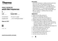
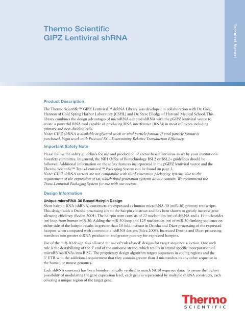
Protocol V – Puromycin Selection7Determining Antibiotic Dose Response (Kill Curve)In order to generate stable cell lines expressing the transgene of interest, it is important to determine the minimum amountof antibiotic required to kill non-transfected cells. A simple procedure to test this is as follows:1. Day 1: Using the same cell type and relative cell densities to be used in subsequent transfection or transductionprocedures, plate cells and culture overnight.2. Day 2: Replace complete growth medium with growth medium supplemented with a range of puromycinconcentrations (0-15 μg/mL), including untreated control cells with no antibiotic added.3. Day 4: Refresh medium and assess viability.4. Replace medium with fresh medium supplemented with the appropriate concentration of puromycin every2-3 days depending on the growth of cells.5. Examine cells daily and identify the minimal concentration of antibiotic that efficiently kills all non-transfected/transduced cells between 3-6 days following addition of puromycin.Puromycin SelectionIf adding antibiotic for selection, use the appropriate concentration as determined based on the above kill curve.1. Add medium containing antibiotic 24 or hours post-transfection or post-transduction, respectively, to begin selectionNote: It is important to wait at least 24 hours after transfection before beginning selection.2. Cells can be harvested for transgene expression 24-72 hours after starting selection.3. If longer selection is required for cells to be confluent, replace selective medium approximately every 2-3 days.4. Monitor the cells daily and observe the percentage of surviving cells. Cells surviving selection will be expressingthe transgene.5. If generating stable cell lines (optional), select and grow for 10-15 days.6. Once non-transfected cells are eliminated and/or you have selected for stably transfected cell lines if desired, you canproceed to assay for transgene expression. RT-qPCR, western blot analysis or other appropriate functional assay can beused; compare treated samples to untreated, reporter alone, non-silencing control, or other controls as appropriate.Protocol VI – Packaging LentivirusThe p<strong>GIPZ</strong> vector is tat dependant, so you must use a packaging system that expresses the tat gene. For packaging ourlentiviral <strong>shRNA</strong> constructs, we recommend the Trans-<strong>Lentiviral</strong> <strong>shRNA</strong> Packaging Kit (Cat #TLP5912 or TLP5917). TheTrans-<strong>Lentiviral</strong> <strong>shRNA</strong> Packaging System allows creation of a replication-incompetent, HIV-1-based lentivirus which can beused to deliver and express your <strong>shRNA</strong> of interest in either dividing or non-dividing mammalian cells. The Trans-<strong>Lentiviral</strong><strong>shRNA</strong> Packaging System uses a replication-incompetent lentivirus based on the trans-lentiviral system developed by Kappes(Kappes 2001). For protocols and information on packaging p<strong>GIPZ</strong> with our Trans-<strong>Lentiviral</strong> <strong>shRNA</strong> Packaging System,please see the product manual available on our website.Protocol VII – TiteringViral TiteringFollow the procedure below to determine the titer of your lentiviral stock using the mammalian cell line of choice IF YOUHAVE PRODUCED VIRAL PARTICLES YOURSELF. This protocol uses the HEK293T cell line that is available as part ofour Trans-<strong>Lentiviral</strong> <strong>shRNA</strong> Packaging. Kit (Cat #TLP5918) or separately (Cat #HCL4517). You can use a standardHEK293T cell line as an alternative.Note: If you have generated a lentiviral stock of theexpression control (such as <strong>GIPZ</strong> non-silencingconstruct), we recommend titering this stock as well.1. The day before transduction, seed a 24-well tissueculture plate with HEK293T Cells at 5 × 10 4 cellsper well in DMEM (10% FBS, 1% pen-strep).Note: The following day, each well should be nomore than 40-50% confluent.Figure 6. Five-fold serial dilutions of virus stock.
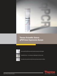
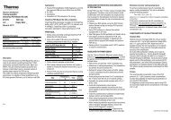
8 2. Make dilutions of the viral stock in a round bottom 96-well plate using serum-free media. Utilize the plate asshown in (Figure 6) using one row for each virus stock to be tested. Use the procedure below (starting at step 4)for dilution of the viral stocks. The goal is to produce a series of 5-fold dilutions to reach a final dilutionof 390,625-fold.3. Add 80 µL of serum-free media to each well.4. Add 20 µL of thawed virus stock to each corresponding well in column 1 (five-fold dilution).Note: Pipette contents of well up and down 10-15 times. Discard pipette tip.5. With new pipette tips, transfer 20 µL from each well of column 1 to the corresponding well in column 2.Note: Pipette up and down 10-15 times and discard pipette tip.6. With new pipette tips, transfer 20 µL from each well of column 2 to the corresponding well in column 3.Note: Pipette up and down 10-15 times and discard pipette tip.7. Repeat transfers of 20 µL from columns 3 through 8, pipetting up and down 10-15 times and changing pipettetips between each dilution.Note: It is strongly recommended that you use a high quality multichannel pipettor when performing multipledilutions. Pre-incubate the dilutions of the virus stock for 5 minutes at room temperature.8. Label 24-well plate as shown in (Figure 7) using one row for each virus stock to be tested.9. Remove culture media from the cells in the 24-well plate.10. Add 225 µL of serum-free media to each well.1 2 3 4 5 6Virus stock 1AVirus stock 2BVirus stock 3CVirus stock 4DFigure 7. Twenty-four well tissue culture plate, seededwith HEK293T cells, used to titer the virus.11. Transduce cells by adding 25 µL of diluted virus from the original 96-well plate (Figure 6) to a well on the24-well destination plate (Figure 7) containing the cells. For example, transfer 25 µL from well A2 of the 96-wellplate into well A1 in the 24-well plate (Table 6).12. Incubate transduced cultures at 37 °C for 4 hours.13. Remove transduction mix from cultures and add 1 mL of DMEM (10% FBS, 1% Pen-Strep).14. Culture cells for 48 hours.15. Count the TurboGFP expressing cells or colonies of cells (Figure 8).Note: Count each multi-cell colony as 1 transduced cell, as the cells will be dividing over the 48 hour cultureperiod. Figure 8 illustrates this principle of cell counting.16. Transducing units per mL (TU/mL) can be determined using the following formula:# of TurboGFP positive colonies counted × dilution factor × 40 = # TU/mL:Example: 55 TurboGFP positive colonies counted in well A3 55 (TurboGFP positive colonies) × 625(dilution factor) × 40 = 1.38 × 10 6 TU/mL
Table 6. Example of set up for dilutions.9Well (Row A, B, C, or D)Originating(96-well plate)Destination(24-well plate)Volume Diluted Virus UsedDilution FactorA1 25 μL 5 *A2 A1 25 μL 25A3 A2 25 μL 125A4 A3 25 μL 625A5 A4 25 μL 3125A6 A5 25 μL 15625A7 A6 25 μL 78125A8 25 μL 390625 **Please note that when expecting very high or very low titers, it would be advisable to include either well 8or well 1 respectively.ABFigure 8. Examples of individual colonies.Once you have generated a lentiviral stock with a suitable titer, you are ready to transduce the lentiviral vector into themammalian cell line of choice and assay for gene silencingMultiplicity of Infection (MOI)To obtain optimal silencing of your <strong>shRNA</strong>, you will need to transduce the lentiviral vector into your mammalian cell lineof choice using a suitable MOI. MOI is defined as the number of transducing units per cell.Determining the Optimal MOIAlthough this is cell line dependent, this generally correlates with the number of integration events per cell and as a result,level of transgene expression. A number of factors can influence determination of an optimal MOI including the nature ofyour mammalian cell (actively-dividing versus non-dividing), its transduction efficiency, your application of interest, and thenature of your gene of interest. If you are transducing your lentiviral construct into the mammalian cell line of choice for thefirst time, after you have titered the lentivirus, we recommend using a range of MOIs (for example, 0, 0.5, 1, 2, 5, 10, 20) todetermine the MOI required to obtain optimal expression for your particular application. It should be noted that to achievesingle copy knockdown, an MOI of 0.3 is generally used, as less than 4% of your cells will have more than one insert.Protocol VIII – TransductionTransduction of Target CellsThe protocol below is optimized for transduction of the lentiviral particles into HEK293T, OVCAR-8 or MCF7 cells in a24-well plate using serum-free media. If a different culture dish is used, adjust the number of cells, volumes, and reagentquantities in proportion to the change in surface area (Table 7). It is strongly recommended that you optimize transductionconditions to suit your target cell line to provide for the highest transduction efficiency possible.It is preferable that transduction be carried out in medium that is serum free and antibiotic free. A reduction in transductionefficiency occurs in the presence of serum; however it is possible to carry out successful transductions with serum present; youwill have to optimize the protocol according to your cells of interest.1. On day 0, plate 5 × 10 4 cells per well in a 24-well plate. Incubate overnight. You will be using full growth medium(with serum) at this stage.
10 2. The next day (day 1), remove the medium and add an appropriate amount of the virus to acheive the MOI youwish to use. Set up all desired experiments and controls in a similar fashion. Bring the total volume of liquidup so that it just covers the cells efficiently with serum-free media (see Table 7 for guidelines). If you are usingconcentrated virus you are likely to use a small volume of virus if you are using unconcentrated virus, you willfind you need more virus volume.Table 7. Suggested volumes of media per surface area per well of adherent cells.Tissue Culture Dish Surface Area per Well (cm 2 )Suggested total serum-free mediumvolume per well (mL)100 mm 56.0 5.060 mm 20.0 2.035 mm 8.0 1.06-well 9.4 1.012-well 3.8 0.524-well 1.9 0.2596-well 0.3 0.13. Approximately 4-6 hours post-transduction, add an additional 1 mL of full medium (serum plus Pen-Strep if youare using it) to your cells and incubate overnight.Note: We have experienced low toxicity with transduction in the cell lines tested, therefore removal of virus is notrequired for many cell lines. In our experience, higher transduction efficiencies have been achieved if the virus isnot removed after 6 hours. However, if toxicity is a problem, aspirate the mixture after 4-6 hours and replacewith fresh growth medium. Additionally, fresh growth medium should be replenished as required for continuedcell growth.4. At 48 hours post-transduction examine the cells microscopically for the presence of reporter gene (TurboGFP)expression as this will be your first indication as to the efficiency of your transduction.Note: When visualizing TurboGFP expression, if less than 90% of all cells are green, it is recommended in thesecases to utilize puromycin selection in order to reduce background expression of your gene of interest fromuntransduced cells.a. If adding puromycin, use the appropriate concentration as determined based on the kill curve (see Protocol V).Incubate cells with the selection medium.b. Approximately every 2-3 days replace with freshly prepared selective medium.c. Monitor the cells daily and observe the percentage of surviving cells. Once the non-transduced controlcells are dead, the surviving cells in the transduced wells will be expressing the <strong>shRNA</strong>. Optimum effectivenessof the puromycin selection should be reached in 4-6 days with puromycin dependent upon the concentration ofpuromycin chosen from the kill curve.Note: The higher the MOI you have chosen, the more copies of the <strong>shRNA</strong> and puromycin resistancegene you will have per cell. When selecting with puromycin, it is worth remembering that at higher MOIs, cellscontaining multiple copies of the resistance gene can withstand higher puromycin concentrations than thoseat lower MOIs. Adjust the concentration of puromycin to a level that will select for the population oftransduced cells you require for your application without going below the minimum antibiotic concentrationyou have established in your kill curve.5. Once your transduction efficiency is at an acceptable level (with or without puromycin selection performedpost-transduction), you can proceed to assay cells for reduction in gene expression or fluorescent reporteractivity by reverse transcription quantitative/real-time PCR (RT-qPCR), western blot analysis or other appropriatefunctional assay. Compare target gene to untreated, reporter alone (empty vector), non-silencing <strong>shRNA</strong>, orother negative controls.Note: Optimal length of incubation from the start of transfection to analysis is dependent on cell type, geneof interest, and the stability of the mRNA and/or protein being analyzed. RT-qPCR generally gives the bestindication of mRNA expression and gene silencing. The use of western blot analysis to determine knockdown isdependent on quantity and quality of the protein, its half-life, and the sensitivity of the antibody and detectionsystems used.Protocol IX – Determining Relative Transduction EfficiencyFollow the procedure below to determine the relative transduction efficiency of purchased <strong>GIPZ</strong> lentiviral particles(Cat #VGH5518, VGM5520, VGH5526). This protocol should only be used with purchased <strong>GIPZ</strong> <strong>shRNA</strong> individualclones in viral particle format.Prior to transducing with purchased <strong>GIPZ</strong> <strong>shRNA</strong> individual clones in viral particle format, we recommend determiningthe relative transduction efficiency of your cell type. <strong>Lentiviral</strong> titers provided with purchased <strong>GIPZ</strong> lentiviralparticles have been calculated by transducing HEK293T cells. Transduction efficiencies vary significantly by cell type.
The relative transduction efficiency of your cells may be estimated by determining the functional titer of a control virussuch as <strong>GIPZ</strong> Non-silencing control viral particles (Cat #RHS4348) in your cells of interest.11Follow the procedure below to determine the functional titer of the <strong>GIPZ</strong> Non-silencing control <strong>shRNA</strong> viral stock inthe mammalian cell line of your choice. The following conditions have been optimized for transduction of HEK293Tcells. When determining the relative transduction efficiency of your cell type, use the transduction conditions that havebeen optimized for your cells of choice interest.1. The day before transduction (day 0), seed a 24-well tissue culture plate with your cells at 5 × 10 4 cells perwell in their respective medium.Note: The following day, each well should be no more than 40-50% confluent.2. Make dilutions of the Non-silencing control <strong>shRNA</strong> viral stock in a round bottom 96-well plate using serumfreemedium. Utilize the plate as shown in Figure 9 with one row for each replicate (we recommend performingat least two replicates). Use the procedure below for dilution of the viral stock. The goal is to produce a series offive-fold dilutions to reach a final dilution of 390,625-fold.Figure 9. Five-fold serial dilutions of virus stock.3. Add 40 uL of serum-free media to each well in column.4. Add 80 uL of serum-free media to each well of columns 2-8.Note: If desired, include 8 μg/mL polybrene in the dilution medium.5. Add 10 µL of thawed control <strong>shRNA</strong> virus stock to each well in column 1 (five-fold dilution).Note: Pipette contents of well up and down 10-15 times. Discard pipette tip.1 2 3 4 5 6Virus stock 1AVirus stock 2BVirus stock 3CVirus stock 4DFigure 10. Twenty-four well tissue culture plate, seededwith HEK293T cells, used totiter the virus.6. With new pipette tips, transfer 20 µL from each well of column 1 to the corresponding well in column 2.Pipette up and down 10-15 times and discard pipette tip.7. With new pipette tips, transfer 20 µL from each well of column 2 to the corresponding well in column 3.Pipette up and down 10-15 times and discard pipette tip.8. Repeat transfers of 20 µL from columns 3 through 8, pipetting up and down 10-15 times and changing pipettetips between each dilution. It is strongly recommended that you use a high-quality multichannel pipettor whenperforming multiple dilutions. Incubate the dilutions of the virus stock for 5 minutes at room temperature.
12 9. Label a 24-well plate as shown in Figure 10 using one row for each replicate.10. Remove culture medium from the cells in the 24-well plate.11. Add 225 µL of serum-free medium to each well.12. Transduce cells by adding 25 µL of diluted control <strong>shRNA</strong> lentivirus from the original 96-well plate(Figure 9) to a well on the 24-well destination plate (Figure 10) containing the cells. For example, transfer25 µL from well A2 of the 96-well plate into well A1in the 24-well plate (Table 8).13. Incubate transduced cultures at 37 °C for 4-6 hours.14. Add 1 mL of your medium (normal serum concentration).15. Culture cells for 72 hours.16. Count the TurboGFP expressing cells or colonies of cells (Figure 11). Count each multi-cell colony as1 transduced cell, as the cells will be dividing over the 72 hour culture period. Figure11 illustrates thisprinciple of cell counting. Count the number of TurboGFP expressing colonies in wells corresponding toat least two viral dilutions.Table 8. Example of set up for dilutions.Well (Row A, B, C, or D)Originating(96-well plate)Destination(24-well plate)Volume Diluted Virus UsedDilution FactorA1 25 μL 5 *A2 A1 25 μL 25A3 A2 25 μL 125A4 A3 25 μL 625A5 A4 25 μL 3125A6 A5 25 μL 15625A7 A6 25 μL 78125A8 25 μL 390625 **Please note that when expecting very high or very low titers, it would be advisable to include either well8 or well 1 respectively.ABFigure 11. Examples of individual colonies.17. Transducing units per ml (TU/mL) can be determined using the following formula:# of TurboGFP positive colonies counted × dilution factor × 40 = # TU/mLNote: 25 μLof diluted virus was added to the cells. This is 1/40th of a mL.Example: 55 TurboGFP positive colonies counted in well A355 (TurboGFP positive colonies) × 625 (dilution factor) × 40 = 1.38 × 10 6 TU/mL.18. The functional titer calculated for your cell line under your experimental conditions can be used todetermine the relative transduction efficiency of your cell type by using the following formula:Functional titer of Non-silencing control <strong>shRNA</strong> virus stock in your cell line ÷ Titer of Non-silencingcontrol <strong>shRNA</strong> virus stock as calculated in HEK293T = Relative transduction efficiencyFor example, if the titer of the Non-silencing control <strong>shRNA</strong> virus stock in HEK293T (as provided on the productspecification sheet) is 6.9 × 10 6 TU/mL and the functional titer of the control <strong>shRNA</strong> virus stock in your cell lineis 1.38 × 10 6 TU/mL, the relative transduction efficiency of your cell type is 0.2. To extrapolate the averagefunctional titer of the provided <strong>GIPZ</strong> viral particles, multiply the average titer of each plate as provided on the productspecification sheet by the relative transduction efficiency of your cell type. In our example, if the titer of the <strong>GIPZ</strong> viralparticles in HEK293T cells is 2 × 10 6 TU/mL and the relative transduction efficiency of your cell type is 0.2, theextrapolated average functional titer of that plate your cell type is 4 × 10 5 TU/mL.
Once the relative transduction efficiency of the <strong>GIPZ</strong> virus has been established in your cell line, use the optimizedtransduction conditions determined in Protocol VIII to transduce your cell line with the purchased <strong>GIPZ</strong> <strong>shRNA</strong>individual clones in viral particle format. If the titer of the non-silencing control <strong>shRNA</strong> virus is not satisfactoryin your cell line you might consider choosing a different cell line more permissive to transduction by lentivirusbefore proceeding.13Protocol X – qPCRQPCR Experimental RecommendationsOne of the biggest challenges of any qPCR experiment is to obtain reproducible reliable data. Due to the sensitivityof this multi-step technique, care must be taken to ensure results obtained are accurate and trustworthy (see Bustinet al., 2009).1. Experimental samples should be run at least in duplicate. It should be noted that with duplicate experimentsit will not be possible to assign error bars to indicate consistency from experimental sample to experimentalsample. Using triplicate samples or higher will enable error bars to be assigned indicating the level ofexperimental variation.2. Reverse Transcriptase reactions for cDNA synthesis should always include a No Template Control (NTC)and No Reverse Transcriptase (no RT) control to check for reagents contamination and the presence ofcontaminating DNA, respectively. Use a robust reverse transcriptase enzyme for cDNA synthesis such as the<strong>Thermo</strong> <strong>Scientific</strong> Maxima cDNA Synthesis Kit for RT-qPCR (Cat #K1641).3. We have found that normalizing the RNA concentration prior to cDNA synthesis will increaseconsistency downstream.4. qPCR should be done at least in triplicate. Again, it should be noted that with duplicate reactions it will notbe possible to assign error bars to indicate the consistency in your qPCR reactions. Using triplicate samplesor higher will enable error bars to be assigned indicating the level of variation between qPCR reactions. Usevalidated primer sets for SYBR-based assays or primers/probe for probe-based assays, such as <strong>Thermo</strong> <strong>Scientific</strong>Solaris qPCR Gene Expression Assays and Master Mixes.5. Make sure the mRNA you are using as your internal reference control for qPCR is expressed at a level higherthan your target gene's message.6. Use only high-quality calibrated pipettes, in conjunction with well fitting barrier tips.7. When pipetting, take the time to visually inspect the fluid in the pipette tip(s) for accuracy and lack of bubbles,especially when using a multi-channel pipette.8. Be sure to spin your qPCR plate prior to loading in the real-time instrument in order to collect the sample atthe bottom of the well and eliminate any bubbles that may have developed.9. With regard to knockdown experiments using <strong>shRNA</strong>, it is vitally important that you greatly reduce if noteliminate entirely those cells which are not transduced or transfected from the population. This can be done inseveral ways: increase the efficiency of your transfection, use a higher mulitplicity of infection (MOI) for yourtransduction, utilize the puromycin selection marker and select against those cells that do not contain the <strong>shRNA</strong>or utilize fluorescent sorting to select against those cells that do not contain the <strong>shRNA</strong>.10. Always utilize the non-silencing control as a reference for target gene expression, as opposed to an untreatedsample. The Non-silencing treated samples will most accurately reproduce the conditions in your experimentalsamples. The Non-silencing best controls for changes in qPCR internal reference gene expression.11. You may also use an untreated sample to indicate substantial changes in target gene expression as seen in thenon-silencing control due to generic consequences of viral transduction and transgene expression. However,it should be noted that small changes in expression levels between an untreated sample and the Non-silencingcontrol are to be expected.12. Cq values greater than 35 should be avoided as they tend to be more variable. Samples with such high Cq valuesshould be repeated at higher cDNA concentrations and with a lower expressing qPCR internal reference control(such as TBP).13. Cq values less than 11 for the qPCR internal reference control should be avoided as it is difficult to determinea proper background subtraction using these values. If this occurs, use Cq values from both your internalreference control as well as your experimental target to determine an optimum cDNA concentration.14. It may be necessary to change internal reference controls if conditions in steps 12 and 13 cannot besimultaneously met.
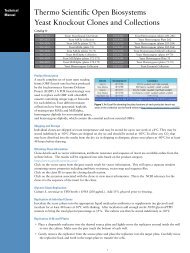
14 Controls and ValidationRNAintro <strong>shRNA</strong> Starter KitsThe use of vector-based RNAi for gene silencing is a powerful and versatile tool. Successful gene silencing in vitrois dependent on several variables including 1) The target cell line being studied, 2) transfection and transductionefficiency, 3) abundance of the mRNA or protein of interest in the target cell line, 4) half life of the protein, and 5)robust experimental protocols. For all these reasons, it is important to run controlled experiments where thetransfection and transduction efficiencies are as high as possible and measurable.Controls are a critical part of a gene silencing experiment. They enable accurate representation of knockdown dataand provide confidence in the specificity of the response. Changes in the mRNA or protein levels in cells treatedwith negative or Non-silencing controls reflect non-specific responses in cells and can be used as a baseline againstwhich specific knockdown can be measured. Positive controls are useful to demonstrate that your experimental systemis functional.120%Residual GAPDH Expression100%100%100%100%100%80%60%Figure 12. Non-silencing lentiviral <strong>shRNA</strong> control does not knockdowncommon endogenous genes. The above data represents the baselineamount of GAPDH, EG5 or PP1B mRNA set at 100% in the control. Therelative amounts of each of these mRNAs are then represented aftertreatment with non-silencing <strong>shRNA</strong> of these genes.40%20%0%UntransducedControl34%28%12%NS#1 NS#1 NS#1 GAPDH GAPDH GAPDHFigure 13. HEK293T cells were transduced with lentiviralparticles expressing GAPDH or Non-silencing <strong>shRNA</strong> atvariable MOIs ranging from 9-48. The graph depicts theresidual levels of GAPDH relative to Non-silencing control.ControlsThe EG5 and GAPDH <strong>GIPZ</strong> lentiviral <strong>shRNA</strong> vectors have been validated as positive controls for RNAi experimentsperformed using the <strong>GIPZ</strong> <strong>shRNA</strong>-containing lentiviral vectors. These <strong>shRNA</strong> have been tested in transductionbased experiments and have shown efficient knockdown at both mRNA and protein levels. The EG5 control has beenvalidated to knockdown human EG5 by RT-qPCR (Figure 14 and 15) and in situ hybridization of cells in tissue culture.The GAPDH control has been validated to knockdown human and mouse GAPDH by RT-qPCR (Figure 13).The <strong>GIPZ</strong> Non-silencing lentiviral <strong>shRNA</strong> vector has been validated as anegative control for RNAi experimentsperformed using the <strong>GIPZ</strong> <strong>shRNA</strong>-containing lentiviral vectors (Figure 12).120%Residual EG5ExpressionOVERLAYANTI-TUBULIN100%100%100%100%80%ANTI-EG5DAPI60%40%20%0%NS#1MOI 3.5NS#1MOI 8.5NS#1MOI 1729%EG5MOI 3.518%EG5MOI 8.517%EG5MOI 17Figure 14. HEK293T cells were transduced with lentiviral particlesexpressing EG5 or non-silencing <strong>shRNA</strong> at MOIs of 3.5, 8.5 and 17.The graph depicts the residual levels of EG5 relative to its nonsilencingcontrol.Figure 15. The characteristic phenotype observed by the targetingof the EG5 (KIF11) gene results in the formation of half spindles,mitotic arrest and monoastral microtubular arrays (green, see thecell on the left). By contrast, normal cells show bipolar spindlesand microtubule networks in mitosis and in interphase (see thecell on the right). The comparative expression of EG5 (red) betweenthe cell on the left and the right shows the extensive knockdownof EG5 in the cell displaying the phenotype (left). The cells werevisualized at 100x magnification using aLeica DMIRB fluorescencemicroscope. HEK293T cells were stained for tubulin (anti-tubulin,green), DNA (DAPI, blue) and EG5 (anti-EG5, red).
7. Editors of Nature Cell Biology (2003). Whither RNAi? Nat Cell Biol 5(6): 489-90.8. Elbashir, S. M., J. Harborth, et al. (2001). Duplexes of 21-nucleotide RNAs mediate RNA interference in culturedmammalian cells. Nature 411(6836): 494-8.9. Fire, A., S. Xu, et al. (1998). Potent and specific genetic interference by double-stranded RNA in Caenorhabditis elegans.Nature 391(6669): 806-11.10. Gregory, R. I., T. P. Chendrimada, et al. (2005). Human RISC couples microRNA biogenesis and posttranscriptionalgene silencing. Cell 123(4): 631-40.11. Kappes, J. C. and X. Wu (2001). Safety considerations in vector development. Somat Cell Mol Genet26(1-6):147-58.12. Kappes, J. C., X. Wu, et al. (2003). Production of trans-lentiviral vector with predictable safety. Methods Mol Med76: 449-65.13. Paddison, P. J., J. M. Silva, et al. (2004). A resource for large-scale RNA-interference-based screens in mammals.Nature 428(6981): 427-31.14. Shimada, T., et. al. (1995). Development of vectors utilized for gene therapy for AIDS. AIDS 4.15. Silva, J. M., M. Z. Li, et al. (2005). Second-generation <strong>shRNA</strong> libraries covering the mouse and human genomes.Nat Genet 37(11): 1281-8.17Limited Use LicensesLimited Use Label License: Evrogen Fluorescent ProteinsThis product contains a proprietary nucleic acid(s) coding for a proprietary fluorescent protein being, including its derivatives ormodifications, the subject of pending U.S. and foreign patent applications and/or patents owned by Evrogen JSC (hereinafter“Evrogen Fluorescent Proteins”). The purchase of this product conveys to the buyer the non-transferable right to use EvrogenFluorescent Proteins for (i) not-for-profit internal Research conducted by the buyer (whether the buyer is an academic orfor-profit entity), where “Research” means non-commercial uses or activities which (or the results of which) do not generaterevenue, and (ii) evaluation of Evrogen Fluorescent Proteins for the purpose of testing its appropriateness for development of atherapeutic, clinical diagnostic, vaccine or prophylactic product, provided that Evrogen Fluorescent Proteins are not used in thedevelopment or manufacture of such product. Offer of Evrogen Fluorescent Proteins for resale; distribution, transfer, or otherwiseproviding access to Evrogen Fluorescent Proteins to any third party for any purpose, or any use of Evrogen Fluorescent Proteinsother than for Research is strictly prohibited. The buyer cannot sell or otherwise transfer materials made by the employment ofEvrogen Fluorescent Proteins to a third party or otherwise use Evrogen Fluorescent Proteins for Commercial Purposes. The buyermay transfer information made through the use of this product solely for research and not for Commercial Purposes. CommercialPurposes means any activity by a party for consideration and may include, but is not limited to: (1) use of Evrogen FluorescentProteins in manufacturing; (2) use of Evrogen Fluorescent Proteins to provide a service, information, or data; (3) use of EvrogenFluorescent Proteins for therapeutic, diagnostic or prophylactic purposes. The purchase of this product does not convey a licenseunder any method claims in the foregoing patents or patentapplications.For information on the foregoing patents or patent applications or on purchasing a license to use Evrogen Fluorescent Proteinsfor purposes other than those permitted above, contact Licensing Department, Evrogen JSC, Miklukho-Maklaya street 16/10,Moscow, 117997, Russian Federation license@evrogen.com.<strong>shRNA</strong> Limited Use Agreement Permitted Use. Portions of this product are covered by published patent applications(the “<strong>shRNA</strong> IP Rights”) owned by Cold Spring Harbor Laboratory, referred to herein as “CSHL.” This product, and/or anycomponents or derivatives of this product, and/or any materials made using this product or its components, are referred toherein as the “Product.” Subject to the terms of this Limited Use Agreement, sale of the Product by (“OBS”) conveys the limited,nonexclusive, nontransferable right to any company or other entity that orders, pays for and takes delivery of the Product (the“Customer”) from OBS to use the Product solely for its internal research and only at such Customer’s facility where the Productis delivered by OBS.Restrictions. Customer obtains no right to transfer, assign, or sublicense its use rights, or to transfer, resell, package, or otherwisedistribute the Product, or to use the Product for the benefit of any third party or for any commercial purpose. The Product orcomponents of the Product may not be used in vitro or in vivo for any diagnostic, preventative, therapeutic or vaccine application,or in the manufacture or testing of a product therefore, or used (directly or indirectly) in humans for any purpose.Compliance. Customer may only use the Product in compliance with applicable local, state and federal laws, regulations andrules, including without limitation (for uses in the United States) EPA, FDA, USDA and NIH guidelines. Customer may not(directly or indirectly) use the Product, or allow the transfer, transmission, export or re-export of all or any part of the Product, inviolation of any export control law or regulation of the United States or any other relevant jurisdiction.Other Uses. Except for the permitted use expressly specified above and subject to the restrictions set forth above, no other use ispermitted. CSHL shall retain all right, title and interest in and to the <strong>shRNA</strong> IP Rights. Nothing herein confers to Customer (byimplication, estoppel or otherwise) any right or license under any patent of CSHL, including, for avoidance of doubt, under anypatent that may issue from or relate to the <strong>shRNA</strong> IP Rights. For information on obtaining an agreement to use this Product forpurposes other than those expressly permitted by this Limited Use Agreement, or to practice under CSHL patent rights, pleasecontact the CSHL Office of Technology Transfer at (516) 367-8301.
Termination of Rights. Upon issuance of any patent covering any portion of the Product, any and all rights conveyedto any Customer (other than a research or educational institution that is a tax-exempt organization under section501(c)(3) of the U.S. IRS code) under this Limited Use Agreement shall terminate and such Customer shall need alicense from CSHL to continue to use any Product that was previously purchased, or to purchase from OBS or anyother vendor or use any product claimed by such issued patent. At such time, such Customer may contact the CSHLOffice of Technology Transfer at (516) 367-8301 and seek a license to use this Product under CSHL’s patent rights.YOUR USE OF THIS PRODUCT CONSTITUTES ACCEPTANCE OF THE TERMS OF THIS LIMITEDUSE AGREEMENT.If you are not willing to accept the limitations of this agreement, OBS is willing to accept return of the Product for afull refund.<strong>Technical</strong> <strong>Manual</strong>Limited Label License: BenitecThis product is sold solely for use for research purposes in fields other than plants. This product is not transferable.If the purchaser is not willing to accept the conditions of this label license, supplier is willing to accept the return ofthe unopened product and provide the purchaser with a full refund. However, if the product is opened, then thepurchaser agrees to be bound by the conditions of this limited use statement. This product is sold by supplier underlicense from Benitec Australia Ltd and CSIRO as co-owners of U.S. Patent No. 6,573,099 and foreign counterparts.For information regarding licenses to these patents for use of ddRNAi as a therapeutic agent or as a method to treat/prevent human disease, please contact Benitec at licensing@benitec.com. For the use of ddRNAi in other fields, pleasecontact CSIRO at www.pi.csiro.au/RNAi.thermoscientific.com/onebio© 2013 <strong>Thermo</strong> Fisher <strong>Scientific</strong> Inc. All rights reserved. Fluorescent Protein and TurboGFP are is a registered trademark of Evrogen.HXB2 is a registered trademark of GenBank. Zeocin is a registered trademark of Invitrogen. All other trademarks are the property of<strong>Thermo</strong> Fisher <strong>Scientific</strong> Inc. and its subsidiaries.EuropeCustomer Servicecs.molbio.eu@thermofisher.com<strong>Technical</strong> Supportts.molbio.eu@thermofisher.comTel 00800 222 00 888Fax 00800 222 00 889Rev_5-23-13United StatesCustomer Servicecs.molbio@thermofisher.com<strong>Technical</strong> Supportts.molbio@thermofisher.comTel 800 235 9880Fax 800 292 6088CanadaCustomer Servicecs.molbio@thermofisher.com<strong>Technical</strong> Supportts.molbio@thermofisher.comTel 800 340 9026Fax 800 472 8322