Botany - Telangana University
Botany - Telangana University
Botany - Telangana University

Create successful ePaper yourself
Turn your PDF publications into a flip-book with our unique Google optimized e-Paper software.
B.Sc. I Year (PRACTICAL) Model PaperSubject : BOTANYPaper – I(Microbial Diversity, Cryptogams and Gymnosperms)[Effective from the academic year 2008-09]Time: 3 Hours Max. Marks. 50Note: Answer All questions. Draw well labelled diagrams wherever necessary.I. Identify the algal components (A,B,C) in the given mixture. Draw labelleddiagrams, classify and identify giving important characters [Diagrams – 1;classification – 1; characters – 2](3x4=12 Marks)II. Describe the procedure of bacterial staining and identify the givenBacterium (D) [Procedure – 2;Description – 1;Identification – 1] (4)III. Prepare T.S. of the diseased material as a temporary mount (E). Identify thepathogen giving reasons and describe with the help of diagrams.(Preparation – 2; Identification – 1; Diagram – 1; Description – 1;Classification- 1) (6)IV. Prepare T.S. of the given material – Pteridophyte/Gymnosperm (F) as a singlestain temporary mount and identify with diagnostic features and providesuitable diagrams.(Slide Preparation – 2; Classification – 1; Diagram – 2; Description – 3) (8)V. Identify giving reasons the specimens and slides ( G,H,I,J,K and L)(Viruses/Fungi – 1;Bryophtya – 2;Pteridophyta&Gymnosperms – 3)(6x2½=15)VI. Record (5)
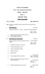
FACULTY OF SCIENCEB.Sc. I Year (Practical) ExaminationSubject : BOTANYPaper – I(Microbial Diversity, Cryptogams and Gymnosperms)QUESTION BANKW.E.F. Annual 2009Time : 3 Hours} {Max. Marks: 50Note : Answer All questions. Draw well labeled diagrams wherever necessary.I. Identify the algal components (A,B,C) in the given mixture. Draw labeleddiagrams, classify and identify giving important characters [Diagrams – I:classification – 1; characters – 2](3x4=12 marks)1. Oscillatoria2. Nostoc3. Volvox4. Oedogonium5. coleochaete6. Chara7. Ectocarpus8. PolysiphoniaII.Describe the procedure of bacterial staining and identify the givenBacterium (D) [Procedure – 2 ; Description – 1 Identification – 1] (4)9. Gram + Bacteria10. Gram – BacteriaIII.Prepare T.S. of the diseased material as a temporary mount (E).Identify the pathogen giving reasons and describe with the help ofdiagrams.(Preparation – 2; Identification – 1; Diagram – 1; Description – 1;Classification – 1) (6)11. White rust on crucifers12. Rust on sorghum13. Tikka disease of groundnutIV.Prepare T.S. of the given material – Pteridophyte / Gymnosperm (F)as a single stain temporary mount and identify with diagnostic featuresand provide suitable diagrams.(Slide Preparation – 2; Classification – 1; Diagram – 2; Description – 3) (8)14. Lycopodium stem15. Equisetum stem16. Marsilea Petiole / Rhizome17. Pinus needle18. Gnetum stem19. Gnetum leaf..2
..2..V. Identify giving reasons the specimens and slides (G, H, I, J, K and L)(viruses /Fungi – 1; Bryophtya – 2; Pteridophyta / Gymnosperms – 3)(6x2½=15)SPECIMENS :20. Tobacco Mosaic virus21. Bhendi Yellow Vein clearing22. Papaya leaf curl23. Ergot of Bajra24. Puccinia rust on wheat25. Puccinia rust on Barberry26. Head smut of sorghum27. Whip smut of sugarcane28. Tikka disease of Groundnut29. Brown leaf spot of rice30. Blast of Rice (Paddy)31. crustose lichen32. Foliose lichen33. Fruticose lichen34. Marchantia thallus with Gemma cups35. Marchantia thallus with Antheridiopshore36. Marchantia thallus with Archegonionphore37. Anthoceros Thallus38. Anthoceros with Sporophyte39. Polytrichium with Sporophyte40. Lycopodium with cone41. Equisetum with cone42. Marsilea with sporocarp43. Pinus male cone44. Pinus female cone45. Gnetum twig46. Gnetum male cone47. Gnetum female coneSLIDES:48. Albugo conidia49. Albugo oospores50. Saccharomyces vegetative / budding51. Penicillium conidia52. Pencillium ascocarp53. Puccinia uredial stage54. Puccinia telial stage55. Puccinia pycnial stage56. Puccinia aecial stage57. Alternaria conidia58. Marchantia thallus V.S.59. Marchantia thallus with Gemma cups60. Marchantia antheridiophore L.S.61. Marchantia archegoniophre L.S.62. Marchantia sporophyte V.S.63. Anthoceros thallus V.S.64. Anthoceros thallus with antheridia65. anthoceros thallus with archegonia66. Anthoceros sporophyte L.S.67. Anthoceros sporophyte T.S.68. Polytrichum leaf T.S.69. Polytrichum stem T.S.70. Polytrichum antheridial branch71. Polytrichum archegonial branch
72. Polytrichum capsule L.S.73. Polytrichum protonema74. Rhynia (Fossil slide)75. Lycopodium strobiuls L.S.76. Equisetum strobilus L.S.77. Marsilea sporocarp V.S.78. Cycadeoidea (Fossil Slide)79. Pinus male cone V.S.80. Pinus pollen grains81. Pinus female cone V.S.82. Pinus ovule V.S.83. Gnetum male cone V.S.84. Gnetum female cone V.S.85. Gnetum ovule V.S...3....3..VI. Record (5)