Mechanisms and neural substrate of spasticity
Mechanisms and neural substrate of spasticity
Mechanisms and neural substrate of spasticity

Create successful ePaper yourself
Turn your PDF publications into a flip-book with our unique Google optimized e-Paper software.
<strong>Mechanisms</strong> & NeuralSubstrate <strong>of</strong> SpasticityDr. Sharon L. Juliano
DefinitionSpasticty - A velocity dependent increase in muscletone ( hypertonia) with exaggerated increase in reflexesreflexes (hyperreflexia).Hypertonia is associated with an increasedresistance to passive stretch.Also associated with the clasp knife phenomen<strong>and</strong> clonus.Caused by damage to descending pathways thatinfluence gamma or alpha motor neurons.
UMNCerebral CortexBrain StemUMNDesc.TractstoSpinalCordSpinal CordEffectorTissueSkeletal Muscle <strong>of</strong>the BodyInterneuronsLMN(AlphaMotorNeurons)
LOWER MOTOR NEURON LESIONS UPPER MOTOR NEURON LESIONS• CAUSES: CAUSES:••1) Degeneration <strong>of</strong> Anterior Horn Cells•2) Transection <strong>of</strong> Ventral Roots1) Degeneration <strong>of</strong> Nerve Cell Bodies inthe Motor / Somatosensory Cortices•3) Transection <strong>of</strong> Spinal Nerves 2) Damage to Axons <strong>of</strong> DescendingSystems particularly CorticospinalFibers•• SYMPTOMS: SYMPTOMS:•1) Flaccid Paralysis•2) Loss <strong>of</strong> Myotatic Reflexes•3) Hypotonia•4) Muscle Fasciculations•5) Atrophy <strong>of</strong> Denervated Muscles1) Spastic Paralysis2) Hyperactive Myotatic Reflexes3) Hypertonia4) Clonus5) Muscle Atrophy--if it occurs, it is verylate <strong>and</strong> results from disuse• 6) Babinski Sign (Corticospinal Damage)• 7) Loss <strong>of</strong> Superficial Abdominal Reflex<strong>and</strong> Cremasteristic Reflex (males)
UMNUMN/Descending Tracts LesionA. does NOT denervate target muscleB. results in spastic paralysisDesc.Tract1. resting tension <strong>of</strong> muscle is(hypertonia)2. hyperactive myotatic reflexesLMNSpinalNerveSkeletal Muscle <strong>of</strong>the Body3. atrophy, if occurs, is a late event-fromdisuse <strong>of</strong> spastic muscleLMN/Spinal Nerve LesionA. does denervate target muscleB. results in flaccid paralysis1. absent or diminished muscle tone(hypotonia)2. loss <strong>of</strong> myotatic reflex3. atrophy <strong>of</strong> denervated muscle/s
cutaneous
IIIIIIIVVIIIIIIVIVIIXVIIIXIXVIII
Flexors=dorsal in Lam. IXDistal MusclesProximal MusclesAxial (Trunk) MusclesExtensors=ventral in Lam. IX
MEDIALANDLATERALDESCENDING SYSTEMS
Medial Descending TractsAnterior FuniculusLateral Descending TractsLateral FuniculusSynapse inLam. VII, VIII &IX (one tract)Innervate axial& proximalmusclesIXIXIIXIIIIIIVVVIVIIVIIIMedialDescendingTractsIIIIIIVVVIVIIVIIIIIXIXLateralDescendingTractsIXSynapse inLam. V, VI, VII,VIII & IX(one tract)InnervatedistalmusclesUE=FlexorsDistalProximalAxialUE=Extensors
RLCORTICOSPINAL TRACTSMotor & Sensory CortexCSTACSTRLLCSTLCSTVIIXVVIIVIIVVIIXIXVIIIVIIIIXACST
RLRUBROSPINAL TRACTRed Nucleus-MidbrainRSTRLIXIXVVIVIIVIIIVIIVIIIVVIIXIX
RLL. VESTIBULOSPINAL TRACTFunction: facilitatesextension <strong>of</strong> all 4extremitiesLateral VestibularNucleus (Pons)LVSTRLVIXVIVIIVIIVVIIXIXVIIIVIIIIXLVST
RLM. VESTIBULOSPINAL TRACTFunction: controls neckmovements to stabilize headfor eye gaze movementsMedial VestibularNucleus (Medulla)RLMVSTVIXVIVIIVVIVIIIXIXVIIIVIIIIX
RLM. RETICULOSPINAL TRACTFunction: controls muscleatonia during sleepReticularFormation(Medulla)RLMRSTIXVVIVIIVIIVVIIXIXVIIIVIIIIXMRSTMRST
RLP. RETICULOSPINAL TRACTFunction: facilitatesaxial/proximal musclesReticular Formation(Pons)PRSTRLIXIXVVIVIIVIIIVIIVIIIVVIIXIX
VIIXIXVVIIVIIIR LR LVVIIXIXVIIIReticular Formation(Pons)PRSTVII
RLP. RETICULOSPINAL TRACTXPRSTReticular Formation(Pons)RLIXIXVVIVIIVIIIVIIVIIIVVIIXIXX
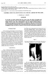
••AREA DAMAGED: Transection <strong>of</strong> the Spinal Cord•STRUCTURES & TRACTS INVOLVED:• 1) Disruption <strong>of</strong> all Spinal Cord TractsSPINAL CORD TRANSECTION•NEUROLOGICAL DEFICITS:• 1) Period <strong>of</strong> Spinal Shock (Avg = 3 wks)• 2) Termination <strong>of</strong> Spinal Shock = Appearance <strong>of</strong>bilateral Babinski signs at ~ 3 wks• 3) Period <strong>of</strong> Minimal Reflex Activity (3-6 wks)• a) muscles flaccid, no myotatic reflexes• b) weak flexor responses that begin distally inresponse to nocioceptive stimuli• 4) Flexor Spasms (6-16 wks)• a) Increasing tone in flexors <strong>and</strong> strongerflexor responses involving more proximalmuscles in response to nocioceptive stimuli• b) Triple Flexion Response is first seen <strong>and</strong>consists <strong>of</strong> flexion <strong>of</strong> the lower extremity at the hip, knee <strong>and</strong> ankle inresponse to mild nocioceptive stimulus• c) Mass Reflex--powerful triple flexion reflex inresponse to non-specific stimulus• d) Paradoxical sweating below lesion level• 5) Alternate Flexor & Extensor Spasms (4 mo-1 yr)• 6) Predominant Extensor Spasms (> 1 yr)• a) marked extensor tone (<strong>spasticity</strong> & claspknifephenomenon)• b) clonus• c) bilateral Babinski signs• d) loss <strong>of</strong> sensation below the lesion level• e) increased myotatic reflexes• f) bowel, bladder <strong>and</strong> sexual functiondisturbances• g) reflex spinal sweating
InhibitoryInterneuronD. RootAfferents[1]ExcitatoryInterneuronDescending TractAxonAlpha Motor Neuron
InhibitoryInterneuronD. RootAfferentsExcitatoryInterneuronDescending TractAxonAlpha Motor Neuron
XInhibitoryInterneuronD. RootAfferentsExcitatoryInterneuronDescending TractXAxonAlpha Motor Neuron
Changes at the level <strong>of</strong> the spinal cord:oAlpha motor neurons appear to be the most involved.oDecrease in the amount <strong>of</strong> presynaptic inhibition <strong>of</strong> 1a afferents.oPost activation repression <strong>of</strong> 1a afferents is decreased.oReciprocal 1a inhibition is reduced.oControl <strong>of</strong> inhibitory interneurons impaired.oIntraspinal sprouting.
XKatz, 1999Decrease in the amount <strong>of</strong> presynaptic inhibition <strong>of</strong> 1a afferents.Lack <strong>of</strong> this inhibition can contribute to increased stretch reflexes.
Reciprocal 1a inhibition is reduced
Reciprocal 1a inhibition is reduced.Gracies, 2005
Lesion at Spinal CordLesion aboveSpinal CordIntraspinal sprouting.
Lack <strong>of</strong> corticospinal controlAbnormal descending influenceAbnormal afferent influenceChanges in inhibitory circuitry
• Lack <strong>of</strong> corticospinal control• Abnormal descendinginfluence• Abnormal afferent influence• Changes in inhibitorycircuitry
The End!
Reciprocal 1a inhibition is reduced
Muscle SpindlesMuscle spindles consist <strong>of</strong> intrafusalfibers & specialized sensory <strong>and</strong> motornerve fibers. These specialized fibersare anchored to the extrafusal musclefibers <strong>and</strong> are stretched when themuscle is stretched. Responsible for themyotatic reflex.Types <strong>of</strong> intrafusal fibers:Nuclear bag – responds to velocity &changes in muscle length (dynamic).Nuclear chain – responds to changes inmuscle length (static).Nerve supply:Sensory – Group 1a -Bag & Chain(dynamic).Group II – Bag & Chain – staticMotor – Gamma motor neurons –innervate ends <strong>of</strong> intrafusal fibers.
DIFFERENCESFUNICULUSTARGET CELLSPATTERN OFINTERNEURONPROJECTIONSMUSCLESINNERVATEDMUSCLE ACTIONFUNCTIONSMEDIALDESCENDINGSYSTEMANTERIORFUNICULUSMEDIAL VII-IXLONG AXONSMANY SC SEG.MUSC. GROUPSAXIAL &PROXIMALFL.=UE;EXT.=LEPOSTURELATERALDESCENDINGSYSTEMLATERALFUNICULUSLATERAL V-IXSHORT AXONSFEW SC SEG.INDIV. MUSC.DISTAL LIMBMUSCLESEXT.=UE;FL.=LESKILLED MOVE.
Gracies, 2005Post activation repression <strong>of</strong> 1a afferents is decreased.