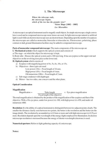
Unit I
Unit I
Unit I
Create successful ePaper yourself
Turn your PDF publications into a flip-book with our unique Google optimized e-Paper software.
Mitochondrion Golgi apparatusSmooth (Agranular)endoplasmic reticulum and Rough (Granular) endoplasmic reticulumLysosomes4
Microtubules Free Ribosomes/PolysomesMicrofilaments Centriole6
. Nonmembrane bound: These organelles are free in the cytoplasm.Free Ribosomes/Polysomes: Free ribosomes contain RNA and protein and show small and large subunits.They are present in all body cells except in mature RBC. Polyribosomes or polysome is a cluster of 10 to20 RNA attached to a long distance molecule of mRNA. Their function is to synthesize the integral proteinsand enzymes to be used intracellularly (not transport proteins cf: rER). If large in amount, the cell showsbasophilic granules in the cytoplasm.Microtubules: These are slender very long and straight filamentous structures 250Aº in diameter havingrigidity and elasticity. They can bend without breaking. They are made up of tubulin protein (Mol. wt.55000) the monomer of which unites to form dimers. Microtubules act as cytoskeleton and maintain theshape of the cell. In neuron they give direction to flow of material synthesized close to the nucleus to theprocesses i.e.Trans-axonal flow. Cilia, flagella and centriole are basically composed of microtubules. Theyplay a major role in mitosis. Colchicine arrests mitosis by binding to the dimers of the tubulin.Microfilaments: These are elongated thread like structures having 40Aº to 60Aº diameter. Actin andmyosin filaments seen in the muscle cells are responsible for contraction. In other cells they change theshape and produce cell movement. In skin cells, filaments are in bundles called tonofilaments and formdesmosomes and hemidesmosomes. In absorptive cells they form the core of microvilli.Intermediate filaments: Intermediate filaments as the name implies are intermediate in size betweenmicrofilaments and microtubules. They are 100 Aº to 150 Aº in diameter and have a purely structuralfunction i.e. they bind intracellular structures to each other and to plasma membrane proteins. In humansthere are more than 50 different types of intermediate filaments seen. These can be divided into fivedifferent classes eg. cytokeratin (characteristic of epithelial cells), vimentin (cells of mesodermal origin),desmin (seen in muscle cells), neurofilament proteins (seen in nerve cells) and glial fibrillary acid protein(seen in glial cell).Centriole: It is a short cylindrical structure usually present as a pair in dividing cells. The wall of eachcentriole is composed of nine longitudinally disposed parallel bundles of microtubules and each bundleconsists of three microtubules i.e. peripheral nine triplets. The triplets are held together by fibrillar materialin the form of a cylinder. Transverse section of the shaft of a cilium shows two singlets in the centre and ninedoublets at periphery. Highly developed single cilium is flagella. In human the only cell having a flagella isthe sperm.Inclusions/Pigment/Granules: They are of two types - exogenous and endogenous. Exogenous inclusionsinclude carotene, minerals like Ag and Pb, tattoo marks (inorganic pigments in macrophages), dust in lungcells etc. Endogenous inclusions include melanin, lipofuscin, haemoglobin, haemosiderin, bilirubin, biliverdinetc. End products of digestion are stored in some cells in the form of glycogen and lipids as cell inclusions.Notes:7
Nucleus (Nuclear membrane, chromatin, nucleolus, nuclear pores, Euchromatic and Heterochromatic)8
Nucleus: It is made up of deoxyribonucleic acid and ribonucleic acids so it takes basic (Hematoxylin)stain and always appears violet or blue under light microscope . It may be oval, round, spherical or flat inshape and may be central or eccentric in location within the cell. Most of the cells in the human body arenucleated (exception-RBC) and a single nucleus is present. Normally, striated muscles and osteoclastshow multinucleated appearance and hepatocytes and transitional epithelium may show binucleated cells.Depending upon the physical state of the chromosomes in an interphase cell, nucleus is of following types:Euchromatic: The chromatin matter is granular in appearance. The chromosomes are coiled at places butuncoiled at other. The uncoiled portion is so thin that it is invisible under light microscope. The uncoilingindicates that RNA synthesis is going on and this is a feature of working cell or active cell or normal cell,hence the name euchromatic. Nucleolus is usually seen clearly within the granular chromatin.Heterochromatic: The chromatin matter is in a condensed or coiled form. The nucleus is more uniformlyviolet after staining. In resting cell the RNA synthesis is very slow in the nucleus so the uncoiled part of thechromosome is negligible. Nucleolus is usually not seen as it is hidden within the dark condensed chromatin.Vesicular: The chromatin matter is very much extended or uncoiled; as a result it can not be seen under lightmicroscope. Nucleolus is the only structure seen within the nucleus. Prominent nucleolus surrounded byclear unstained white zone within the nucleus gives it an owl’s eye appearance. Extended chromatin indicatesthat rapid RNA synthesis is going on showing that the cell is very active e.g. neurons.Pyknotic: Nucleus is shrunken and chromatin matter is very much condensed so that on staining the nucleardetails are not visible. The dark violet and small sized nucleus indicates that the cells is degenerating, dyingor showing some pathological process e.g. epidermis of skin.Nucleus (E/M structure): It shows a nuclear membrane as two parallel lines each of 70Aº thickness with250 Aº distance between the two. A total thickness of the membrane is 40nm and it shows nuclear poreseach about 100-200 nm in size. Through the pore, the pore complex extends for a short distance into thecytoplasm and nuclear sap. Nuclear pores are closed by diaphragm. Within the chromatin material nucleolusis present which is metabolically active site in the nucleus. Nuclear organizers are the parts of acrocentricchromosomes with satellites (No. 13, 14, 15, 21, 22) which are associated with the formation of nucleoli.Thus a cell can have maximum 10 nucleoli but because of plane of section all cannot be seen at one time.Notes:9
Macula adherens (Desmosome)Zonula adherens (Intermediate junction)Zonula occludens (Tight junction)Macula communicans (Gap junction)10
Intercellular contactsThe plasma membrane is the surface, which establishes contact with other cells and promotes variouskinds of cellular interaction. Structurally there are two main classes of contact:- Nonspecialised andSpecialised. The nonspecialised contacts are more common, which enable them to form loose aggregationat the same time permitting some movements. Here membrane surfaces are separated by 20nm. Followingare the types of specialised cell junctional complexes.They are seen only under electron microscope and are the regions of reciprocally adherent cellsIntercellular contactsNonspecialised Specialised/ Junctional ComplexesOccluding Adhesive CommunicatingZonula occludens Zonula adherens Macula adherens Macula communicans(Tight junction) (Intermediate junction) (Desmosome) (Gap junctions)Zonula occludens: It is a continuous zone present around the apices of epithelial cells, endothelial cells andat other sites where there is a barrier to prevent diffusion through the intercellular space. Colloidal tracersfail to move from the lumen of the intestine into the spaces between the epithelial cells. Here the membranesof the adjacent cells come into contact obliterating the gap between them. Contacts lie along branching andanastomosing ridges formed by the particles within the membrane. These junctions prevent the leakage oftoxic substances from the lumen into the tissue.Zonula adherens:It is a continuous belt formed around the apices of epithelial cells. Microfilaments areoften inserted into a dense coat on the cytoplasmic side of the membrane. No filaments span the gapbetween the cell surfaces but an adhesive material may intervene at this point which anchor the cells e.g.Intercalated discs of cardiac muscles.Macula adherens:It is a plaque like structure where the plasma membrane is coated internally with alayer of dense proteins in to which intracytoplasmic tonofilaments are inserted. The intercellular gap isbridged by thin interlocked filaments and is rich in glycoprotein (desmoglea). Single sided orhemidesmosome are also found e.g. epidermis.Macula communicans: The opposed membranes are separated by an apparent gap of 3mm. This gap istraversed by numerous dense beads arranged in hexagonal arrays on the two surfaces. Such junctionsform limited attachment of plaques rather than continuous zones thereby allowing free passage of substancesalong the cleft between the cells e.g. Hepatocytes and connective tissue.11
3. Epithelial tissueSimple squamous Simple cuboidalSimple columnar Simple columnar ciliatedSimple columnar with goblet cells Simple columnar with microvilli12
3. Epithelial tissueHuman body is made up of four basic tissues, epithelium, connective tissue, muscle and nerve tissue.Epithelium is a covering or lining tissue of all body surfaces, membranes and viscera. At many sites it formsglands which open and secrete on the epithelial surfaces. Epithelium is protective, secretory and absorptivein function. It rests on a basement membrane. It is classified according to the number of layers and shapeof the surface cells.Simple epithelium: It consists of only a single layer of cells arranged compactly in a contiguous manner..According to the shape of the epithelial cells it is subclassified into squamous, cuboidal and columnarvariety.Simple squamous (Squamous = scale like): It consists of single layer of very thin cells.Nuclei are thickerthan cytoplasm and create bulges on the membrane, easily seen under microscope in transverse section.The cytoplasm is so thin that it sometimes can not be seen under low power e.g. endothelial lining of bloodvessels, mesothelial lining of serous cavities and lining of thin segment of loop of Henle.Simple cuboidal:They do not actually have the shape of cubes but when cut at right angles to surface, theyappear cubical. There is less difference in the height and breadth of the cell (In a squamous cell length ismore and thickness is negligible) e.g. small collecting tubules in the medulla of the kidney, lining of inactivethyroid follicle, lining of the ovary.Simple columnar: Here the cells are more taller than wider. Surface view is hexagonal as that in cuboidalcells. Nucleus is basal, oval or elongated in shape.i) Simple columnar : It is unmodified variety, which provides protection and also secretes along wetsurfaces. Cytoplasm is light eosinophilic e.g. lining epithelium of gastrointestinal tract, gall bladder andlarger ducts of many glands.ii) Simple columnar ciliated: Luminal surface of the cells show cilia which beat to move the mucous alongthe membrane. Goblet cells are intermingled with ciliated cells e.g. lining of prepubertal uterus, centralcanal of spinal cord.iii) Simple columnar with goblet cells: Mucous-secreting cells are present in between the columnar liningcells. Due to mucous synthesis and accumulation they assume a bowl-like shape and indent the cytoplasmof neighboring cell. Its nucleus is in the narrow stem like portion close to the base. Mucous is washedduring H&E staining so they appear frothy, vacuolated or empty e.g. large intestine.iv) Simple columnar with microvilli: Apical surface of the cell shows the presence of microvilli. Underlight microscope it is described as cells having striated border or brush border. It increases the absorptivesurface area by 30 times. e .g. small intestine.Notes:13
Stratified squamousi) Keratinised ii) NonkeratinisedStratified cuboidal Stratified columnar ciliatedTransitional epitheliumi) Stretched ii) Relaxed14
Compound epithelium: They have two or more layers of cells. They withstand more wear and tear andare chiefly protective in nature. The wear and tear requires constant replenishment of surface cells fromdeeper layers. Such compound epithelia are called ‘Stratified’. According to the type of surface cell theyare further subclassified.Stratified Squamous:There are 5 to 6 layers of cells. The superficial cells are of squamous type. Accordingto the presence or absence of keratin it is further divided into two groups.i) Stratified Squamous Nonkeratinised: The deepest layer consists of a single row of columnar cells. Theintermediate layers show polyhedral cells but as the surface is reached they become flattened so that at thefree surface cells are squamous. Keratin is absent on the surface e.g. mouth, pharynx, oesophagus, vagina,cornea etc.ii) Stratified Squamous Keratinised: The layers from deep to superficial are same as in nonkeratinisedvariety. The superficial cells are dead and hence non-nucleated. They appear as eosinophilic fibrillar strands.This is a tough nonliving layer of keratin e.g. skin.Stratified Cuboidal: It consists usually of only 2-3 layers of epithelial cells in which deeper layers arepolyhedral and superficial layer is cuboidal in shape. e.g. duct of sweat gland, ovarian follicle.Stratified Columnar : It consists of 2-3 layers as in the above variety except for the more superficial cells,which are columnar type. It is present at the transitional zone between different types of epithelia e.g. largeducts of exocrine glands, male urethra, fornix of conjunctiva.Transitional Epithelium: It is also known as urothelium.It is not a transition between two different types ofepithelia. It is a form of stratified epithelium. It is also 5 to 6 layers thick with columnar basal cell layer. Theintermediate cells are pear shaped and the surface cells are umbrella like. Most of the layers show similarcells with rounded nuclei. This similarity of cells differentiates this epithelium from stratified squamousnonkeratinised variety. The base of the epithelium is not indented by connective tissue papillae, thus itpresents an even contour e.g. ureter, urinary bladder.i) Transitional epithelium stretched: Transitional epithelium can be stretched as in full bladder without thesuperficial cells pulling apart from one another. The surface cell have an extra reserve of glycosylatedplates embeded into the cell membrane on the luminal side, which gets stretched out and continues to keepthe epithelium urine-proof.(Hence the other name -Urothelium). They merely become drawn out intobroader thinner cells.ii) Transitional epithelium relaxed: When the transitional epithelium is not stretched, the more superficialcells appear rounded. The surface cells are commonly polyploid or multinucleated.Notes:15
Pseudostratified epithelium: Single layer of columnar cells all resting on the basement membrane. Theyare of different heights and so the nuclei of the cells appear to be placed one above the other, giving astratified appearance. e.g. lining of the respiratory tract and male genital tract.Pseudo stratified ciliated columnar epithelium: In this epithelium, some of the columnar cells show presenceof cilia eg. Respiratory tract, uterine tube. The pseudostratified ciliated columnar epithelium of the respiratorytract also shows presence of goblet cells and referred as Respiratory epithelium.In this variety, the mucoussecreted by the goblet cells forms a film on the luminal surface and the ciliary beating moves the mucousladen with inspired dust particles. The basal cells, which do not reach the surface, probably serve as stemcells for the taller kind of cells when they are lost from the membrane. The short motile cilia are morenumerous and close together. Cilia arise from basal bodies located just beneath the surface membrane ofthe columnar cells. The basal bodies of adjacent cells give the appearance of continuous membrane e.g.Trachea, Bronchus.Pseudostratified columnar epithelium with stereocilia: In this variety the columnar cells which reach thelumen show very long stereocilia. They are nonmotile or false cilia, which are actually long and branchingmicrovilli. These columnar cells apparently function primarily as absorptive cells. e.g. Duct of epididymis,Dutus deferens.Notes:17
4. Connective tissueFibroblast. Fibrocyte.Adipose cell Macrophage.Mast cell. Plasma cell.18
4. Connective/ Supporting TissueIt connects the other tissues and is mainly mesodermal in origin. Histologically, it contains cellular andnoncellular components. The cells of the connective tissue produce nonliving intercellular substancewhich consists of fibres and ground substance.EmbryonicCONNECTIVE / SUPPORTING TISSUEAdultMucoid Mesenchyme General SpecialLoose DenseRegular IrrregularCells: They are divided into fixed and wandering cells. Some of them can be identified undermicroscope in H & E staining.Fibroblast: They are fusiform or spindle shaped with branching processes. They have abundant basophiliccytoplasm. Cell processes are eosinophilic. Nucleus has prominent nucleolus and chromatin is extended(euchromatic) type. They are present in all types of connective tissue.Fibrocyte: Eventually after a fibroblast has surrounded itself with the intercellular substance it hasproduced, it loses its ability to divide and synthesize. This old cell that has almost completed the task ofproducing intercellular substance is fibrocyte. They are elongated pink cells with rod shaped nucleus andsmall amount of cytoplasm. Nucleus is heterochromatic. They are present in all types of connectivetissue.Adipose cell: These are oval or spherical in shape containing a large globule of fat surrounded by thin rimof cytoplasm. Fat is dissolved during staining therefore empty spaces appear in the cell, giving it acharacteristic signet ring appearance. They are present in the superficial fascia in large amount.Macrophage: The shape varies (pleomorphic) with the content. The cytoplasm is granular vacuolated andbasophilic. Ingested particles are present in the vacuole, which is unstained. The nucleus is small roundedand may be eccentric in location. The cell is phagocytic in function. Theyare most numerous in looseconnective tissue.Mast cells (Histaminocytes): These are large, round or oval cells showing metachromatic granules. Nucleusis small and obscured by the granules. The granules are of heparin, histamine or serotonin. They aredifficult to identify in H & E stain and require dyes like toluidine blue or alcian blue which make the nucleistain blue, but the granules stain purple to red (metachromatic staining). They are most frequently seenalong the blood vessels and nerves in serous membranes and loose areolar tissue.Plasma cells: They are oval or round with basophilic cytoplasm within which round acidophilic Russellbodies may be seen. Nucleus is eccentric and chromatin is characteristically cartwheel in appearance.They are derived from lymphocyte-B and produce antibodies.Other cells: Blood cells, marrow cells, cells of the bone and cartilage and pericytes are the otherexamples of connective tissue cells.Notes:19
Collagen fibres Elastic fibresLoose areolar tissueAdipose tissueTendon L.STendon T.S20
Noncellular componentsFibres: There are two types of connective tissue fibres; each type for a specific function. All these fibresare synthesized by the connective tissue cells (fibroblast) and are deposited outside the cell. Fibrillarmaterial secreted by the cell gets polymerized outside the cell to form fibres.i) Collagen fibres: They are wide, wavy kind composed of the protein collagen. The fibres are eosinophilicwith a maximum width of 10µm. Each collagen fibre is made up of fibrils. Unfixed fibres are white sothey are also called as white fibres. Collagen is a tough protein and makes meat tough. If it is boiled inwater it becomes hydrated and is converted into gelatin, which is soft. Most of the flexibility of thetissue is due to collagen. Under the microscope they appear in bundles and some bundles appearwavy. One big bundle may be seen dividing into two bundles but true branching is absent e.g. tendon,aponeurosis. Reticular fibres: These type of collagen fibres are argyrophilic and are seen in basementmembrane,glands,blood vessels and in highly parenchymatous organs like liver, lymphoid organs.ii) Elastic fibres: They show branching and maximum diameter is 1µm. They can be stretched bytensile force and on being released they snap back like rubber bands to their original length.Because of their different refractive index unstained fibres appear darker and yellow. They branchand anastomose freely and run singly. e.g. ligamentum flava and ligamentum nuchae, capsules andligaments of joints of ear ossicles.Matrix (Nonfibrous, amorphous ground substance): The matrix is of the nature of soft gel andamorphous and contains Glycosaminoglcans or GAGs (formerly called as acid mucopolysaccharides).Some GAGs are complexed with protein to form proteoglcans. The GAGs may be nonsulphated (e.g.Hyaluronic acid) and sulphated or neutral (e.g.Heparan sulphate, Chondroitin-4-sulphate, Chondroitin-6-sulphate, Dermatan sulphate and Keratan sulphate) and shows metachromasia. It appears pink in H& E and cannot be differentiated from surrounding content. Special stains like PAS and Toludine bluemay be used for staining the matrix.Connective tissue slides: Following are some of the slides studied in this practical.The slide ofthe loose areolar tissue is the single best slide to study the connective tissue.Loose areolar tissue: The slide shows most of the constituents of the connective tissue i.e. cells, fibrousand nonfibrous parts. Collagen fibre bundles are wavy, thick and eosinophilic. Elastic fibres are singlebranched and may be straight or wavy when cut. They are highly refringent. A group of adipose cells canbe seen with vacuolated cytoplasm. Most of the nuclei in the slide are of fibroblasts and fibrocytes. Plasmacells, lymphocytes and eosinophils can also be identified however identification of other cells requiresspecial stainingAdipose tissue: This tissue is formed entirely of fat cells organised into lobules. Fat lobules are separatedby connective tissue septa, which also conduct blood vessels and nerves into the adipose cell. Within thelobule individual fat cells are supported by stroma that consists of nets of delicate reticular and collagenfibres containing abundant capillaries in their meshes. Fat cell under microscope appears as a signet ringonly if the section passes through the nucleus. Fat is unstained so the cell appears as empty or vacuolated.The diameter of the fat cell may be as much as120µ. White fat is abundant in adults while brown fat is seenin neonates.Tendon: It is a dense regular collagenous connective tissue having great tensile strength. In longitudinalsection it shows compact parallel bundles of collagen with a thin partition of loose connective tissue inbetween the bundles. There are row of fibroblasts (tendon cells). In the transverse section collagen bundlesare seen covered by interfascicular connective tissue. Fibroblasts are seen in this interfascicular connectivetissue along with blood vessels and nerves.21
5. Muscular TissueStriated muscle L.S.:(LP) (Striations, Perimysium, Bundles of myofibrils)Striated muscle L.S.: (HP)(Dark and light bands, Myofibrils, Nuclei, Endomysium, and Capillaries)Striated muscle T.S.:(Epimysium, Perimysium, Endomysium)22
Sarcomere(Relaxed): (A and I band, M and H zones, Z line, Actin and Myosin filaments)(Use black lead pencil)Sarcomere(Contracted) (Use black lead pencil)24
Sarcomere (Relaxed): Each myofibril is made up of myofilaments. They are of two types Actin and Myosinthat are made up of actin and myosin proteins respectively. Actin is thin filament with 8nm diameter andmyosin is the thick filament with 12 nm diameter. Myosin filaments are confined to the A band i.e. the lengthof the myosin filament is equal to the width of the A band. Actin filaments are attached at one end to the Zband (German - Zwischenschiebe = between disc) through á actinin protein. Actinin protein is present inthe Z band. The second end of the actin filament is free in the I band and also extends in the outer parts ofA band where they interdigitate with myosin filament. I band is made up of only actin filaments alone.H band is (named after Hensen) the part of the band into which the actin filament do no extend.M band (Mittleschiebe = middle (German) is seen due to fine interconnections between adjacentmyosin filaments. In the relaxed condition (uncontracted) the overlapping of the actin and myosin isminimal. Sarcomere is the part of a myofibril between two consecutive Z lines. The Z line is at thejunction of two sarcomeres. Sarcomere is the ultimate contractile unit of the striated muscle and is 2.6µin length in a contracted state.Sarcomere (Contracted): During muscle contraction two Z lines come closer due to sliding in of actinfilaments in to the intervals between the myosin filaments. The width of A band remains unchanged but thewidth of the I band decreases. The H band disappears in fully contracted sarcomere.Two twisted subfilaments of actin are made up of globular molecules of G - actin. Each actinfilament has a head end that extends into the A band and a tail end that is anchored to the Z line. Largenumber of myosin molecules form myosin filament. Each molecule has two subunits and each unit has ahead and a tail. All the heads project outside the filaments and tails are attached to the M line. Myosinfilaments establish bonds with adjoining actin filaments and head of myosin bends due to bond. Thisbonding drags the actin with it. Head then unbends and establishes another band with the next part of theactin filament. These bands are made and unmade in rapid succession dragging the actin filament in to theintervals between the myosin, leading to muscle contraction.Notes:25
Smooth muscle (LP) : (Nucleus, Sarcoplasm, Myofibrils, Connective tissue)L.S. T.S.Cardiac Muscle: (Myofibril Intercalated discs (HP): (Branching bundles-Connective tissue-capillaries) & anastomosis of myofibres)26
Smooth muscle (LP): Smooth muscle fibre is a single cell with single central nucleus. Layers of smoothmuscles are commonly subdivided into bundles of fibres each of which is invested with connective tissuesupplying it with capillaries and nerve fibres. The ends of fibres in one bundle interdigitate with those inother bundles. Muscle fibres are elongated and tapered at the ends having length 30µ to 500µ. Averagelength is 200µ with 8µ diameter while longest are seen in pregnant uterus. The nucleus lies in the widest partof the fibre i.e. in the middle. The cytoplasm is eosinophilic and uniform. Relaxed smooth muscle is spindleshaped with smooth outline while contracted muscle assumes a more ellipsoid shape and the cell membraneand cytoplasm bulge out into bubble like expansions. They lack striations because filaments overlap eachother in no definite pattern and Z lines are absent. This sacrifies speed of contraction for relativeindefatiguability.Cardiac Muscle: It is present in the heart. It is striated and is involuntary in action. The structure of thesarcomere is the same as that in the skeletal muscle.Cardiac Muscle: Muscle fibres are not strictly parallel but they branch and anastomose to form anetwork . Each muscle cell is about 80µm length and l5µm in diameter with single central nucleus. Themyofibrils are relatively few so that the striations are not as distinct as in skeletal muscles.The slit likespaces between anastomosing fibres contain endomysium that carries capillaries and lymphatics close tothe muscle fibres. These spaces show the nuclei of fibroblast.Cytoplasm of the cardiac fibre iseosinophilic.Intercalated discs (HP): The junctions between adjacent muscle cells are seen as dark staining transverselines running across the fibres called intercalated discs. They may appear broken into number of steps.These discs lie close to the I band of the striations. The cytoplasm near the cell junctions shows denseappearance with actin filaments embedded in it. These cell junctions consists of numerous desmosomes,gap junctions and tight junctions. Gap junctions allow electrical continuity thus cardiacmuscle is a physiological syncytium but not an anatomical one.Notes:27
6. Nervous TissueTypes of NeuronPseudounipolar Bipolar MultipolarCell body or Perikaryon : Nucleus - Nissl’s substance - Cytoplasm - Axon hillock)(Label the diagram)28
Astrocyte (Fibrous) Astrocyte (Protoplasmic)Oligodendroglial cell Microglial cellSchwann cell Ependymocyte30
Neuroglial cells: In H & E stain neurons and neuroglia cannot be identified separately all the time. Neuronsare supported by neuroglia in the nervous system. In their supporting role they seem to “glue” neuronstogether by being present between neuronal cell bodies and fibres holding them in places hence the name.It is important to note that the general connective tissue is present only alongthe blood vessels in the CNS.Astrocyte (Fibrous): (Gr. Astros = star) This neuroglial cell has many processes extending out in a star likefashion from its cell body and is seen better in Cajal stain. Processes are long and straight with very little orno branching. They are located in the white matter.Astrocyte (Protoplasmic): They have branching cytoplasmic processes extending from all aspects of theircell bodies. In metallic staining they resemble bushy shrubs. Their processes are shorter andbranch extensively.Processes of both the types of astrocytes widen at the end and form an astrocyte foot along the capillaries.This is the blood brain barrier. Astrocyte feet also end on the membrane that separates brain from piamatter. They play a major role in the cohesion of nervous system.Oligodendroglial cell: These cells have few tree like processes. They are responsible for the myelination inthe CNS. Their processes wrap nerve fibres to form myelin. A single cell can wrap several nerve fibres.Microglial cell: They are not true neuroglial cells but derived from general connective tissue (mesoderm).They are phagocytes derived from blood monocytes. They are also called as brain macrophages arestanied by silver carbonate method. They do not divide. Relative to the other these are the smallest glialcells with flattened body and short processes and present in more number in the grey matter.Schwann cell: They are responsible for the myelination of the peripheral nervous system. They cover theinternode region of a peripheral nerve fibre and form myelin of that internode region. Schwann cells areessential for the repair of the damaged peripheral nerves.Ependymocyte: They line the lumen of all the cavities inside the CNS viz. ventricles, aqueduct and centralcanal. According to the location they vary from squamous to columnar in shape. Their surfaces havenumerous microvilli and cilia contributing to the flow of CSF. The nucleus is heterochromatic. Tanycytesare the ependymoglial cells (ependymal astrocytes) and have the function of transport of substances betweenthe vascular system and ventricular system.Notes: 31
Peripheral nerve T.S: (Epineurium - Perineurium - Blood vessels)Peripheral nerve single fibre L.S :(Showing Node - Internode - Myelin - Axons - Schwann cells)Use black lead pencil32
7. CartilageHyaline proper (LP) :(Fibrous & chondrogeniclayer of perichondrium- territorial and interinterterritorial matrix- chondrocytes -chondroblasts)Hyaline (H.P) cartilage showing lacunae andcell nestsArticular hyaline cartilageCellular cartilage34
7. CartilageIt is a specialised connective tissue having cells and matrix (extracellular component), where matrix is solid.In blood the matrix is liquid. Depending upon the content of the matrix it is divided into three types, hyalinecartilage, fibrocartilage and elastic cartilage. The hyaline cartilage is classified into the articular hyaline andnon articular hyaline cartilage. The cellular cartilage is present in embryonic life.Hyaline cartilage: The matrix is glistening, smooth, glass like and is large in amount. It is covered byperichondrium, which has outer fibrous and inner chondrogenic cell layer. It is avascular. The fibrouslayer of perichondrium shows ordinary dense regularly arranged connective fibres with flattenedfibroblast and fibrocytes. Chondrogenic layer shows chondrogenic cells with mitotic figures appositionalgrowth). This layer may be absent in fully developed cartilage. Inside the cartilage there arechondrocytes and chondroblast. The chondroblast secretes matrix (Interstitial growth) and can stilldivide. Cells are present in lacunae. The matrix consists of collagen fibres and nonfibrous amorphoussubstance. The refractive index of collagen is the same as that of matrix so they are not seen separately.Each chondroblast secretes the matrix around itself thus forming its own (house) lacuna. When onechondroblast divides then 2 or more daughter cells are seen in the lacuna (primary lacuna). These daughtercells continue to secrete the matrix around themselves so that the primary lacuna is divided into 2 or 4 smallsecondary lacunae. This group of cells in the lacunae is called as a cell nest.Non articular hyaline cartilage: It shows perichondrium where chondrogenic layer may not be seen afterfull development. Chondrocytes are large cells with eosinophilic cytoplasm and prominent nucleus. Thecell number is less in old age. Cells are in groups around the periphery. The cartilage in the thickest partmay show a few large vascular channels (cartilage canals) and medullary elements.Articular hyaline cartilage: This is a type of hyaline cartilage where the perichondrium is absent. At the endsof the long bones it is seen as epiphyseal cartilage on the free joint surface. Superficial cells are morescattered and lie parallel to the surface. In the next zone mitotic figures can be seen in the deeper parallelcell layers. In the deeper column cells are large and secrete matrix. The deepest zone is calcified. Articularcartilageisextremely smooth with coefficient of friction 0.01 to 0.02 e.g.articular surfaces of the bones..Cellular cartilage: This is a type of hyaline cartilage not seen in the adult human tissue but present in pinnaof rabbit. Chondrocytes are very large in number and matrix is less in amount. Sometimes this is groupedas a fourth type of the cartilage and not as subtype of hyaline.35
White fibrocartilage: (Bundles of collagen fibres- narrow area of nonfibrous matrix- cartilage cells)Yellow elastic cartilage: (Perichondrium- Cartilage cells- Branching elastic fibres)36
White fibrocartilage: It shows bundle of collagen fibres arranged not so parallel with small interfasciculargroups of chondrocytes. The tissue has great tensile strength. It can resist repeated pressure and friction.The chondrocytes are small, ovoid, surrounded by concentrically striated matrix. Perichondrium is notablyabsent in the fibrocartilage, because this type of cartilage occurs in close proximity to a bone. e.g. acetabularlabrum, glenoidal labrum, articular discs of the temporomandibular joint and sternoclavicular joint, menisciof knee joint, intervertebral discs, cartilage between manubrium and sternum and the symphyseal cartilageof symphysis pubis.Elastic cartilage: To some extent hyaline cartilage is elastic but at certain sites where more elasticity isrequired elastic fibres are present in large amount in the cartilage. Elastic cartilage is firm and elastic.Chondrocytes produce elastic fibres as well as nonfibrous intercellular substance. These chondrocytes arelarge and occur singly. Elastic fibres are best stained with Orcein. The ground substance contains abundanceof branching and anastomosing elastic fibres. Chondrocytes are larger than those of hyaline cartilage andare present singly or in small groups. Perichondrium is present and shows two layers. e.g. Cartilage ofepiglottis, cartilage of pinna and external auditory meatus, alar cartilage of the nose, tips of corniculate andcuneiform cartilages of the larynx.Notes:37
8. BoneGround bone (Dried compact bone) T.S: (Haversian canal- Lacunae- Canaliculi- Inner and outercircumferential lamellae- Interstitial and concentric lamellae)( Use black lead pencil).Ground compact bone L.S: (Haversian canal- Volkmann’s canal- Lacunae- Lamellae- Canaliculi)( Use black lead pencil).38
Decalcified bone T.S.: (Fibrous and osteogenic layers of periosteum- Osteoblasts and osteocytes inlacunae- matrix)Developing bone LS.: (Resting- Proliferation- Hypertrophy- Calcification and Ossification zones).40
Decalcified compact bone T.S.: If the calcium is removed from the matrix then stains can enter the celland the remaining matrix and cells can be studied. This process of removal of calcium by prolonged treatmentwith weak inorganic acids is called as decalcification. Such a bone loses its rigidity and becomes flexible.Calcination is the process of destroying organic matrix by burning the bone in air whereas inorganic matrix isleft behind. Such a bone is brittle (crumbles on touch) like glass. A section of decalcified bone shows themicroanatomy of compact bone in the outer aspect and that of cancellous bone and marrow spaces inner tothe compact bone. In the decalcified bone Haversian systems are seen in the periphery inner to the periosteum..Periosteum shows outer fibrous layer and contains collagen fibres, blood vessels and inner osteogenic layershows flat osteogenic cells. Blood vessels and nerves are seen in the Haversian canal, nuclei of osteocytesand endothelial cells are stained. Osteocytes are generally shrunken away from the walls of lacunae theyinhabit. The cytoplasm is poorly stained.Developing bone L.S.:The process of bone formation is ossification. Inendochondral ossification, a cartilage model is replaced by bone. The epiphyseal plate of long bone is the bestexample of growing bone. The slide of developing bone shows the following zones.Zone of resting cartilage: Chondrocytes are small and flat. This is a reserve zone of normal hyaline cartilagein which chondrocytes are distributed in lacunae singly or in groups.Zone of proliferation: This is a zone of cartilage growth. Cells are larger and show mitotic activity. Theymultiply and arrange in parallel columns separated by bars of matrix.Zone of maturation: Cells increase in size and secrete alkaline phosphatase. Chondrocytes hypertrophy byswelling of nucleus and cytoplasm and lacunae enlarge.Zone of calcification: Calcium salts are deposited in the matrix of the cartilage.Calcification arrests diffusionand thus nutrition to chondrocytes. So they die leaving lacunae or empty spaces, with dead and dying cells.This is chondrolysis.Zone of ossification: Blood vessels from the periosteum grow inside and the calcified matrix is replaced bybone matrix. Chondrogenic layer of periosteum is stimulated to differentiate into osteogenic layer fromwhich osteoblast are formed. They deposit the bone.Bone cellsFibroblast in periosteum: They are present in the periosteum. They are flat cells with cytoplasmic processesat tapering ends. The cytoplasm is basophilic however after the formation of fibres they mayappear eosinophilic. Nucleus is euchromatic.Osteogenic cells in the periosteum: They are present in the osteogenic layer of periosteum. They divide andredivide to form other varieties of bone cells. They are plump and long but still spindleshaped. Cytoplasm is basophilic.Osteocyte: The osteoblasts after completing their role in bone formation remain as osteocytes. They arequiescent cells present in lacunae with processes in canaliculi. They are imprisoned in the intercellular matrixand show eosinophilic or sometimes less basophilic system.Osteoblasts: They are present where bone deposition is going on. Osteoblast shows many cell cytoplasmicprocesses and heavily basophilic cytoplasm. Nucleus is round and eccentric. Cell cytoplasmic processescome in contact with the similar processes of the adjacent cells without anastomosis. Usually they do notdivide but synthesize and secrete matrix. They appear as round or polygonal cells with the nucleus beinglocated away from the cell surface where bone deposition isgoing on.Osteoclasts: These are bone eroding (resorption) cells mainly present at site where bone resorption is goingon (e.g. endosteal surface). They are multinucleated giant cells with eosinophilic cytoplasm and vacuoles.They are located in lacunae of erosion or Howship’s lacunae. The bony surface of the cell shows brushborder and opposite surface shows nuclei commonly 5 to l0 in number but the cellmay have more than 100 nuclei.41
9. Blood VesselsElastic artery: (Tunica intima- media- adventitia- Vasa vasorum- Subendothelial intima- Elastic laminae)Muscular artery: (Tunica intima- media- adventitia- Internal and external elastic laminae- Smoothmuscle)42
9. Blood VesselsHistologically blood vessels show three basic layers tunica intima, media and adventitia. In the artery themedia is thicker than other two coats, while in the vein the adventitia is thicker than the other two coats.Depending upon the amount of elastic fibres or smooth muscles,the arteries are either elastic or musculartype.Elastic artery: Compared with the intima in other vessels elastic artery shows very thick intima, one fifthof total thickness of the wall. It is pale, showing incomplete laminae of elastic fibres embedded along withcells in an amorphous intercellular substance. The luminal surface is lined by the simple squamous endothelium.Internal elastic lamina is not easy to distinguish. Nuclei of fibroblast and macrophages are seen in theintima. The bulk of media is formed by concentrically arranged fenestrated laminae of elastic fibres 40- 70in number. Nuclei of smooth muscle cells are seen in the adjacent laminae. The media is limited externallyby distinct external lamina. Adventitia shows, thin irregularly arranged connective tissue containing bothcollagen and elastic fibres and vasa vasorum. Blood vessels and lymphatics are seen up to outer part of themedia. Unmyelinated sympathetic fibres supply the muscles in the arterial wall. During systole the elasticarterial wall is stretched and during diastole because of elastic recoil the artery contracts passively andmaintains the blood pressure during diastole e.g. Ascending aorta, arch of aorta, thoracic aorta, pulmonaryartery etc.Muscular artery: The size of lumen of this type of artery is regulated by the muscles, so it is also called asa distributing artery. Blood supply is under sympathetic control. Tunica intima shows simple squamousepithelium (endothelium) with a small amount of subendothelial connective tissue. Internal elastic lamina isseen as a wavy bundle at the junction of intima and media. It is usually in contracted condition giving awavy appearance to the intima and is easily seen as bright pink line. Split internal elastic lamina may beseen. The media shows spirally disposed smooth muscle cells with intercellular substance secreted by themuscle cells containing chiefly elastin. External elastic lamina made up of elastin is a substantial lamina at thejunction of the media and adventitia. The adventitia is half or two third the thickness of media and showscollagen and elastic fibres. Vasa vasorum and lymphatics are also seen e.g. Subclavian, axillary, femoral,popliteal artery etc.Arteriole: Smaller branches of an artery with less than 100µ diameter are called as arterioles.Tunicaintima shows simple squamous endothelial layer lying on base of intervening tissue. Internal elastic laminamarks the end of intima. Media shows circularly arranged smooth muscle fibres and is limited on its outersurface by external elastic lamina. Adventitia shows the mixture of collagen and elastic fibres. In smallerbranches of arterioles internal elastic lamina is not seen, while only one or two smooth muscle cells are seenin the media. If the diameter is less than 50µ then it is terminal arteriole and lateral branches of terminalarterioles are called metaarterioles, which supply a capillary bed. Arterioles are best seen in the slide ofspleen.Notes:43
VeinCapillaries: (Endothelium- Basal lamina- Pericyte- Pericapillary connective tissue)a. Continuous type b. Fenestrated typeSinusoids: (Endothelial cells- Reticuloendothelial cells-Reticular fibres)44
Vein: Venules join to form small veins. Blood flows under low pressure so the wall is not thick. The flow ofthe blood is slow, so the lumen is larger than arteries. Out of the total blood in the human body at any giventime 70% is in venous system. Tunica intima shows simple squamous epithelial lining resting on poorlydefined internal elastic lamina, which may be seen separated from the endothelium by scanty subendothelialcollagenous connective tissue. Tunica media is thinner than that seen in the artery. It shows circularlydisposed smooth muscle fibres with more collagen and very few elastic fibres. Cerebral and meningealveins are so protected that they do not have muscles in their wall. Tunica adventitia is the thickest coatmade up of mainly collagen with the nuclei of fibroblast. As there is deoxygenated blood in the lumen, vasavasorum are many more in veins compared with those in the arteries and they reach up to intima to nourishthe wall. This explains why tumours that spread by lymphatics invade the walls of veins but never walls ofarteries. Veins can be best studied in the slides of liver and glands.Capillaries: They not only form but also, absorb the tissue fluid. They can be best studied in the slide ofstriated muscle L.S. and T.S. Pericytes are the cells located around the capillaries but derived from themesoderm and these are relatively undifferentiated cells. Whenever required they can be converted intoosteogenic, chondrogenic cell or fibroblast etc.Continuous type: Simple squamous epithelial cells line the capillaries. Nucleus of these cells appears as ablue crescent partly bulging and encircling the lumen. One, two or sometimes more cells encircle the lumen,which is 7 to 9µm in diameter. Epithelial cells under E/M shows many pinocytotic vesicles. Edges of thesecells are joined by tight junctions, macular occludens type. In brain it is zonula occludens type formingeffective blood brain barrier. Epithelial cells rest on the basement membrane, which also encircles thepericytes. These capillaries are for exchange of materials e.g. capillaries in the muscle, skin and lung.Fenestrated type: The walls of the capillaries have apertures in their endothelial lining. These aperturesmay be closed by cytoplasmic diaphragms of the endothelial cells. Some fenestrations are present withinthe endothelial cell cytoplasm. These are the cytoplasmic pores which pass through the entire thickness ofthe cell. Capillaries are meant for diffusion of substances e.g. Renal glomeruli, intestinal villi, pancreas.White face of fear and blush of embarrassment is due to sympathetic (autonomic) control over the capillarycirculation.Sinusoids: These are large and thin walled and are irregular in shape. They are lined by endothelium andmacrophages. Lining simple squamous cells may show scattered reticuloendothelial cells. Sinusoids do nothave adventitial support and basal lamina like that in capillaries. However they are supported by a thinlayer of reticular fibres. Sinusoids are seen in liver, spleen, adrenal, parathyroid, pituitary etc. Sinusoids areof two types - fenestrated and discontinuous types. RE cells are the phagocytic cells.Notes:45
10. Lymphoid TissueLymph node (LP):(Cortex - Medulla - Capsule - Subcapsular sinus - Trabeculae - Medullary cords)Lymphatic follicle and the Germinal centre (HP)46
10. Lymphoid TissueTissue fluid enters the lymph capillaries and then it is called lymph. The larger lymphatics enter the lymphnode and finally lymph reaches the blood. The lymphocytes are nongranular leucocytes. T lymphocytes arematured in the thymus while B lymphocytes are formed in the bone marrow.Lymph nodeLymph node (LP): They are covered by the connective tissue capsule containing the adipose tissue. Theafferents are many and penetrate the capsule around the convex surface while efferent comes out from theconcave hilum. There are valves in both the vessels. The capsule is thick at the hilum and here it gives offtrabeculae to support and carry blood vessels. Trabeculae also enter inside along the convex surface. Theinternal structure shows a cortex and a medulla. The cortex is packed with the lymphocytes.The lymphmoves (filters) from the cortex to medulla. Through the trabeculae blood vessels enter inside.The internalsupport is provided by the reticular fibres formed by the reticular cells. There are pyramidal areas of theloose mesh with the base towards the capsule and the apex towards medulla. The cellular parenchymaexists as spherical cortical nodules in the cortex. The islands of cellular parenchyma between the lymphaticsinuses are called medullary cords. The afferent vessels empty the lymph in the subcapsular sinus, whichcontinues as subcortical sinuses between the pyramids and further continue as the medullary sinuses betweenthe medullary cords.The midcortical and deep cortical zones are called the thymus dependant zone of the lymph node.The subcapsular space contains the RE cells and free cells like the lymphocytes, macrophages and plasmacells. It is lined by the endothelium while the sinuses have a discontinuous wall.The cortical/primary nodules(follicles) are the areas of greater concentration of lymphocytes. The cytoplasm exists as a thin rim so ismostly not visible under low power. It appears as a greatly concentrated nuclear area.The lymphatic follicle (nodule) and the germinal centre (HP): A lymphatic nodule not showing a ralativelypale staining germinal centre is called primary nodule. That showing a germinal centre is calledsecondary or reactionary nodule. It develops after the exposure to the antigen. Lymphocytes becomeblast cells and multiply. It is a rounded mass of cells with basophilic cytoplasm. This is a light staining(faint violet) area within the dark basophilic primary nodule. It is a lymphopoietic centre. It contains largelymphocytes and plasma cells. Nuclei are larger and so appear as less concentrated and widelyseparated from each other, compared with surrounding zone of the primary centre.Depending upon the packing density, the cortex is subdivided into three zones. The germinalcentre is seen in zone III.Notes:47
Spleen (LP): ( Capsule - Trabeculae - Red and white pulp)Spleen (HP): (Arteriole - Lymphatic sheath - Sinusoids - Cords of Billroth)48
Spleen:The spleen is a blood filtering lymphoid organ. It is covered by a capsule consisting of a connectivetissue and smooth muscle fibres. The capsule sends extensions known as trabeculae into the substance ofspleen. The parenchyma of the spleen is arranged as red and white pulp The white pulp consists of lymphoidnodules with an eccentrically placed arteriole. These are called splenic corpuscles or Malphigian corpuscles.Red pulp consists of splenic cords of Bilroth which have linearly arranged groups of lymphocytes andmacrophages. Between the splenic cords are the sinusoids of spleen lined by discontinuous endothelium.The arteries branch in the trabeculae and then they enter the white pulp. Smaller trabeculae showonly the veins. In the white pulp the arteries are covered by reticular sheaths (not seen in the slide) whichare heavily the infiltrated with the lymphocytes. These are the lymphatic nodules, which may show thegerminal centre. White pulp is distributed along arteries that leave trabeculae. When a sheath is expandedto form the nodule, the artery gives off a branch to supply the nodule called follicular artery. A follicularartery leaves white pulp and enters the red pulp. Here it divides into 2 to 6 branches, which radiate indifferent directions. These are straight or penicillar arteries. Each of these again divides in to 2 to 3 arterioles.They enter curious little structures called as ellipsoids. Perivascular lymphatic sheaths of the arteries andarterioles are the thymus dependent zone of the spleen. The border between the red and white pulp is notsharp. This is the transitional or marginal zone.In the red pulp there are the sinusoids lined by long narrow endothelial cells showing slits. The redpulp between two sinusoids is like a cord so called Bilroth cords. The red pulp contains RBCs, WBCs,plasma cells, macrophages and reticular cells. The basic cells of the germinal centre are activated B cells.The rim that surrounds the centre is composed of the T cells. From this position the plasma cells movereadily into the red pulp.Notes:49
Thymus: (Capsule-Septa-Lobules-Cortex-Medulla-Hassall’s corpuscles)50
Thymus: It is the central lymphoid organ. It feeds the T lymphocytes to all the peripheral lymphoid organs.It is bilobed and its shape is like a‘thyme leaf`. This lymphoid organ is covered by a capsule which sendsincomplete septa into the substance dividing it into lobes. Each lobe shows an outer darker with an innerpaler medulla. Presence of Hassall’ s corpuscles is the main feature of the slide.The cortex is concentrated with the lymphocytes while the more central medulla is pale and containsless lymphocytes.The epithelial cells (not seen in H & E) form a network by their processes, which arelinked with the desmosomes. Outer cortex shows the large lymphocytes and rapid mitosis give origin to thesmall lymphocytes, which are in the deeper zone of the cortex. The cortical nodules are absent so that thewhole the cortex is uniformly dark violet. The lobulation of the thymus affects only the cortex; the medullaof the adjacent lobules is confluent. A completely isolated lobule containing a central core of medulla is anartifact due to the plane of the section.The plasma cells are never seen in the cortex as the B lymphocytes never come across any antigen inthe thymus due to blood-thymic barrier. However the plasma cells may be seen in the medulla. All thelymphoid organs have a reticulum formed by the reticular fibres but the thymus has a reticulum formed bythe epithelial cells. The macrophages may be seen in the capsule, at the corticomedullary junction and inthe medulla. Each Hassall’ s corpuscle has a central core formed by the epithelial cells that have undergonedegeneration. It is a pink staining hyaline mass around which there are concentrically arranged epithelialcells, stained bright pink with H & E stain. Hassall’s corpuscles increase in number and size as age advances.These are keratinised epithelial cells.Blood-Thymic barrier: The arterioles from the corticomedullary junction ascend into the cortex where theyform the anastomosing capillary arcades from which the capillaries return to corticomedullary junction anddrain into the venules of medulla. There is a continuous epithelium surrounding the capillaries of the cortexformed by epithelial cells. There is a space between the capillary pericytes and the epithelial membrane.Both the capillary endothelium and epithelial cells have separate basement membranes. The flow is towardsthe medulla and thus the antigen is washed towards the medulla and the cortex is protected from antigen.The component of the barrier are as follows:i) Capillary endothelium iv) Epithelial basement membraneii) Basement membrane v) Epithelial celliii) Perivascular spaceNotes:51
Palatine Tonsil (LP): (Stratified squamous nonkeratinised epithelium-Lymphatic nodules- Tonsillarcrypts- Germinal centres-Mucous glands-T.S of striated muscle)52
Palatine Tonsil (LP): A lymphoid tissue covered by a mucous membrane is called as a tonsil. There arelingual, pharyngeal, palatine and abdominal tonsils in the body. The tonsil usually means palatine tonsilunless specified otherwise.The tonsil is covered on the oral side by the stratified squamous nonkeratinised epithelium, whichdips into the underlying lymphatic tissue to form 10 to 20 little pits or primary crypts. A primary pit mayshow many secondary pits. In the lamina propria the lymphatic tissue is arranged in the form of lymphaticnodules that extend along the sides of the crypts. In these lymphatic nodules there is loose lymphaticconnective tissue with many plasma cells. In the lamina propria some mucous glands are seen and theirducts open on the surface and not at the bottom of the tonsillar crypts. As the crypts are difficult to beflushed out, tonsillitis occurs commonly due to accumulation of debris Part of striated muscle coat ofpharynx may be seen in transverse section.Notes:53
Morphological classification of exocrine gland11. Exocrine Glands54
11. Exocrine GlandsGeneral Consideration: The epithelial tissue is divided into the covering or the lining membrane and the glands.The glands are specialised epithelial tissues suitably modified to produce some secretion. The secretory cells arepresent singly or in groups and thus constitute the unicellular or multicellular glands. Unicellular glands areinterspersed among other nonsecretory epithelial cells. They are called the glands because of the shape like anacorn (L - gland = acorn).Multicellular glands develop as a diverticulum from the epithelial surface. The distal partof the diverticulum forms the secretory element. The proximal part either disappears or forms a duct.Exocrine (Acinus and duct):These are the glands which pour their secretion onto the epithelial surface (milieuexterior) through the ducts (Gr - Exo = out). It shows terminal secretory unit having columnar cells and centrallumen, which continues with a small duct.Endocrine (Cell and capillary): These glands pour their secretion into the adjacent capillaries (milieu interior). Theyare ductless glands (Gr. Endo = Inside) and the secretion is hormone. It shows a group of cells placed around thecapillaries. Except in the thyroid, extra cytoplasmic storage of hormone is not seen.Paracrine: The secretion is neither released into blood nor into the duct but it is released locally. It is swift andlocalized in the induction of response. The secretion can act on the contiguous cells or on groups of nearby cellsbrought by diffusion.Autocrine: Secretion acts on the cell which secretes.Juxtacrine: Secretion acts on the immediate adjacent cell.Morphological classification of exocrine glands:The exocrine glands have two basic parts: a secretory unitand a duct. If the secretory unit is elongated in shape, it is called a tubular gland. If the secretory unit is like a bunchof grapes or flask- shaped, it is called acinar or alveolar. The classification is based on whether duct is branched ornot, and on whether the secretory unit is branched or not. If the duct is not branched, the prefix ‘simple’ is used. Ifthe duct system is a branched one, then the prefix compound is used. The secretory unit ‘tubular’ may be branchedor not branched. The secretory unit ‘acinar’ may be branched or not branched. Accordingly the exocrine glandscan be classified as:Simple tubular gland: Duct is not branched; the tubular secretory unit is not branched. (eg) Mucosal glands oflarge intestine.Simple coiled tubular gland: Duct is not branched; the tubular secretory unit is coiled. (eg) Sweat glands of theskin.Simple branched tubular gland: Duct is not branched; the tubular secretory unit is branched.(eg) Gastric glands.Simple acinar gland: Duct is not branched and the secretory unit acinar shaped and not branched.(eg) Mucosalglands of penile urethra (urethral glands of Littre).Simple branched acinar gland: Duct is not branched but the acinar secretory unit is branched. (eg) Sebaceousglands.Compound tubular gland: Duct system is branched and the tubular secretory unit is also branched.(eg)Brunner’s glands of duodenum.Compound acinar gland: Duct is branched and the acinar secretory unit is also branched. (eg)Exocrine glandsof pancreas.Compound tubuloalveolar gland: Duct system is branched and the secretory units are branched and partlyacinar and partly tubular. (eg) Salivary glands.Classification according to the mode of secretion: On the basis of the manner in which the secretion of theexocrine gland is poured out, they are classified into 3 groups.Apocrine : There is an accumulation of secretion in the apical or luminal part of the cytoplasm. During its secretionthe apical part of the cytoplasm is lost. However there is no cell death e.g. Apocrine sweat glands (specializedsweat glands of axilla and groin).Holocrine: (Gr - Hols = all). The synthesized secretion accumulates in the cytoplasm so much so that all theorganelles occupy a very small area and the major part of the cell is loaded with the secretory material. When thesecretion is released, cell lysis occurs and the cell no longer exists. There are basal cells in such gland, whichundergo continuous mitosis and replace the lysed cells e.g. Sebaceous gland.Merocrine: It is also called as the eccrine or epicrine. The secretory product is synthesized in a form of themembrane bound vesicles. These vesicles join to the apical cell membrane. When the cell membrane ruptures thecontent of the secretory vesicle is released into the lumen. No cytoplasm is lost during this process e.g. Pancreas.55
Serous salivary gland e.g Parotid gland ( LP): (Interlobular and intralobular connective tissue -Interlobular and intralobular ducts - Adipose cells- Blood vessels)Serous alveolus (HP)56
Salivary glands: There are 3 pairs of major salivary glands and many small (minor) scattered salivaryglands. The saliva is a mixed secretion of the salivary glands. Depending upon the type of secretion unitsalivary glands are either serous, mucous or mixed type.The capsule of collagenous connective tissuecovers the gland. This collagenous capsule sends septa or partitions inside the gland which carry the bloodvessels and nerves and support the duct system. They also divide the gland substance into many lobules.The connective tissue is divided into interlobular and intralobular. Interlobular connective tissue is easilyseen in the form of septa containing blood vessels and ducts. Intralobular connective tissue is scanty,indistinct, and is present in the interalveolar areas. At places adipose tissue is present within the connectivetissue and inside the lobules.Serous salivary gland: Secretion is watery and is rich in digestive enzymes. The transverse section of thegland resembles a pie cut in to pieces but not yet served. Each piece of the pie corresponds to a singlesecretory cell.e.g Parotid salivary gland.The parotid gland is a compound tubuloalveolar or racemose gland. Leading from the secretory endpiecesare intercalated, striated and excretory ducts. All the excretory ducts finally open into the main parotidduct. Within each lobule are masses of serous alveoli is lined by the pyramidal simple columnar cells. Thelumen of the alveoli is scarcely seen. The nucleus are prominent and rounded. Serous alveoli show basalbasophilia and apical eosinophilic cytoplasm. The intralobular ducts, blood vessels and adipose tissue arealso seen within the lobule.Serous alveolus (H.P): Large triangular columnar cells rest on a basement membrane. A small lumen ispresent in the centre of each acinus but the lumen is difficult to see under L/M. The cytoplasm of the basalaspect of the cell is basophilic as it is rich in rER. The apical cytoplasm contains eosinophilic zymogengranules which are membrane bound vesicles containing secretion. The nucleus of myoepithelial cell is seenaround each alveolus.Intercalated duct: They are lined by cuboidal to simple squamous epithelium with prominent nuclei. Cellscontain long mitochondria,small rough endoplasmic reticulum, golgi apparatus, lysosomes and secretorygranules. It is involved in the addition of water and electrolytes to saliva. Lumen of the duct is small.Striated duct: Cells show basal striations due to packed columns of elongated mitochondria. Between thecolumns, the cytoplasm is divided into basal processes by infolded basal plasmalemma, lateral aspect ofwhich may be extensively plicated to interdigitate with the adjacent processes. These cells are engaged intransport of K+ into the saliva and Na + from saliva, thus make saliva hypotonic. The luminal surface formsmicrovilli. The duct lining is simple cuboidal to columnar cells with central nuclei. The lumen of duct isdistinct.Intralobular duct: All striated and intercalated ducts are intralobular ducts as they are seen only inside alobule and never in the interlobular tissue. Large number of these ducts and their prominent appearance isthe feature by which parotid can be differentiated from pancreas. All these ducts are distinctly seen scatteredwithin a lobule and surrounded by many secretory alveoli.Interlobular duct: The interlobular duct is seen within the interlobular tissue along with blood vessels. Theyare cut in all possible planes and seen accordingly in the slide. The lumen becomes progressively wider andthe lining epithelium is simple columnar and as the duct becomes wider it is lined by pseudostratifiedcolumnar nonciliated epithelium. The larger ducts are lined by stratified columnar epithelium.57
Mucous salivary gland (LP): (Interlobular and intralobular duct - Interlobular and intralobular connectivetissue -Adipose cell- Blood vessels)Mucous alveolus (HP)58
Mucous salivary gland: The secretion is predominantly mucous in character. The duct system is similarto that in the parotid gland. The mucous secretory units are packed within a lobule. They are largercompared to the serous alveoli and are lined by simple columnar cells resting on a basement membrane.The mucous cells are pale staining because the mucinogen granules is washed during staining. The lumen islarger and easily seen while the nuclei are flat near the basement membrane.Mucous alveolus: The columnar cells of a mucous alveolus rest on a basement membrane. The nucleus ofeach cell is flat with a long axis parallel to the basement membrane. The nucleus is disk like and is squeezedup against the bases of the cells.The basal cytoplasm is less basophilic. The larger part of the cytoplasm issupranuclear containing membrane bound vesicles of mucous as in unicellular goblet cells. The mucous isa glycoprotein so it does not stain with H & E and hence the cytoplasm is pale and vacuolated. Thesevacuoles are best stained with PAS staining method.Notes:59
Mixed salivary gland (LP): A salivary gland showing both the serous and mucous secretory units iscalled mixed salivary gland. In the section of submandibular gland we get this picture but because of moreserous secretory units and many serous demilunes, submandibular is sometimes called as predominantlyserous gland. The duct system is similar to that of parotid salivary gland showing intercalated, striated andexcretory ducts, which can be seen under microscope as intralobular and interlobular ducts.Inside a lobule many secretory alveoli are seen with some adipose tissue and ducts. Presence of manymucous secretory units differentiates the slide of submandibular from parotid. Similarly presence of manyserous secretory units differentiates it from sublingual gland. Many mucous secretory units show serousdemilune. A serous secretory unit should be identified separately from mucous unit by the student. Serousalveolus stains dark violet, is small in size, with hardly any lumen, round basal nucleus, basal basophiliaand apical eosinophilia. Mucous alveolus stains faint pink and is large in size with a prominent lumen. Aflat basal nucleus is situated very close to the cell membrane and cytoplasm is vacuolated. Myoepithelialcells and demilunes are also seen.The demilune is a half moon shaped cluster of serous secreting cells around a mucous secretoryunit. The mucous alveolus is capped by a crescent shaped group of serous cells. The nuclei and thecytoplasm of the serous cells are like that of other serous cells lining the serous alveolus. Secretions ofthese cells reach the lumen of the mucous alveolus to which they cap, through tiny intercellular canals. Themucous secretory units are cradled in a loose basket made up of cytoplasmic processes of the myoepithelialcells lying between the bases of the secretory cells. The basement membrane of these cells lies betweenthe secretory unit and the surrounding connective tissue. The nucleus of the myoepithelial cell is central.61
12. Integumentary SystemThin skin : (Epidermis-Dermis- Arrector pilorum-Sweat gland- Sebaceous glands- Hair follicle)Thick skin : (Strata basale- spinosum- granulosum- lucidum- corneum of epidermis-Papillary andreticular layers of dermis)62
12. Integumentary SystemSkin, the largest organ of the body consists of two layers, namely, epidermis and dermis which are firmlyattached to each other.Epidermis is made up of keratinised stratified squamous epithelium. It is avascular and is nourished bydiffusion. The basal layer of columnar cells rest on a basement membrane and is called stratum basale.Nuclei are distinct and show mitosis. Melanocytes are derived from melanoblasts and they are situated inthe basal layer of epidermis. They produce the pigment melanin, which is taken up by epithelial cells ofepidermis by phagocytosis. Langerhans cells are seen in metallic preparation showing branching processes.Next layer is stratum spinosum of polyhedral cells. Cells show many spine like processes loaded withtonofilaments. Third layer is stratum granulosum, which is 2 to 4 cell thick. They show keratohyalinegranules in the cytoplasm, which is basophilic in nature. Fourth layer is stratum lucidum; it is thin, clear,bright and homogenous line consists of eleidin substance. Eleidin is the transformed product of keratohyaline.Outermost layer is stratum corneum or layer of keratin. Here nuclei and cytoplasmic organelles disappearand keratohyaline granules fade away due to intracellular activity of lysosome-derived enzymes. The granulesare converted into a homogenous matrix in which all the previously formed filamentous material of the cellbecomes embedded. Each cell is transformed into one of the squames of keratin. In some specieskeratinisation can be in the absence of keratohyaline granules.Deepest cell layers proliferate throughout lifeand newer cells are pushed towards the surface. Here they die and are transformed into horny materialcalled as keratin. Keratin is water-proof and is a barrier for pathogenic micro-organisms. Keratin is toughfibrous protein highly resistant to chemical changes.Dermis is made up of connective tissue showing dermal papillae extending into epidermis, and epidermisshows epidermal papillae extending into dermis. Outer layer of dermis is called papillary layer because itshows the papillae. Deeper layer of dermis is called reticular layer, which is made up of thicker denseirregularly arranged connective tissue. Elastic fibres form network of very fine fibres in papillary layer whilein reticular layer they form a network of randomly distributed coarse fibres. Apart from various skinappendages and fibres, the dermis shows many fibroblasts, few macrophages and adipose cells.Theintegument consists of skin, hair, nail, sweat and sebaceous glands. The thickness of skin varies from0.5mm to 4 mm. It rests on the subcutaneous tissue. Depending on the thickness of the epidermis, skin isclassified into ‘Thin Skin’ and ‘Thick Skin.’Thin Skin: The epidermis is thin in ‘Thin Skin.’ Only three layers of epidermis nly. stratum corneum,stratum granulosum and stratum basale are clearly seen. Sections of sweat and sebaceous glands, hairfollicles, blood vessels are also seen. This type of skin is seen in the scalp, flexor aspect of limbs etc.Thick Skin: The epidermis is thick in ‘Thick Skin.’ All the five layers of epidermis are clearly seen. Thickskin may be hairy e.g. extensor aspect of limbs, back. Thick skin which is not hairy or ‘Glabrous’ is seenin the skin of palm, sole of foot.Notes:63
Nail: (Eponychium- Hyponychium- Germinal matrix- Sterile matrix- Root and body of nail- Lunule)64
Hair: In humans, hairs are not important to keep body warm as in other mammals but are important inrepair of skin. In humans, skin is not very hairy. No hair follicles are formed after birth. Terminal hairsdevelop at puberty.Hair follicle (LP): Structure of hair is different from hair follicle. Hair has shaft or visible part, and a root,which is embedded in the skin within the follicle. Hair is composed of central medulla, middle cortex andoutermost cuticle. Epidermal down growth is the hair follicle and its terminal (deeper) expansion is the hairbulb. Invagination of the dermis in the concavity of the bulb is the hair papilla.A hair follicle shows twocoats inner and outer coats. Inner coat is epithelial and outer coat is dermis of the skin. The inner coatshows inner root sheath and outer root sheath. Inner root sheath is made up of outer Henle’s layer, middleHuxley’s layer and outermost cuticle of inner root sheath. Outer root sheath is the continuation of stratumbasale and stratum spinosum layers of the epidermis. Arrector pilorum is the smooth muscle extendingfrom the hair follicle to the dermis.Hair follicle (HP):Melanocytes close to hair papilla produce melanin which is taken by cortex formingepithelial cells which are undergoing process of keratinisation. Following layers can be studied under highpower.i) Medulla: It is made up of cornified cells forming two or three layers. Air spaces are present in betweenand sometimes within the cells. Medulla may be absent in the cell. Keratin of the medulla is soft.ii) Cortex: It shows many layers of cuboidal or cornified cells. Pigment granules are present in these cells.Keratin of cortex is hard.iii) Cuticle of hair: There are thin flat scale like single layer of cells arranged like shingles on the side of ahouse except that their free edges point upward. These free edges interlock with those of inner root sheath.This makes it difficult to pull out a hair without atleast a part of inner root sheath coming out with it. This isof medicolegal interest.iv) Henle’s layer: It is formed by a single layer of cuboidal cells. It is the outermost layer. Nuclei areflattened.v) Huxley’s layer: It consists of a number of rows of elongated cells containing tricohyaline granules, whichappear eosinophilic.vi) Cuticle of inner epithelial root sheath: This layer is similar to the cuticular layer of the hair. It liesagainst the cuticle of the hair and consists of flattened cornified cells.Nail: A nail shows root, body and free edge. The nail root is covered by proximal nail fold and rests on thegerminal matrix while the body of the nail lies on the sterile matrix. Epidermis of the skin from the nail lieson the sterile matrix. Epidermis of the skin from the nail fold continues as germinal matrix and then furtheras sterile matrix. The nail substance is formed by the proliferation of cells in the germinal matrix, which isthicker than the sterile matrix. Usual dermal papillae are not seen in the sterile matrix. The stratum corneumlining the deep surface of the proximal nail fold extends for a short distance on to the surface of the nail.This is eponychium. Similarly, hyponychium is seen on the undersurface of the free distal edge of the nail.The slide may show the distal phalanx (bone) covered by articular cartilage and vessels, nerves,muscles, fibrous tissue etc.Notes:65
Sebaceous gland: (Duct and secretory part)Sweat gland: (Duct and secretory part)66
Sebaceous gland: It is seen on the side of the hair follicle towards which hair slants. Secretion is ofholocrine type. Basal cells resting on the basement membrane show mitosis. Newer cells are pushed fromthe inner layer towards the center of the gland. They synthesize sebum, a fatty material that accumulates inthe cytoplasm. As cells go further from nourishment they undergo necrosis releasing sebum.The gland is seen between the hair follicle and arrector pilorum muscle like a bunch of grapes.Cells are polyhedral containing fat, indicated by vacuolated cytoplasm. The duct is lined by stratifiedsquamous epithelium, which is continuous with the outer root sheath of the follicle. It is of simple branchedalveolar gland type. Duct of the sebaceous glands may usually open into the hair follicle but at places mayopen independent of hair follicle.Sweat gland: They are of two types: apocrine and merocrine.Apocrine sweat glands are seen in the axillaand in the groin. Sweat gland consists of a single long tube the lower end of which is highly coiled. Thecoiled part is called body of the gland and proximal uncoiled part is the duct. The duct follows a spiralcourse in the epidermis and opens on the surface as sweat pore.Because of coiled nature single gland is cut at various levels so that clusters of secretory units areseen and above that duct is also seen. The secretory units are lined by simple cuboidal epithelium with anarrow lumen. Secretory cells are covered from outer side by myoepithelial cells. Their nuclei can be seendeep to the nuclei of secretory cells. The duct is lined by stratified cuboidal epithelium.Notes:67
Ground tooth (LP): Enamel-Dentine-Cementum- Pulp cavity - root canal) (Use black lead pencil).Developing tooth (LP) : (Ameloblasts-Odontoblasts-Cementoblasts)68
Tooth: The tooth shows a projected part called the crown and the part within the gum and socket calledas the root. Enamel which covers the crown is the hardest substance in the human body. This slide isunstained like ground bone as the tooth matrix is calcified.Ground section of tooth (L.P): The central part of the slide shows a large cavity towards crown called pulpcavity and a narrow cavity towards the root called root canal. Both the cavities are continuous with eachother and it opens by a root pore through which vessels, nerves and connective tissue enter the canal. Thecavity is surrounded from all sides by dentine. The dentine is covered from outside by enamel in the regionof the crown and by cementum in the region of root. Dentine is made up of wavy parallel dentinal tubules.The enamel is composed of rods or prisms held together by a small amount of interprismatic cementingsubstance. Incremental growth lines or dark striae of Retzius represent variations in the rate of enameldeposition. Between the dark striae there are light striae or band of Schreger seen due to refraction of lightfrom enamel rods.Developing tooth (L.P): A stained slide of decalcified developing tooth shows various types of cells.Ameloblasts are the enamel producing cells columnar in nature with apical surface towards the dentine andthe basement membrane to the opposite side. Odontoblasts are simple columnar cells lining the pulp orpulp cavity. Their apical surface is towards the enamel and basement membrane is towards the pulp. In theroot part, the dentine is covered by cementoblasts, which secrete cementum. Cementum is firmly adherentwith the periosteum of the alveolar socket by strong periodontal ligament.Enamel-dentine junction (H.P): Dried tooth under microscope shows the enamel in the form of elongatedrods while at the enamel-dentine junction many enamel spindles are seen. These are extensions of dentinalmatrix penetrating for a short distance into the enamel. There are groups of poorly calcified twisted enamelrods called enamel tufts. They extend from the dentine-enamel junction into the enamel. Interglobularspaces are seen in the dentine appearing as black spaces.Dentinal canals are also seen in the dentine.Interglobular spaces are filled with incompletely calcified dentine and are called as interglobular dentine.Dentine-cementum junction (H.P): At the dentine-cementum junction the granular layer of Tomes is seen.Dentine shows large irregular interglobular air filled black spaces. Cementum shows lacunae and canaliculi,which were occupied by cementoblasts in living. The granular layer of Tomes is same as interglobularspaces but are smaller and closer together.Notes:69


