Unit I
Unit I
Unit I

You also want an ePaper? Increase the reach of your titles
YUMPU automatically turns print PDFs into web optimized ePapers that Google loves.
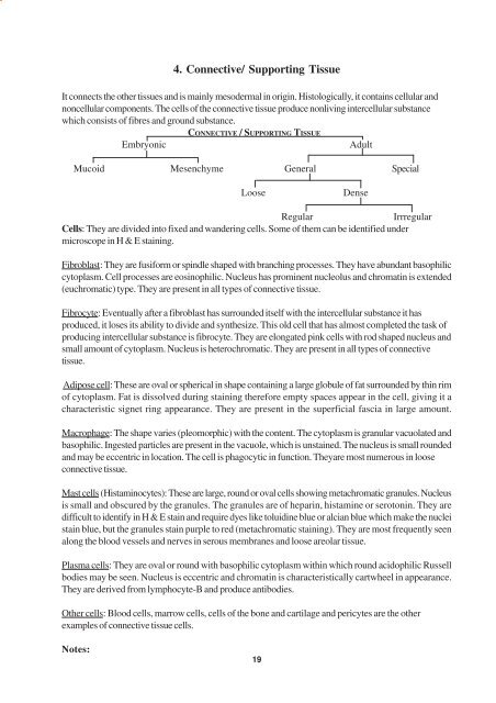
4. Connective/ Supporting TissueIt connects the other tissues and is mainly mesodermal in origin. Histologically, it contains cellular andnoncellular components. The cells of the connective tissue produce nonliving intercellular substancewhich consists of fibres and ground substance.EmbryonicCONNECTIVE / SUPPORTING TISSUEAdultMucoid Mesenchyme General SpecialLoose DenseRegular IrrregularCells: They are divided into fixed and wandering cells. Some of them can be identified undermicroscope in H & E staining.Fibroblast: They are fusiform or spindle shaped with branching processes. They have abundant basophiliccytoplasm. Cell processes are eosinophilic. Nucleus has prominent nucleolus and chromatin is extended(euchromatic) type. They are present in all types of connective tissue.Fibrocyte: Eventually after a fibroblast has surrounded itself with the intercellular substance it hasproduced, it loses its ability to divide and synthesize. This old cell that has almost completed the task ofproducing intercellular substance is fibrocyte. They are elongated pink cells with rod shaped nucleus andsmall amount of cytoplasm. Nucleus is heterochromatic. They are present in all types of connectivetissue.Adipose cell: These are oval or spherical in shape containing a large globule of fat surrounded by thin rimof cytoplasm. Fat is dissolved during staining therefore empty spaces appear in the cell, giving it acharacteristic signet ring appearance. They are present in the superficial fascia in large amount.Macrophage: The shape varies (pleomorphic) with the content. The cytoplasm is granular vacuolated andbasophilic. Ingested particles are present in the vacuole, which is unstained. The nucleus is small roundedand may be eccentric in location. The cell is phagocytic in function. Theyare most numerous in looseconnective tissue.Mast cells (Histaminocytes): These are large, round or oval cells showing metachromatic granules. Nucleusis small and obscured by the granules. The granules are of heparin, histamine or serotonin. They aredifficult to identify in H & E stain and require dyes like toluidine blue or alcian blue which make the nucleistain blue, but the granules stain purple to red (metachromatic staining). They are most frequently seenalong the blood vessels and nerves in serous membranes and loose areolar tissue.Plasma cells: They are oval or round with basophilic cytoplasm within which round acidophilic Russellbodies may be seen. Nucleus is eccentric and chromatin is characteristically cartwheel in appearance.They are derived from lymphocyte-B and produce antibodies.Other cells: Blood cells, marrow cells, cells of the bone and cartilage and pericytes are the otherexamples of connective tissue cells.Notes:19