

Primary Hepatic Gastrinoma: An Unusual Case of Zollinger-Ellison ...
Primary Hepatic Gastrinoma: An Unusual Case of Zollinger-Ellison ...
Primary Hepatic Gastrinoma: An Unusual Case of Zollinger-Ellison ...

Create successful ePaper yourself
Turn your PDF publications into a flip-book with our unique Google optimized e-Paper software.
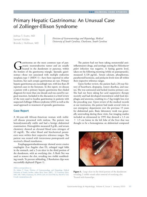
G & H C l i n i c a l C a s e S t u d i e s<strong>Primary</strong> <strong>Hepatic</strong> <strong>Gastrinoma</strong>: <strong>An</strong> <strong>Unusual</strong> <strong>Case</strong><strong>of</strong> <strong>Zollinger</strong>-<strong>Ellison</strong> SyndromeJoshua T. Evans, MDSamuel NicklesBrenda J. H<strong>of</strong>fman, MDDivision <strong>of</strong> Gastroenterology and Hepatology, MedicalUniversity <strong>of</strong> South Carolina, Charleston, South Carolina<strong>Gastrinoma</strong>s are the most common type <strong>of</strong> pancreaticneuroendocrine tumor and are usuallylocated in the duodenum or pancreas, withinthe borders <strong>of</strong> the gastrinoma triangle. Sporadic gastrinomas—thosenot associated with multiple endocrineneoplasia type 1 (MEN 1)—have been reported in otherlocations, but such ectopic gastrinomas are rare. <strong>Primary</strong>hepatic gastrinomas are exceedingly rare, with less than 20reported cases in the literature. In this report, we discussa patient with a primary hepatic gastrinoma that eludeddiagnosis for more than one decade and was cured by surgicalresection. Included in the discussion is a brief review<strong>of</strong> the tests used to localize gastrinomas in patients withsuspected <strong>Zollinger</strong>-<strong>Ellison</strong> syndrome (ZES) as well as theusual approach to treatment <strong>of</strong> sporadic gastrinomas.<strong>Case</strong> ReportA 46-year-old African-American woman with sicklecelldisease presented with melena. The patient washemodynamically stable and had a benign abdominalexamination. Hemoglobin measured 8 g/dL, and serumchemistry showed an elevated blood urea nitrogen <strong>of</strong>43 mg/dL. The other blood and biochemical parameterswere within their respective reference ranges. Thepatient was treated with intravenous pantoprazole andreceived a blood transfusion.Esophagogastroduodenoscopy showed severe erosiveesophagitis (Los <strong>An</strong>geles class D), enlarged rugal foldsin the stomach, and a 2-cm ulcer in the third portion <strong>of</strong>the duodenum, with an overlying clot. A Roth Net wasused to remove the clot, revealing two visible nonbleedingvessels. To prevent rebleeding, 2 Resolution clips weresuccessfully deployed (Figure 1).The patient had not been taking nonsteroidal antiinflammatorydrugs, and serologic testing for Helicobacterpylori infection was negative. A fasting gastrin level,taken on the following morning (while on pantoprazole),measured 3,120 pg/mL. Serum calcium, phosphorous,parathyroid hormone, and prolactin levels were all withintheir respective reference ranges.Upon further review, the patient had a 20-year history<strong>of</strong> heartburn, dyspepsia, watery diarrhea, and nausea.She was uninsured and lacked routine primary care.She had not been taking her acid suppressive therapyrecently and had developed intermittent solid food dysphagiaand anorexia, resulting in a 5-kg weight loss overthe preceding year. Upon review <strong>of</strong> the medical recordsat our institution, the patient had made several visits toour emergency department over the previous 15 yearsfor abdominal pain. Basic laboratory work was generallyunrevealing during these visits, but imaging studiesincluded an ultrasound in 1995 that showed a 1.3 cm× 1.5 cm lesion in the left lobe <strong>of</strong> the liver that wasthought to be a hemangioma; an abdominal computedAddress correspondence to:Dr. Joshua T. Evans, Division <strong>of</strong> Gastroenterology and Hepatology, MedicalUniversity <strong>of</strong> South Carolina, Ashley River Tower (ART) - 25 Courtenay Drive,MSC 290, 7100A, Charleston, SC 29455; Cell: 314-323-6790; Fax: 843-876-7232;E-mail: evansjos@musc.eduFigure 1. Large ulcer in the third portion <strong>of</strong> the duodenumrevealing 2 visible vessels after removal <strong>of</strong> overlying clot.Treated with hemoclips.Gastroenterology & Hepatology Volume 6, Issue 1 January 2010 53
E V A N S e t a lFigure 2. Magnetic resonance image showing a 4.1 cm × 3.5cm well-circumscribed mass in the left lobe <strong>of</strong> the liver.tomography scan in 2004 that showed a 2.5 cm × 2.6cm lesion in the left lobe <strong>of</strong> the liver; and an abdominalultrasound in 2005 that showed a similar lesion measuring2.5 cm × 2.6 cm. At that time, magnetic resonanceimaging was recommended, but not performed.After 72 hours <strong>of</strong> intravenous proton pump inhibitortherapy, the patient began to eat, her dysphagia resolved,and she regained her appetite. She was placed on 60 mg<strong>of</strong> oral pantoprazole twice daily. Magnetic resonanceimaging was performed and showed a 4.1 cm × 3.5 cmwell-circumscribed mass in the left lobe <strong>of</strong> the liver(Figure 2). Somatostatin-receptor scintigraphy showed afocus <strong>of</strong> markedly increased octreotide uptake in the lefthepatic lobe corresponding to the lesion seen on magneticresonance imaging and consistent with a neuroendocrinetumor (Figure 3). Endoscopic ultrasonography showedseveral very mildly enlarged lymph nodes in the periportalregion but no other lesions in the gastrinoma triangle orpancreas. Fine-needle aspiration <strong>of</strong> the periportal lymphnodes showed normal tissue.<strong>An</strong> exploratory laparotomy was performed. Thoroughbimanual palpation <strong>of</strong> the pancreas, intraoperativeultrasound <strong>of</strong> the pancreas, kocherization <strong>of</strong> the duodenum,and removal <strong>of</strong> several mildly enlarged periportaland omental lymph nodes did not identify evidence <strong>of</strong>alternative primary sites <strong>of</strong> the gastrinoma or evidence <strong>of</strong>metastases. Frozen sections <strong>of</strong> the lymph nodes showednormal tissue. A left lateral segmentectomy was performedto remove the liver lesion, which was not palpableintraoperatively. Pathology showed a 2.9 cm × 4.0 cmneuroendocrine tumor consistent with a gastrinoma.Stains were positive for synaptophysin and chromogranin(Figure 4).Thirty-six hours postoperatively, the patient’s serumgastrin level was 41 pg/mL, consistent with a surgical cureFigure 3. Somatostatin-receptor scintigraphy (octreotidescan) showing markedly increased octreotide uptake in the lefthepatic lobe corresponding to the lesion seen on the magneticresonance image.<strong>of</strong> ZES. Two months postoperatively, she remains eugastrinemic(gastrin level, 55 pg/mL). The patient has dramaticallyimproved: her symptoms have entirely resolved,she has regained 5 kg <strong>of</strong> weight, and her proton pumpinhibitor therapy has now been decreased to pantoprazole40 mg twice daily. We plan to repeat an upper endoscopyto monitor the mucosal response to acid suppression andexclude Barrett esophagus.DiscussionZES was first described in 1955 and affects approximately1% <strong>of</strong> patients with peptic ulcer disease. Classically, thesyndrome is characterized by primary ulcerations in atypicallocations such as the second or third portions <strong>of</strong> theduodenum or jejunum; severe gastric acid hypersecretion;and the presence <strong>of</strong> a non-beta islet cell tumor <strong>of</strong> the pancreasor duodenum. However, ulcers in patients with ZESare more commonly normal in size and location (typicallyless than 1 cm in size and located in the duodenal bulb). 1Diarrhea in patients with gastrinomas is common andresults from a variety <strong>of</strong> mechanisms, including gastricacid hypersecretion; inactivation <strong>of</strong> pancreatic enzymes(leading to steatorrhea); epithelial cell damage; and inhibition<strong>of</strong> sodium and water absorption, leading to bothmalabsorptive and secretory states. 1 Diagnosis <strong>of</strong> a gastrinomarequires a high level <strong>of</strong> suspicion, as the presentingsymptoms (diarrhea, heartburn, dyspepsia) are so common.The average time between initial presentation anddiagnosis <strong>of</strong> ZES is 6–9 years. 1 A markedly elevated serumgastrin level <strong>of</strong> greater than 1,000 pg/mL in the absence <strong>of</strong>54 Gastroenterology & Hepatology Volume 6, Issue 1 January 2010
P r i m a r y H e p a t i c G a s t r i n o m aABFigure 4. Pathology showing a 2.9 cm × 4.0 cmneuroendocrine tumor consistent with a gastrinoma. Stainswere positive for synaptophysin (A) and chromogranin (B).achlorhydria is virtually diagnostic <strong>of</strong> a gastrinoma. Exclusion<strong>of</strong> achlorhydric conditions such as atrophic gastritisand pernicious anemia (which are much more commonthan gastrinomas) is essential, as severe hypergastrinemiato this degree can occur in these diseases.If nonachlorhydric hypergastrinemia is confirmed,a secretin stimulation test is performed. The paradoxicalrise <strong>of</strong> serum gastrin level in response to a secretin level <strong>of</strong>greater than 120 pg/mL is 94% sensitive and nearly 100%specific for gastrinoma (again, in the absence <strong>of</strong> achlorhydria).1 Although a greater than 200 pg/mL rise in gastrinhas been widely considered to be a positive test, this valuemay <strong>of</strong>fer slightly lower sensitivity. 1,2 Controversial issuesinclude whether (and if so, when) acid suppression shouldbe stopped prior to secretin stimulation testing. Manyexperts believe that continuing acid suppression therapydoes not alter the results <strong>of</strong> secretin provocation testing,and in some patients (particularly those with a history <strong>of</strong>complicated or perforated peptic ulcer disease), discontinuingacid suppression may be dangerous. 1A negative secretin stimulation test is extremely rarein patients with ZES. If there is a high index <strong>of</strong> suspicionfor ZES and the secretin test is negative, the test caneither be repeated or a calcium stimulation test can beperformed. This test is used less commonly, is less sensitive(63% vs 94%), and has the potential for serious sideeffects from the intravenous infusion <strong>of</strong> calcium. 2 However,reports <strong>of</strong> patients with ZES who test negative tosecretin stimulation and positive to calcium stimulationexist. Therefore, the test is considered highly specificfor gastrinoma.When biochemical studies suggest that a gastrinomais present, its location must be identified. A number <strong>of</strong>tests are used to localize gastrinomas, including ultrasonography,computed tomography, magnetic resonanceimaging, selective arterial secretin or calcium stimulationstudies, and endoscopic ultrasound (EUS). In general, acombination <strong>of</strong> these tests allows for pre-operative localization<strong>of</strong> gastrinomas in approximately 80% <strong>of</strong> cases. 1,3With the advent <strong>of</strong> EUS, the sensitivity for detectingsporadic gastrinomas prior to surgical exploration hasincreased to approximately 85%. 4 In general, the noninvasiveimaging modalities tend to have slightly highersensitivities in patients with MEN 1. 3Although somatostatin-receptor scintigraphy (octreotidescan) is touted for its high sensitivity in localizinggastrinomas, the use <strong>of</strong> this imaging modality is highlydependent on the size <strong>of</strong> the gastrinoma, ranging from30% for gastrinomas smaller than 1.1 cm to 96% forgastrinomas larger than 2 cm. 5 In cases where noninvasiveimaging is negative, EUS becomes highly valuable withthe ability for real-time cytologic evaluation <strong>of</strong> primarytumors and suspicious lymph nodes. The sensitivity <strong>of</strong>EUS depends upon gastrinoma location as well, rangingfrom 43% for duodenal gastrinomas to 85% for pancreaticgastrinomas. 4,6,7Still, some cases require surgical exploration to locatethe gastrinoma. The surgical tenets <strong>of</strong> exploration forgastrinoma include a Kocher maneuver to allow carefulexamination <strong>of</strong> the head <strong>of</strong> the pancreas and uncinateprocess, bimanual palpation <strong>of</strong> the body and tail <strong>of</strong> thepancreas, intraoperative ultrasonography, duodenotomyand exploration <strong>of</strong> the duodenal mucosa, and sampling<strong>of</strong> lymph nodes in the gastrinoma triangle. 1 The definition<strong>of</strong> a biochemical cure following surgery for ZESis a normal fasting and secretin stimulated gastrin levelpostoperatively. The initial postoperative cure rate isapproximately 60% for sporadic gastrinomas, but thisfigure decreases significantly to 34% at 10-year followup.3 In patients with gastrinomas associated with MEN 1syndrome, disease-free survival rates are much lower andGastroenterology & Hepatology Volume 6, Issue 1 January 2010 55
E V A N S e t a lsurgery remains controversial. 3 Therefore, it is essential toexclude MEN 1 in patients with gastrinoma.Approximately one third <strong>of</strong> patients with MEN 1present with ZES initially, followed by other manifestations<strong>of</strong> MEN 1 later, usually primary hyperparathyroidism.Indeed, the first surviving ZES patient was diagnosedwith hyperparathyroidism some 40 years after the initialdiagnosis <strong>of</strong> ZES. 8,9 Gene mutation analysis for the MENgene is available but costly, and there are no guidelinesfor genetic testing in these patients. It is generally recommendedthat all patients with gastrinomas are screenedfor MEN 1 by measuring serum calcium, phosphorous,intact-parathyroid hormone, and prolactin levels. Thesemarkers should be followed after surgical resection atregular intervals to monitor for recurrence.Medical therapy for ZES consists primarily <strong>of</strong> acidsuppression therapy with high-dose proton pump inhibitors.Although the somatostatin analogue octreotide hasbeen used with success to treat other neuroendocrinetumors, its success in treating gastrinomas has not beenconsistent. At present, octreotide remains a second-linetherapy in ZES for treating extreme hypersecretory statesleading to voluminous diarrhea and dehydration. 1Prior to the widespread use <strong>of</strong> proton pumpinhibitors, most <strong>of</strong> the morbidity in ZES resulted fromsevere and recurrent peptic ulcerations. Partial and totalgastrectomy were commonly performed, and patientsfrequently required multiple operations. In the post−proton pump inhibitor era, the natural history <strong>of</strong> thedisease appears to have changed and studies have shownthat the number <strong>of</strong> surgical procedures in these patientshas dropped dramatically. 8Although proton pump inhibitors effectively treatthe acid hypersecretion in patients with ZES, they maydelay the diagnosis. One study showed that in the pre−proton pump inhibitor era (prior to 1985), the incidence<strong>of</strong> patients presenting with metastatic disease was 19%,with a 5-year survival <strong>of</strong> 29%, whereas in the post−protonpump inhibitor era (from 1985 to 1995), the incidence<strong>of</strong> patients presenting with metastatic gastrinomas was55%, with a 5-year survival rate <strong>of</strong> 5%. 10 This provideseven more impetus for the clinician to have a high index<strong>of</strong> suspicion when evaluating patients with heartburn,abdominal pain, and diarrhea, particularly if there is ahistory <strong>of</strong> peptic ulcer disease.Even with total resection <strong>of</strong> the gastrinoma, patientsmay continue to have gastric acid hypersecretion due toan excess <strong>of</strong> gastric parietal cells resulting from the trophiceffect <strong>of</strong> gastrin preceding the resection. Approximately40% <strong>of</strong> patients require acid suppression up to 4 yearspostoperatively. 1 The dose <strong>of</strong> proton pump inhibitortherapy should be gradually reduced postoperatively,according to symptoms and endoscopic healing. Alternatively,the proton pump inhibitor may be dose-adjustedby measuring the basal acid output, with the goal <strong>of</strong> abasal acid output <strong>of</strong> less than 10 mEq/hr in men and lessthan 5 mEq/hr in women. 1SummaryThis unusual case emphasizes the need for a high index <strong>of</strong>clinical suspicion in evaluating patients who present withsymptoms compatible with ZES. In retrospect, our patienteluded the diagnosis <strong>of</strong> ZES for many years. Measurement<strong>of</strong> the serum gastrin level earlier would have allowedtimely diagnosis and prevented significant morbidity. Weplan to measure the fasting serum gastrin level at regularintervals postoperatively in this patient. The possibility<strong>of</strong> occult lymph node or other primary locations missedby EUS and surgical exploration still exists, and recurrenceis possible. It is noted that a duodenotomy was notperformed during the operation, and the possibility <strong>of</strong> atiny duodenal gastrinoma was not definitively excluded.However, the rapid fall in serum gastrin level followingresection <strong>of</strong> the hepatic gastrinoma argues against this.References1. <strong>Ellison</strong> EC, Johnson JA. The <strong>Zollinger</strong>-<strong>Ellison</strong> syndrome: a comprehensivereview <strong>of</strong> historical, scientific, and clinical considerations. Curr Probl Surg.2009;46:13-106.2. Berna MJ, H<strong>of</strong>fmann KM, Long SH, Serrano J, Gibril F, Jensen RT. Serumgastrin in <strong>Zollinger</strong>-<strong>Ellison</strong> syndrome: II. Prospective study <strong>of</strong> gastrin provocativetesting in 293 patients from the National Institutes <strong>of</strong> Health and comparison with537 cases from the literature. Evaluation <strong>of</strong> diagnostic criteria, proposal <strong>of</strong> newcriteria, and correlations with clinical and tumoral features. Medicine (Baltimore).2006;85:331-364.3. Wells SA. Surgery for <strong>Zollinger</strong>-<strong>Ellison</strong> syndrome. N Engl J Med. 1999;341:688-690.4. Norton JA, McLean AM, Fairclogh PD. Endoscopic ultrasound in the localization<strong>of</strong> pancreatic islet cell tumors. Best Pract Res Clin Endocrinol Metab.2004;19:177-193.5. Alexander HR, Fraker DL, Norton JA, Bartlett DL, Tio L, et al. Prospectivestudy <strong>of</strong> somatostatin receptor scintigraphy and its effect on operative outcome inpatients with <strong>Zollinger</strong>-<strong>Ellison</strong> syndrome. <strong>An</strong>n Surg. 1998;228:228-238.6. Norton A, Jensen RT. Resolved and unresolved controversies in the surgicalmanagement <strong>of</strong> patients with <strong>Zollinger</strong>-<strong>Ellison</strong> syndrome. <strong>An</strong>n Surg.2004;240:757-773.7. <strong>An</strong>derson MA, Carpenter S, Thompson NW, Nostrant TT, Elta GH, ScheimanJM. Endoscopic ultrasound is highly accurate and directs management inpatients with neuroendocrine tumors <strong>of</strong> the pancreas. Am J Gastroenterol. 2000;95:2271-2277.8. <strong>Ellison</strong> EC. <strong>Zollinger</strong>-<strong>Ellison</strong> syndrome: a personal perspective. Am Surg.2008;74:563-571.9. Melvin WS, Johnson JA, Sparks J, Innes JT, <strong>Ellison</strong> EC. Long-term prognosis<strong>of</strong> <strong>Zollinger</strong>-<strong>Ellison</strong> syndrome in multiple endocrine neoplasia. Surgery.1993;114:1183-1188.10. Corleto VD, <strong>An</strong>nibale B, Gibril F, <strong>An</strong>geletti S, Serrano J, et al. Does the widespreaduse <strong>of</strong> proton pump inhibitors mask, complicate and/or delay the diagnosis<strong>of</strong> <strong>Zollinger</strong>-<strong>Ellison</strong> syndrome? Aliment Pharmacol Ther. 2001;15:1555-1561.56 Gastroenterology & Hepatology Volume 6, Issue 1 January 2010