

PDF (0K) - World Journal of Gastroenterology
PDF (0K) - World Journal of Gastroenterology
PDF (0K) - World Journal of Gastroenterology

Create successful ePaper yourself
Turn your PDF publications into a flip-book with our unique Google optimized e-Paper software.
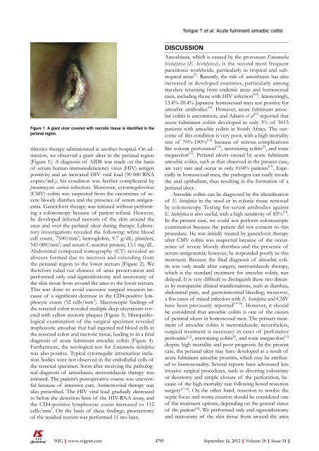
Torigoe T et al . Acute fulminant amoebic colitisFigure 1 A giant ulcer covered with necrotic tissue is identified in theperianal region.tibiotics therapy administered at another hospital. On admission,we observed a giant ulcer in the perianal region(Figure 1). A diagnosis <strong>of</strong> AIDS was made on the basis<strong>of</strong> serum human immunodeficiency virus (HIV) antigenpositivity and an increased HIV viral load (90 000 RNAcopies/mL); his condition was further complicated bypneumocystis carinii infection. Moreover, cytomegalovirus(CMV) colitis was suspected from the occurrence <strong>of</strong> severebloody diarrhea and the presence <strong>of</strong> serum antigenemia.Ganciclovir therapy was initiated without performinga colonoscopy because <strong>of</strong> patient refusal. However,he developed infected necrosis <strong>of</strong> the skin around theanus and over the perianal ulcer during therapy. Laboratoryinvestigations revealed the following: white bloodcell count, 7500/mm 3 ; hemoglobin, 9.7 g/dL; platelets,545 000/mm 3 ; and serum C-reactive protein, 13.1 mg/dL.Abdominal computed tomography (CT) revealed anabscess formed due to necrosis and extending fromthe perianal region to the lower rectum (Figure 2). Wetherefore ruled out chances <strong>of</strong> anus preservation andperformed only end-sigmoidostomy and necrotomy <strong>of</strong>the skin tissue from around the anus to the lower rectum.This was done to avoid excessive surgical invasion because<strong>of</strong> a significant decrease in the CD4-positive lymphocytecount (52 cells/mm 3 ). Macroscopic findings <strong>of</strong>the resected colon revealed multiple deep ulcerations coveredwith yellow necrotic plaques (Figure 3). Histopathologicalexamination <strong>of</strong> the surgical specimen revealedtrophozoite amoebae that had ingested red blood cells inthe resected colon and necrotic tissue, leading to in a finaldiagnosis <strong>of</strong> acute fulminant amoebic colitis (Figure 4).Furthermore, the serological test for Entamoeba histolyticawas also positive. Typical cytomegalic intranuclear inclusionbodies were not observed in the endothelial cells <strong>of</strong>the resected specimen. Soon after receiving the pathologicaldiagnosis <strong>of</strong> amoebiasis, metronidazole therapy wasinitiated. The patient’s postoperative course was uneventfulbecause <strong>of</strong> intensive care. Antiretroviral therapy wasalso prescribed. The HIV viral load gradually decreasedto below the detection limit <strong>of</strong> the HIV-RNA assay, andthe CD4-positive lymphocyte count increased to 112cells/mm 3 . On the basis <strong>of</strong> these findings, proctectomy<strong>of</strong> the residual rectum was performed 11 mo later.DISCUSSIONAmoebiasis, which is caused by the protozoan Entamoebahistolytica (E. histolytica), is the second most frequentparasitosis worldwide, particularly in tropical and subtropicalareas [7] . Recently, the risk <strong>of</strong> amoebiasis has alsoincreased in developed countries, particularly amongtravelers returning from endemic areas and homosexualmen, including those with HIV infection [8,9] . Interestingly,13.4%-20.4% Japanese homosexual men test positive foramoebic antibodies [10] . However, acute fulminant amoebiccolitis is uncommon, and Adams et al [11] reported thatacute fulminant colitis developed in only 3% <strong>of</strong> 3013patients with amoebic colitis in South Africa. The outcome<strong>of</strong> this condition is very poor, with a high mortalityrate <strong>of</strong> 70%-100% [1,2] because <strong>of</strong> serious complicationslike colonic perforation [3,4] , necrotizing colitis [5] , and toxicmegacolon [12] . Perianal ulcers caused by acute fulminantamoebic colitis, such as that observed in the present case,are very rare and occur in only 0.04% patients [11] . Especiallyin homosexual men, the pathogen can easily invadethe anal epithelium, thus resulting in the formation <strong>of</strong> aperianal ulcer.Amoebic colitis can be diagnosed by the identification<strong>of</strong> E. histolytica in the stool or in colonic tissue removedby colonoscopy. Testing for serum antibodies againstE. histolytica is also useful, with a high sensitivity <strong>of</strong> 85% [13] .In the present case, we could not perform colonoscopicexamination because the patient did not consent to thisprocedure. He was initially treated by ganciclovir therapyafter CMV colitis was suspected because <strong>of</strong> the occurrence<strong>of</strong> severe bloody diarrhea and the presence <strong>of</strong>serum antigenemia; however, he responded poorly to thistreatment. Because the final diagnosis <strong>of</strong> amoebic colitiswas only made after surgery, metronidazole therapy,which is the standard treatment for amoebic colitis, wasdelayed. It is very difficult to distinguish these two diseasesby nonspecific clinical manifestations, such as diarrhea,abdominal pain, and gastrointestinal bleeding; moreover,a few cases <strong>of</strong> mixed infection with E. histolytica and CMVhave been previously reported [14-16] . However, it shouldbe considered that amoebic colitis is one <strong>of</strong> the causes<strong>of</strong> perianal ulcers in homosexual men. The primary treatment<strong>of</strong> amoebic colitis is metronidazole; nevertheless,surgical treatment is necessary in cases <strong>of</strong> perforativeperitonitis [1,2] , necrotizing colitis [5] , and toxic megacolon [12]despite high mortality and poor prognosis. In the presentcase, the perianal ulcer may have developed as a result <strong>of</strong>acute fulminant amoebic proctitis, which may be attributedto homosexuality. Several reports have advocated lessinvasive surgical procedures, such as diverting colostomyor ileostomy and simple closure <strong>of</strong> the perforation, because<strong>of</strong> the high mortality rate following bowel resectionsurgery [17,18] . On the other hand, resection to resolve theseptic focus and stoma creation should be considered one<strong>of</strong> the treatment options, depending on the general status<strong>of</strong> the patient [19] . We performed only end-sigmoidostomyand necrotomy <strong>of</strong> the skin tissue from around the anusWJG|www.wjgnet.com4795 September 14, 2012|Volume 18|Issue 34|


