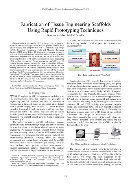
Fabrication of Tissue Eng Using Rapid Prototyp Fabrication of ...
Fabrication of Tissue Eng Using Rapid Prototyp Fabrication of ...
Fabrication of Tissue Eng Using Rapid Prototyp Fabrication of ...

Create successful ePaper yourself
Turn your PDF publications into a flip-book with our unique Google optimized e-Paper software.
World Academy <strong>of</strong> Science, <strong>Eng</strong>ineering and Technology 59 2011the existing limitation and future prospects.II. RAPID PROTOTYPING TECHNIQUES FOR TISSUE ENGINEERINGSCAFFOLDS FABRICATIONA. The extrusion-based RP techniquesThe extrusion-based RP technique is also known as FusedDeposition Modelling (FDM) in which a thin thermoplasticfilaments are melted by heating and guided by an extrudercontrolled by a computer, to form 3D objects. The materialleaves the extruder in a liquid form and hardens immediately.The previously formed layer, which is the substrate for thenext layer, must be maintained at a temperature just below thesolidification point <strong>of</strong> the thermoplastic material to assuregood interlayer adhesion [5]. The major limitations <strong>of</strong> FDMare the use <strong>of</strong> filament-based materials and the high heat effecton raw material. To overcome some <strong>of</strong> these limitations,alternative extrusion-based processes have been proposed like3D Fiber Deposition (3DF) [6], Bioplotting [7], precisionextruding deposition(PED) [8] and other techniques we willdescribe through the review context.The feasibility <strong>of</strong> FDM to fabricate porous customizedfreeform structures <strong>of</strong> medical-grade polymethylmethacrylate(PMMA) was investigated by Espalin et al. [9]. It was foundthat, by enabling the use <strong>of</strong> PMMA in FDM, medical implantssuch as custom crani<strong>of</strong>acial implants can be directly fabricatedfrom medical imaging data improving the current state <strong>of</strong>PMMA use in medicine. Yen et al. [10], also employed FDMin the production <strong>of</strong> poly (D,L-lactide-co-glycolide) (PLGA)scaffolds filled with type II collagen and evaluated the cellularproliferation and matrix deposition <strong>of</strong> these hybrid scaffolds.SEM <strong>of</strong> the pure and hybrid scaffolds are shown in Fig. 3.technique in which a molten hydrogels, thermoplasticpolymers and biomaterial pastes are extruded from a CAMcontrolled robotic unit on a stage in the form <strong>of</strong> a fibre. 3DFwas utilized to produce poly (ethylene glycol)-terephthalate/poly (butylenes terephthalate) (PEGT/PBT)block co-polymer scaffolds with fully interconnecting porenetwork for engineering <strong>of</strong> articular cartilage. By varying theco-polymer composition, porosity and pore geometry,scaffolds were produced with a range <strong>of</strong> mechanical propertiesclose to articular cartilage. The scaffolds seeded with bovinechondroccytes supported a homogeneous cell distribution andsubsequent cartilage like tissue formation [6]. Li et al. [13],On the other hand, produced a porous Ti6Al4V scaffolds,(Fig. 4) with fully interconnected pore networks, highlycontrollable porosities, and pore sizes by 3DF. The Ti6Al4Vpowders (68 vol %) were mixed with an aqueous solution <strong>of</strong>methylcellulose (0.3 wt %) as binder and satiric acid (0.2 wt%) to improve the rheological properties <strong>of</strong> the slurry. Theyconcluded that 3DF is a promising technique for the designand fabrication <strong>of</strong> custom made Ti6Al4V scaffoldarchitectures for orthopaedic implant applications.bacFig. 3 SEM <strong>of</strong> pure PLGA scaffolds (a, b) and hybrid scaffolds (c, d)produced by FDM [10].Recently, Tellis et al. [11] produced polybutyleneterephthalate (PBT) scaffolds groups with various porestructures by FDM. They used compression testing and MicroCT to compare compressive stiffness, porosity, connectivitydensity, and trabecular separation <strong>of</strong> each scaffold to a naturalbone sample. In another approach, Geffre et al. [12] comparedbone ingrowths into macroscopic PBT porous scaffoldsfabricated by FDM, which had either a simple pore structureor a complex pore structure mimicking the native tissuearchitecture.Woodfield et al. [6], on the other hand, developedthe 3D Fiber Deposition (3DF) system. Which is an FDM-likedFig. 4 Surface morphologies <strong>of</strong> 3DF Ti6Al4V scaffolds built withvarying fiber spacing from 0.2 to 0.7 mm in increments <strong>of</strong> 0.1 [13].Woodfield et al. [14] fabricated anatomical femoral andtibial cartilage constructs by 3DF. They evaluated producedscaffolds in vitro and in vivo in an autologous rabbit modeland found that Porous, interconnected 3DF scaffoldarchitectures enhanced chondrocyte attachment and redifferentiationcapacity while exhibiting mechanical propertiessimilar to native articular cartilage explants.To overcome filament preparation problem in FDM, avariation <strong>of</strong> FDM called precision extruding deposition (PED)for fabrication <strong>of</strong> bone tissue scaffolds were developed byWang et al. [8]. In PED , material in pellet or granule form isfed into a chamber where it is liquefied. Pressure from arotating screw forces the material down a chamber and outthrough a nozzle tip. This process was used by Shor et al [15]to directly fabricate polycaprolactane (PCL) and (PCL–hydroxyapatite, HA) composite tissue scaffolds. Similar workwas conducted by Yildirim et al. [16] to fabricate (PCL)scaffolds with a 0/90° strut configuration with 300 µm pore1873
World Academy <strong>of</strong> Science, <strong>Eng</strong>ineering and Technology 59 2011size, 250 µm strut width and were treated with oxygen-basedplasma in order to increase the cellular activity.Low temperature deposition manufacturing (LDM) isanother modified version <strong>of</strong> FDM developed by Xiong et al.[17] to overcome the heating and liquefying processing <strong>of</strong>materials. The system comprises a multi-nozzle extrusionprocess and a thermally induced phase separation process.LDM was recently used by Li et al. [18] to fabricateindividualized tissue engineering PLGA/ tricalcium phosphate(TCP) composite scaffolds based on alveolar bone defects.Mäkitie et al. [19], also assessed the viability <strong>of</strong> (PLGA/TCP)composite scaffold generated with LDM (Fig. 5) in a 3D cellcultivation, in vitro.plotter, in order to increase the elastic modulus and yieldstrength <strong>of</strong> the strand in the scaffold.Fig. 6 Cell attachment <strong>of</strong> bioplotted PCL after 7 days (a) on thenormal strand and (b) on the modified strand produced with avibration <strong>of</strong> 30 Hz.[ 25], [26]Fig. 5 PLGA–TCP scaffold manufactured by LDM [19]Another variants <strong>of</strong> the FDM technique is Bioplotting,developed separately by Freiburg Materials Research Centre,Germany, and marketed by Envision Technologies GmBH,Germany, [20]. In bioplotter technique, a micro needle isemployed as the extrusion nozzle where liquids, pastes, melts,solutions, hot melts, reactive oligomers or dispersions whichare initially stored in a heated cartridge, are extruded into atemperature controlled liquid dispensing medium. Thedispensing medium induces solidification <strong>of</strong> the depositedmaterial by cooling, heating or through chemical reaction.Also, by using a dispensing solution <strong>of</strong> similar density as thebuilding material, the buoyancy exerted by the medium on thebuild can prevent the collapse <strong>of</strong> complex structures thuseliminating the need for sacrificial support structures whichare typical in conventional FDM systems [20]. Most recently,Various studies were carried out using bioplotting technique tocontrol the scaffold architecture in order to obtain betterresults in terms <strong>of</strong> combining enhanced tissue growth withadequate mechanical properties [21]-[24]. In these studies,Scaffolds mechanical properties, cell growth, and structuremorphology were evaluated and characterized. Son and coworker[25],[26 ] , modified the bottom plate <strong>of</strong> a commercialbioplotter so that it was vibrated by a piezoelectric transducer(PZT). By this modification, scaffolds with rough surfacestrands can be obtained and as a result they fabricate 3-Dpolymeric scaffolds with enhanced compressive modulus,initial cell attachment and proliferation (as shown in Fig. 6)without any chemical or biological treatment. Anothermodification in 3D bioplotter was also introduced by Hee et al[27]. They designed an oscillating nozzle system for the 3DDaoud et al. [28], developed a bioplotted micr<strong>of</strong>abricatedPLGA Scaffolds with controlled pore structures. Theyoptimized the structural integrity and pore size required forpancreatic islet culture and seeding. Ye et al. [29] used aBioplotting system to fabricate nano biocomposite scaffolds <strong>of</strong>non-stoichiometric apatite (ns-AP) and poly(ε-caprolactone)(PCL) scaffolds. They reported that, scaffold with 40 wt% ns-AP contained open and well interconnected pores with a size<strong>of</strong> 400–500 µm, and exhibited a maximum porosity <strong>of</strong> 76%.Additionally, Oliveira et al. [30] Studied the nucleation andgrowth <strong>of</strong> biomimetic apatite layers on the surfaces <strong>of</strong>bioplotted starch/polycaprolactone SPCL scaffolds with a0°/90°strut structure. Haberstroh et al. [31] Investigated theosteogenic effect <strong>of</strong> three different cell-seeded 3D-bioplottedscaffolds with 3 different biomaterials in an ovine calvarialcritical-size defect model.Robocasting, also is an extrusion-based RP process inwhich a colloidal suspension, or ink, is extruded through amicron-sized nozzle in a defined trajectory to form a threedimensionalstructure [32] and is referred to in the literature asrobotic deposition and direct-write assembly [33]. Recently,this technique has been used to fabricate porous β- Tricalciumphosphate (TCP) scaffolds with a controlled architecture [34].The compressive strength <strong>of</strong> the fabricated TCP scaffolds wasenhanced by polymer infiltration. The authors reported thatinfiltrating polymers into the porous robocasted ceramicstructure was shown to considerably boost the strength andtoughness <strong>of</strong> the material. The fracture modes and the strength<strong>of</strong> robocasted HA and TCP scaffolds was also identified bythe same group in another related work [35].Another novel and modified deposition techniques havebeen also introduced in the last 3 years. These methods weredeveloped to increase manufacturing flexibility by enhancingdeposition capability in achieving optimum scaffoldrequirements. The new methods include Multi-head depositionsystem (MHDS) [36], [37], screw extrusion system(SES)[38],[39], BioExtruder [40],Combined FDM and1874
World Academy <strong>of</strong> Science, <strong>Eng</strong>ineering and Technology 59 2011electrospinning (ESP) system [41], [42], Combined plottingand (ESP) [43], [44], 3DF and (ESP) [45]. Combined rapidfreezing and plotting system [46], [47], porogen-basedextrusion system [48] and modified plotting system [49].B. Three Dimensional Printing (3DP)3D printing was the first RP technique to be proposed forbiomedical and tissue engineering purposes [50]. 3D Printeruses ink-jet printing approach to accurately writes a ‘‘binder’’solution like polymer latex or silica colloid , which moves inaccordance to the CAD cross-sectional data through the inkjetprint head, onto metallic, ceramics or composites powder [51].The first step in 3D Printing is the spreading <strong>of</strong> powder onto aplatform with a roller, followed by the inkjet print headprinting a two-dimensional pattern, onto the powder layer.Then the next powder layer is spread and the process isrepeated until the part is finished. The unused powder acts, assupport for the part and is brushed or blown <strong>of</strong>f afterwards.The piston chamber is lowered and refilled with another layer<strong>of</strong> powder and the process repeated. The process usuallyfollowed by a temperature treatment to burn the binder <strong>of</strong>f anda final sintering step [52].Recent researches on 3D printed scaffolds focus onevaluating Mechanical and in vivo and in vitro performance <strong>of</strong>scaffolds. Recently, Detsch et al. [53] fabricated samples frompure HA and β-TCP as well as a biphasic calcium phosphatesBCP mixture with~60 wt% HA by 3DP. They studied celldevelopment on manufactured scaffolds surfaces by analyzingcell proliferation, differentiation, and activation. Shanjani et al[54] fabricated a calcium polyphosphate structures with a 3DPsystem and used SolidWorks® s<strong>of</strong>tware in the design <strong>of</strong> theporous samples. They reported that structures fabricated usingthe direct 3DP method may be more advantageous comparedto the conventionally sintered CPP structures <strong>of</strong> equivalentpercent porosity due to higher compressive strength and largerpores. Klammert et al. [55] introduced powder-printedmagnesium ammonium phosphate (struvite) structures for thefirst time.Ge et al. [56] investigated the mechanical properties andmicro-environment <strong>of</strong> 3D printed poly-lactic-co-glycolic acid(PLGA) scaffolds and they evaluated the proliferation anddifferentiation <strong>of</strong> human fetal osteoblasts after 3 weeks <strong>of</strong> invitro culture on the produced scaffolds. The results showedthat the PLGA scaffolds examined had mechanical propertiessimilar to that <strong>of</strong> trabecular bone, but was still much weakercompared to cortical bone. In addition to general porosity, thePLGA scaffolds also had micropores within macropore wallsand the cultured human osteoblasts could proliferate uponseeding on the PLGA scaffolds. Warnke and co-workers [57],[58], investigated the biocompatibility <strong>of</strong> HA and TCPscaffolds produced by 3DP printing/sintering techniques andtheir ability to support and promote the proliferation <strong>of</strong> humanosteoblasts compared with the commonly used bonereplacement material, Bovine hydroxyapatite(BioOss) in vitro.They noted that both versions <strong>of</strong> 3D printed and sinteredscaffolds were colonized by human osteoblasts, however morecells were seen on HA scaffolds than TCP scaffolds. Cellvitality staining and biocompatibility tests also showedsuperior biocompatibility <strong>of</strong> HA scaffolds to BioOss , whileBioOss was more compatible than TCP. The group alsoevaluated the biocompatibility and osteoinductivity <strong>of</strong>individually designed HA and TCP blocks compared toBioOss for heterotopic bone induction in a rat model as shownin Fig. 7. It was found that, designed HA and TCP blockstested as well as the BioOss blocks are suitable as matrices forendocultivation as they showed good biocompatibility in vivo.Fig. 7 Insertion <strong>of</strong> individually designed 3D-printed HA scaffold intothe rat [58].Klammert et al. [59] established a novel 3D powder printedmaterial using calcium phosphate cement chemistry as a cellculture scaffold for osteoblastic cells. In another study,Lowmunkong et al. [60] investigated the possibility <strong>of</strong>fabricating 3D scaffolds from pure plaster <strong>of</strong> Paris (POP)powder (calcium sulfate hemihydrates) with an averageparticle size <strong>of</strong> 10 µm–20 µm by 3DP and to transform thefabricated object from POP to HA or other bioceramic. AfterHA-transformed, specimen was sintered at 1150° C for 3 h,the compressive strength increased four times when comparedwith HA specimen. However, its crystal structure wastransformed to β-TCP due to the chemical reaction <strong>of</strong>transformed HA with remaining phosphate in the specimen.Gbureck et al. [61] Fabricated a custom made TCP/calciumpyrophosphate bone substitutes with a well-definedarchitecture using 3DP. They characterized the mechanicalperformance and porosity contained within the fabricatedsamples. The feasibility <strong>of</strong> 3DP to fabricate porous pureTitanium dental scaffold was recently investigated by Wiria etal. [62]. The 3D printed Titanium dental implant prototype hasbeen successfully fabricated and shown to have elasticmodulus <strong>of</strong> 4.8–13.2 GPa. This elastic modulus is much lowerthan the modulus <strong>of</strong> the bulk commercially pure Titanium andis in the range <strong>of</strong> elastic modulus <strong>of</strong> natural bone.Another processing system based on inkjet printingtechnology is the 3D phase change inkjet printer. This processutilises droplet deposition technique in which thermoplasticbuilding material and a wax like support material aredeposited from separate jets onto a working surface. As aresult <strong>of</strong> heat conduction, the droplets induce local melting onthe underlying layer and causes bonding to occur. After eachlayer hardens, uniform thickness is maintained by a millinghead [63]. <strong>Using</strong> this printing system, Park et al. [64] designed1875