Final Programme
Final Programme
Final Programme

You also want an ePaper? Increase the reach of your titles
YUMPU automatically turns print PDFs into web optimized ePapers that Google loves.
: imaging generations<br />
myESR.org<br />
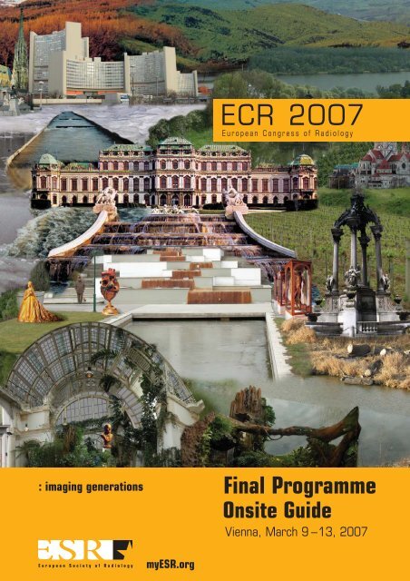
<strong>Final</strong> <strong>Programme</strong><br />
Onsite Guide<br />
Vienna, March 9 –13, 2007
GE Healthcare<br />
Re-think. Re-discover.<br />
Re-invent. Re-imagine.<br />
We have a shared passion. A desire to transform healthcare. By listening to your needs and putting the<br />
strength of the world’s leading scientists, engineers and business people together, anything can happen.<br />
The future of healthcare can change forever. Predict, diagnose, treat, monitor and inform in ways never<br />
thought possible. Help patients experience what we call early health, which focuses on early prevention<br />
rather than late diagnosis. If we can find disease sooner, we can help people live longer, fuller lives.<br />
Together we can re-think, re-discover and re-invent. Healthcare Re-imagined.<br />
To see Radiology Re-imagined, come visit us at ECR (Booths 202 & 213/Expo B) or visit<br />
www.gehealthcare.com/re-imagine<br />
GE imagination at work<br />
© 2007 General Electric Company GE Medical Systems, a General Electric Company doing business as GE Healthcare
welcomes you<br />
to its annual meeting,<br />
the European Congress<br />
of Radiology 2007<br />
under the patronage of<br />
Dr. Heinz Fischer, Federal President of Austria<br />
European Society of Radiology<br />
19 th European Congress of Radiology<br />
Sessions in joint sponsorship with<br />
CIRSE<br />
Cardiovascular and Interventional<br />
Radiological Society of Europe<br />
EuroPACS<br />
European Association for the Promotion of<br />
Information Exchange on PACS Research<br />
EUSOBI<br />
European Society of Breast Imaging<br />
ESCR<br />
European Society of Cardiac Radiology<br />
ESGAR<br />
European Society of Gastrointestinal and<br />
Abdominal Radiology<br />
ESHNR<br />
European Society of Head and Neck Radiology<br />
ESSR<br />
European Society of Musculoskeletal Radiology<br />
ESNR<br />
European Society of Neuroradiology<br />
ESPR<br />
European Society of Paediatric Radiology<br />
ESTI<br />
European Society of Thoracic Imaging<br />
ESUR<br />
European Society of Urogenital Radiology<br />
EFOMP<br />
European Federation of Organisations for<br />
Medical Physics<br />
European Working Group on Contrast Agents<br />
ISRRT<br />
International Society of Radiographers and<br />
Radiological Technologists<br />
ECR accepts no responsibility<br />
for errors or misprints.<br />
Congress venue<br />
Austria Center Vienna<br />
Bruno Kreisky Platz 1<br />
AT – 1220 Vienna, Austria<br />
Congress language<br />
English<br />
Onsite Opening Hours<br />
Registration<br />
Thursday, March 8<br />
09:00 – 19:00<br />
Friday, March 9 to Saturday, March 10<br />
07:00 – 19:00<br />
Sunday, March 11 to Monday, March 12<br />
07:00 – 18:00<br />
Tuesday, March 13<br />
07:00 – 14:00<br />
Preview centre – EDIPS<br />
EDIPS – ECR’s Digital Preview System<br />
Thursday, March 8<br />
12:00 – 19:00<br />
Friday, March 9 to Monday, March 12<br />
07:00 – 19:00<br />
Tuesday, March 13<br />
07:00 – 14:00<br />
Scientific exhibition – EPOSTM Friday, March 9<br />
14:00 – 19:00<br />
Saturday, March 10 to Monday, March 12<br />
07:00 – 19:00<br />
Technical exhibition<br />
Friday, March 9<br />
14:00 – 19:00<br />
Saturday, March 10 to Monday, March 12<br />
10:00 – 18:00<br />
The final programme is printed on<br />
environment-friendly paper.<br />
myESR.org
Friday, March 9<br />
ECR Opening<br />
Concert<br />
Inaugural<br />
Lecture<br />
17:45–19:15<br />
Saturday, March 10<br />
Sunday, March 11<br />
Monday, March 12<br />
Tuesday, March 13<br />
08:30 09:00 09:30 10:00 10:30 11:00 11:30 12:00<br />
State-of-the-Art Symposium<br />
SA 1<br />
Special Focus Sessions<br />
SF 1a, SF 1b, SF 1c, SF 1d<br />
Categorical Courses<br />
CC 116, CC 117<br />
Mini Courses<br />
MC 118, MC 119<br />
Refresher Courses, E3 Session<br />
(100)<br />
State-of-the-Art<br />
Symposium<br />
SA 5<br />
Special Focus<br />
Session<br />
SF 5<br />
Categorical<br />
Courses<br />
CC 516, CC 517<br />
Mini Courses<br />
MC 518, MC 519<br />
New Horizons<br />
Session<br />
NH 9<br />
Special Focus<br />
Sessions<br />
SF 9a, SF 9b<br />
Categorical<br />
Courses<br />
CC 916, CC 917<br />
Mini Courses<br />
MC 918, MC 919<br />
State-of-the-Art<br />
Symposium<br />
SA 13<br />
Special Focus Session<br />
SF 13<br />
Categorical Courses<br />
CC 1316, CC 1317<br />
Refresher Courses,<br />
E3 Session,<br />
Hands-on Workshops<br />
(1300)<br />
Special Focus Session<br />
SF 17<br />
Categorical Course<br />
CC 1717<br />
EuroAIM<br />
EA 1725<br />
Refresher Courses,<br />
E3 Session<br />
(1700)<br />
Refresher<br />
Courses,<br />
E 3 Session,<br />
Hands-on<br />
Workshops<br />
(500)<br />
ESR Session<br />
ER 526<br />
EFOMP–Workshop<br />
Hospital Manager<br />
Symposium<br />
Refresher<br />
Courses,<br />
E3 Session,<br />
Hands-on<br />
Workshops<br />
(900)<br />
ESR Session<br />
ER 926<br />
European Society of Radiology<br />
E 3 Session,<br />
Hands-on Workshops,<br />
Scientific Sessions<br />
(200)<br />
Siemens Symposium<br />
ECR meets Austria<br />
E3 Session,<br />
Hands-on Workshops,<br />
Scientific Sessions<br />
(600)<br />
EFOMP – Workshop<br />
Hospital Manager Symposium<br />
GE Healthcare Symposium<br />
Siemens Symposium<br />
ECR meets the Czech Republic<br />
E3 Session,<br />
Hands-on Workshops,<br />
Scientific Sessions<br />
(1000)<br />
Kodak Symposium<br />
Guerbet Symposium<br />
E 3 Session,<br />
Hands-on Workshops,<br />
Scientific Sessions<br />
(1400)<br />
E 3 Session,<br />
Hands-on Workshops,<br />
Scientific Sessions<br />
(1800)<br />
Time Schedule<br />
12:15 13:00 13:30 14:00 14:00 14:30 15:00 15:30 16:00 16:30 17:00 17:30<br />
Opening Ceremony<br />
Presentation of<br />
Honorary Members<br />
and<br />
Gold Medallists<br />
Special Presidential Award<br />
Wilhelm Conrad Röntgen<br />
Honorary Lecture<br />
Hospital Manager Symposium<br />
Bracco Symposium<br />
Siemens Symposium<br />
GE Healthcare Symposium<br />
Bright or not so Bright? The Future<br />
of Biomedical Imaging and Imaging<br />
Research<br />
Bracco Symposium<br />
Toshiba Symposium<br />
Philips Symposium<br />
GE Healthcare Symposium<br />
Felix Fleischner<br />
Honorary Lecture<br />
Bracco Symposium<br />
GE Healthcare Symposium<br />
Philips Symposium<br />
Junior Image Interpretation Quiz<br />
Closing Ceremony<br />
E 3 Session,<br />
Hands-on Workshops,<br />
Scientific Sessions<br />
(300)<br />
Schering Symposium<br />
Special Focus Session<br />
SF 7<br />
E3 Session,<br />
Hands-on Workshops,<br />
Scientific Sessions<br />
(700)<br />
Schering Symposium<br />
Image Interpretation Quiz<br />
Hands-on Workshop<br />
ECR meets China<br />
Special Focus Sessions<br />
SF 15a, SF 15b, SF 15c, SF 15d<br />
Categorical Course<br />
CC 1516<br />
Refresher Courses,<br />
E 3 Sessions,<br />
Hands-on Workshops<br />
(1500)<br />
myESR.org<br />
New Horizons Session<br />
NH 4<br />
Special Focus Sessions<br />
SF 4a, SF 4b, SF 4c<br />
Categorical Course<br />
CC 416<br />
Refresher Courses,<br />
E3 Session<br />
(400)<br />
ESR Session<br />
ER 426<br />
New Horizons<br />
Session<br />
NH 8<br />
Special Focus<br />
Session<br />
SF 8<br />
Categorical<br />
Courses<br />
CC 816, CC 817<br />
State-of-the-Art<br />
Symposium<br />
SA 12<br />
Special Focus<br />
Session<br />
SF 12<br />
Categorical<br />
Courses<br />
CC 1216, CC 1217<br />
Categorical Course<br />
CC 1617<br />
Refresher Courses,<br />
E3 Sessions<br />
(1600)<br />
Refresher<br />
Courses,<br />
E3 Session<br />
(800)<br />
RTF Highlighted<br />
Lectures<br />
ESR Session<br />
ER 826<br />
Siemens<br />
Workshop<br />
Refresher<br />
Courses,<br />
E 3 Session<br />
(1200)<br />
ESR Session<br />
ER 1226<br />
Siemens<br />
Symposium
© stefan liewehr<br />
As the first Austrian and even Viennese President of<br />
ECR, I am equally delighted and proud to welcome<br />
you to this year’s meeting in my hometown! ECR<br />
2007 is the second official congress of the ESR, and<br />
the thirteenth consecutive meeting to take place in<br />
Vienna. Although I am not superstitious, I view this<br />
as a good omen. The fact that ECR has never left<br />
Vienna after the initiation of the new format in 1991<br />
reflects the growing success of this meeting. This<br />
is related to several factors, among them the highly<br />
effective meeting organisation – the ECR/ESR office –,<br />
its drive for excellence, and the astonishingly<br />
high number of dedicated people working for this<br />
meeting. It is also a consequence of continuous<br />
improvements and innovations in the educational<br />
and scientific programme, in meeting organisation,<br />
meeting design, and style.<br />
We have made extraordinary efforts to develop<br />
a superb, comprehensive educational programme<br />
which offers more sessions than ever, illuminates<br />
all significant areas and touches upon all relevant<br />
innovations in radiology. This programme will be<br />
presented by outstanding young and seasoned<br />
international speakers, both from within and outside<br />
Europe. All in all, ECR 2007 offers an educational<br />
gourmet selection that caters for every taste.<br />
Likewise, the scientific programme has been<br />
expanded, but not at the expense of sacrificing<br />
the high quality of scientific presentations. While<br />
maintaining our selectively low acceptance rate,<br />
we have added four more sessions to entertain<br />
high quality science provided by European and<br />
international researchers. Looking at the final<br />
scientific programme, it will be difficult for me (and<br />
hopefully for everyone else) to choose between so<br />
many attractive sessions.<br />
Moreover, we have established a new interactive<br />
learning centre which will allow you not only to<br />
expand your knowledge using innovative interactive<br />
teaching methods, but also to test your own<br />
knowledge in certain fields. This should be both<br />
educational and fun!<br />
Dear delegates and visitors of ECR,<br />
dear colleagues!<br />
myESR.org<br />
As interactive teaching was so well received within<br />
the last years, we have made all efforts to expand<br />
the interactive part of the teaching programme<br />
to offer more of those successful and attractive<br />
elements to a wider audience at ECR 2007. In this<br />
context, we have also increased the seating for the<br />
interactive teaching courses to alleviate the distress<br />
of overcrowded rooms and hot temperatures.<br />
In regards to the social programme, ECR and Vienna<br />
offer a huge variety of cultural events, sophisticated<br />
entertainment and culinary delights. ECR 2007<br />
has a distinguished Viennese touch but one which<br />
does not solely reflect traditional taste. We have<br />
strived to blend historic and modern elements, and<br />
integrate these into a ‘Viennese Melange’. The official<br />
ECR poster, created by a contemporary multimedia<br />
artist, is a visible symbol of this philosophy.<br />
Vienna is not your everyday city, and ECR is not your<br />
everyday meeting. Clear-sighted and bold leaders in<br />
European Radiology as well as the brilliant ECR office<br />
have developed a meeting format which combines<br />
first rate education and science with modern style,<br />
exquisite design, and entertainment. Our goal has<br />
always been and will always be to increase the joy<br />
in learning, to celebrate the art of science, and<br />
at the same time, to entertain our visitors during<br />
long and exhausting meeting days. The results<br />
are visible to everyone: ECR is among the most<br />
innovative meetings in this world, and takes great<br />
pride in using style, design and entertainment as<br />
supportive media to transport and deliver education<br />
and science as our dearest core values.<br />
Welcome at ECR 2007!<br />
Christian J. Herold<br />
ECR 2007 President<br />
3
Hitachi Medical Systems:<br />
Visit us at booth n°519/<br />
ExpoE and get inspired!<br />
www.hitachi-medical-systems.com
Committees & Dignitaries<br />
<strong>Programme</strong> Overviews &<br />
Floor Plans<br />
ECR Information from A – Z<br />
<strong>Programme</strong> by Topics<br />
Educational &<br />
Scientifi c <strong>Programme</strong><br />
European Society of Radiology<br />
6 – 7 Committees<br />
8 – 9 Subcommittees<br />
11 Topic Coordinators<br />
12 – 19 Dignitaries<br />
20 Special Presidential Award<br />
24 – 33 <strong>Programme</strong> Overviews<br />
34 – 40 Floor Plans<br />
41 Metro Map<br />
44 Publish Europe – ‘European Radiology’<br />
45 – 53 ECR Information A – Z<br />
54 – 55 Claim CME at ECR 2007<br />
56 – 57 e-Learning Tools<br />
59 – 60 ‘ECR meets’ China, Czech Republic & Austria<br />
61 Hospital Manager Symposium<br />
62 – 70 (Junior) Image Interpretation Quiz<br />
72 – 73 Plenary Sessions<br />
74 – 75 Registration and Payment<br />
76 – 77 Scientifi c Exhibition – EPOS / SE Jury<br />
79 IMAGINE<br />
80 – 81 Special Exhibition<br />
84 New Horizons Sessions<br />
85 State-of-the-Art Symposia<br />
86 – 90 Special Focus Sessions<br />
91 – 93 Categorical Courses<br />
93 – 94 Mini Courses<br />
95 – 114 Refresher Courses / Scientifi c Sessions<br />
115 – 117 E³ – European Excellence in Education<br />
118 EuroAIM<br />
119 – 120 ESR Sessions: Challenges for European Radiology<br />
121 RTF – Radiology Trainees Forum <strong>Programme</strong><br />
122 Radiology Integrated Training Initiative (R-ITI)<br />
123 – 128 Hands-on Workshops<br />
129 – 134 Satellite Symposia<br />
137 Contents<br />
138 – 257 Postgraduate Educational <strong>Programme</strong><br />
258 – 340 Scientifi c Sessions<br />
341 – 419 Scientifi c and Educational Exhibits<br />
420 – 462 List of Authors and Co-authors<br />
463 – 465 List of Moderators<br />
466 – 470 Personal Planner<br />
myESR.org<br />
5<br />
Committees &<br />
Dignitaries<br />
<strong>Programme</strong> Overviews<br />
& Floor Plans<br />
ECR Information<br />
from A – Z<br />
<strong>Programme</strong> by Topics<br />
Educational &<br />
Scientifi c <strong>Programme</strong>
Committees<br />
ESR EXECUTIVE BOARD<br />
ESR President<br />
N. Gourtsoyiannis, Iraklion/GR<br />
ESR Vice-President<br />
A. Chiesa, Brescia/IT<br />
ESR Secretary-General<br />
G. Frija, Paris/FR<br />
ESR Treasurer<br />
B. Marincek, Zurich/CH<br />
Members<br />
A. Adam, London/UK<br />
A.L. Baert, Leuven/BE<br />
J.I. Bilbao, Pamplona/ES<br />
L. Bonomo, Rome/IT<br />
É. Breatnach, Dublin/IE<br />
C.J. Herold, Vienna/AT<br />
G.P. Krestin, Rotterdam/NL<br />
J. Lissner, Munich/DE ✝<br />
I.W. McCall, Shropshire/UK<br />
Y. Menu, Le Kremlin-Bicêtre/FR<br />
A. Palkó, Szeged/HU<br />
M.F. Reiser, Munich/DE<br />
B. Silberman, Paris/FR<br />
M. Szczerbo-Trojanowska, Lublin/PL<br />
ECR EXECUTIVE COMMITTEE<br />
Chairman<br />
A. Adam; London/UK<br />
Congress President<br />
C.J. Herold; Vienna/AT<br />
First Congress Vice-President<br />
M.F. Reiser; Munich/DE<br />
Second Congress Vice-President<br />
B. Marincek; Zurich/CH<br />
Honorary President<br />
J. Lissner; Munich/DE ✝<br />
EAR President<br />
N. Gourtsoyiannis; Iraklion/GR<br />
Editor-in-Chief ‘European Radiology’<br />
A.L. Baert; Leuven/BE<br />
Members<br />
Y. Menu; Le Kremlin-Bicêtre/FR<br />
M. Szczerbo-Trojanowska; Lublin/PL<br />
PROGRAMME PLANNING COMMITTEE<br />
Postgraduate Educational <strong>Programme</strong><br />
Chairman: Y. Menu; Le Kremlin-Bicêtre/FR<br />
Members: A. Besim; Ankara/TR<br />
I.M. Björkman-Burtscher; Lund/SE<br />
A.K. Dixon; Cambridge/UK<br />
R. Hermans; Leuven/BE<br />
M.G.M. Hunink; Rotterdam/NL<br />
A.H. Karantanas; Iraklion/GR<br />
A. Palkó; Szeged/HU<br />
Scientific Papers<br />
Chairman: B. Marincek; Zurich/CH<br />
Members: A. Borges; Lisbon/PT<br />
T.H. Helbich; Vienna/AT<br />
K. Malagari; Athens/GR<br />
Scientific Exhibition<br />
Chairman: M. Szczerbo-Trojanowska; Lublin/PL<br />
Members: L. Martí-Bonmatí; Valencia/ES<br />
V.E. Sinitsyn; Moscow/RU<br />
Categorical Courses<br />
Coordinators:<br />
C. Catalano; Rome/IT<br />
J.E. Husband; London/UK<br />
M. Prokop; Utrecht/NL<br />
M.F. Reiser; Munich/DE<br />
E 3 – Interactive Teaching Coordination<br />
J. Cáceres; Barcelona/ES<br />
Foundation Course<br />
C.D. Claussen; Tübingen/DE<br />
Interactive Teaching Course<br />
J. Cáceres; Barcelona/ES<br />
B. Hamm; Berlin/DE<br />
E-Learning<br />
D. Caramella; Pisa/IT<br />
P. Pokieser; Vienna/AT<br />
6 European Society of Radiology
E-SAP (Self Assessment <strong>Programme</strong>)<br />
I.W. McCall; Oswestry/UK<br />
Cases-of-the-Day<br />
P. Schnyder; Lausanne/CH<br />
Physics <strong>Programme</strong><br />
J. Damilakis; Iraklion/GR<br />
Image Interpretation Quiz<br />
A.A. Bankier; Vienna/AT<br />
Junior Image Interpretation Quiz<br />
R.A. Novelline; Boston, MA/US<br />
Evaluation<br />
K. Kinkel; Chêne-Bougeries/CH<br />
Liaison ‘European Radiology’<br />
A.L. Baert; Leuven/BE<br />
ECR Onsite Media<br />
M.J. Lee; Dublin/IE<br />
C. Roche; Galway/IE<br />
Technical Exhibition<br />
M.F. Reiser; Munich/DE<br />
TECHNICAL EXHIBITION COMMITTEE<br />
Chairman:<br />
M.F. Reiser, First Congress Vice-President<br />
Members:<br />
P. Baierl, ESR Executive Director<br />
A. Baccarini, GE Healthcare<br />
H.C. Blied, Siemens AG, Medical Systems<br />
C. Burm, AGFA HealthCare<br />
M.C. Cedrini, Bracco<br />
Z. Dakic, Philips Medical Systems<br />
N. Denjoy, COCIR<br />
R. Wies, IPCAA<br />
myESR.org<br />
Advisory Board<br />
AGFA HealthCare<br />
Bracco<br />
Eastman Kodak Company<br />
GE Healthcare<br />
Philips Medical Systems<br />
Bayer Schering Pharma<br />
Siemens Medical Solutions<br />
INTERNATIONAL RELATIONS COMMITTEE<br />
B.J.J. Abdullah; Kuala Lumpur/MY<br />
Y. Akhmetov; Almaty/KZ<br />
P. Alderson; Leesburg, VA/US<br />
J. Chalaoui; Montréal, QC/CA<br />
B.I. Choi; Seoul/KR<br />
J.-P. Dai; Beijing/CN<br />
S.S. Doda; New Delhi/IN<br />
A.A.S. dos Santos; Rio de Janeiro/BR<br />
M. El Fortia; Misurata/LY<br />
M.R. Elmeligy; Agouza/EG<br />
C.J. Garcia; Santiago/CL<br />
R.D. Garcia-Mónaco; Buenos Aires/AR<br />
M. Graif; Tel Aviv/IL<br />
L. Kenny; Sydney/AU<br />
J. Labuscagne; Pretoria/ZA<br />
L. Leong; Hong Kong/CN<br />
E. Mainero Crespo; Mexico D.F./MX<br />
T. McLoud; Boston, MA/US<br />
A. Mishra, New Delhi/IN<br />
P. Min; Chengdu/CN<br />
W.C.G. Peh; Singapore/SG<br />
J. Qi; Tianjin/CN<br />
P.R. Ros; Boston, MA/US<br />
G. Sato Giordani; Santiago/CL<br />
E.J. Stern; Seattle, WA/US<br />
M.E. Stoopen; Mexico D.F./MX<br />
S. Tamura; Miyazaki/JP<br />
K.R. Thomson; Prahran/AU<br />
J.H. Thrall; Boston, MA/US<br />
C. Zhou; Beijing/CN<br />
Committees<br />
The scientific programme of ECR 2007 is dedicated to Prof. Dr. Dr. Josef Lissner (1923 – 2006).<br />
7
Subcommittees<br />
SCIENTIFIC SUBCOMMITTEES<br />
Abdominal and Gastrointestinal<br />
Chairmen: G.J. Antes; Kempten/DE<br />
S.C. Efremidis; Ioannina/GR<br />
R. Lencioni; Pisa/IT<br />
Members: R.G.H. Beets-Tan; Maastricht/NL<br />
N. Elmas; Izmir/TR<br />
T.K. Helmberger; Lübeck/DE<br />
P. Lefere; Roeselare/BE<br />
D.J. Lomas; Cambridge/UK<br />
D.E. Malone; Dublin/IE<br />
N. Papanikolaou; Iraklion/GR<br />
P. Pokieser; Vienna/AT<br />
J. Puig Domingo; Sabadell/ES<br />
G.A. Rollandi; Genoa/IT<br />
W. Schima; Vienna/AT<br />
P. Taourel; Montpellier/FR<br />
I. Távora; Lisbon/PT<br />
V. Vilgrain; Clichy/FR<br />
D. Weishaupt; Zurich/CH<br />
S.D. Yarmenitis; Iraklion/GR<br />
Breast<br />
Chairman: M.G. Wallis; Coventry/UK<br />
Members: C. Boetes; Nijmegen/NL<br />
R. Chersevani; Gorizia/IT<br />
J. Danes; Prague/CZ<br />
G. Esen; Istanbul/TR<br />
C.K. Kuhl; Bonn/DE<br />
P. Skaane; Oslo/NO<br />
A. Van Steen; Leuven/BE<br />
B. Vitak; Linköping/SE<br />
Cardiac<br />
Chairman: M.R. Rees; Gwynedd/UK<br />
Members: T. Bader; Vienna/AT<br />
J. Bremerich; Basle/CH<br />
M. Gutberlet; Berlin/DE<br />
D. Kuijpers; The Hague/NL<br />
A.J.B.S. Madureira; Porto/PT<br />
L. Natale; Rome/IT<br />
O. Stukalova; Moscow/RU<br />
S. Tuma; Prague/CZ<br />
Chest<br />
Chairman: T. Franquet; Barcelona/ES<br />
Members: A.A. Bankier; Vienna/AT<br />
G. Ferretti; Grenoble/FR<br />
F. Gleeson; Oxford/UK<br />
N. Howarth; Chêne-Bougeries/CH<br />
M.L. Storto; Chieti/IT<br />
D. Tack; Brussels/BE<br />
I.E. Tyurin; Moscow/RU<br />
D. Wormanns; Berlin/DE<br />
Computer Applications<br />
Chairman: E. Bellon; Leuven/BE<br />
Members: M. Langer; Freiburg/DE<br />
W. Leodolter; Graz/AT<br />
E.R. Ranschaert; ‘s-Hertogenbosch/NL<br />
J. Reponen; Oulu/FI<br />
P. Sacco; Siena/IT<br />
D.R. Voellmy; Berne/CH<br />
Molecular Imaging and Contrast Media<br />
Chairman: G. Morana; Treviso/IT<br />
Members: J.O. Barentsz; Nijmegen/NL<br />
C. Bremer; Münster/DE<br />
E. Canet Soulas; Bron/FR<br />
K.A. Miles; Brighton/UK<br />
C. Moonen; Bordeaux/FR<br />
A.R. Padhani; Northwood/UK<br />
H.S. Thomsen; Herlev/DK<br />
G.K. von Schulthess; Zurich/CH<br />
J.K. Willmann; Zurich/CH<br />
Genitourinary<br />
Chairman: K. Kinkel; Chêne-Bougeries/CH<br />
Members: C.S. Balleyguier; Villejuif/FR<br />
T.M. Cunha; Lisbon/PT<br />
F. Frauscher; Innsbruck/AT<br />
G.J. Jager; Hertogenbosch/NL<br />
P.A. McCarthy; Galway/IE<br />
J. McHugo; Birmingham/UK<br />
U.G. Müller-Lisse; Munich/DE<br />
R. Pozzi-Mucelli; Verona/IT<br />
8 European Society of Radiology
Head and Neck<br />
Chairman: M.G. Mack; Frankfurt a. Main/DE<br />
Members: C. Czerny; Vienna/AT<br />
J.E. Kabala; Bristol/UK<br />
K. Karlinger; Budapest/HU<br />
M.M. Lemmerling; Gent/BE<br />
N. Martin-Duverneuil; Paris/FR<br />
S. Robinson; Helsinki/FI<br />
M. Scaglione; Naples/IT<br />
M. Stajgis; Poznan/PL<br />
Interventional Radiology<br />
Chairman: A.A. Hatzidakis; Iraklion/GR<br />
Members: A.-M. Belli; London/UK<br />
L. Boyer; Clermont-Ferrand/FR<br />
R. Gandini; Rome/IT<br />
W.R. Jaschke; Innsbruck/AT<br />
M. Köcher; Olomouc/CZ<br />
V. McDermott; Cork/IE<br />
S. Müller-Hülsbeck; Kiel/DE<br />
I. Rozanes; Istanbul/TR<br />
Musculoskeletal<br />
Chairman: F. Kainberger; Vienna/AT<br />
Members: G.K.O. Åström; Uppsala/SE<br />
J.-L. Drapé; Paris/FR<br />
M. Maas; Amsterdam/NL<br />
C. Martinoli; Genoa/IT<br />
M. Padrón; Madrid/ES<br />
F.M.H.M. Vanhoenacker; Antwerp/BE<br />
K. Wörtler; Munich/DE<br />
M. Zanetti; Zurich/CH<br />
Neuro<br />
Chairman: A. Rovira-Cañellas; Barcelona/ES<br />
Members: B. Appel; Antwerp/BE<br />
P. Barsi; Budapest/HU<br />
L. Biscoito; Lisbon/PT<br />
R. Gasparotti; Brescia/IT<br />
N. Girard; Marseille/FR<br />
K.-O. Løvblad; Geneva/CH<br />
M. Schumacher; Freiburg/DE<br />
R. Siemund; Lund/SE<br />
myESR.org<br />
Pediatric<br />
Chairman: V. Donoghue; Dublin/IE<br />
Members: K. Darge; Würzburg/DE<br />
M.P. García-Peña; Barcelona/ES<br />
I. Gassner; Innsbruck/AT<br />
C. Kellenberger; Zurich/CH<br />
E. Kis; Budapest/HU<br />
M.L. Lobo; Lisbon/PT<br />
C. Owens; London/UK<br />
G.-H. Sebag; Paris/FR<br />
Physics in Radiology<br />
Chairman: J. Geleijns; Leiden/NL<br />
Members: H. Aget; Tours/FR<br />
S. Christofides; Nicosia/CY<br />
K. Faulkner; Wallsend/UK<br />
C. Leidecker; Bucharest/RO<br />
F. Schick; Tübingen/DE<br />
W. van der Putten; Galway/IE<br />
U. Zdesar; Ljubljana/SI<br />
Radiographers<br />
Chairmen: A. Borges; Lisbon/PT<br />
K. Eklund; Lund/SE<br />
Members: G. Einarsson; Reykjavik/IS<br />
F. Friedl; Wiener Neustadt/AT<br />
A. Hartvig Sode; Odense/DK<br />
M. Jaronen; Tampere/FI<br />
D. Koumarianos; Athens/GR<br />
A. Lukken; Tartu/EE<br />
Vascular<br />
Subcommittees<br />
Chairman: M. Sapoval; Paris/FR<br />
Members: M. Bekiesinska-Figatowska; Warsaw/PL<br />
E. Brountzos; Athens/GR<br />
E.A. Eracleous; Nicosia/CY<br />
C. Loewe; Vienna/AT<br />
H.I. Manninen; Kuopio/FI<br />
J.H. Peregrin; Prague/CZ<br />
T. Sabharwal; London/UK<br />
H.-J. Wagner; Marburg/DE<br />
9
Categorical Courses<br />
What I Should Know About Imaging<br />
for Staging Cancer<br />
J.E. Husband; London/UK<br />
M.F. Reiser; Munich/DE<br />
Multidetector CT Made Easy<br />
C. Catalano; Rome/IT<br />
M. Prokop; Utrecht/NL<br />
Mini Courses<br />
Women’s Imaging:<br />
Practical Questions, Useful Answers<br />
H. Hricak; New York, NY/US<br />
Molecular Imaging<br />
O. Clément; Paris/FR<br />
E³ – European Excellence in Education<br />
Interactive Image Teaching Sessions<br />
J. Cáceres; Barcelona/ES<br />
Interactive Teaching Course:<br />
Useful Signs in Imaging<br />
J. Cáceres; Barcelona/ES<br />
B. Hamm; Berlin/DE<br />
Foundation Course: Cardiac Radiology<br />
C.D. Claussen; Tübingen/DE<br />
BI-RADS – Training in Breast Imaging<br />
T.H. Helbich; Vienna/AT<br />
myESR.org<br />
Hands-on Workshops<br />
Image Guided Breast Biopsy: How to do it<br />
C.S. Balleyguier; Paris/FR<br />
T.H. Helbich; Vienna/AT<br />
Experience Vascular Procedures Using Simulators<br />
T. Sabharwal; London/UK<br />
Tips and Tricks in Radiofrequency Ablation<br />
R. Lencioni; Pisa/IT<br />
Dancing with Workstations<br />
D. Caramella; Pisa/IT<br />
W.J. Niessen; Rotterdam/NL<br />
B.M. ter Haar Romeny; Eindhoven/NL<br />
Topic Coordinators<br />
ESR Sessions: Challenges for European Radiology<br />
N. Gourtsoyiannis; Iraklion/GR<br />
11
Dignitaries<br />
Special<br />
Presidential<br />
Award<br />
ECR 2007 DIGNITARIES<br />
Honorary Members<br />
Robert R. Hattery; Tucson, AZ/US<br />
Kaori Togashi; Kyoto/JP<br />
Gold Medallists<br />
Philippe A. Grenier; Paris/FR<br />
Robert E. Steiner; London/UK<br />
Honorary Lecturers<br />
William R. Brody; Baltimore, MD/US<br />
Martine Rémy-Jardin; Lille/FR<br />
Inaugural Lecturer<br />
Stephen Swensen; Rochester, MN/US<br />
SPECIAL PRESIDENTIAL AWARD<br />
Elias A. Zerhouni; Bethesda, MD/US<br />
12 European Society of Radiology
Robert R. Hattery was born in Phoenix, Arizona, in<br />
1939, but his roots are in Indiana.<br />
After receiving his B.A. in Anatomy/Physiology from<br />
Indiana University, he went on to obtain his M.D.<br />
from the same institution in 1964. An internship<br />
at Parkland Memorial Hospital in Dallas, TX, was<br />
followed by two years of military service before Dr.<br />
Hattery moved to Rochester, Minnesota, to finish his<br />
residency at the Mayo Medical School.<br />
On completion of his radiology residency, Dr. Hattery<br />
accepted a position as coordinator and teacher of<br />
skeletal radiology and anatomy, paediatric radiology,<br />
and diagnostic radiology at Mayo Medical School<br />
and Mayo Clinic, where he would find a home for his<br />
entire distinguished academic career. He discovered<br />
his niche in diagnostic radiology starting in 1973,<br />
when he was appointed as an instructor. He quickly<br />
moved up the academic ladder at Mayo, reaching<br />
the rank of professor in 1982. In 1981, he was<br />
named chairman of the Department of Diagnostic<br />
Radiology, a post he held for the next 5 years.<br />
From 1994 to 1998, he served as chairman of the<br />
Mayo Clinic Board of Governors, thus becoming the<br />
chief executive officer for all of the Mayo Clinic’s<br />
operations in Rochester.<br />
At present, Dr. Hattery is immediate past president<br />
of the Radiological Society of North America (RSNA)<br />
and executive director of the American Board of<br />
Radiology (ABR), a position he has held since 2002.<br />
He maintains professor emeritus status at the Mayo<br />
Clinic College of Medicine, where he devoted 30<br />
years to his teaching and administrative duties. He is<br />
currently a clinical professor of diagnostic radiology<br />
at the University of Arizona in Tucson, where he is<br />
involved in educating radiology and urology residents<br />
in genitourinary imaging.<br />
Dr. Hattery has devoted most of his career to<br />
imaging of the genitourinary tract, with particular<br />
emphasis on computed tomography (CT), CT<br />
urography, ultrasonography, and contrast agents.<br />
Furthermore, his interests lie in the fields of<br />
organisational dynamics, transitions through change,<br />
continuous quality improvement, board certification<br />
and professionalism.<br />
He has been a much sought-after speaker in the<br />
area of genitourinary imaging, having fulfilled visiting<br />
professorships and lectureships at institutions around<br />
the world, from Japan, China, and India to Germany,<br />
myESR.org<br />
Robert R. Hattery<br />
Tucson, AZ/US<br />
Dignitaries<br />
Italy, Spain, and Scotland. He<br />
has participated in 17 named<br />
lectures or visiting professorships.He has authored<br />
or co-authored more than 150 publications,<br />
including peer-reviewed articles, abstracts and book<br />
chapters. Most relate to his interest and expertise<br />
in genitourinary radiology, although in recent years,<br />
in his position at the ABR he has been writing and<br />
speaking more frequently on resident training and<br />
maintenance of certification (MOC). Dr. Hattery has<br />
served on the editorial boards of RadioGraphics,<br />
Journal of Computed Body Tomography, Urologic<br />
Radiology, and Abdominal Imaging. In addition, he<br />
has contributed as a scientific reviewer to Radiology,<br />
RadioGraphics, Urology, Urologic Radiology,<br />
Investigative Radiology, Abdominal Imaging, and<br />
American Journal of Roentgenology.<br />
Dr. Hattery is a past-president of the Society of<br />
Computed Body Tomography (SCBT), the Society<br />
of Uroradiology (SUR) and the American Board of<br />
Radiology (ABR). An RSNA member for more than<br />
20 years, he was elected to the RSNA Board of<br />
Directors as liaison-designate for Publications and<br />
Educational Materials at the Society’s 1998 annual<br />
meeting. Dr. Hattery has served on the Board in<br />
the following capacities: liaison for Publications<br />
and Educational Materials (1999-2002); liaison for<br />
Publications and Communications (2002-2003);<br />
chairman of the Board (2003-2004); president-elect<br />
(2004-2005) and became president at the Society’s<br />
annual meeting in December, 2005.<br />
Dr. Hattery has been honoured by many organisations<br />
for his contributions. He received the Hartman Gold<br />
Medal of the Minnesota Radiological Society in<br />
1998, the gold medal of the American Roentgen Ray<br />
Society in 2000, and the gold medal of the Society of<br />
Uroradiology in 2005.<br />
In recognition of his outstanding contributions in the<br />
field of genitourinary imaging and computed body<br />
tomography, as well as of his efforts in educating and<br />
tutoring young radiologists over decades, Dr. Hattery<br />
is awarded Honorary Membership of the European<br />
Congress of Radiology and the European Association<br />
of Radiology.<br />
13
Dignitaries<br />
Kaori Togashi<br />
Kyoto/JP<br />
was born in 1954 in Kyoto, Japan.<br />
After receiving her M.D. from Kyoto University in<br />
1979 and completing her residencies at the Kyoto<br />
University Hospital and the Hospital of Shiga Medical<br />
School in Shiga, Japan, she became assistant<br />
professor in radiology at the Shiga Medical School<br />
in 1980.<br />
From 1983 to 1987 Dr. Togashi attended the<br />
Postgraduate School of Medicine at Kyoto University,<br />
where she graduated with a degree of Doctor of<br />
Medical Science. From 1987 to 1989 she worked<br />
as clinical associate at Kyoto Teishin Hospital in<br />
Kyoto, and from 1989 to 1992 as Director in<br />
Radiology at Otsu Municipal Hospital in Shiga,<br />
before she moved back to Kyoto to become assistant<br />
professor in radiology and nuclear medicine at the<br />
Kyoto University Faculty of Medicine. After a short<br />
stint as clinical associate at the Osaka Red Cross<br />
Hospital in Osaka, Japan, in 1997/98, she then took<br />
an appointment as associate professor in Hitachi<br />
Medical Corporation Chair at the Department of<br />
Diagnostic and Interventional Imageology. In 2003<br />
Dr. Togashi went back to Kyoto as associate<br />
professor at the department of radiology, where<br />
in 2004 she was promoted to her current position<br />
as professor and chairperson at the Department<br />
of Diagnostic Imaging and Nuclear Medicine at<br />
Kyoto University’s Graduate School of Medicine.<br />
Dr. Togashi thus became the first woman to chair<br />
a department at the medical school of Kyoto<br />
University, one of the most prestigious universities<br />
in Japan, known by its distinguished tradition and<br />
outstanding achievements in basic science, and the<br />
first woman in Japan to chair a radiology department<br />
in a national university.<br />
Apart from her vast œuvre in Japanese, Dr. Togashi<br />
has published 117 original articles, 16 invited<br />
reviews and 7 books in English, most notably ‘MRI of<br />
the female pelvis’, and ‘Magnetic Resonance imaging<br />
in cervical cancer’, two virtual standards in her field<br />
of expertise. Due to her reputation in this field, in<br />
1997 Dr. Togashi was invited to become visiting<br />
assistant professor in radiology at Harvard Medical<br />
School, Beth Israel Deaconess Medical Center, in<br />
Harvard, Massachusetts/US. Dr. Togashi is now<br />
striving to extend the horizon of MR imaging with<br />
the use of cine MR imaging technique for the uterus.<br />
This technique has attracted interests not only from<br />
radiology, obstetrics, and gynaecology, but also from<br />
the bioengineering society as a new tool to elucidate<br />
biomechanics of the uterine function.<br />
Dr. Togashi is a member of the Japan Radiological<br />
Society (JRS), the Japanese Society of Magnetic<br />
Resonance in Medicine, the Japanese Society of<br />
Obstetrics and Gynaecology, the Japanese Society<br />
of Infertility, the Radiological Society of North<br />
America, the International Society for Magnetic<br />
Resonance in Medicine, the Society of Uroradiology,<br />
and the Society for the advancement of Woman’s<br />
Imaging. In these societies, Dr. Togashi has not only<br />
presented numerous academic achievements, but<br />
also contributed to the educational programmes<br />
such as the refresher course in the annual meeting<br />
of the Radiological Society of North America.<br />
Dr. Togashi’s remarkable achievements have been<br />
duly honoured by her peers, leading to a string of<br />
awards, such as the prize for excellent manuscript<br />
at the 49th Annual Meeting of the Japan Radiological<br />
Society (Kobe, 1990), five Certificates of Merit<br />
at RSNA (1998, 1999, 2000, 2001, 2002), two<br />
Excellence in Design Awards at RSNA (2002,<br />
2003), the ESUR Gold Medal in 2002, the Prize for<br />
excellent subject at the 57th Scientific Meeting of<br />
the Japan Oesophageal Society (Kyoto, 2003), two<br />
International Jump Awards at ISMRM (2004, 2005),<br />
two Educational Exhibit Awards (Magna Cum Laude<br />
and Cum Laude) at ECR 2005, and two Certificate<br />
of Merits awards at ECR 2006. Many of these<br />
attainments are also the results of her assiduous<br />
effort to train young academic radiologists.<br />
In recognition of her extraordinary accomplishments,<br />
especially in the field of women’s<br />
imaging, and of her pioneering role as female<br />
radiologist and shining example for her fellow<br />
scientists, particularly in her home country,<br />
Dr. Togashi is awarded Honorary Membership of the<br />
European Congress of Radiology and the European<br />
Association of Radiology.<br />
14 European Society of Radiology
Philippe Grenier was born in 1949 in Paris, France.<br />
He received his medical degree from the school<br />
of medicine at the University of Paris in 1972. He<br />
then completed a residency in diagnostic radiology<br />
at the Assistance Publique – Hôpitaux de Paris<br />
and a fellowship in the Department of Radiology of<br />
the Hôpital Beaujon, Faculté de Médecine Xavier<br />
Bichat. Upon completion of his fellowship in 1982,<br />
Dr. Grenier remained at Faculté de Médecine Xavier<br />
Bichat as an associate professor of radiology. From<br />
there, he went on to the Faculté de Médecine de<br />
Bobigny as professor of radiology in 1988. The<br />
following year he accepted his present position as<br />
professor of radiology with the Faculté de Médecine<br />
Pitié-Salpêtrière, Université Pierre et Marie Curie,<br />
where he served as vice-president of the university<br />
from 1998 to 2001. Since 1989, he has been<br />
chairman of the Department of Diagnostic Radiology<br />
at the Hôpital Pitié-Salpêtrière in Paris.<br />
A pioneer in the diagnosis of chronic airway<br />
disease and chronic diffuse infiltrative lung disease<br />
using high-resolution CT, Dr. Grenier is currently<br />
contributing to the evaluation of chest disease with<br />
multislice CT. A respected expert in chest imaging<br />
and respiratory disease, Dr. Grenier is author or<br />
coauthor of 175 peer-reviewed articles, 57 books<br />
or book chapters. He has contributed to more than<br />
400 scientific presentations and exhibits at national<br />
and international congresses. He is currently<br />
a member of the editorial boards for European<br />
Radiology, Journal of Thoracic Imaging and Academic<br />
Radiology. In addition Dr. Grenier reviews for several<br />
national and international scientific journals on<br />
medical imaging and respiratory diseases, amongst<br />
them European Respiratory Journal, Investigative<br />
Radiology, Journal de Radiologie, New England<br />
Journal of Medicine, Thorax, and Radiology, for<br />
which he received an ‘Editor’s recognition award for<br />
reviewing with distinction’ in 2004.<br />
Dr. Grenier has obtained 18 grants for research<br />
and several awards for scientific exhibitions and<br />
outstanding scientific research from international<br />
societies including the Radiological Society of North<br />
America and the European Congress of Radiology<br />
(2002, 2004, 2005). He has given invited lectures<br />
at more than one hundred international scientific<br />
meetings.<br />
myESR.org<br />
Philippe A. Grenier<br />
Paris/FR<br />
Dignitaries<br />
Dr. Grenier is an honorary member of the Swedish<br />
Society of Medical Radiology, the Japan Radiological<br />
Society, the Radiological Society of North America,<br />
the Société Canadienne Française de Radiologie,<br />
the Austrian Society of Radiology, the Italian Society<br />
of Radiology, and he is also Honorary Fellow of the<br />
Royal College of Radiologists. Furthermore, he<br />
is a founding member of the Société d’Imagerie<br />
Thoracique and the European Society of Thoracic<br />
Imaging.<br />
Dr. Grenier has been actively involved in the<br />
organisation of the European Congress of Radiology<br />
since 1991. He was president of ECR 2002 and the<br />
chairman of the ECR Executive Committee in 2003.<br />
He has served as chairman of the EAR Committee for<br />
Subspecialties (1998-1999) and the EAR Education<br />
Committee (2002-2005). In 2003 Dr. Grenier was<br />
president of the prestigious Fleischner Society, a<br />
group in which he was elected a member more than<br />
15 years ago. He is currently President Elect of the<br />
European Society of Thoracic Imaging, and has been<br />
recently elected General Secretary of the Société<br />
Française de Radiologie.<br />
In recognition of his exceptional achievements<br />
in radiology, particularly in Thoracic Imaging, Dr.<br />
Philippe Grenier is awarded the Gold Medal of the<br />
European Congress of Radiology and the European<br />
Association of Radiology.<br />
15
Dignitaries<br />
Robert E. Steiner<br />
London/UK<br />
was born in 1918 in Prague, then Austria. He began<br />
his medical studies at the University of Vienna in<br />
1935. In 1938 he left for Dublin in Ireland where<br />
he completed his studies at the national University<br />
College and graduated in 1941. He started his<br />
medical career in the United Kingdom Emergency<br />
Medical Services where he served from 1941<br />
to 1945. His radiological training began in the<br />
United Sheffield Hospitals in 1944 and continued<br />
until 1950, when he finished as a junior registrar<br />
and tutor in diagnostic radiology. From 1950 to<br />
1983 he worked in the Department of Diagnostic<br />
Radiology at Hammersmith Hospital, London, first<br />
as deputy Director and then as Director when he<br />
was appointed the first Professor of Diagnostic<br />
Radiology of the University of London at the Royal<br />
Postgraduate Medical School at Hammersmith<br />
Hospital. He retired in 1983, but continued to work<br />
part time in the MRI unit now named after him. Dr.<br />
Steiner is now Emeritus Professor of Diagnostic<br />
Radiology at the University of London.<br />
Dr. Steiner held distinguished offices in radiological<br />
societies in the UK and abroad; he was President<br />
of the British Institute of Radiology (1972-1973),<br />
President of the Royal College of Radiologists (1977-<br />
1980), and President of the Fleischner Society<br />
(1974-1975), to name some. He is an Honorary<br />
Fellow of many national societies in Europe, America,<br />
East Asia and Australia and has also attained many<br />
distinctions. Amongst those are Commander of the<br />
British Empire from the UK government; the Gold<br />
Medal of the Royal College of Radiologists; the<br />
Barclay Medal of the British Institute of Radiology;<br />
the Rajewsky Medal of the European Association<br />
of Radiology; and he is an Honorary Fellow of<br />
radiological societies throughout the world. He has<br />
also given a large number of named lectures such as<br />
the Olle Olsen lecture in Lund/SE, the Chamberlain<br />
lecture in Philadelphia/US, the Fleischner lecture in<br />
Boston/US, the Sosman lecture in Boston/US, and<br />
the Schinz lecture in Bern/CH.<br />
Professor Steiner’s research interests were largely<br />
in the cardiovascular field, the pulmonary circulation<br />
and pulmonary diseases. In all these areas he<br />
worked very closely with his clinical colleagues. Since<br />
1979 he was involved with the development of the<br />
clinical application of Magnetic Resonance Imaging<br />
as a part-time member of a team supported by the<br />
Medical Research Council, the University of London,<br />
and the British Department of Health and Industry.<br />
Publications number over 250, with the main emphasis<br />
on the pulmonary and cardiovascular systems, and<br />
Magnetic Resonance Imaging. Professor Steiner<br />
has been editor of 4 editions of Recent Advances<br />
in Radiology and Imaging (Churchill Livingstone) and<br />
of Clinical disorders of the pulmonary circulation<br />
(Churchill Livingstone); he was editor of the British<br />
Journal of Radiology from 1965 to 1970, and has<br />
written chapters in a number of textbooks.<br />
For his numerous influential contributions to the<br />
science of radiology, particularly in MRI and in<br />
cardiovascular and pulmonary diseases, Dr. Robert<br />
E. Steiner is awarded the Gold Medal of the European<br />
Congress of Radiology and the European Association<br />
of Radiology.<br />
16 European Society of Radiology
William R. Brody was born in 1944 in Stockton,<br />
California. He received his B.S. and M.S. degrees<br />
in electrical engineering from the Massachusetts<br />
Institute of Technology, and his M.D. and Ph.D., also<br />
in electrical engineering, from Stanford University.<br />
Following post-graduate training in cardiovascular<br />
surgery and radiology at the Stanford University<br />
School of Medicine, the National Institutes of Health in<br />
Bethesda, Maryland, and the University of California,<br />
San Francisco, Dr. Brody was professor of radiology<br />
and electrical engineering at Stanford University<br />
from 1977 to 1986. From 1987 to 1994, he was<br />
the Martin Donner Professor and director of the<br />
Department of Radiology, professor of electrical and<br />
computer engineering, and professor of biomedical<br />
engineering at Johns Hopkins, and radiologist-inchief<br />
of The Johns Hopkins Hospital in Baltimore,<br />
Maryland/US. From 1994 to 1996 Dr. Brody served<br />
as provost of the Academic Health Center at the<br />
University of Minnesota. And on September 1,<br />
1996, he became the 13th president of The Johns<br />
Hopkins University, a most prestigious position,<br />
which he still holds today.<br />
He has been a co-founder of three medical device<br />
companies, and served as the president and chief<br />
executive officer of Resonex, Inc. from 1984 to<br />
1987. He has over 100 publications and one U.S.<br />
patent in the field of medical imaging and has<br />
made contributions in medical acoustics, computed<br />
tomography, digital radiography and magnetic<br />
resonance imaging.<br />
Dr. Brody serves as a trustee of The Commonwealth<br />
Fund and of the Baltimore Community Foundation,<br />
and sits on the governing committee of the Whitaker<br />
Foundation. He serves on the Board of Directors of<br />
Medtronic Inc. and Mercantile Bankshares. He is a<br />
member of the executive committee of the Council<br />
on Competitiveness; the International Academic<br />
Advisory Committee, Singapore; the selection<br />
committee of the Goldseker Foundation; and the<br />
FBI’s National Security Higher Education Advisory<br />
Board. He formerly served on the President’s<br />
Foreign Intelligence Advisory Board, on the board<br />
of the Minnesota Orchestra Association, and on<br />
the Corporation of the Massachusetts Institute of<br />
Technology.<br />
myESR.org<br />
William R. Brody<br />
Baltimore, MD/US<br />
Dignitaries<br />
Dr. Brody is a member of the Institute of Medicine of<br />
the National Academy of Sciences, and a fellow of the<br />
Institute of Electrical and Electronic Engineers, the<br />
American College of Radiology, the American College<br />
of Cardiology, the American Heart Association,<br />
the International Society of Magnetic Resonance<br />
in Medicine, the American Institute of Biomedical<br />
Engineering, and the American Academy of Arts<br />
and Sciences.<br />
In recognition of his exceptional accomplishments<br />
in medical imaging as well as in electrical and<br />
biomedical engineering, and in appreciation of his<br />
influential contributions to science, Dr. William Brody<br />
is invited as Honorary Lecturer of the European<br />
Congress of Radiology and the European Association<br />
of Radiology. He will present the Wilhelm Conrad<br />
Röntgen – Honorary Lecture, entitled “Healthcare<br />
and the Ford Model T: Unsafe at any speed. How to<br />
make hospitals safer places for patients”.<br />
Wilhelm Conrad Röntgen – Honorary Lecture<br />
Saturday, March 10, 12:15 – 12:45, Room B<br />
‘Healthcare and the Ford Model T: Unsafe<br />
at any speed. How to make hospitals safer<br />
places for patients’<br />
17
Dignitaries<br />
Martine Rémy-Jardin<br />
Lille/FR<br />
was born in 1956 in Meknès, Morocco. She received<br />
her M.D. in 1981 from the Montpellier School<br />
of Medicine. From 1981 to 1986 she completed<br />
her radiologic and pneumologic residency at the<br />
University Centers of Toulouse and Lille before<br />
she obtained her Specialty Board Certification in<br />
Radiology in 1986. In the same year Dr. Rémy-Jardin<br />
moved on to the Hôpital Calmette of the University<br />
Center of Lille, as a fellow in radiology until 1989,<br />
when she became associate professor of radiology,<br />
and finally in 1991 professor of radiology. In 1998<br />
she was promoted to her current position as head<br />
of the Department of Radiology and Chairman of<br />
the Department of Thoracic Imaging at the Hôpital<br />
Calmette of the University Center of Lille. In addition<br />
Dr. Rémy-Jardin received her Ph.D. in 1993 from the<br />
University of Lille.<br />
Dr. Rémy-Jardin names her main fields of interest<br />
as HRCT of diffuse infiltrative lung diseases,<br />
interventional vascular procedures, cardiac and<br />
pulmonary functional imaging, and reduction in<br />
radiation dose. However, the major focus of her<br />
research lies probably in spiral CT imaging, where<br />
she covers technical developments, postprocessing<br />
techniques, and CT angiography as well as<br />
multidetector CT. Those special areas of interest<br />
make up the core of her published works, which<br />
comprise 166 peer-reviewed publications, 68<br />
postgraduate publications, 245 scientific papers, 28<br />
book chapters, and 4 books, of which ‘CT Angiography<br />
of the Chest’ is most likely the best-known.<br />
Dr. Rémy-Jardin was on the editorial boards of<br />
Radiology (1995-2000) and European Radiology<br />
(1995-2002). She acts currently as journal reviewer<br />
for European Radiology, European Respiratory Journal,<br />
Revue Française des Maladies Respiratoires, Journal<br />
de Radiologie, American Journal of Respiratory<br />
and Critical Care Medicine, Thorax, and Radiology.<br />
For her significant contributions to Radiology she<br />
received the Editor’s Recognition Award with Special<br />
Distinction for Manuscript Reviewing in 1993.<br />
Dr. Rémy-Jardin is a member of the Fleischner<br />
Society, the American Thoracic Society, the Society<br />
of Thoracic Radiology, the European Society of<br />
Thoracic Imaging, the European Respiratory Society,<br />
the Radiological Society of North America, the<br />
European Society of Radiology, the French Society<br />
of Thoracic Imaging, and the French Society of<br />
Radiology, for which she served as President of the<br />
Scientific Committee from 1995 to 2006.<br />
In recognition of her outstanding achievements,<br />
particularly in the fields of thoracic imaging and<br />
spiral CT imaging, Dr. Martine Rémy-Jardin is invited<br />
as Honorary Lecturer of the European Congress of<br />
Radiology and the European Association of Radiology.<br />
She will present the Felix Fleischner – Honorary<br />
Lecture, entitled “Why thoracic imaging must evolve<br />
toward cardiothoracic imaging”.<br />
Felix Fleischner – Honorary Lecture<br />
Monday, March 12, 12:15 – 12:45, Room B<br />
‘Why thoracic imaging must evolve toward<br />
cardiothoracic imaging’<br />
18 European Society of Radiology
Stephen Swensen was born in 1955 in Ames,<br />
Iowa/US. He received his M.D. from the University<br />
of Wisconsin in Madison in 1981. His residency<br />
training was at the Mayo Clinic College of Medicine<br />
in Rochester, MN/US, and his Pulmonary Radiology<br />
Fellowship was at Brigham and Women’s Hospital,<br />
Harvard Medical School. In addition to his scientific<br />
education, Dr. Swensen received a Masters of<br />
Medical Management degree from Carnegie Mellon<br />
University’s Heinz School of Public Policy and<br />
Management in 2004.<br />
From 1986 to 1987 Dr. Swensen served as<br />
instructor of radiology at the Harvard Medical School<br />
in Boston, Massachusetts, before he moved to the<br />
Mayo Clinic College of Medicine, where he is still<br />
working today. From 1989 to 1994 he was assistant<br />
professor of radiology. He then became associate<br />
professor of radiology, and finally in 1998 professor<br />
and chair of the department of radiology, a position<br />
he held until 2006 when he became the Mayo Clinic<br />
Director for Quality and Safety. He is a member of<br />
the Mayo Clinic Management Oversight Group and<br />
the Mayo Medical Ventures Executive Board. He has<br />
headed up three National Institutes of Health grants,<br />
worth more than $10 million, that have led to<br />
advancements in the early detection, understanding<br />
and treatment of cancers and lung disease.<br />
Dr. Swensen has published more than 110 peerreviewed<br />
articles, ten book chapters, and six books,<br />
most notably ‘Thoracic Radiology: A Teaching File.’<br />
and of course ‘High-Resolution CT of the Chest:<br />
Comprehensive Atlas.’ He has been on the editorial<br />
boards of Mayo Clinic Proceedings, Academic<br />
Radiology, and RSNA News, and has been a reviewer<br />
for Chest, American Journal of Roentgenology,<br />
Journal of Bronchology, Journal of Thoracic Imaging,<br />
and Radiology.<br />
myESR.org<br />
He is a fellow of the American College of Chest<br />
Physicians and of the American College of Radiology,<br />
past president of the American Society of Clinic<br />
Radiologists and the Society of Thoracic Radiology,<br />
and the current president of the Fleischner Society.<br />
In recognition of his outstanding accomplishments as<br />
teacher and researcher, in particular in the field of<br />
thoracic imaging, Dr. Swensen is invited as Inaugural<br />
Lecturer of the European Congress of Radiology<br />
and the European Association of Radiology, and will<br />
present his lecture ‘Lessons from our European<br />
Motherlands: What we forgot on the boat trip<br />
across the Atlantic’.<br />
Inaugural Lecture<br />
Stephen Swensen<br />
Rochester, MN/US<br />
Friday, March 9, 17:45 – 18:15, Room A<br />
Dignitaries<br />
‘Lessons from our European Motherlands:<br />
What we forgot on the boat trip across<br />
the Atlantic’<br />
19
Special Presidential Award<br />
Elias A. Zerhouni<br />
Bethesda, MD/US<br />
Dr. Elias A. Zerhouni, world renowned radiologist<br />
and researcher, Director of the National Institutes<br />
of Health in Bethesda, Maryland, USA, shall receive<br />
a special personal award from the president of ECR<br />
2007, Professor Christian J. Herold, in recognition<br />
of his many outstanding achievements.<br />
Sunday, March 11, 12:15 – 13:15, Room B<br />
‘Bright or not so bright? The future of<br />
biomedical imaging and imaging research’<br />
• ‘The role of imaging sciences in medicine<br />
of the future: View of the NIH’<br />
Elias A. Zerhouni, Bethesda, MD/US<br />
• ‘Time for a new perspective in medical<br />
imaging research: View of the European<br />
Medical Research Councils (EMRC)’<br />
Liselotte Højgaard, Copenhagen/DK<br />
20 European Society of Radiology
Friday, March 9, 17:45<br />
Austria Center Vienna, Room A<br />
Inaugural Lecture<br />
‘Lessons from our European<br />
Motherlands: What we forgot on<br />
the boat trip across the Atlantic’<br />
Stephen S, Rochester, MN/US<br />
Opening Concert<br />
Paul Gulda & Joe Zawinul (piano)<br />
Johannes Brahms ‘Variations on<br />
a theme by Joseph Haydn, op. 56b’<br />
Joe Zawinul and The Zawinul Syndicate<br />
An evening of Jazz<br />
myESR.org<br />
Inaugural Lecture and Concert<br />
21
Welcome to ECR Arts & Culture!<br />
ESR is proud and happy to present to you again an extensive and distinguished<br />
cultural programme during your time at ECR 2007.<br />
Vienna fulfils anew its reputation as cultural capital of the world, providing a<br />
boundless variety of artistic endeavours. Be it the virtuosity of Vienna’s famous<br />
orchestras led by the world’s leading conductors, the transcendent beauty of the<br />
opera, or the sheer abundance of fine arts ranging from ancient Greek sculptures<br />
to Baroque paintings, from Art Nouveau furniture to installations representing<br />
visionary views of the 21st century – there is sure to be found something for every<br />
artistic taste.<br />
Pick up your personal brochure at the Arts & Culture booth in the entrance hall<br />
and find out more about what Vienna has to offer …<br />
Enjoy your stay in Vienna!<br />
22 European Society of Radiology
Committees & Dignitaries<br />
<strong>Programme</strong> Overviews &<br />
Floor Plans<br />
ECR Information from A – Z<br />
<strong>Programme</strong> by Topics<br />
Educational &<br />
Scientifi c <strong>Programme</strong><br />
6 – 7 Committees<br />
8 – 9 Subcommittees<br />
11 Topic Coordinators<br />
12 – 19 Dignitaries<br />
20 Special Presidential Award<br />
24 – 33 <strong>Programme</strong> Overviews<br />
34 – 40 Floor Plans<br />
41 Metro Map<br />
44 Publish Europe – ‘European Radiology’<br />
45 – 53 ECR Information A – Z<br />
54 – 55 Claim CME at ECR 2007<br />
56 – 57 e-Learning Tools<br />
59 – 60 ‘ECR meets’ China, Czech Republic & Austria<br />
61 Hospital Manager Symposium<br />
62 – 70 (Junior) Image Interpretation Quiz<br />
72 – 73 Plenary Sessions<br />
74 – 75 Registration and Payment<br />
76 – 77 Scientifi c Exhibition – EPOS / SE Jury<br />
79 IMAGINE<br />
80 – 81 Special Exhibition<br />
84 New Horizons Sessions<br />
85 State-of-the-Art Symposia<br />
86 – 90 Special Focus Sessions<br />
91 – 93 Categorical Courses<br />
93 – 94 Mini Courses<br />
95 – 114 Refresher Courses / Scientifi c Sessions<br />
115 – 117 E³ – European Excellence in Education<br />
118 EuroAIM<br />
119 – 120 ESR Sessions: Challenges for European Radiology<br />
121 RTF – Radiology Trainees Forum <strong>Programme</strong><br />
122 Radiology Integrated Training Initiative (R-ITI)<br />
123 – 128 Hands-on Workshops<br />
129 – 134 Satellite Symposia<br />
137 Contents<br />
138 – 257 Postgraduate Educational <strong>Programme</strong><br />
258 – 340 Scientifi c Sessions<br />
341 – 419 Scientifi c and Educational Exhibits<br />
420 – 462 List of Authors and Co-authors<br />
463 – 465 List of Moderators<br />
466 – 470 Personal Planner<br />
European Society of Radiology myESR.org<br />
23<br />
23<br />
<strong>Programme</strong> Overviews<br />
& Floor Plans
Time Schedule<br />
07:00<br />
registration<br />
room A<br />
2nd level<br />
room B<br />
2nd level<br />
room C<br />
2nd level<br />
room E1<br />
entr. level<br />
room E2<br />
entr. level<br />
room F1<br />
entr. level<br />
room F2<br />
entr. level<br />
room G/H<br />
lower level<br />
24 European Society of Radiology<br />
room I<br />
lower level<br />
07:30 07:30<br />
08:00 08:00<br />
08:30<br />
CC 117<br />
SA 1<br />
SF 1b<br />
08:30<br />
Multidetector<br />
State-of-<br />
RC 101<br />
SF 1a<br />
RC 104<br />
Special Focus RC 115<br />
CT Made<br />
the-Art RC 110 Abdominal<br />
Special Focus<br />
CC 116<br />
Chest<br />
Session Vascular<br />
Easy<br />
Symposium Musculo- and Gastro-<br />
Session<br />
The Staging of<br />
A practical<br />
The patient Non-invasive<br />
09:00 MDCT<br />
Pulmonary skeletalintestinal CT and<br />
09:00<br />
Cancer<br />
approach for<br />
with elevated vascular<br />
implementation:<br />
embolism: Imaging Imaging of<br />
radiation dose:<br />
TNM staging:<br />
interpreting<br />
PSA: When imaging:<br />
Making<br />
Frequently of the ankle malignant<br />
How to make<br />
Overview<br />
the chest<br />
and how to use Methodology<br />
the best of the<br />
asked and foot<br />
liver<br />
this devil an<br />
09:30 (p. 138)<br />
radiograph<br />
multimodality update<br />
technology<br />
questions (p. 140) lesions<br />
angel?<br />
09:30<br />
(p. 141)<br />
imaging (p. 144)<br />
race<br />
(with answers)<br />
(p. 141)<br />
(p. 142)<br />
(p. 143)<br />
(p. 138)<br />
(p. 139)<br />
10:00 10:00<br />
10:30<br />
10:30<br />
11:00<br />
11:30<br />
SS 210<br />
Musculoskeletal<br />
Arthritis<br />
imaging<br />
(p. 258)<br />
SS 201<br />
Abdominal<br />
Viscera<br />
Liver lesions:<br />
CT and MR<br />
imaging<br />
(p. 259)<br />
SY 1<br />
Siemens<br />
Satellite<br />
Symposium<br />
Molecular<br />
medicine in<br />
oncology<br />
(p. 129)<br />
SS 203<br />
Cardiac<br />
Cardiac<br />
imaging:<br />
From basic<br />
science<br />
to bedside<br />
(p. 260)<br />
SS 204<br />
Chest<br />
Lung cancer:<br />
Screening,<br />
diagnosis and<br />
staging<br />
(p. 261)<br />
SS 208<br />
Head<br />
and Neck<br />
Diffusion<br />
and perfusion<br />
imaging<br />
(p. 262)<br />
SS 202<br />
Breast<br />
Diagnosis and<br />
intervention<br />
(p. 263)<br />
SS 215<br />
Vascular<br />
Peripheral<br />
arteries<br />
(p. 264)<br />
11:00<br />
11:30<br />
12:00 12:00<br />
12:30<br />
Opening<br />
Ceremony/<br />
Presentation<br />
of Gold Medal<br />
Awards and<br />
12:30<br />
Honorary<br />
13:00 Members<br />
13:00<br />
13:30 13:30<br />
14:00<br />
14:00<br />
SY 2<br />
SS 308<br />
SS 301 Bayer<br />
SS 304<br />
SS 310<br />
SS 303<br />
Head and<br />
GI Tract Schering<br />
Chest<br />
SS 302 SS 315<br />
Musculo-<br />
Cardiac<br />
Neck<br />
14:30 From the Pharma<br />
Mutidetector<br />
Breast Vascular 14:30<br />
skeletal<br />
Advances<br />
Angiography<br />
esophagus Satellite<br />
CT: Airways<br />
Digital Carotid<br />
Osteoporosis<br />
in cardiac<br />
and high<br />
to the small Symposium<br />
and lung<br />
mammography imaging<br />
and tumors<br />
MRI<br />
resolution<br />
bowel Innovative<br />
parenchyma<br />
(p. 275) (p. 276)<br />
15:00 (p. 270)<br />
(p. 272)<br />
imaging<br />
(p. 271) imaging<br />
(p. 273)<br />
15:00<br />
(p. 274)<br />
(p. 129)<br />
15:30 15:30<br />
16:00<br />
SF 4a<br />
16:00<br />
NH 4<br />
RC 401<br />
SF 4b<br />
Special Focus<br />
CC 416 New Horizons<br />
Abdominal<br />
Special Focus<br />
Session<br />
RC 404<br />
RC 415<br />
The Staging of Session<br />
and Gastro-<br />
Session RC 402<br />
The potential<br />
Chest<br />
Vascular<br />
16:30 Cancer New insights<br />
intestinal<br />
Polytrauma Breast<br />
16:30<br />
role of MDCT<br />
Imaging of the<br />
Imaging<br />
Staging nodal into brain<br />
Diseases<br />
imaging: Digital<br />
in the triage<br />
pleura and<br />
in carotid<br />
and distant morphology<br />
of the<br />
Solving Mammography<br />
of patients<br />
chest wall<br />
disease<br />
metastases and brain<br />
mesentery and<br />
problems in (p. 155)<br />
17:00 with acute<br />
(p. 153)<br />
(p. 156)<br />
(p. 150) function<br />
peritoneum<br />
the fi rst hour<br />
17:00<br />
chest pain<br />
(p. 151)<br />
(p. 153)<br />
(p. 154)<br />
(p. 152)<br />
EPOS TM – scientifi c exhibition<br />
technical exhibition<br />
17:30 17:30<br />
18:00 Inaugural<br />
Lecture<br />
(p. 161)<br />
18:00<br />
18:30 ECR Opening<br />
Concert<br />
18:30<br />
19:00 19:00<br />
07:00
oom K<br />
lower level<br />
room L/M<br />
1st level<br />
room N/O<br />
1st level<br />
room R1<br />
1st level<br />
room R2<br />
1st level<br />
myESR.org<br />
room R3<br />
1st level<br />
room W & U<br />
entr. & 2nd<br />
room X<br />
2nd level<br />
Friday, March 9<br />
room Z & Y<br />
2nd level<br />
room Z<br />
2nd level<br />
07:00 07:00<br />
07:30 07:30<br />
08:00 08:00<br />
08:30<br />
SF 1c<br />
MC 118 SF 1d<br />
08:30<br />
Special<br />
Women’s Special Focus<br />
E³ 120 MC 119<br />
Focus<br />
Imaging Session RC 108 Foundation Molecular<br />
Session<br />
Current The quality Head and Course: Imaging<br />
09:00 Imaging in<br />
09:00<br />
concepts revolution: Neck Cardiac Molecular<br />
dementia:<br />
and practical Best practice Imaging Radiology imaging<br />
An emerging<br />
approaches to and patient of the orbit Getting and cell<br />
role<br />
09:30 imaging safety in (p. 147) acquainted ... therapies<br />
for the<br />
09:30<br />
the uterus radiology<br />
(p. 148) (p. 148)<br />
radiologist?<br />
(p. 146) (p. 146)<br />
(p. 145)<br />
10:00 10:00<br />
10:30<br />
10:30<br />
11:00<br />
11:30<br />
SS 205<br />
Computer<br />
Applications<br />
CAD and<br />
automated<br />
image<br />
analysis<br />
(p. 265)<br />
SS 206 SS 207<br />
SS 211<br />
Contrast Media Genitourinary<br />
Neuro<br />
New contrast Technical<br />
Diffusion tensor<br />
agents progress<br />
imaging/<br />
and adverse in prostate<br />
Functional MRI<br />
events<br />
MRI<br />
(p. 266)<br />
(p. 267) (p. 268)<br />
E³ 220<br />
Foundation<br />
Course:<br />
Cardiac<br />
Radiology<br />
Design<br />
and function<br />
(p. 149)<br />
SS 209<br />
Interventional<br />
Radiology<br />
Hepatobiliary<br />
interventions<br />
(p. 269)<br />
WS 222<br />
Experience<br />
Vascular<br />
Procedures<br />
Using<br />
Simulators<br />
(p. 125)<br />
11:00<br />
11:30<br />
12:00 12:00<br />
12:30 12:30<br />
13:00 13:00<br />
13:30 13:30<br />
14:00<br />
14:30<br />
15:00<br />
SS 305<br />
Computer<br />
Applications<br />
Interactive<br />
image<br />
viewing and<br />
PACS<br />
(p. 277)<br />
SS 311a<br />
Neuro<br />
Brain perfusion<br />
and diffusion<br />
in ischemia<br />
(p. 278)<br />
SS 311b<br />
Neuro<br />
Disorders<br />
of the<br />
brain<br />
and the<br />
pituitary<br />
gland<br />
(p. 279)<br />
E³ 320<br />
SS 307 Foundation<br />
Genitourinary Course:<br />
CT of the urinary Cardiac<br />
tract and Radiology<br />
adrenals Tubes, pipes<br />
(p. 280) and muscles<br />
(p. 149)<br />
SS 309<br />
Interventional<br />
Radiology<br />
Musculoskeletal<br />
interventions<br />
(p. 281)<br />
WS 322<br />
Experience<br />
Vascular<br />
Procedures<br />
Using<br />
Simulators<br />
(p. 125)<br />
WS 323<br />
Tips and<br />
Tricks in<br />
Radiofrequency<br />
Ablation<br />
(p. 126)<br />
WS 24A1<br />
Dancing<br />
with workstations<br />
(p. 127)<br />
WS 24A2<br />
Dancing<br />
with workstations<br />
14:00<br />
14:30<br />
15:00<br />
15:30 (p. 127) 15:30<br />
16:00<br />
RC 406<br />
16:00<br />
16:30<br />
17:00<br />
ER 426<br />
SF 4c<br />
ESR Session:<br />
Special Focus<br />
Challenges<br />
Session<br />
for European<br />
Multimodality<br />
Radiology<br />
small bowel<br />
Management<br />
wall imaging<br />
of change<br />
(p. 157)<br />
(p. 156)<br />
Molecular<br />
Imaging<br />
MR contrast<br />
agents in<br />
clinical<br />
and<br />
experimental<br />
use<br />
(p. 158)<br />
RC 407<br />
Genitourinary<br />
Urogenital<br />
imaging<br />
techniques<br />
(p. 159)<br />
E³ 420<br />
Foundation<br />
Course:<br />
Cardiac<br />
Radiology<br />
The engine<br />
(p. 159)<br />
RC 409<br />
Interventional<br />
Radiology<br />
Treatment<br />
of acute<br />
hemorrhage<br />
(p. 160)<br />
16:30<br />
17:00<br />
17:30 17:30<br />
18:00 18:00<br />
18:30 18:30<br />
19:00 19:00<br />
25
Time Schedule<br />
07:00<br />
registration<br />
EPOS- scientifi c exhibition<br />
room A<br />
2nd level<br />
room B<br />
2nd level<br />
room C<br />
2nd level<br />
room E1<br />
entr. level<br />
room E2<br />
entr. level<br />
room F1<br />
entr. level<br />
room F2<br />
entr. level<br />
room G/H<br />
lower level<br />
26 European Society of Radiology<br />
room I<br />
lower level<br />
07:30 07:30<br />
08:00 08:00<br />
08:30<br />
SA 5<br />
08:30<br />
CC 516<br />
SF 5<br />
ER 526<br />
State-of-the-Art<br />
EF 1<br />
The Staging<br />
Special Focus RC 501<br />
RC 504<br />
ESR Session:<br />
CC 517<br />
Symposium<br />
EFOMP<br />
of Cancer<br />
Session Abdominal and Chest<br />
RC 502 Challenges<br />
Multidetector<br />
Acute stroke:<br />
Workshop<br />
09:00 Primary tumor<br />
It does not Gastrointestinal Thoracic imaging<br />
Breast for European 09:00<br />
CT Made Easy<br />
Imaging, time<br />
State-of-the-art of<br />
staging (1):<br />
hurt! Modern Liver after surgery<br />
Ultrasound and Radiology<br />
The heart: A<br />
windows and<br />
instrumentation<br />
Hemato-<br />
dental imaging transplantation: and organ<br />
breast disease The European<br />
new found land<br />
implications for<br />
for molecular<br />
oncological<br />
for the general Role of imaging transplantation<br />
(p. 167) Directive on<br />
09:30 (p. 162)<br />
the radiology<br />
imaging<br />
malignancy<br />
radiologist (p. 165)<br />
(p. 166)<br />
MR safety 09:30<br />
department<br />
(p. 166)<br />
(p. 162)<br />
(p. 164)<br />
(p. 168)<br />
(p. 163)<br />
10:00<br />
10:30<br />
SY 3<br />
GE Healthcare<br />
10:30<br />
11:00<br />
11:30<br />
EM 1<br />
"ECR Meets<br />
Austria"<br />
Imaging of<br />
lifestyle induced<br />
disorders<br />
(p. 172)<br />
SS 610<br />
Musculoskeletal<br />
Hurting muscles<br />
and joints<br />
(p. 282)<br />
SS 601<br />
Abdominal<br />
Viscera<br />
Whole-body<br />
imaging<br />
(p. 283)<br />
Satellite<br />
Symposium<br />
The expanding<br />
role of volume<br />
ultrasound in<br />
radiology: Stateof-the-art<br />
and<br />
new challenges<br />
(p. 129)<br />
SS 603<br />
Cardiac<br />
Advances in<br />
imaging of<br />
coronary artery<br />
disease<br />
(p. 284)<br />
SY 4<br />
Siemens<br />
Satellite<br />
Symposium<br />
Innovations in<br />
angiography<br />
(p. 129)<br />
EF 2<br />
EFOMP<br />
Workshop<br />
Molecular<br />
imaging<br />
applications<br />
(p. 173)<br />
SS 602<br />
Breast<br />
Advanced<br />
ultrasound<br />
imaging<br />
(p. 285)<br />
SS 615<br />
Vascular<br />
Whole body<br />
angiography<br />
(p. 286)<br />
11:00<br />
11:30<br />
12:00 12:00<br />
Honorary<br />
SY 5<br />
SY 6<br />
12:30<br />
Lecture 1<br />
(p. 175)<br />
Bracco<br />
Satellite<br />
Siemens and<br />
Bayer Schering SY 7<br />
12:30<br />
Symposium Pharma joint GE Healthcare<br />
Impact of Satellite Satellite<br />
13:00<br />
MDCT on the<br />
management of<br />
Symposium<br />
Dual source CT<br />
Symposium<br />
MR Re-imagined<br />
13:00<br />
cardiovascular (DSCT) in clinical (p. 130)<br />
diseases<br />
practice<br />
13:30 (p. 130)<br />
(p. 130)<br />
13:30<br />
14:00<br />
SY 8<br />
SF 7<br />
14:00<br />
14:30<br />
15:00<br />
SS 710<br />
SS 701a<br />
Musculoskeletal<br />
Abdominal<br />
Knee: Innovations<br />
Viscera<br />
in cartilage<br />
Liver MR imaging<br />
imaging<br />
(p. 292)<br />
(p. 291)<br />
SS 701b<br />
GI Tract<br />
GI infl ammation<br />
and pelvic fl oor<br />
disorders<br />
(p. 293)<br />
Bayer Schering<br />
Pharma Satellite<br />
Symposium<br />
Practical and<br />
clinical aspects<br />
of MRA with<br />
Vasovist®<br />
SS 703<br />
Cardiac<br />
Advances in<br />
the imaging<br />
of myocardial<br />
infarction<br />
(p. 294)<br />
SS 704<br />
Chest<br />
Functional MR<br />
imaging<br />
(p. 295)<br />
Special Focus<br />
Session<br />
Ask the editors:<br />
Discuss your<br />
most burning<br />
questions and<br />
problems<br />
SS 702<br />
Breast<br />
Screening and<br />
CAD<br />
(p. 296)<br />
SS 713<br />
Physics in<br />
Radiology<br />
New technologies<br />
(p. 297)<br />
14:30<br />
15:00<br />
(p. 130)<br />
(p. 176)<br />
15:30 15:30<br />
16:00<br />
16:30<br />
17:00<br />
CC 817<br />
Multidetector CT<br />
Made Easy<br />
The chest: A<br />
high resolution<br />
country<br />
(p. 177)<br />
CC 816<br />
The Staging of<br />
Cancer<br />
Primary tumor<br />
staging (2):<br />
Gynecological<br />
cancers<br />
(p. 178)<br />
NH 8<br />
RC 810<br />
New Horizons<br />
Musculoskeletal<br />
Session<br />
MR imaging of<br />
Latest advances<br />
the spine<br />
in breast imaging<br />
(p. 179)<br />
(p. 178)<br />
RC 801<br />
Abdominal and<br />
Gastrointestinal<br />
Imaging of the<br />
pancreas: Key<br />
questions<br />
(p. 180)<br />
RC 803<br />
Cardiac<br />
Basic aspects of<br />
cardiac anatomy<br />
and physiology<br />
(p. 181)<br />
RC 811<br />
Neuro<br />
Functional MR<br />
imaging<br />
(p. 181)<br />
RC 802<br />
Breast<br />
Challenges in<br />
breast imaging<br />
(p. 182)<br />
ER 826<br />
ESR Session:<br />
Challenges<br />
for European<br />
Radiology<br />
Biomedical<br />
imaging<br />
research: A<br />
multidisciplinary<br />
cooperation<br />
(p. 183)<br />
16:00<br />
16:30<br />
17:00<br />
technical exhibition<br />
17:30 17:30<br />
18:00 18:00<br />
18:30 18:30<br />
19:00 19:00<br />
07:00<br />
10:00
oom K<br />
lower level<br />
room L/M<br />
1st level<br />
room N/O<br />
1st level<br />
room R1<br />
1st level<br />
room R2<br />
1st level<br />
myESR.org<br />
room R3<br />
1st level<br />
room W & U<br />
entr. & 2nd<br />
Saturday, March 10<br />
room X<br />
2nd level<br />
room Z & Y<br />
2nd level<br />
room Z<br />
2nd level<br />
07:00 07:00<br />
07:30 07:30<br />
08:00 08:00<br />
08:30<br />
MC 518<br />
08:30<br />
Women’s WS 521<br />
MC 519<br />
Imaging Image Guided<br />
E³ 520 Molecular<br />
09:00<br />
HS 1<br />
Hospital<br />
Changing<br />
paradigms in the<br />
Breast Biopsy:<br />
How to do it<br />
RC 507<br />
Genitourinary<br />
Foundation<br />
Course: Cardiac<br />
Imaging<br />
Molecular<br />
09:00<br />
Manager evaluation and "Hands-on" Female pelvis Radiology imaging in<br />
Symposium management Workshop (p. 170) The valves cardiovascular<br />
09:30<br />
Management of breast and<br />
ovarian cancer<br />
(p. 169)<br />
Lecture<br />
(p. 123)<br />
(p. 171) disorders<br />
(p. 171)<br />
09:30<br />
10:00 10:00<br />
10:30<br />
10:30<br />
11:00<br />
11:30<br />
HS 2<br />
Hospital<br />
Manager<br />
Symposium<br />
IT<br />
SS 611<br />
Neuro<br />
Manifestations<br />
and sequelae of<br />
trauma/Diffusion<br />
(p. 287)<br />
SS 606<br />
Contrast Media<br />
Liver imaging<br />
(p. 288)<br />
SS 609a<br />
Interventional<br />
Radiology<br />
Liver tumor<br />
ablation<br />
(p. 289)<br />
E³ 620<br />
Foundation<br />
Course: Cardiac<br />
Radiology<br />
Pickles and<br />
bumps<br />
(p. 174)<br />
SS 609b<br />
Interventional<br />
Radiology<br />
Carotid and<br />
peripheral<br />
vascular<br />
interventions<br />
(p. 290)<br />
WS 621<br />
Image Guided<br />
Breast Biopsy:<br />
How to do it<br />
(p. 123)<br />
WS 622<br />
Experience<br />
Vascular<br />
Procedures<br />
Using<br />
Simulators<br />
(p. 125)<br />
WS 624<br />
Dancing with<br />
Workstations<br />
(p. 127)<br />
11:00<br />
11:30<br />
12:00 12:00<br />
12:30<br />
HS 3<br />
Hospital<br />
Manager<br />
Symposium<br />
Finance<br />
12:30<br />
13:00 13:00<br />
13:30 13:30<br />
14:00<br />
14:30<br />
15:00<br />
SS 715<br />
Vascular<br />
Vessel wall<br />
imaging<br />
(p. 298)<br />
SS 711<br />
Neuro<br />
Metabolic and<br />
neuropsychiatric<br />
disorders<br />
(p. 299)<br />
SS 706<br />
Molecular<br />
Imaging<br />
Oncological<br />
molecular<br />
imaging: From<br />
animal to<br />
human<br />
(p. 300)<br />
SS 709a<br />
Interventional<br />
Radiology<br />
Ablation beyond<br />
the liver<br />
(p. 301)<br />
E³ 720<br />
Interactive<br />
Teaching<br />
Course<br />
Signs in<br />
abdominal<br />
imaging<br />
(p. 176)<br />
SS 709b<br />
Interventional<br />
Radiology<br />
Aorta and<br />
abdominal<br />
vessels<br />
(p. 302)<br />
WS 721<br />
Image Guided<br />
Breast Biopsy:<br />
How to do it<br />
(p. 123)<br />
WS 722<br />
Experience<br />
Vascular<br />
Procedures<br />
Using<br />
Simulators<br />
(p. 125)<br />
WS 24B1<br />
Dancing with<br />
WS 723 Workstations<br />
Tips and Tricks in (p. 127)<br />
Radiofrequency<br />
Ablation<br />
(p. 126)<br />
WS 24B2<br />
Dancing with<br />
Workstations<br />
14:00<br />
14:30<br />
15:00<br />
15:30 (p. 127) 15:30<br />
16:00<br />
WS 1<br />
E³ 820<br />
SF 8<br />
RC 808<br />
TF 1<br />
Siemens<br />
Interactive RC 809<br />
Special Focus<br />
Head and Neck<br />
RTF - Radiology<br />
Workshop<br />
Image Teaching Interventional<br />
16:30 Session<br />
Practical head<br />
16:30<br />
Trainees Forum<br />
Increase your<br />
Traps in imaging Radiology<br />
Bone marrow<br />
and neck<br />
Highlighted<br />
effi ciency in<br />
of cirrhosis Carotid<br />
edema: Terra<br />
imaging and<br />
Lectures<br />
radiography and<br />
and portal interventions<br />
incognita?<br />
reporting<br />
17:00 (p. 184)<br />
fl uoroscopy<br />
hypertension (p. 187)<br />
(p. 185)<br />
(p. 186)<br />
17:00<br />
(p. 134)<br />
(p. 186)<br />
17:30 17:30<br />
18:00 18:00<br />
18:30 18:30<br />
19:00 19:00<br />
16:00<br />
27
Time Schedule<br />
07:00<br />
EPOS- scientifi c exhibition<br />
registration<br />
room A<br />
2nd level<br />
room B<br />
2nd level<br />
room C<br />
2nd level<br />
room E1<br />
entr. level<br />
room E2<br />
entr. level<br />
room F1<br />
entr. level<br />
room F2<br />
entr. level<br />
room G/H<br />
lower level<br />
28 European Society of Radiology<br />
room I<br />
lower level<br />
07:30 07:30<br />
08:00 08:00<br />
08:30<br />
ER 926 08:30<br />
SF 9a<br />
CC 916<br />
ESR Session:<br />
CC 917<br />
NH 9<br />
RC 903<br />
Special Focus<br />
The Staging<br />
RC 901<br />
Challenges<br />
Multidetector<br />
New Horizons RC 910<br />
Cardiac RC 911 Session<br />
of Cancer<br />
Abdominal and<br />
for European<br />
09:00 CT Made Easy<br />
Session Musculoskeletal<br />
How to perform a Neuro Hip pain in<br />
09:00<br />
Primary tumor<br />
Gastrointestinal<br />
Radiology<br />
The guts:<br />
Imaging the Bone marrow<br />
complete CT/MR Interventional adults: What the<br />
staging (3):<br />
Imaging of the<br />
Monitoring<br />
Growing<br />
kidneys: The malignancies<br />
examination of neuroradiology radiologist (and<br />
Urological<br />
acute abdomen<br />
therapy and<br />
opportunities<br />
future is function! (p. 190)<br />
the heart (p. 192) the clinician)<br />
09:30 cancers<br />
(p. 191)<br />
follow-up in<br />
(p. 188)<br />
(p. 189)<br />
(p. 192)<br />
needs to know<br />
09:30<br />
(p. 189)<br />
cancer<br />
(p. 193)<br />
(p. 194)<br />
10:00<br />
10:30<br />
SY 10 SS 1005<br />
10:30<br />
SY 9<br />
EM 2<br />
Guerbet Computer<br />
Kodak<br />
"ECR meets the SS 1010 SS 1001<br />
Satellite Applications<br />
Satellite SS 1003<br />
SS 1002 SS 1013<br />
Czech Republic" Musculoskeletal Abdominal<br />
Symposium Radiological<br />
11:00 Symposium Cardiac<br />
Breast Physics in 11:00<br />
Highlights of Diagnosis and Viscera<br />
MR imaging reporting and<br />
What defi nes Cardiac CT:<br />
Clinical MR Radiology<br />
diagnostic treatment of Pancreas and<br />
for your at-risk teaching<br />
progress in The cutting edge<br />
applications Dose assessment<br />
imaging in the spinal disorders biliary tract<br />
patients: From Radiological<br />
the radiology (p. 305)<br />
(p. 307) (p. 308)<br />
11:30 centre of Europe (p. 303) (p. 304)<br />
the newborn reporting and<br />
environment?<br />
11:30<br />
(p. 200)<br />
to the elderly teaching<br />
(p. 131)<br />
(p. 131) (p. 306)<br />
12:00<br />
Bright or not<br />
SY 14<br />
so bright?<br />
SY 11<br />
The future of<br />
SY 12<br />
GE Healthcare<br />
12:30 Bracco<br />
SY 13<br />
biomedical<br />
Toshiba<br />
Satellite<br />
12:30<br />
Satellite<br />
Philips<br />
imaging<br />
Satellite<br />
Symposium<br />
Symposium<br />
Satellite<br />
and imaging<br />
Symposium<br />
Everyday<br />
research<br />
MRI for the<br />
Symposium<br />
Breakthrough<br />
challenges in CT:<br />
13:00 (p. 201)<br />
assessment<br />
Simplicity in<br />
13:00<br />
imaging<br />
Recognising the<br />
of organ<br />
MR and CT<br />
innovations 2007<br />
risks and fi nding<br />
vascularisation<br />
(p. 132)<br />
(p. 131)<br />
solutions<br />
(p. 131)<br />
13:30 (p. 132)<br />
13:30<br />
14:00<br />
14:30<br />
IIQ<br />
Image<br />
Interpretation<br />
Quiz<br />
14:30<br />
15:00 15:00<br />
15:30 15:30<br />
16:00<br />
16:00<br />
ER 1226<br />
CC 1216<br />
CC 1217<br />
SY 15<br />
SF 12 ESR Session:<br />
The Staging of<br />
RC 1204 RC 1211<br />
Multidetector<br />
SA 12<br />
Siemens<br />
Special Focus Challenges<br />
Cancer<br />
RC 1210<br />
Chest<br />
Neuro<br />
16:30 CT Made Easy<br />
State-of-the-Art<br />
Satellite<br />
Session for European 16:30<br />
Primary tumor<br />
Musculoskeletal<br />
Oncologic and MR spectroscopy<br />
The abdomen:<br />
Symposium<br />
Symposium<br />
Fetal and Radiology<br />
staging (4):<br />
Elbow/Wrist<br />
iatrogenic lung of the brain in<br />
The realm of<br />
CT colonography<br />
Advances in<br />
maternal MR How to cope with<br />
Gastrointestinal<br />
(p. 204)<br />
disorders clinical practice<br />
MDCT<br />
(p. 203)<br />
breast imaging<br />
imaging new challenges<br />
17:00 tumors<br />
(p. 205) (p. 206)<br />
(p. 202)<br />
(p. 132)<br />
(p. 207) in radiology 17:00<br />
(p. 203)<br />
(p. 208)<br />
technical exhibition<br />
17:30 17:30<br />
18:00 18:00<br />
18:30 18:30<br />
19:00 19:00<br />
07:00<br />
10:00<br />
14:00
oom K<br />
lower level<br />
room L/M<br />
1st level<br />
room N/O<br />
1st level<br />
room R1<br />
1st level<br />
room R2<br />
1st level<br />
myESR.org<br />
room R3<br />
1st level<br />
room W & U<br />
entr. & 2nd<br />
room X<br />
2nd level<br />
Sunday, March 11<br />
room Z & Y<br />
2nd level<br />
room Z<br />
2nd level<br />
07:00 07:00<br />
07:30 07:30<br />
08:00 08:00<br />
08:30<br />
08:30<br />
09:00<br />
09:30<br />
RC 913 MC 918 SF 9b RC 908<br />
Physics in Women’s Special Focus Head and Neck<br />
Radiology Imaging Session Imaging after<br />
Display quality Controversies in Hot topics in treatment of<br />
and diagnostic breast cancer interventional head<br />
accuracy screening radiology and neck tumors<br />
(p. 195) (p. 196) (p. 197) (p. 198)<br />
E³ 920<br />
Imaging<br />
Management<br />
of Common<br />
Clinical<br />
Situations<br />
Lung cancer<br />
(p. 198)<br />
MC 919<br />
Molecular<br />
Imaging<br />
Molecular<br />
imaging in<br />
cancer<br />
(p. 199)<br />
WS 921<br />
Image Guided<br />
Breast Biopsy:<br />
How to do it<br />
(p. 123)<br />
09:00<br />
09:30<br />
10:00 10:00<br />
10:30<br />
10:30<br />
11:00<br />
11:30<br />
SS 1012<br />
Pediatric<br />
Chest<br />
(p. 309)<br />
SS 1011<br />
SS 1006<br />
Neuro<br />
SS 1009<br />
Molecular SS 1007 E³ 1020<br />
WS 1021<br />
Subarachnoid<br />
Interventional<br />
Imaging Genitourinary Interactive<br />
Image Guided<br />
hemorrhage<br />
Radiology<br />
From basic to Advances in Image Teaching<br />
Breast Biopsy:<br />
and treatment of<br />
Non-vascular<br />
stem women’s imaging Prostate cancer<br />
How to do it<br />
neurovascular<br />
interventions<br />
cell to vascular (p. 312) (p. 201)<br />
(p. 123)<br />
lesions<br />
(p. 313)<br />
(p. 311)<br />
(p. 310)<br />
WS 1022<br />
Experience<br />
Vascular<br />
Procedures<br />
Using<br />
Simulators<br />
(p. 125)<br />
WS 1024<br />
Dancing with<br />
Workstations<br />
(p. 127)<br />
11:00<br />
11:30<br />
12:00 12:00<br />
12:30 12:30<br />
13:00 13:00<br />
13:30 13:30<br />
14:00<br />
WS 24C1<br />
Dancing with<br />
Workstations<br />
14:00<br />
14:30 (p. 127) 14:30<br />
15:00<br />
WS 24C2<br />
Dancing with<br />
Workstations<br />
15:00<br />
15:30 (p. 127) 15:30<br />
16:00<br />
16:00<br />
16:30<br />
17:00<br />
RC 1205<br />
RC 1213<br />
E³ 1220<br />
Computer<br />
Physics in<br />
RC 1212<br />
RC 1207 Interactive<br />
RC 1214 Applications<br />
Radiology<br />
Pediatric<br />
Genitourinary Teaching<br />
Radiographers What can<br />
Quality control<br />
Gastrointestinal<br />
Imaging of the Course<br />
Education computer-aided<br />
and dose<br />
tract<br />
male genitalia Signs in GU and<br />
(p. 210) diagnosis (CAD)<br />
reduction in<br />
(p. 209)<br />
(p. 211) breast imaging<br />
do today?<br />
digital radiology<br />
(p. 212)<br />
(p. 210)<br />
(p. 213)<br />
16:30<br />
17:00<br />
17:30 17:30<br />
18:00 18:00<br />
18:30 18:30<br />
19:00 19:00<br />
29
Time Schedule<br />
07:00<br />
EPOS- scientifi c exhibition<br />
registration<br />
room A<br />
2nd level<br />
room B<br />
2nd level<br />
room C<br />
2nd level<br />
room E1<br />
entr. level<br />
room E2<br />
entr. level<br />
room F1<br />
entr. level<br />
room F2<br />
entr. level<br />
room G/H<br />
lower level<br />
30 European Society of Radiology<br />
room I<br />
lower level<br />
07:30 07:30<br />
08:00 08:00<br />
08:30<br />
SA 13<br />
08:30<br />
SF 13<br />
RC 1313<br />
CC 1316 State-of-the-Art<br />
CC 1317<br />
RC 1301<br />
RC 1311 Special Focus Physics in<br />
The Staging of Symposium<br />
RC 1303<br />
Multidetector CT<br />
RC 1310 Abdominal and<br />
Neuro<br />
Session Radiology<br />
Cancer PET/CT and<br />
Cardiac<br />
09:00 Made Easy<br />
Musculoskeletal Gastrointestinal<br />
Diffusion and PACS: Recipes Justifi cation and 09:00<br />
Primary tumor SPECT/CT:<br />
The<br />
Bones and joints:<br />
Soft tissue Small nodules in<br />
perfusion MR for understanding optimisation of<br />
staging (5): Indications and<br />
atherosclerotic<br />
CT is still alive!<br />
tumors the cirrhotic liver:<br />
imaging of the and dealing multidetector<br />
Thoracic limitations in<br />
plaque<br />
(p. 214)<br />
(p. 216) What to do<br />
brain with the new CT (MDCT)<br />
09:30 malignancy cardiology and<br />
(p. 217)<br />
(p. 217)<br />
(p. 218) challenges examinations 09:30<br />
(p. 215)<br />
oncology<br />
(p. 219 )<br />
(p. 220)<br />
(p. 215)<br />
10:00<br />
ECR<br />
10:30<br />
10:30<br />
General<br />
SS 1410<br />
Assembly<br />
SS 1404<br />
SS 1413<br />
Musculoskeletal<br />
SS 1408<br />
SS 1401a SS 1401b<br />
SS 1403<br />
Chest<br />
SS 1402 Physics in<br />
Knee and<br />
Head and Neck<br />
11:00 Abdominal GI Tract<br />
Cardiac Pulmonary<br />
Breast Radiology 11:00<br />
hip: Internal<br />
Detection,<br />
Viscera CT and MR<br />
The heart in embolism and<br />
Advances in MR Quality<br />
derangement and<br />
diagnosis and<br />
Liver ultrasound colonography<br />
systemic disease cardiovascular<br />
imaging assurance and<br />
ischaemia<br />
staging<br />
(p. 315)<br />
(p. 316)<br />
(p. 317)<br />
diseases<br />
(p. 320) optimisation<br />
11:30 (p. 314)<br />
(p. 319)<br />
(p. 318)<br />
(p. 321) 11:30<br />
12:00 12:00<br />
SY 18<br />
Honorary<br />
SY 16<br />
Lecture 2<br />
Bracco<br />
GE Healthcare<br />
Satellite<br />
(p. 225)<br />
SY 17<br />
Satellite<br />
12:30 12:30<br />
Symposium<br />
Philips<br />
Symposium<br />
Achieving optimal<br />
Technology<br />
diagnostic Satellite<br />
innovations<br />
yield through Symposium<br />
the use of real<br />
and clinical<br />
13:00 Evolutions in<br />
13:00<br />
time contrast<br />
experience<br />
enhanced<br />
hybrid imaging<br />
in functional<br />
ultrasound (p. 133) imaging (FI)<br />
(CEU)<br />
and CT<br />
13:30 (p. 133)<br />
13:30<br />
(p. 133)<br />
14:00<br />
14:00<br />
SF 15a<br />
RC 1513<br />
CC 1516<br />
EM 3<br />
Special Focus<br />
SF 15b<br />
SF 15c<br />
Physics in<br />
The Staging of<br />
RC 1504<br />
RC 1502<br />
"ECR meets<br />
Session RC 1511 Special Focus<br />
Special Focus<br />
Radiology<br />
Cancer<br />
Chest<br />
Breast<br />
14:30 China"<br />
MDCT from the Neuro<br />
Session<br />
Session<br />
Physics and 14:30<br />
Primary tumor<br />
Imaging of<br />
Diagnosis and<br />
China and Europe:<br />
esophagus to Pediatric Whole body<br />
New frontiers<br />
technology of<br />
staging (6):<br />
obstructive lung<br />
staging of small<br />
Tradition and<br />
the duodenum: neuroradiology imaging: Panacea<br />
in imaging and<br />
multi-modality<br />
Head and neck<br />
disease<br />
breast cancer<br />
future<br />
The new kids on (p. 228)<br />
or fallacy<br />
intervention<br />
imaging<br />
15:00 tumors<br />
(p. 230)<br />
(p. 232)<br />
(p. 225)<br />
the block<br />
(p. 229)<br />
(p. 231)<br />
systems 15:00<br />
(p. 226)<br />
(p. 227)<br />
(p. 233)<br />
15:30 15:30<br />
16:00<br />
CC 1617<br />
E³ 1620a<br />
RC 1601<br />
RC 1613<br />
Multidetector CT<br />
RC 1603 RC 1611<br />
How to do a<br />
RC 1610 Abdominal and<br />
Physics in<br />
Made Easy<br />
Cardiac<br />
Neuro<br />
RC 1602<br />
16:30 successful<br />
Musculoskeletal Gastrointestinal<br />
Radiology 16:30<br />
CT of the brain,<br />
Imaging of Infections of the Breast<br />
presentation:<br />
CT/MR of CT colonography:<br />
MR-guided<br />
head and neck:<br />
From abstract to EAR<br />
myocardial central nervous MRI<br />
articular cartilage Principles and<br />
cardiovascular<br />
Is there anything<br />
slide show General<br />
infarction<br />
system<br />
(p. 242)<br />
(p. 239) practical aspects<br />
interventions<br />
17:00 new?<br />
(p. 239) Assembly<br />
(p. 241)<br />
(p. 241)<br />
(p. 240)<br />
(p. 243) 17:00<br />
(p. 238)<br />
technical exhibition<br />
17:30 17:30<br />
18:00 18:00<br />
18:30 18:30<br />
19:00 19:00<br />
07:00<br />
10:00<br />
16:00
oom K<br />
lower level<br />
room L/M<br />
1st level<br />
room N/O<br />
1st level<br />
room R1<br />
1st level<br />
room R2<br />
1st level<br />
myESR.org<br />
room R3<br />
1st level<br />
room W & U<br />
entr. & 2nd<br />
room X<br />
2nd level<br />
Monday, March 12<br />
room Z & Y<br />
2nd level<br />
room Z<br />
2nd level<br />
07:00 07:00<br />
07:30 07:30<br />
08:00 08:00<br />
08:30<br />
E³ 1320<br />
08:30<br />
09:00<br />
09:30<br />
RC 1312 RC 1314<br />
Pediatric Radiographers<br />
Chest imaging CT<br />
(p. 221) (p. 221)<br />
RC 1315<br />
Interactive<br />
RC 1308<br />
RC 1309 WS 1321<br />
Vascular<br />
Teaching<br />
Head and Neck<br />
Interventional Image Guided<br />
Non-invasive<br />
Course<br />
Traps and pitfalls<br />
Radiology Breast Biopsy:<br />
vascular<br />
Signs in<br />
in head and<br />
Renal arterial How to do it<br />
imaging: How<br />
neuroradiology<br />
neck imaging<br />
interventions (p. 123)<br />
I do it<br />
and head and<br />
(p. 223)<br />
(p. 224)<br />
(p. 222)<br />
neck imaging<br />
(p. 223)<br />
09:00<br />
09:30<br />
10:00 10:00<br />
10:30<br />
10:30<br />
SS 1414<br />
SS 1412<br />
Radiographers<br />
11:00 Pediatric<br />
Old problems<br />
Fetal and neuro<br />
new solutions:<br />
imaging<br />
CT and MR<br />
(p. 322)<br />
11:30 (p. 323)<br />
E³ 1420<br />
SS 1407<br />
SS 1409<br />
SS 1411<br />
Interactive<br />
Genitourinary<br />
Interventional<br />
Neuro<br />
Image Teaching<br />
Diagnosing and<br />
Radiology<br />
What’s new<br />
Classifying<br />
staging prostate<br />
Percutaneous<br />
in brain tumor<br />
breast lesions:<br />
cancer: What’s<br />
embolisation<br />
imaging?<br />
The BI-RADS<br />
new?<br />
therapy<br />
(p. 324)<br />
(TM) exercise<br />
(p. 325)<br />
(p. 326)<br />
(p. 225)<br />
WS 1424<br />
Dancing with<br />
Workstations<br />
(p. 127)<br />
11:00<br />
11:30<br />
12:00 12:00<br />
12:30 12:30<br />
13:00 13:00<br />
13:30 13:30<br />
14:00<br />
14:30<br />
15:00<br />
RC 1512<br />
Pediatric<br />
Radiation<br />
protection/<br />
Miscellaneous<br />
(p. 233)<br />
SF 15d<br />
Special Focus<br />
RC 1507<br />
RC 1514 Session<br />
Genitourinary<br />
Radiographers Contrast Media:<br />
Imaging of<br />
Pediatric Hottest news<br />
prostate cancer<br />
(p. 234) and burning<br />
(p. 236)<br />
questions<br />
(p. 235)<br />
E³ 1520<br />
Interactive<br />
Teaching<br />
Course<br />
Signs in the<br />
chest and<br />
vessels<br />
(p. 236)<br />
RC 1509<br />
Interventional<br />
Radiology<br />
Endovascular<br />
treatment<br />
of aortoiliac<br />
occlusive<br />
disease<br />
(p. 237)<br />
WS 24D1<br />
Dancing with<br />
Workstations<br />
(p. 127)<br />
WS 24D2<br />
Dancing with<br />
Workstations<br />
14:00<br />
14:30<br />
15:00<br />
15:30 (p. 127) 15:30<br />
16:00<br />
16:00<br />
RC 1606<br />
E³ 1620b RC 1609<br />
Molecular<br />
Interactive Interventional<br />
RC 1612 RC 1614 Imaging and RC 1607<br />
Image Teaching Radiology<br />
16:30 Pediatric Radiographers Contrast Media Genitourinary<br />
16:30<br />
Musculoskeletal Percutaneous<br />
Cardiovascular Radiation Angiogenesis Imaging of the<br />
lesions: Mistakes interventions in<br />
imaging protection imaging in pelvic fl oor<br />
you shouldn’t musculoskeletal<br />
(p. 243) (p. 244) clinical practice (p. 245)<br />
17:00 make disease<br />
today<br />
17:00<br />
(p. 246) (p. 247)<br />
(p. 245)<br />
17:30 17:30<br />
18:00 18:00<br />
18:30 18:30<br />
19:00 19:00<br />
31
Time Schedule<br />
07:00<br />
registration<br />
room A<br />
2nd level<br />
room B<br />
2nd level<br />
room C<br />
2nd level<br />
room E1<br />
entr. level<br />
room E2<br />
entr. level<br />
room F1<br />
entr. level<br />
room F2<br />
entr. level<br />
room G/H<br />
lower level<br />
32 European Society of Radiology<br />
room I<br />
lower level<br />
07:30 07:30<br />
08:00 08:00<br />
08:30<br />
08:30<br />
RC 1715<br />
CC 1717<br />
SF 17<br />
Vascular<br />
Multidetector CT<br />
RC 1704 RC 1711 RC 1702<br />
Special Focus RC 1710<br />
Non-invasive<br />
Made Easy<br />
Chest<br />
Neuro<br />
Breast<br />
09:00 Session Musculoskeletal<br />
vascular 09:00<br />
MDCT<br />
Volumetric HRCT Imaging of Breast<br />
Imaging of the Sports injuries of<br />
imaging: Which<br />
angiography:<br />
of diffuse lung vascular brain interventional<br />
abdomen: Where the knee<br />
developments<br />
Do we need<br />
diseases disease procedures<br />
MR hits back (p. 249)<br />
will infl uence our<br />
09:30 anything else?<br />
(p. 250) (p. 251) (p. 252)<br />
(p. 248)<br />
clinical practice? 09:30<br />
(p. 248)<br />
(p. 252)<br />
10:00 10:00<br />
10:30<br />
10:30<br />
SS 1803a<br />
SS 1804<br />
11:00<br />
11:30<br />
SS 1810<br />
Musculoskeletal<br />
Shoulder and<br />
hand<br />
(p. 327)<br />
SS 1801a<br />
Cardiac SS 1803b Chest<br />
SS 1815<br />
Abdominal SS 1801b Cardiac rhythm Cardiac Pulmonary<br />
Vascular<br />
Viscera GI Tract diseases: Congenital and nodules:<br />
Aorta and<br />
Liver Colorectal cancer Cardiomyopathy degenerative Detection,<br />
visceral vessels<br />
transplantation (p. 329)<br />
and disorders characterisation<br />
(p. 333)<br />
(p. 328)<br />
electrophysiology (p. 331) and volumetry<br />
SS 1806<br />
Contrast Media<br />
Cardiovascular<br />
(p. 334)<br />
SS 1813<br />
Physics in<br />
Radiology<br />
Computed<br />
tomography<br />
(p. 335)<br />
11:00<br />
11:30<br />
(p. 330)<br />
(p. 332)<br />
12:00 12:00<br />
12:30<br />
JIIQ<br />
Junior Image<br />
Interpretation<br />
Quiz<br />
12:30<br />
13:00 Closing<br />
Ceremony<br />
13:00<br />
13:30 13:30<br />
14:00 14:00<br />
14:30 14:30<br />
15:00 15:00<br />
15:30 15:30<br />
16:00 16:00<br />
16:30 16:30<br />
17:00 17:00<br />
17:30 17:30<br />
18:00 18:00<br />
18:30 18:30<br />
19:00 19:00<br />
07:00
oom K<br />
lower level<br />
room L/M<br />
1st level<br />
room N/O<br />
1st level<br />
room R1<br />
1st level<br />
room R2<br />
1st level<br />
myESR.org<br />
room R3<br />
1st level<br />
room W & U<br />
entr. & 2nd<br />
room X<br />
2nd level<br />
Tuesday, March 13<br />
room Z & Y<br />
2nd level<br />
room Z<br />
2nd level<br />
07:00 07:00<br />
07:30 07:30<br />
08:00 08:00<br />
08:30<br />
08:30<br />
E³ 1720<br />
EA 1725<br />
Interactive RC 1709<br />
RC 1714 EuroAIM:<br />
RC 1708 Teaching Interventional<br />
RC 1712 Radiographers European<br />
09:00 Head and Neck Course Radiology<br />
09:00<br />
Pediatric Quality network for the<br />
The thyroid/ Signs in Non-malignant<br />
Neuro imaging assurance/ Assessment<br />
parathyroid musculoskeletal hepatic<br />
(p. 253) Management of Imaging in<br />
(p. 255) and pediatric interventions<br />
09:30 (p. 254) Medicine<br />
imaging (p. 256)<br />
09:30<br />
(p. 254)<br />
(p. 256)<br />
10:00 10:00<br />
10:30<br />
SS 1814<br />
SS 1811<br />
E³ 1820<br />
Radiographers<br />
SS 1807<br />
SS 1812<br />
Neuro<br />
BI-RADS<br />
New insights<br />
Genitourinary<br />
Pediatric<br />
Neuro-<br />
on radiation<br />
Renal and<br />
Abdominal<br />
degenerative<br />
protection,<br />
functional<br />
imaging and<br />
and<br />
educational<br />
urinary imaging<br />
radiation dose<br />
demyelinating<br />
and managing<br />
(MR/US)<br />
(p. 336)<br />
diseases<br />
issues<br />
(p.339)<br />
(p. 338)<br />
(p. 337)<br />
TM<br />
10:30<br />
SS 1809<br />
WS 1821<br />
- Training in Interventional<br />
11:00 Image Guided<br />
11:00<br />
breast imaging: Radiology<br />
Breast Biopsy:<br />
What every Exciting new<br />
How to do it<br />
radiologist techniques<br />
(p. 123)<br />
11:30 needs to know (p. 340)<br />
11:30<br />
(p. 257)<br />
12:00 12:00<br />
12:30 12:30<br />
13:00 13:00<br />
13:30 13:30<br />
14:00 14:00<br />
14:30 14:30<br />
15:00 15:00<br />
15:30 15:30<br />
16:00 16:00<br />
16:30 16:30<br />
17:00 17:00<br />
17:30 17:30<br />
18:00 18:00<br />
18:30 18:30<br />
19:00 19:00<br />
33
Floor Plan U2 – Lower Level<br />
MASSAGE<br />
POINT<br />
MASSAGE<br />
POINT<br />
Room<br />
G/H<br />
to entrance level to entrance level<br />
408<br />
LOWER LEVEL<br />
407 406<br />
COAT CHECK<br />
SIEMENS<br />
EPOS TM<br />
34 European Society of Radiology<br />
409<br />
EXPO D<br />
EXPO FOYER D<br />
405<br />
TO EXPO C / B<br />
EPOS <br />
OFFICE<br />
404<br />
403<br />
402<br />
Room<br />
I<br />
to entrance level to entrance level<br />
401<br />
Room<br />
K<br />
TO<br />
EXPO E
SOCIETY BOOTHS<br />
SOCIETY BOOTHS<br />
CONGRESS<br />
OFFICE<br />
SOCIETY BOOTHS<br />
SOCIETY BOOTHS<br />
Room W<br />
MEETING POINT<br />
to lower level<br />
SOCIETY BOOTHS<br />
PRESS AND<br />
BUSINESS CENTRE<br />
to lower level<br />
Room<br />
E1<br />
SOCIETY BOOTHS<br />
SMOKING<br />
AREA<br />
Room<br />
E2<br />
LEAD<br />
SYSTEM<br />
NEW REGISTRATION<br />
SHOP<br />
SOCIETY<br />
BOOTHS<br />
BAGS<br />
to 1st to 1<br />
level<br />
st level<br />
AUSTRIAN AIRLINES<br />
COFFEE<br />
SHOP<br />
ENTRANCE LEVEL<br />
TRAVEL SERVICE<br />
JET CAR<br />
SOCIETY BOOTHS<br />
ECR MEETS<br />
EXPO<br />
SERVICES<br />
AND<br />
EXHIBITORS<br />
REGISTRATION<br />
myESR.org<br />
ECR MEETS<br />
ECR MEETS<br />
ESR<br />
BOOTH<br />
MAIN ENTRANCE<br />
Floor Plan OE – Entrance Level<br />
SOCIETY BOOTHS<br />
ACV<br />
INFO<br />
to 1st to 1<br />
level<br />
st level<br />
Room<br />
F1<br />
PRE REGISTRATION<br />
MONT<br />
BLANC<br />
MONDIAL<br />
ARTS & CULTURE<br />
CHINA<br />
WELCOME DESK<br />
CONGRESS DINING<br />
AND SHOPPING GUIDE<br />
SHOE<br />
SHINE<br />
EXPRESS<br />
LANE<br />
Room<br />
F2<br />
BAGS<br />
SOCIETY BOOTHS<br />
Cases-ofthe-Day<br />
/<br />
Quiz<br />
to lower level<br />
SOCIETY BOOTHS<br />
to lower level<br />
PHOTO COMPETITION<br />
SOCIETY BOOTHS<br />
OUTSIDE SMOKING<br />
R-ITI<br />
= CASH DISPENSER / ATM<br />
TO UNDERGROUND<br />
35
EXPO A<br />
ENTRANCE LEVEL<br />
AUSTRIA CENTER<br />
VIENNA<br />
SATURN TOWER<br />
TO / FROM UNDERGROUND<br />
MAIN ENTRANCE<br />
128<br />
126<br />
127<br />
TO EXPO B<br />
125<br />
EXPO A<br />
124<br />
15<br />
24 22 21 17 16<br />
25<br />
1 2 3<br />
103<br />
5<br />
23<br />
26<br />
18<br />
14<br />
6 4<br />
20<br />
27<br />
36 European Society of Radiology<br />
11<br />
12<br />
13<br />
19<br />
28<br />
123<br />
104<br />
31<br />
32<br />
29<br />
41<br />
39 40<br />
30<br />
122<br />
42 43 44 45<br />
102 101<br />
121<br />
106<br />
33 34 35 36 37 38<br />
120<br />
105<br />
107<br />
EXTENSION EXPO A<br />
111 112 113 114 115 116 117 118<br />
108 109 110<br />
‘ECR CITY’<br />
N<br />
TECH GATE<br />
W E<br />
ARES TOWER<br />
S
Booksellers<br />
AUSTRIA CENTER<br />
VIENNA<br />
LOWER LEVEL<br />
TO<br />
554 553 552 551 550<br />
EXPO D<br />
549<br />
548<br />
537<br />
536<br />
539<br />
543 542 541 540 538<br />
539a<br />
544 545 547<br />
546<br />
514<br />
512<br />
513<br />
535<br />
515<br />
511<br />
myESR.org<br />
510<br />
516<br />
534<br />
509<br />
518<br />
517<br />
533<br />
508<br />
TO<br />
EXPO D<br />
EXPO C EXPO E<br />
504 505 506 507<br />
302 303 304 304a 501 502 503<br />
347 346 345<br />
351 350 349 348<br />
352<br />
532<br />
301<br />
531<br />
519<br />
305<br />
306<br />
307<br />
308<br />
344<br />
342 343<br />
341<br />
530<br />
523<br />
340<br />
520<br />
521<br />
522<br />
317 309 310<br />
329<br />
330<br />
529<br />
524<br />
339<br />
528<br />
525 526 527<br />
EXPO B<br />
328<br />
338<br />
TO EXPO A<br />
311<br />
318<br />
331<br />
326<br />
327<br />
203<br />
316<br />
337<br />
201<br />
202<br />
ECR meets China<br />
325<br />
332<br />
319<br />
EXPO B – C – E<br />
312<br />
315<br />
323<br />
324<br />
333<br />
209<br />
208<br />
336<br />
217<br />
214<br />
213<br />
207<br />
322<br />
313<br />
321 320 314<br />
335 334<br />
216<br />
210<br />
204<br />
215<br />
205 206 211 212 214<br />
37
Floor Plan 1 st Level<br />
Room<br />
L/M<br />
OUTSIDE SMOKING<br />
PREVIEW<br />
CENTRE<br />
EDIPS<br />
to 2 nd level<br />
to 2 nd level<br />
FREE PUBLICATIONS<br />
Room<br />
R3<br />
to entrance level<br />
to entrance level<br />
INTERNET POINTS<br />
1 st<br />
LEVEL<br />
FUTURE MEETINGS<br />
Room<br />
R2<br />
FREE PUBLICATIONS<br />
INTERNET POINTS<br />
MEDITATION<br />
ROOM<br />
Room<br />
R1<br />
BRACCO<br />
ÖSTERREICH<br />
LOUNGE<br />
38 European Society of Radiology<br />
to entrance level<br />
to entrance level<br />
SPECIAL EXHIBITION<br />
to 2 nd level<br />
to 2 nd level<br />
Room<br />
N/O
Room<br />
X<br />
1 EFOMP<br />
2 ESMRMB<br />
3 ESUR<br />
3<br />
2<br />
Room<br />
Y<br />
1<br />
to 1 st level<br />
to 3 rd level<br />
4 ESCR<br />
5 ESPR<br />
4<br />
to 3 rd level<br />
6 ESGAR<br />
7 ESTI<br />
Room<br />
B<br />
to 1 st level<br />
Room<br />
U<br />
ePACS<br />
2 nd<br />
myESR.org<br />
LEVEL<br />
Room<br />
A<br />
IMAGINE<br />
8 EUSOBI<br />
9 CIRSE<br />
Room<br />
C<br />
Room<br />
Z<br />
Floor Plan 2 nd Level<br />
to 3 rd level<br />
10 EuroPACS<br />
11 ESHNR<br />
5 6 7 8 9 10 11<br />
to 3 rd level<br />
to 1 st level to 1 st level<br />
Meeting<br />
Room<br />
XI<br />
14<br />
WORKSHOP<br />
“DANCING WITH<br />
WORKSTATIONS”<br />
RESTAURANT<br />
13<br />
12<br />
Meeting<br />
Room<br />
IX<br />
12 ESSR<br />
13 ESNR<br />
14 MIR<br />
39
Floor Plan 3 rd Level<br />
OFFICES 1-18<br />
to 2 nd level<br />
to 2 nd level<br />
VIII<br />
VII<br />
VI<br />
MEETING ROOMS<br />
V<br />
3 rd LEVEL<br />
PRESIDENT’S<br />
OFFICE<br />
MEETING ROOMS<br />
OFFICES 19–33<br />
40 European Society of Radiology<br />
IV<br />
III<br />
II<br />
I<br />
to 2 nd level<br />
to 2 nd level
Underground line<br />
Local railway<br />
Vienna International Airport / City Airport Train<br />
Local train line<br />
Park and Ride<br />
myESR.org<br />
Metro Map<br />
41
������������������<br />
Postgraduate Educational <strong>Programme</strong><br />
�����������������������<br />
����������������������<br />
42<br />
European Society of Radiology<br />
���������������������� ������������������������
Committees & Dignitaries<br />
<strong>Programme</strong> Overviews &<br />
Floor Plans<br />
ECR Information from A – Z<br />
<strong>Programme</strong> by Topics<br />
Educational &<br />
Scientifi c <strong>Programme</strong><br />
6 – 7 Committees<br />
8 – 9 Subcommittees<br />
11 Topic Coordinators<br />
12 – 19 Dignitaries<br />
20 Special Presidential Award<br />
24 – 33 <strong>Programme</strong> Overviews<br />
34 – 40 Floor Plans<br />
41 Metro Map<br />
44 Publish Europe – ‘European Radiology’<br />
45 – 53 ECR Information A – Z<br />
54 – 55 Claim CME at ECR 2007<br />
56 – 57 e-Learning Tools<br />
59 – 60 ‘ECR meets’ China, Czech Republic & Austria<br />
61 Hospital Manager Symposium<br />
62 – 70 (Junior) Image Interpretation Quiz<br />
72 – 73 Plenary Sessions<br />
74 – 75 Registration and Payment<br />
76 – 77 Scientifi c Exhibition – EPOS / SE Jury<br />
79 IMAGINE<br />
80 – 81 Special Exhibition<br />
84 New Horizons Sessions<br />
85 State-of-the-Art Symposia<br />
86 – 90 Special Focus Sessions<br />
91 – 93 Categorical Courses<br />
93 – 94 Mini Courses<br />
95 – 114 Refresher Courses / Scientifi c Sessions<br />
115 – 117 E³ – European Excellence in Education<br />
118 EuroAIM<br />
119 – 120 ESR Sessions: Challenges for European Radiology<br />
121 RTF – Radiology Trainees Forum <strong>Programme</strong><br />
122 Radiology Integrated Training Initiative (R-ITI)<br />
123 – 128 Hands-on Workshops<br />
129 – 134 Satellite Symposia<br />
137 Contents<br />
138 – 257 Postgraduate Educational <strong>Programme</strong><br />
258 – 340 Scientifi c Sessions<br />
341 – 419 Scientifi c and Educational Exhibits<br />
420 – 462 List of Authors and Co-authors<br />
463 – 465 List of Moderators<br />
466 – 470 Personal Planner<br />
European Society of Radiology myESR.org<br />
43<br />
43<br />
ECR Information<br />
from A – Z
Publish Europe – European Radiology<br />
The activities of ‘European Radiology’, official organ of<br />
the ESR as well as of several European Societies of<br />
Radiological Subspecialties, were highly satisfactory<br />
during 2006.<br />
The number of manuscripts submitted to ‘European<br />
Radiology’ will reach new heights for 2006. Indeed,<br />
we expect to receive more than 1,300 manuscripts<br />
compared to 1,177 manuscripts in 2005. It is<br />
especially pointed out that more than 88% of these<br />
manuscripts deal with original research. This is<br />
proof of the attraction of our journal for researchers<br />
from all over the world to publish their material.<br />
During the year 2005, articles previously published<br />
in ‘European Radiology’ were cited 5,662 times; an<br />
increase of 15% compared to 2004. The impact<br />
factor now stands at 2.437.<br />
The electronic manuscript handling system is<br />
functioning to the great satisfaction of authors, peer<br />
reviewers and editorial staff. I am particularly proud<br />
that we were able to reach a mean time interval of<br />
approximately 6 weeks between acceptance of the<br />
manuscripts and their online publication.<br />
De to the high inflow of manuscripts and the financial<br />
constraints of the available printing space, the time<br />
interval between manuscript acceptance and the<br />
printed publication has unfortunately increased. In<br />
the future all efforts will be made to improve this<br />
situation.<br />
For the future, the editorial board of ‘European<br />
Radiology’ plans to remain at the forefront of<br />
technological and editorial innovation in scientific<br />
publishing, in line with the advanced policies<br />
developed by ESR and its office staff.<br />
This annual report is a very welcome opportunity to<br />
express my deep appreciation and gratitude to the<br />
deputy editors, the assistant editors, and the entire<br />
editorial board for their enthusiastic and excellent<br />
support. I also wish to thank the more than 900<br />
reviewers from Europe and overseas, who kindly<br />
and benevolently invested so much of their time<br />
and expertise in order to achieve our high scientific<br />
standards.<br />
Albert L. Baert<br />
Editor-in-Chief<br />
44 European Society of Radiology
Arts & Culture<br />
Delegates are encouraged to visit the Arts & Culture<br />
counter in the entrance hall to get information<br />
on Vienna’s cultural events such as exclusive<br />
opera performances, delightful concerts, and the<br />
fascinating exhibitions in Vienna’s most important<br />
and remarkable museums. Pick up your personal<br />
Arts & Culture brochure at this counter to find<br />
descriptions of all cultural places and the relevant<br />
data!<br />
Badges<br />
For organisational and security reasons, badges<br />
will have to be worn at the congress venue. Access<br />
to the different areas will only be granted upon<br />
presentation of an adequate badge.<br />
Please note that in order to obtain CME credits,<br />
it is mandatory to affix your PersonaliD – former<br />
ECRiD – stickers to the evaluation forms available<br />
in each scientific session, and to drop these into the<br />
dedicated boxes.<br />
Book of Abstracts – ‘European Radiology<br />
Supplements’ (Supplement 1 to Volume 17)<br />
Professional registrants will find a complimentary<br />
copy in their congress bag.<br />
Business Centre<br />
The Business Centre provides copy and data<br />
processing facilities against payment. In addition,<br />
a wireless LAN is available in the Business Centre<br />
allowing you to access the Internet with your private<br />
laptop. The Business Centre is located on the<br />
entrance level.<br />
myESR.org<br />
ECR Information from A – Z<br />
Cases-of-the-Day<br />
From Friday through Monday, five Cases-of-the-Day<br />
covering different sections of radiology will be shown<br />
in EPOS (ECR’s Electronic Presentation Online<br />
System) in Expo D on the lower level as well as on<br />
dedicated terminals located on the entrance level to<br />
the right of the registration area. Participants are<br />
invited to submit their diagnoses. The winners will<br />
be announced on the ESR website.<br />
We would like to acknowledge the contribution of the<br />
following first authors to the Cases-of-the-Day:<br />
L. Alamo (Germany), P.E. Andersen (Denmark),<br />
C.D. Claussen (Germany), P. Hauser (France),<br />
M. Herman (Belgium), M. Horger (Germany),<br />
E.M. Kamel (Switzerland), E. Koral (Belgium), S.B.<br />
Lachanis (Greece), T. Laswed (Switzerland), A.<br />
León (Spain), P. Maeder (Switzerland), C. Picht<br />
(Switzerland), S. Proietti (Switzerland), V. Provoost<br />
(Belgium), P. Robinson (United Kingdom), L.H. Ros<br />
Mendoza (Spain), A. Tagliafico (Italy), I. Várkonyi<br />
(Hungary).<br />
Categorical Courses<br />
One new Categorical Course on ‘Multidetector<br />
CT Made Easy’ is presented at ECR 2007. The<br />
presentation material will be available at EDIPS<br />
Download after the Congress.<br />
The Categorical Course on ‘What I Should Know<br />
About Imaging for Staging Cancer’ is repeated from<br />
ECR 2006. The material will be presented in an<br />
interactive way with audience participation and self<br />
assessment by use of an electronic voting system<br />
(key-pads). This course has been filmed onsite<br />
ECR 2006 and the material is available in ECR’s<br />
Electronic Congress – eECR.<br />
Attendance places will be allocated on a first-come,<br />
first-served basis.<br />
Please refer to pages 91-93 for the courses’<br />
programmes.<br />
Churches and Religious Communities in Vienna<br />
Vienna is a multi-religious, multi-ethnic city. We will<br />
be pleased to provide you with information on your<br />
religious community at the Travel Service Desk.<br />
45
ECR Information from A – Z<br />
CME Accreditation System<br />
See pages 54-55.<br />
Coat Check<br />
The coat check service is located on the lower level<br />
and offered free of charge to ECR 2007 delegates.<br />
Coffee / Snack Bars<br />
Bright, welcoming and modern – these are the words<br />
that best describe the recently completed renovation<br />
and increase of recreational facilities, resulting in the<br />
introduction of a mobile bar and a total of eight foyer<br />
cafés throughout the whole congress venue and<br />
across all levels of the building, setting new technical<br />
and visual design standards.<br />
The foyer cafés serve as communication and<br />
meeting areas with a lounge-type atmosphere where<br />
participants may regain their strength and energies.<br />
Freshly brewed delicious Italian coffee, sweet and<br />
sour snacks for every taste and hot dishes to suit<br />
guests from a variety of cultural backgrounds are<br />
offered.<br />
Within the technical exhibition area, ECR has<br />
additionally built its own cafés in Expo A, Extension<br />
Expo A and Expo E.<br />
Please refer to the ‘coffee-cup’ sign on the floor plans<br />
on pages 34-40 of this programme to locate the<br />
various foyer-cafés.<br />
Confirmation of payment and attendance / CME<br />
accreditation<br />
Any congress-related confirmation will be available<br />
during and after the congress at the ESR website<br />
(myESR.org) in the MyPersonal area (login with<br />
your last name and your PersonaliD as printed on<br />
your badge). Internet access will be provided at<br />
the registration terminals (from Sunday, March 11<br />
onwards) as well as at the internet points.<br />
Congress Language<br />
English<br />
Congress Venue<br />
Austria Center Vienna<br />
Bruno Kreisky Platz 1<br />
AT – 1220 Vienna, Austria<br />
Phone: (+43 1) 260 69-0<br />
To reach the ACV by public transport from the city<br />
centre (Stephansplatz) take the U1 underground<br />
line (red line – direction ‘Leopoldau’). Get off at<br />
Vienna International Centre/Kaisermühlen and take<br />
the exit marked Schüttaustraße. Travelling time:<br />
Approximately eight minutes.<br />
ECR Cares<br />
ECR 2007 will spoil delegates again with a number<br />
of services: Free apples and free water bottles<br />
will be distributed during breaks. In addition, shoe<br />
shining services will be available on the entrance<br />
level, and free massage will be offered on the lower<br />
level within the relax area.<br />
‘ECR Meets’ Sessions<br />
The purpose of ‘ECR meets’ is to forge closer ties<br />
between the invited countries and ECR. The host<br />
nation is the country of the President: Austria. The<br />
two guest nations of this year’s ECR are China and<br />
the Czech Republic. There are dedicated sessions<br />
for the radiological communities of these nations<br />
to demonstrate the excellence of radiology in their<br />
countries to congress attendees. Attendance places<br />
will be allocated on a first-come, first-served basis.<br />
Please refer to pages 59-60 for the programme of<br />
the sessions.<br />
ECR Today<br />
Upon popular demand the daily congress newspaper<br />
ECR Today is back this year. It is published from<br />
Friday to Monday and distributed in the entrance<br />
hall of the congress venue as well as in 50 selected<br />
hotels throughout Vienna.<br />
EFOMP (European Federation of Organisations for<br />
Medical Physics) Workshop<br />
The 9 th EFOMP workshop at ECR will focus on stateof-the-art<br />
instrumentation for molecular imaging<br />
and molecular imaging applications (please refer to<br />
page 112).<br />
46 European Society of Radiology
Emergency Information / First Aid<br />
For fire, medical or police assistance, please contact<br />
the ACV Information, internal telephone number<br />
155, or dial the emergency number 123. All<br />
elevators are provided with telephones. A medical<br />
specialist trained in emergency medicine will be<br />
present throughout the congress.<br />
ePACS<br />
ePACS is a new e-learning tool for both novices<br />
and experts. It intends to support lectures and<br />
courses by enabling participants to improve their<br />
performance in diagnosing and reporting of cases<br />
presented with clinical and radiological information<br />
only. See page 56.<br />
ESR Booth<br />
Visit the ESR booth in the entrance hall! Find all<br />
relevant information about ESR Membership, CME<br />
Accreditation, ESOR – European School of Radiology<br />
and EIBIR – European Institute for Biomedical<br />
Imaging Research. Become a Member!<br />
ESR Office<br />
Neutorgasse 9<br />
AT – 1010 Vienna, Austria<br />
Phone: (+43-1) 533 40 64-0<br />
Fax: (+43-1) 533 40 64-448<br />
myESR.org<br />
During ECR 2007 the ESR Office will be onsite at<br />
the Austria Center Vienna.<br />
ESR Sessions ‘Challenges for European Radiology’<br />
The concept of these sessions is to communicate<br />
and exchange issues on radiological training and<br />
education, research networking, radiological<br />
management and professional developments. This<br />
year’s topics are: 1) Management of change, 2)<br />
The European Directive on MR safety, 3) Biomedical<br />
imaging research: A multidisciplinary cooperation, 4)<br />
Monitoring therapy and follow-up in cancer, 5) How<br />
to cope with new challenges in radiology.<br />
Attendance places will be allocated on a first-come,<br />
first-served basis. Please refer to pages 119-120<br />
for the sessions’ programme.<br />
myESR.org<br />
ECR Information from A – Z<br />
‘European Radiology’ – Publish Europe<br />
See page 44.<br />
<strong>Final</strong> <strong>Programme</strong> / Onsite Guide<br />
Professional registrants will find a complimentary<br />
copy in their congress bag.<br />
Foundation Course<br />
(E³ – European Excellence in Education)<br />
This basic teaching course, which will take place<br />
under the heading of E³ – European Excellence in<br />
Education in room R2 on the first level, is designed<br />
to be suitable for trainees and for those who wish<br />
to refresh their knowledge. The motto of the course<br />
is: “All you need to know about cardiac imaging<br />
in 12 easy lessons. This course answers your<br />
questions!” The 30-minute lectures are followed by<br />
15-minute interactive Q&A sessions with the use of<br />
an electronic voting system (key-pads).<br />
The course will be concluded with a self-marking<br />
electronic examination that will allow the participants<br />
to assess what they have learned. The self<br />
assessment test will take place in the EPOS<br />
area on Saturday, March 10, 12:15–13:15. The<br />
presentation material will be available at EDIPS<br />
Download.<br />
Attendance places will be allocated on a first-come,<br />
first-served basis. Please refer to pages 115-116<br />
for the course programme.<br />
Free Publications<br />
This year for the first time, ECR presents a ‘Free<br />
Publications’ booth located on the 1 st level. Pick up<br />
your free copies of radiology journals and magazines<br />
and get free access to online radiology journals! Free<br />
bags are provided for your convenience.<br />
General Assemblies<br />
The ECR General Assembly will be held on Monday,<br />
March 12, 10:15–11:15, in Room E1.<br />
Access to the ECR 2007 General Assembly will be<br />
granted only to full members in good standing.<br />
47
ECR Information from A – Z<br />
The EAR General Assembly will be held on Monday,<br />
March 12, from 16:00–18:00 in Room C. Each EAR<br />
Member Society was invited to attend the meeting<br />
with a delegation of up to three persons including<br />
ideally the President and members of the Executive<br />
Committee. Only pre-registered delegates are thus<br />
allowed to participate. There will be a cocktail<br />
reception afterwards with all delegates to the EAR<br />
General Assembly from 18:00–18:45 in Foyer B.<br />
‘Hands-on’ Workshops<br />
The following ‘Hands-on’ Workshops are presented<br />
at ECR 2007:<br />
– Image Guided Breast Biopsy: How to do it<br />
– Experience Vascular Procedures Using<br />
Simulators<br />
– Tips and Tricks in Radiofrequency Ablation<br />
– Dancing with Workstations<br />
The number of participants of each workshop is<br />
restricted. Please refer to pages 123-128 for the<br />
workshops’ programmes.<br />
Hospital Manager Symposium<br />
Do not miss out on the 4th edition of the Hospital<br />
Manager Symposium to be held on Saturday, March<br />
10, on hot topics related to ‘Management, IT<br />
and finance for hospitals and radiologists’. The<br />
symposium is presented and organised by ECR and<br />
European Hospital.<br />
See page 61.<br />
Opening hours:<br />
Friday, March 9 to Monday, March 12:<br />
08:00–18:00<br />
Tuesday, March 13<br />
08:00–12:00<br />
Interactive Teaching Sessions<br />
(E³ – European Excellence in Education)<br />
The sessions on ‘Musculoskeletal lesions: Mistakes<br />
you shouldn’t make’ and ‘BI-RADS – Training in<br />
breast imaging’ are repeated from ECR 2006.<br />
The following new topics will be introduced: ‘Traps<br />
in imaging of cirrhosis and portal hypertension’,<br />
‘Prostate cancer’, ‘Classifying breast lesions’, ‘The<br />
imaging management of lung cancer’ and ‘How to do<br />
a successful presentation: From abstract to slide<br />
show’ (simultaneous translation from English to<br />
Chinese for this session).<br />
The material will be presented in an interactive way<br />
with audience participation and self assessment by<br />
use of an electronic voting system (key-pads).<br />
Attendance places will be allocated on a first-come,<br />
first-served basis. Please refer to pages 115 for the<br />
programme of the sessions.<br />
Internet Points<br />
Computer terminals with internet connection are<br />
available on the 1 st level and can be used for multiple<br />
purposes. Delegates can compile their personal<br />
session itineraries using the programme planner,<br />
send emails and browse through the www.<br />
IMAGINE<br />
ECR’s popular high-tech specialty exhibit is back. On<br />
the 2nd (Junior) Image Interpretation Quiz<br />
The Image Interpretation Quizzes are two traditional<br />
level in Foyer A new technological developments highlights of every ECR. This year’s slogans are<br />
in the field of diagnostic and interventional radiology ‘Couples for Europe’ and ‘Emergency Room’ (see<br />
are shown.<br />
pages 62-70).<br />
The focus is on CAD, interactive visualization, image The unknown cases of the Image Interpretation Quiz<br />
guided interventions & robotics, and computer-based are displayed in EPOS as well as on dedicated<br />
training.<br />
terminals located on the entrance level to the right<br />
of the registration area. Deadline for submitting your<br />
diagnoses: Sunday, March 11, 14:00.<br />
48 European Society of Radiology
Liability<br />
The ECR/ESR and the Austria Center Vienna are<br />
free from all liabilities which may arise from the<br />
delegates’ and presenters’ participation in ECR<br />
2007 and its activities.<br />
Lost and Found<br />
Lost and Found articles may be picked up at the ACV<br />
Information desk located in the entrance area.<br />
Meditation Room<br />
The Meditation room is located on the 1 st level,<br />
close to EDIPS. You will find it mentioned on the<br />
floor plan.<br />
Meeting Rooms<br />
Meeting rooms I (1) – VIII (8) are located on the 3 rd<br />
level of the Austria Centre. Meeting rooms IX (9)<br />
and XI (11) are located on the 2 nd level, next to the<br />
restaurant. If you have a meeting scheduled in one<br />
of these rooms please contact the Info Service Desk<br />
on the 3 rd level.<br />
Membership<br />
For ESR membership application, please contact the<br />
ESR booth located in the entrance hall.<br />
Mini Courses<br />
Two new Mini Courses are presented at ECR 2007:<br />
‘Women’s Imaging: Practical Questions, Useful<br />
Answers’ and ‘Molecular Imaging’. The presentation<br />
material will be available at EDIPS Download after<br />
the congress.<br />
Attendance places will be allocated on a first-come,<br />
first-served basis.<br />
Please refer to pages 93-94 for the courses’<br />
programmes.<br />
myESR.org<br />
ECR Information from A – Z<br />
New Horizons Sessions<br />
The aim of a New Horizons Session is to bring to<br />
the attention of a practitioner an overview of the<br />
new developments in a specific area of practice e.g.<br />
specialty, technique, or disease. These developments<br />
may become routine within a few years, or may<br />
indicate a new direction for research and clinical<br />
application. Attendance places will be allocated on a<br />
first-come, first-served basis. Please refer to page<br />
84 for the programme of the sessions.<br />
Photo Competition<br />
The winning photographs of the 4 th ECR Photo<br />
Competition are on display in the entrance area. The<br />
competition was open to amateur photographers.<br />
Any photographs from landscapes to portraits were<br />
welcome and considered by the jury.<br />
Plenary Sessions<br />
See pages 72-73.<br />
Press<br />
A press conference takes place on Friday, March<br />
9, at 09:00 at the Austria Center Vienna, Meeting<br />
Room V (3 rd level). For press accreditation, please<br />
contact the Press Office on the entrance level. To<br />
obtain a press badge, an international press ID or<br />
a confirmation letter from the relevant medium has<br />
to be presented.<br />
Additional media events are scheduled to take place<br />
during ECR. For more information, please contact<br />
the Press Office or visit the ESR website.<br />
Delegates and exhibitors may display their press kits<br />
in the Press Office.<br />
49
ECR Information from A – Z<br />
Preview Centre – EDIPS<br />
EDIPS, ECRs digital preview system allows for fast<br />
and easy presenter identification through badge<br />
scanning. Big screens in the waiting area display<br />
the names of the presenters asked to proceed<br />
to the Check-In counter where they hand in their<br />
presentations. At the Preview Stations, presenters<br />
have the possibility to do a final check on their<br />
presentations. Again for this year, presenters were<br />
offered the possibility to submit their material prior<br />
to the congress!<br />
The preview centre is located on the 1 st level, Foyer<br />
L/M.<br />
Tickets for public transportation are available at the<br />
Travel Service Desk in the entrance area.<br />
Special ECR tickets:<br />
5 days (valid from March 9): € 14.40<br />
4 days (valid from March 9): € 12.90<br />
72-hour ticket: € 12.00<br />
Single tickets (one way only): € 1.50<br />
CAT (city airport train) single ticket:<br />
Metro map see page 41.<br />
€ 7.00<br />
Radiology Trainees Forum (RTF)<br />
RTF promotes and coordinates the efforts of radiology<br />
trainees at a European level in order to improve the<br />
progress of radiology and related sciences. Apart<br />
from that, one of RTF’s most important goals is to<br />
provide an equal level of radiological knowledge and<br />
skills for radiology trainees all over Europe in close<br />
collaboration with the national Radiology Trainees<br />
Forums and EAR.<br />
‘Highlighted Lectures’ organised by the RTF will be<br />
given on Saturday, March 10, 16:00–17:30 in Room<br />
K (see page 121).<br />
A cocktail reception will take place on Saturday<br />
evening.<br />
The RTF General Assembly will be held on Sunday,<br />
March 11, 16:30–18:00, Meeting Room V<br />
(3rd level). For more detailed information please visit<br />
the RTF booth on the entrance level.<br />
Opening hours:<br />
Thursday, March 8: 12:00–19:00<br />
Friday, March 9 through<br />
Monday, March 12: 07:00–19:00<br />
Tuesday, March 13: 07:00–14:00<br />
Please note that only digital material will be allowed<br />
for oral presentations.<br />
The material must be in English and must be provided<br />
on CD-ROM, DVD, ZIP disk or USB devices so that<br />
it can be transferred to a central server onsite. It<br />
is mandatory that the data carriers are delivered to<br />
the audiovisual preview centre 2 hours prior to the<br />
session at the latest. Computers connected to data<br />
Recording<br />
projectors are provided in each lecture room for the<br />
speaker to retrieve the saved data. Private laptops/ Video or audio recording of presentations is not<br />
notebooks may not be used.<br />
allowed without the speaker’s/exhibitor’s as well<br />
as ECR’s prior permission. Please contact the ECR<br />
The material remains the property of the speakers<br />
Press Office in case of enquiries.<br />
and will not be reused without permission. The data<br />
will be deleted after the congress. Presenters will<br />
Refresher Courses<br />
be asked to give their permission for showing their<br />
presentation electronically after the congress at 75 refresher courses have been organised by the<br />
EDIPS Download.<br />
various subcommittees for ECR 2007.<br />
Attendance places will be allocated on a first-come,<br />
Public Transport<br />
first-served basis.<br />
Please refer to pages 95-114 for the programme<br />
of the sessions.<br />
50 European Society of Radiology
Registration Opening Hours<br />
Thursday, March 8 09:00 – 19:00<br />
Friday, March 9 07:00 – 19:00<br />
Saturday, March 10 07:00 – 19:00<br />
Sunday, March 11 07:00 –18:00<br />
Monday, March 12 07:00 –18:00<br />
Tuesday, March 13 07:00 –14:00<br />
For further information please refer to pages<br />
74-75.<br />
Restaurant at the Austria Center Vienna<br />
Besides the availability of hot and cold snacks in<br />
the various foyer cafés and coffee shops as listed<br />
under ‘Coffee’ in this section of the programme,<br />
a restaurant offering a buffet lunch is available in<br />
the foyer C on the 2 nd level of the Austria Center<br />
Vienna:<br />
Opening hours<br />
Friday, March 9 until Monday March 12:<br />
11:30–14:00<br />
Restaurant Reservations<br />
Our staff at the ‘Congress Dining & Shopping’ Desk<br />
will be pleased to recommend to you places to eat<br />
situated close to your hotel or to a certain theatre<br />
and will be happy to reserve a table of your choice.<br />
Satellite Symposia<br />
Industrial satellite symposia (see pages 129-134)<br />
have been organised by international companies.<br />
Attendance at these symposia will also be certified<br />
– on condition that the relevant evaluation form has<br />
been received by the organisation duly completed.<br />
Attendance places will be allocated on a first-come,<br />
first-served basis.<br />
Scientific Exhibition – EPOS<br />
In the course of ECR 2007, EPOS, the Electronic<br />
Presentation Online System, has grown by around<br />
another 930 electronic presentations. A total of 873<br />
posters were accepted for presentation in EPOS<br />
(472 scientific exhibits and 401 educational exhibits).<br />
Additional 103 presentations have been submitted<br />
by speakers of the educational/scientific programme.<br />
170 computer workstations are available.<br />
myESR.org<br />
ECR Information from A – Z<br />
Besides EURORAD, the largest peer-reviewed<br />
database of radiology teaching files on the Internet,<br />
the Cases-of-the-Day, the Image Interpretation<br />
Cases as well as the Self-Assessment in Cardiac<br />
Radiology are also available in EPOS (see pages<br />
76-77).<br />
During ECR 2007, EPOS is located in Expo D on<br />
the lower level.<br />
Opening hours:<br />
Friday, March 9:<br />
14:00–19:00<br />
Saturday, March 10 to Monday, March 12:<br />
07:00–19:00<br />
Note: On Saturday, March 10, 12:15–13:15,<br />
EPOS will be closed for a self assessment test for<br />
the participants of the foundation course on cardiac<br />
radiology.<br />
Scientific Presentation Prizes<br />
The authors of the best scientific papers and<br />
scientific/educational exhibits will be awarded a prize<br />
of € 1,500.–.<br />
Scientific Papers: The prize will be assigned to the<br />
best paper presentation of each topic based on the<br />
evaluation by session moderators and subcommittee<br />
members. Selection criteria will comprise quality<br />
of presentation and images, scientific content and<br />
overall impression of the performance.<br />
The prize winners will be informed after the Congress<br />
and will be published on the ESR website and in the<br />
ESR Newsletter. ECR 2008 registration fees will be<br />
waived for the presenters.<br />
Scientific/educational exhibits: see page 76 (Scientific<br />
Exhibition Awards).<br />
51
ECR Information from A – Z<br />
Scientific <strong>Programme</strong> Planner / Abstract Viewer<br />
All ECR 2007 abstracts can be accessed online via<br />
the Scientific <strong>Programme</strong> Planner tool on the ECR<br />
website. This easy-to-use tool allows you to explore<br />
the entire scientific and educational programme by<br />
session types, topics and dates. You can also search<br />
for abstract texts, authors and abstract titles.<br />
In addition, the system offers a user-friendly feature<br />
to create your personal time schedule for the<br />
upcoming meeting, either for printout or upload to<br />
your PDA. It is very easy to customise your schedule<br />
for ECR 2007!<br />
Security / Safety<br />
The safety of all congress attendees is of utmost<br />
importance to the European Congress of Radiology.<br />
The Austrian Center Vienna and the ECR have<br />
taken security precautions to ensure the maximum<br />
possible safety for all ECR participants. Please<br />
inform our staff, especially our room attendants,<br />
immediately in case security problems occur.<br />
In addition, the ECR reserves the right to check<br />
your identification upon admission to the congress<br />
centre and/or inside the building. You may be asked<br />
at any time to present adequate proof of identity<br />
by showing either your passport, driver’s licence,<br />
national or military identification, or student ID, all<br />
with photograph and signature.<br />
Smoking<br />
Smoking is not permitted inside the Austria Center<br />
Vienna. The ECR is a non-smoking congress. Outside<br />
the building, we kindly ask you to use the ashtrays<br />
provided.<br />
Society Booths<br />
Around 60 national and international radiological<br />
societies present their meetings and societies within<br />
the society booths area, which is located on the<br />
entrance level, next to the registration desks and in<br />
front of Foyer E and Foyer F.<br />
Society Suites<br />
Each of the main ECR topics will be represented<br />
with a room provided to the respective subspecialty<br />
societies. These rooms are intended as meeting<br />
places and communication centres where<br />
radiologists specialised in a certain field of interest<br />
can get together and have professional discussions<br />
and private meetings off the hectic congress life.<br />
Computers with access to internet as well as access<br />
to EPOS will be available.<br />
For the locations of individual society suites, please<br />
refer to the floor plan (2 nd level).<br />
Special Assistance<br />
Delegates with special needs may park on the lower<br />
level as well as on the square in front of the main<br />
entrance where wheelchair access is possible. With<br />
the exception of rooms B and C on the 2 nd level<br />
(where a ramp is provided), all lecture rooms have a<br />
flat entrance and are accessible by wheelchair.<br />
Special Exhibition<br />
In compliance with ECR’s tradition of offering its<br />
delegates an ambitious supporting programme at<br />
the annual congress, ECR is proud to present again<br />
an exhibition by Prof. Vogel from Hamburg/DE in<br />
cooperation with the German Röntgenmuseum.<br />
This year’s exhibit is headed ‘Travel. Transport.<br />
Terrorism.’ and is presented on the 1 st level opposite<br />
Room R1.<br />
Special Focus Sessions<br />
The concept of a Special Focus Session is to deal<br />
with a topic at the cutting edge of development and<br />
clinical application. A traditional didactic approach<br />
is inappropriate for such a session, which should<br />
reflect the lecturer’s forthright personal views on<br />
a developing subject. The topics of these sessions<br />
should be presented in such a way so as to<br />
promote debate, or to give an in-depth analysis.<br />
The chairman will introduce each aspect of the topic<br />
and the panellists will then discuss their different<br />
perspectives and opinions. The audience will have the<br />
opportunity to discuss their ideas with the lecturers.<br />
Attendance places will be allocated on a first-come,<br />
first-served basis. Please refer to pages 86-90 for<br />
the programme of the sessions.<br />
52 European Society of Radiology
State-of-the-Art Symposia<br />
These sessions will inform the audience about the<br />
‘real state-of-the-art’ of a given subject. Each of the<br />
lecturers is an expert in the topic as a whole or in<br />
some specific aspect of the topic, which will be the<br />
subject of the respective session. The presentations<br />
will be followed by a discussion conducted by the<br />
panellists, led by the chairman. Attendance places<br />
will be allocated on a first-come, first-served basis.<br />
Please refer to page 85 for the programme of the<br />
sessions.<br />
Taxi<br />
There is a taxi stand outside the main entrance.<br />
To call a taxi, please dial (+43 1) 40 100.<br />
Technical Exhibition<br />
Opening hours:<br />
Friday, March 9:<br />
14:00–18:00<br />
Saturday, March 10 to Monday, March 12:<br />
10:00–18:00<br />
Detailed information on the Technical Exhibition can<br />
be found in the ‘On-Show exhibition guide – Exhibitor<br />
Directory and Product Information’, which will be<br />
distributed together with the congress bags.<br />
Travel Service<br />
ECR is proud to offer its delegates a series of services<br />
that should facilitate their travel arrangements and<br />
make their stay in Vienna as pleasant as possible.<br />
ECR’s Travel Service Desk will also accommodate<br />
the official travel agent Mondial, and will be open<br />
from Thursday, March 8 until Monday, March 12,<br />
07:00–18:00 and on Tuesday, March 13, 07:00–<br />
12:00. It is located on the entrance level near the<br />
exhibitors’ information desk.<br />
myESR.org<br />
ECR Information from A – Z<br />
Air Travel/Housing<br />
The Austrian Airlines desk in the entrance area<br />
offers the following services for Austrian Airlines and<br />
Star Alliance flights:<br />
Ticket office:<br />
Friday, March 9 to Monday, March 12:<br />
09:00–18:00<br />
Tuesday, March 13:<br />
09:00–14:00<br />
Check-in services:<br />
Sunday, March 11 to Monday, March 12:<br />
09:00–18:00<br />
Tuesday, March 13:<br />
09:00–14:00<br />
At the Vienna Airport<br />
– Check-in for Star Alliance flights with handbaggage<br />
only:<br />
Deadline: Latest passengers to show up at<br />
the respective boarding-gate at Vienna Airport<br />
according to boarding-time shown on boardingpass<br />
– Check-in for Star Alliance flights (except flights to<br />
Tel-Aviv and USA) with baggage:<br />
Deadline: 45 minutes before departure latest<br />
passenger to show up with baggage at dedicated<br />
congress check-in desk / Terminal 1 at Vienna<br />
Airport<br />
Wireless LAN<br />
Free wireless LAN access is available all over the<br />
congress venue.<br />
53
Claim CME at ECR 2007<br />
General Information<br />
• Each ECR delegate receives a confirmation of the<br />
attended activities.<br />
• The maximum of hours to attend at scientific<br />
activities is 40 (please note that this number<br />
differs from the maximum number of UEMS/<br />
EACCME credits).<br />
CME ACCREDITATION<br />
Europe<br />
The ECR – European Congress of Radiology 2007 is<br />
accredited by the European Accreditation Council for<br />
Continuing Medical Education (EACCME) to provide<br />
the following CME activity for medical specialists.<br />
The EACCME is an institution of the European Union<br />
of Medical Specialists (UEMS), www.uems.net.<br />
The ECR 2007 is designated for a maximum of<br />
27 hours of European external CME credits. Each<br />
medical specialist should claim only those hours of<br />
credit that he/she actually spent in the educational<br />
activity. European Accreditation is granted by the<br />
EACCME in order to allow participants to validate<br />
the credits obtained at this activity in their home<br />
European country.<br />
The following countries are members of the UEMS:<br />
Austria, Azerbaijan, Belgium, Croatia, Cyprus,<br />
Czech Republic, Denmark, Estonia, Finland, France,<br />
Germany, Greece, Hungary, Iceland, Ireland, Italy,<br />
Latvia, Lithuania, Luxembourg, Malta, Netherlands,<br />
Norway, Poland, Portugal, Romania, Slovakia,<br />
Slovenia, Spain, Sweden, Switzerland, Turkey, United<br />
Kingdom.<br />
54 European Society of Radiology<br />
USA<br />
The American Medical Association (AMA) has an<br />
agreement of mutual recognition of continuing<br />
medical education (CME) credits with the European<br />
Union of Medical Specialists (UEMS). Under the<br />
terms of this agreement, the AMA will convert<br />
CME credits certified by the European Accreditation<br />
Council for Continuing Medical Education (EACCME),<br />
the accrediting arm of the UEMS, to AMA PRA<br />
Category 1 Credits. Physicians (MDs, DOs or<br />
equivalent international degree) may have their<br />
EACCME credits converted to AMA PRA Category 1<br />
Credits by applying to the AMA.<br />
For further information about the conversion<br />
of EACCME credits into AMA PRA Category 1<br />
Credits, please contact the AMA at pra@amaassn.org<br />
or visit the AMA website at:<br />
http://www.ama-assn.org/ama/pub/category/<br />
14348.html.<br />
Worldwide<br />
CME claimed at the ECR are accepted by almost all<br />
national CME authorities worldwide.<br />
Austria<br />
The ECR is an accredited CME provider of the<br />
Austrian Medical Chamber. The Austrian Medical<br />
Chamber has granted a maximum of 40 DFP (Diplom-<br />
Fortbildungs-Programm der Österreichischen<br />
Akademie der Ärzte) credits for ECR 2007.<br />
Germany<br />
Austrian CME credits (DFP-Punkte) earned during<br />
this meeting are recognized by all German Medical<br />
Chambers 1:1. The key for converting CME hours<br />
into DFP credits is as follows: DFP Credits = number<br />
of hours x 60 / 45. The maximum number of DFP<br />
credits to be earned at ECR 2007 is 40.
CME Acquisition Procedure<br />
CME (continuing medical education) credits will only<br />
be awarded, if<br />
• the questionnaire provided at the entrance of<br />
each session is fully completed,<br />
• your unique personal CME sticker, which you will<br />
receive together with your badge, is affixed,<br />
• and the form is dropped into the provided box<br />
immediately after the relevant scientific session.<br />
The combined participation and evaluation<br />
questionnaire considerably helps the next organising<br />
committee to select subjects for future ECRs.<br />
Evaluation sheets differ depending on the types of<br />
the scientific event.<br />
A maximum of 3 hours of attendance at the scientific<br />
exhibition (SE) will be listed if the participant has<br />
completed and submitted the online SE evaluation<br />
form using EPOS (Electronic Presentation Online<br />
System).<br />
Guidance<br />
Confirmation of participation in the scientific<br />
programme is to be obtained as follows:<br />
Scientific Sessions<br />
1. Participate in the event of your interest.<br />
2. Personalise the relevant questionnaire (evaluation<br />
form) using your CME sticker, since otherwise it<br />
is not possible to sort out the forms afterwards<br />
and to provide confirmation.<br />
3. Fill in this form completely during the session.<br />
4. Drop the completed form into the box provided at<br />
the exit of the room when leaving the session.<br />
Scientific Exhibition<br />
Attendance and evaluation are recorded online in<br />
EPOS.<br />
1. Enter EPOS (Electronic Presentation Online<br />
System) and view the posters of your interest.<br />
The pop-up window “Accreditation & Evaluation”<br />
will appear 10 minutes after logging in, inviting<br />
you to complete the evaluation form.<br />
2. Fill in this form completely and press the<br />
“Submit” button.<br />
myESR.org<br />
Claim CME at ECR 2007<br />
Please note that confirmation of any additional<br />
attendance of the scientific programme, for which<br />
you have not submitted an evaluation form during the<br />
congress, cannot be claimed at a later date as late<br />
requests cannot be processed and can thus not be<br />
included in your record of attendance.<br />
CME Confirmations<br />
Every participant will be able to view, download and<br />
print his/her personal record of attendance from<br />
the ESR website at www.myESR.org (My Personal<br />
Area) on the condition that the above mentioned<br />
procedures have been accomplished. This service is<br />
already available onsite at the numerous computer<br />
terminals and at the ESR booth near the registration.<br />
Please note that your PersonaliD (former ECRiD),<br />
printed on your badge, is required for login.<br />
The printout of your record will be recognised by<br />
the national accreditation society upon submission.<br />
Please note that the record of attendance will be<br />
issued only to the participant. It will not be supplied<br />
to any accreditation agency or other organisation/<br />
health authority.<br />
After the congress CME certificates will still be<br />
available online. To show and print your CME<br />
certificates after the congress, please log into<br />
the “My Personal Area” with your last name and<br />
PersonaliD (former ECRiD).<br />
Although participants may partially attend multiple<br />
concurrent sessions, the total number of hours<br />
printed at the end of the list limits the credit to the<br />
equivalent of a single session during that time slot.<br />
For further information, please contact the ECR CME<br />
Support at cme@ecr.org.<br />
55
e-Learning Tools – Learn from home<br />
ESR is proud to introduce a new e-learning tool for both novices and<br />
experts, which will be presented for the first time at ECR 2007:<br />
ePACS.<br />
ePACS offers a new method of learning on the basis of original<br />
radiological reports with either self directed or guided training in an<br />
emulated radiological working environment using a modified web based<br />
PACS-system.<br />
Attendants will study images in DICOM quality, and will learn how to<br />
diagnose and draw up written results. At the end of each teaching file<br />
an expert report including comprehensive multimedia explanations will<br />
be provided. Participants will have the option to obtain a diploma by<br />
using the supplied self assessment module.<br />
ePACS intends to support lectures and courses by enabling participants<br />
to improve their performance in diagnosing and reporting of cases<br />
presented with clinical and radiological information only.<br />
EDIPS Download is part of ECR’s Digital Preview System. It enables<br />
you for the first time to download Microsoft ® Office PowerPoint<br />
Presentations of given lectures from ECR 2006 throughout the year.<br />
Register for free and enjoy browsing through over 530 presentations<br />
covering a wide variety of radiological topics, and provided by the most<br />
esteemed scientists from all over the world.<br />
More than 1,500 unique users per month!<br />
ECR’s electronic congress tool eECR brings a selection of scientific<br />
highlights of ECR right to your home. View and listen to given talks from<br />
ECR 2004, 2005, and 2006, as well as presentations from ESGAR and<br />
ESMRMB (in total over 300).<br />
Experience ‘ECR meets’ Sessions, Categorical Courses, Foundation<br />
Courses, Interactive Image Teaching Sessions, Special Focus Sessions<br />
and New Horizons Sessions, Refresher Courses, State-of-the-Art<br />
Symposia, the Image Interpretation and Junior Image Interpretation<br />
Quizzes, as well as Satellite Symposia and Hospital Administrator<br />
Symposia presentations.<br />
Search the database easily by topic, title or keywords.<br />
eECR holds something for everyone and can be accessed free of<br />
charge.<br />
56 European Society of Radiology
Years ago ECR has taken a bold step and introduced an allelectronic<br />
scientific exhibition using EPOS, the Electronic Presentation<br />
Online System, thus setting new standards in the medical meeting<br />
industry.<br />
At ECR 2006, the electronic scientific exhibition under EPOS<br />
continued its path of success. 777 scientific and educational exhibits,<br />
13 presentations by invited lecturers and 71 presentations by paper<br />
presenters were displayed on 100 terminals on the traditional ‘EPOS<br />
floor’.<br />
Browse through more than 5,400 electronic exhibits of ECR 2003-<br />
2006 as well as of several subspecialty societies including CIRSE,<br />
ESGAR, ESMRMB, ESCR, ESSR and SIR.<br />
The online EPOS database can be accessed free of charge by<br />
everybody.<br />
The largest peer-reviewed teaching database of radiology on the<br />
internet offers free access to a wealth of medical information and<br />
imaging data, whose accuracy and quality have been validated by some<br />
of the most experienced radiologists in Europe.<br />
EURORAD contains case reports for medical students (simple cases),<br />
residents in radiology (everyday cases) and senior radiologists (complex<br />
cases).<br />
For easy retrieval of required cases, the website offers a new powerful<br />
search engine as well as multi-lingual navigation. EURORAD allows you<br />
to download, save, and print the cases as PDF documents, or e-mail<br />
them via the web-based e-mail client.<br />
All EURORAD cases are registered with a DOI (Digital Object Identifier),<br />
which makes all cases citable.<br />
myESR.org<br />
e-Learning Tools – Learn from home<br />
57
ECR 2007 welcomes its industry partners!<br />
Look forward to ECR’s technical exhibition and its<br />
impressive abundance of radiological novelties!<br />
ECR is traditionally accompanied by an exceptional<br />
technical exhibition spanning 25,000 m². In 2007<br />
we will welcome a total of 276 exhibitors (220<br />
companies and 56 international and national<br />
societies) from 35 nations in Vienna.<br />
We are proud to announce this year’s opening of the<br />
new Expo E concrete hall right next to the Austria<br />
Center Vienna, which will offer new presentation<br />
possibilities. On show will be the newest products<br />
and latest developments in regard to medical<br />
equipment, networking solutions, and innovations in<br />
every radiology-related aspect. Live demonstrations<br />
will round up the picture of an extensive and<br />
comprehensive exhibition where industry people<br />
from all over the world will meet in an inspiring<br />
atmosphere, setting the scene for the future of<br />
medical imaging.<br />
Furthermore, around 19 booksellers and publishers<br />
will display a broad range of publications in the new<br />
exhibition hall Expo E on the lower level.<br />
58 European Society of Radiology
ECR meets China<br />
myESR.org<br />
‘ECR meets’ Sessions<br />
Monday, March 12, 14:00 – 15:30, Room A<br />
EM 3 China and Europe: Tradition and future<br />
Presiding: C.J. Herold; Vienna/AT<br />
J. Qi; Tianjin/CN<br />
• Introduction (A-344)<br />
J. Qi; Tianjin/CN<br />
• Advanced MRI of prostate cancer (A-345)<br />
M. Chen; Beijing/CN<br />
• fMRI study for reorganisation of brain motor cortex function (A-346)<br />
X.-Y. Feng; Shanghai/CN<br />
• Reorganisation of speech production of postoperative laryngeal<br />
carcinoma patients: A follow-up fMRI study (A-347)<br />
J.-Z. Yin; Tianjin/CN<br />
• Characterisation of psychiatric disorders by high-fi eld MRI (A-348)<br />
Q.-Y. Gong; Chengdu, Sichuan/CN<br />
• E3 Sessions (A-344)<br />
J. Qi; Tianjin/CN<br />
Monday, March 12, 2007, 16:00–17:30 (Room B)<br />
1620a How to do a successful presentation: From abstract to slide show<br />
Simultaneous translation: English � Chinese<br />
Monday, March 12, 2007, 17:30 (Foyer B)<br />
Special Welcome Buffet for Chinese delegates<br />
Special China Welcome Desk<br />
with Mandarin speaking staff located in the entrance hall<br />
Special Vienna city maps in Mandarin<br />
59
‘ECR meets’ Sessions<br />
ECR meets the Czech Republic<br />
Sunday, March 11, 10:30 – 12:00, Room A<br />
EM 2 Highlights of diagnostic imaging in the centre of<br />
Europe<br />
Presiding: C.J. Herold; Vienna/AT<br />
M. Mechl; Brno/CZ<br />
• Introduction: Overview of the history of radiology in the<br />
Czech Republic<br />
(A-247)<br />
M. Mechl; Brno/CZ<br />
• Interventional treatment of portal hypertension (A-248)<br />
A. Krajina; Hradec Králové/CZ<br />
• Breast cancer screening programme in the Czech<br />
Republic:<br />
Current status and future objectives (A-249)<br />
M. Skovajsova; Prague/CZ<br />
• The role of PET/CT imaging in the management of<br />
colorectal cancer<br />
(A-250)<br />
J. Votrubová; Prague/CZ<br />
• Which modality should be used in patients with chest<br />
pain: MDCT or MRI? (A-251)<br />
J. Ferda; Plzen/CZ<br />
• Fetal MRI: Current status in the Czech Republic (A-252)<br />
P. Elias; Hradec Králové/CZ<br />
ECR meets Austria<br />
Saturday, March 10, 10:30 – 12:00, Room A<br />
EM 1 Imaging of lifestyle induced disorders<br />
Presiding: C.J. Herold; Vienna/AT<br />
G. Mostbeck; Vienna/AT<br />
• Introduction: Austria between extremes (A-139)<br />
H. Imhof; Vienna/AT<br />
• Swallowing and Wiener Schnitzel (A-140)<br />
P. Pokieser; Vienna/AT<br />
• Sachertorten with whipped cream:<br />
The diffi cult life of Austrian coronary arteries (A-141)<br />
C. Loewe; Vienna/AT<br />
• Overweight (overloaded) heroes (A-142)<br />
F. Kainberger; Vienna/AT<br />
Moving in the mountains<br />
• Rock climbing injuries (A-143)<br />
A. Klauser; Innsbruck/AT<br />
• Mountain biking injuries (A-144)<br />
F. Frauscher; Innsbruck/AT<br />
• Wine and Styrian lifestyle: The 4D liver dilemma (A-145)<br />
E. Sorantin; Graz/AT<br />
60 European Society of Radiology
4 th HOSPITAL MANAGER SYMPOSIUM AT ECR 2007<br />
MANAGEMENT, IT AND FINANCE FOR HOSPITALS AND RADIOLOGISTS<br />
MARCH 10, 2007, 8:30–13:30, AUSTRIA CENTER VIENNA<br />
Presented and organised by:<br />
PROGRAMME<br />
Welcome address and introduction by Professor Christian J. Herold, President of ECR 2007<br />
Moderation by Dr. Wolfgang Brandtner, Austria<br />
Session 1 – Management<br />
–<br />
–<br />
–<br />
–<br />
Consumerism – impact on business models and processes in healthcare<br />
Harald Pitz, MD, Vice President Industry Business Unit Healthcare,<br />
Higher Education & Research, SAP AG, Walldorf, Germany<br />
Management and fi nancing of multi-disciplinary outpatient institutions<br />
Joachim Bovelet, CEO, Vivantes GmbH Berlin, Germany<br />
Business intellegence in healthcare – turning strategies into action<br />
Jan Schillebeeckx, MD, Head of Radiology at Imelda Hospital in Bonheiden, Belgium<br />
Effi ciency and ethics in hospitals – a contradiction?<br />
Stephan Feldhaus, MD, Head Corporate Communications Department,<br />
Siemens Medical Solutions Group, Erlangen, Germany<br />
Session 2 – IT Solutions<br />
– Key trends in medical archive systems<br />
Bernard Algayres, EAMER Business Manager, Eastman Kodak’s Health Group, Herts, UK<br />
– How to integrate IT in hospital groups<br />
N.N.<br />
– Secure and effective archiving of clinical data with Hitachi Content Archive Platform<br />
Georgios Rimikis, MD, Manager Solution Strategy, Hitachi Data Systems GmbH, Dreieich-Buchschlag,<br />
Germany<br />
Session 3 – Finance<br />
– Polish private radiology institutions – management and fi nancing conditions<br />
Jacek Brzeziński, MD, PhD, Clinical Research Director, Helimed Diagnostic Imaging, Katowice, Poland<br />
– What fi nancing models does the healthcare market need and what do leasing companies have to offer?<br />
Sabine Eidmann, Med Finance, Comprendium Leasing (Deutschland) GmbH, Unterföhring, Germany<br />
– Advantages and disadvantages of fi nancing models for healthcare investments – with a special focus on<br />
private funding<br />
Nikos Maniadakis, MD, General Manager & President of the University General Hospital of Irakleion, Crete,<br />
Greece<br />
List of sponsors:<br />
HealthCare
(Junior) Image Interpretation Quiz<br />
The Image Interpretation sessions, two traditional<br />
highlights of every ECR, provide both education<br />
and entertainment. Two panels of distinguished<br />
radiologists will share their knowledge and diagnosis<br />
strategy with you.<br />
In the ‘senior’ quiz, European couples will challenge<br />
each other in an enjoyable and exciting competition,<br />
where they will face tricky cases. The moderator<br />
will guide them, while two charming but impartial<br />
referees will keep track of the scores and make sure<br />
that the audience is entertained.<br />
Registrants can study the cases onsite in EPOS<br />
and on dedicated terminals in the entrance hall and<br />
are encouraged to take part actively and submit<br />
their diagnoses. The winners will be announced on<br />
the ESR website after the congress.<br />
The ‘juniors’’ slogan is: ‘Emergency Room’. The<br />
panellists will be confronted with their cases<br />
just a few hours before the session, which will<br />
raise the suspense even more and the moderator<br />
will guarantee a scientifically challenging and<br />
entertaining session.<br />
Sunday, March 11, 14:00-15:30, Room A<br />
Image Interpretation Quiz<br />
‘Couples for Europe’<br />
Moderator:<br />
A.A. Bankier; Vienna/AT<br />
Panellists:<br />
France/United Kingdom:<br />
L.M. Hertz-Pannier; Paris/FR,<br />
S.R. Desai; London/UK<br />
Greece/Turkey:<br />
A. Oikonomou; Alexandroupolis/GR,<br />
E.T. Tali; Ankara/TR<br />
Germany/Austria:<br />
J. Scheidler; Munich/DE,<br />
P. Reittner; Graz/AT<br />
Italy/Hungary:<br />
S. Magnaldi; Trieste/IT,<br />
I. Lázár; Miskolc/HU<br />
Referees:<br />
H.M.L. Carty; Liverpool/UK<br />
T. McLoud; Boston, MA/US<br />
Tuesday, March 13, 12:15-13:15, Room A<br />
Junior Image Interpretation Quiz<br />
‘Emergency Room’<br />
Moderator:<br />
R.A. Novelline; Boston, MA/US<br />
Panellists:<br />
C. Cronin; Galway/IE<br />
A. Dimopoulou; Uppsala/SE<br />
S. Pans; Leuven/BE<br />
O. Persiva Morenza; Barcelona/ES<br />
K. Sklinda; Warsaw/PL<br />
We would like to acknowledge the contribution of the following authors to the Image Interpretation Quiz<br />
(in alphabetical order): A. Ba-Ssalamah, E. Eisenhuber, D. Prayer, M.T. Schmook; Vienna/AT<br />
62 European Society of Radiology
Case 1<br />
49-year old man; dyspnea on exertion; night sweats<br />
Fig. 1: Postero-anterior radiograph<br />
Fig. 3: Thin-section CT<br />
myESR.org<br />
Image Interpretation Quiz<br />
Fig. 2: Lateral radiograph<br />
Cases are available in EPOS and on dedicated terminals in the entrance hall.<br />
Don’t miss out on submitting your diagnoses until Sunday, March 11, 14:00.<br />
63
Image Interpretation Quiz<br />
Case 2<br />
24-year old woman, anemia<br />
Fig. 1: Postero-anterior<br />
chest radiograph<br />
Fig. 2: Lateral chest radiograph<br />
Cases are available in EPOS and on dedicated terminals in the entrance hall.<br />
Don’t miss out on submitting your diagnoses until Sunday, March 11, 14:00.<br />
64 European Society of Radiology
Case 3<br />
79-year old man referred for upper abdominal pain<br />
Fig. 3: T1-weighted GRE in phase<br />
Fig. 1: Coronal T2*<br />
myESR.org<br />
Fig. 4: T1-weighted GRE opposed phase<br />
Fig. 2: Axial T2-TSE<br />
Cases are available in EPOS and on dedicated terminals in the entrance hall.<br />
Don’t miss out on submitting your diagnoses until Sunday, March 11, 14:00.<br />
Image Interpretation Quiz<br />
65
Image Interpretation Quiz<br />
Case 3 (continued)<br />
Fig. 5: Axial contrast-enhanced<br />
T1-weighted MRI<br />
Case 4<br />
19-year old man; screening chest radiograph for military service. Chest radiograph is followed by CT.<br />
Fig. 1: Chest radiograph<br />
Fig. 2: Contrast-enhanced CT<br />
Cases are available in EPOS and on dedicated terminals in the entrance hall.<br />
Don’t miss out on submitting your diagnoses until Sunday, March 11, 14:00.<br />
66 European Society of Radiology
Case 5<br />
myESR.org<br />
Image Interpretation Quiz<br />
Seven month old girl, normal at birth (gestional week 37, Apgar (10/10/10). One month history of feeding<br />
and sleeping problems, loss of motoric milestones<br />
Fig. 1: Axial T2-weighted MRI<br />
Fig. 3: Saggital non-enhanced<br />
T1-weighted MRI<br />
Fig. 2: Axial T2-weighted MRI<br />
Fig. 4: MR spectroscopy<br />
Cases are available in EPOS and on dedicated terminals in the entrance hall.<br />
Don’t miss out on submitting your diagnoses until Sunday, March 11, 14:00.<br />
67
Image Interpretation Quiz<br />
Case 6<br />
23-year old patient with systemic disease undergoes CT. Six months later, MRI evaluation for further<br />
therapy. At initial CT: Anaemia, leucopenia and thrombocytopenia. At MR: Elevated C-reactive protein.<br />
Fig. 1: Axial non-enhanced CT Fig. 2: Axial GRE MRI<br />
Fig. 3: Axial non-enhanced T1-weighted MRI<br />
Fig. 4: Axial contrast-enhanced<br />
T1-weighted MRI<br />
Cases are available in EPOS and on dedicated terminals in the entrance hall.<br />
Don’t miss out on submitting your diagnoses until Sunday, March 11, 14:00.<br />
68 European Society of Radiology
Case 7<br />
32-year old woman with lower abdominal pain is referred for sonography of the pelvis.<br />
The patient is a doctor, and sonography is extended to the upper abdomen …<br />
myESR.org<br />
Image Interpretation Quiz<br />
Fig. 1: Axial contrast-enhanced CT<br />
Fig. 2: Axial contrast-enhanced CT<br />
Fig. 3: Axial contrast-enhanced CT<br />
Cases are available in EPOS and on dedicated terminals in the entrance hall.<br />
Don’t miss out on submitting your diagnoses until Sunday, March 11, 14:00.<br />
69
Image Interpretation Quiz<br />
Case 8<br />
Two-year old boy; status after mild trauma; increasing frontotemporal swelling.<br />
Fig. 1: Axial CT scan (bone window)<br />
Fig. 2: Axial non-enhanced CT scan<br />
(brain window)<br />
Fig. 3: Axial contrast enhanced<br />
T1-weighted MRI<br />
Fig. 4: Coronal contrast enhanced fat<br />
saturated T1-weighted MRI<br />
Cases are available in EPOS and on dedicated terminals in the entrance hall.<br />
Don’t miss out on submitting your diagnoses until Sunday, March 11, 14:00.<br />
70 European Society of Radiology
European Society of Radiology<br />
Become a member<br />
The bigger we are, the better we can serve you!<br />
Visit the ESR booth in the entrance hall<br />
Nicholas Gourtsoyiannis<br />
ESR President<br />
myESR.org
Plenary Sessions<br />
Friday, March 9<br />
12:15 – 13:30, Room A<br />
Opening Ceremony<br />
Musical performance by the Wiener Virtuosen<br />
Presentation of the ECR Gold Medal Award to<br />
Philippe A. Grenier; Paris/FR<br />
Robert E. Steiner; London/UK<br />
Presentation of Honorary Membership to<br />
Robert R. Hattery; Tucson, AZ/US<br />
Kaori Togashi; Kyoto/JP<br />
Presentation of Special Presidential Award to<br />
Elias A. Zerhouni; Bethesda, MD/US<br />
17:45 – 19:15, Room A<br />
Inaugural Lecture<br />
‘Lessons from our European Motherlands:<br />
What we forgot on the boat trip across the Atlantic’<br />
Stephen Swensen; Rochester, MN/US<br />
ECR Concert<br />
Paul Gulda & Joe Zawinul<br />
Joe Zawinul & The Zawinul Syndicate<br />
Saturday, March 10<br />
12:15 – 12:45, Room B<br />
Wilhelm Conrad Röntgen – Honorary Lecture<br />
‘Healthcare and the Ford Model T: Unsafe at any<br />
speed. How to make hospitals safer places for<br />
patients’<br />
William R. Brody; Baltimore, MD/US<br />
72 European Society of Radiology
Sunday, March 11<br />
12:15 – 13:15, Room B<br />
‘Bright or not so bright? The future of biomedical<br />
imaging and imaging research’<br />
‘The role of imaging sciences in medicine of the<br />
future: View of the NIH’<br />
Elias A. Zerhouni; Bethesda, MD/US<br />
‘Time for a new perspective in medical imaging<br />
research: View of the European Medical Research<br />
Councils (EMRC)’<br />
Liselotte Højgaard; Copenhagen/DK<br />
14:00 – 15:30, Room A<br />
Image Interpretation Quiz ‘Couples for Europe’<br />
Moderator:<br />
Alexander A. Bankier; Vienna/AT<br />
Panellists:<br />
France/United Kingdom:<br />
Lucie M. Hertz-Pannier; Paris/FR,<br />
Sujal R. Desai; London/UK<br />
Greece/Turkey:<br />
Anastasia Oikonomou; Alexandroupolis/GR,<br />
E. Turgut Tali; Ankara/TR<br />
Germany/Austria:<br />
Jürgen Scheidler; Munich/DE,<br />
Pia Reittner; Graz/AT<br />
Italy/Hungary:<br />
Silvia Magnaldi; Trieste/IT,<br />
István Lázár; Miskolc/HU<br />
Referees:<br />
Helen M.L. Carty; Liverpool/UK<br />
Theresa McLoud; Boston, MA/US<br />
myESR.org<br />
Monday, March 12<br />
12:15 – 12:45, Room B<br />
Felix Fleischner – Honorary Lecture<br />
‘Why thoracic imaging must evolve<br />
toward cardiothoracic imaging’<br />
Martine Rémy-Jardin; Lille/FR<br />
Tuesday, March 13<br />
12:15 – 13:30, Room A<br />
Plenary Sessions<br />
Junior Image Interpretation Quiz ‘Emergency Room’<br />
Moderator:<br />
Robert A. Novelline; Boston, MA/US<br />
Panellists:<br />
Carmel Cronin; Galway/IE<br />
Allina Dimopoulou; Uppsala/SE<br />
Steven Pans; Leuven/BE<br />
Oscar Persiva Morenza; Barcelona/ES<br />
Katarzyna Sklinda; Warsaw/PL<br />
Closing Ceremony<br />
Concluding Remarks<br />
Christian J. Herold; Vienna/AT<br />
ECR 2007 President<br />
Address by the ECR 2008 President<br />
Maximilian F. Reiser; Munich/DE<br />
73
Registration and Payment<br />
Registration desk opening hours:<br />
Thursday, March 8 09:00 – 19:00<br />
Friday, March 9 07:00 – 19:00<br />
Saturday, March 10 07:00 – 19:00<br />
Sunday, March 11 07:00 – 18:00<br />
Monday, March 12 07:00 – 18:00<br />
Tuesday, March 13 07:00 – 14:00<br />
Registration fees:<br />
Full Fee Non Member: € 650.-<br />
Full Fee Member: € 450.-<br />
Resident Non Member*: € 440.-<br />
Resident Member*:<br />
Radiographer<br />
€ 260.-<br />
(technical assistant)*: € 180.-<br />
Hospital Manager*:<br />
Single Day Ticket<br />
€ 180.-<br />
(March 9-12):<br />
Single Half Day Ticket<br />
€ 235.-<br />
(March 13): € 150.-<br />
Student**: free of charge<br />
Registration fees (incl. 20% VAT)<br />
* Registration as Resident / Radiographer or<br />
Hospital Manager<br />
For registrations as Resident or Radiographer a<br />
confirmation from your head of department has<br />
to be presented at the registration desk. Hospital<br />
Managers have to present a written confirmation<br />
of their status on official hospital letter paper upon<br />
registration.<br />
** Registration as Student<br />
Registrations categorised as Student are limited<br />
to students without an academic title and must be<br />
accompanied by a valid student ID in English. The<br />
general age limit for students as such is set at 30<br />
years.<br />
To qualify for the reduced ECR member registration<br />
fees at ECR 2007, ECR membership fees for the<br />
year 2006 must have been settled by October 31,<br />
2006. All other participants will have to register as<br />
non-members.<br />
Payment<br />
Registration are to be paid in EURO:<br />
a) in cash<br />
b) by credit card<br />
(Visa or Eurocard/ Mastercard)<br />
No cheques or bank transfer transaction slips will<br />
be accepted.<br />
Name changes<br />
Name changes are not possible. New registration<br />
and payment is required.<br />
Cancellation<br />
ECR offers all pre-registered participants the<br />
possibility to take out a cancellation policy with<br />
our partner ‘Europäische Reiseversicherung’. Thus,<br />
ECR itself will not refund any registration fees. All<br />
requests have to be issued to the ‘Europäische<br />
Reiseversicherung’ directly. Refunds will be given<br />
according to the terms and conditions of the<br />
‘Europäische Reiseversicherung’.<br />
Badges<br />
For organisational and security reasons, badges<br />
will have to be worn at the congress venue. Access<br />
to the different areas will only be granted upon<br />
presentation of an adequate badge.<br />
Please note that in order to obtain CME credits,<br />
it is mandatory to affix your PersonaliD – former<br />
ECRiD – stickers to the evaluation forms available<br />
in each scientific session, and to drop these into the<br />
dedicated boxes.<br />
74 European Society of Radiology
Lost badge / Forgotten badge<br />
In the case of loss, a replacement badge will only<br />
be provided against full payment of the applicable<br />
on-site registration fee.<br />
Forgotten badges will be replaced against a deposit<br />
of the full onsite fee.<br />
Confirmation of payment and attendance /<br />
CME confirmation (Record of attendance)<br />
Any congress-related confirmation will be available<br />
during and after the congress at the ESR website<br />
(www.myESR.org) in the MyPersonal area (login<br />
with your last name and your PersonaliD as printed<br />
on your badge). Internet access will be provided<br />
at the registration terminals (from Sunday, March<br />
11 onwards) as well as via W-LAN throughout the<br />
congress centre.<br />
Name changes<br />
Name changes are not possible. New registration<br />
and payment is required.<br />
myESR.org<br />
Registration and Payment<br />
75
Scientific Exhibition – EPOS<br />
Opening hours:<br />
Friday, March 9<br />
14:00-19:00<br />
Saturday, March 10 to Monday, March 12<br />
07:00-19:00<br />
Location:<br />
Lower level/Expo D<br />
The EPOS staff will be glad to assist you during<br />
these times.<br />
Note:<br />
On Saturday, March 10, 12:15 – 13:15, EPOS<br />
will be closed for a self assessment test for the<br />
participants of the foundation course on cardiac<br />
radiology.<br />
What’s new in EPOS (ECR’s Electronic<br />
Presentation System) at ECR 2007?<br />
− 827 electronic exhibits: 383 educational and<br />
444 scientific exhibits<br />
− 11 invited lectures<br />
− 92 scientific papers<br />
− 20 Cases-of-the-Day (five new cases each day)<br />
− 8 Image Interpretation Cases<br />
The cases will be discussed in the Image<br />
Interpretation Quiz "Couples for Europe" on<br />
Sunday, March 11, 14:00 – 15:30 in Room A.<br />
− Self-Assessment: Cardiac Radiology<br />
− EURORAD – Radiological Case Database<br />
Scientific Exhibition Awards<br />
An independent jury consisting of 111 European<br />
radiologists has judged all scientific and educational<br />
exhibits with regard to their scientific content,<br />
educational value, originality and visual impression.<br />
Taking advantage of EPOS, the rating was carried<br />
out online, prior to the congress, enabling a detailed<br />
and considered assessment.<br />
On this basis, the scientific exhibition committee has<br />
awarded prizes to the best posters, as follows:<br />
Magna Cum Laude (6)<br />
Cum Laude (10)<br />
Certificate of Merit (15)<br />
Each of the Magna Cum Laude winners will be<br />
presented with a monetary prize (€ 1,500) and<br />
a certificate. Winners of the Cum Laude and<br />
Certificate of Merit award will get a certificate and<br />
a small gift, which can be picked up at the EPOS<br />
Office. All awarded posters are flagged in EPOS<br />
and will be published on the ECR website. ECR 2008<br />
registration fees will be waived for presenters of<br />
Magna Cum Laude awarded posters.<br />
Attendance and Evaluation<br />
of the scientific exhibition are recorded online in<br />
EPOS:<br />
1. Enter EPOS and view the posters of your<br />
interest. The pop-up window “Evaluation &<br />
Accreditation” will remind you to complete the<br />
evaluation form.<br />
2. Fill in this form completely and press the<br />
“Submit” button. The evaluation of the electronic<br />
scientific exhibition is very important for future<br />
planning. Your opinion and comments will be highly<br />
appreciated. For those who login and who submit<br />
completed evaluation forms, a maximum of 3<br />
hours attendance at the scientific exhibition will<br />
be listed in the record of attendance.<br />
EPOS at ECR 2007 is kindly sponsored by Siemens.<br />
76 European Society of Radiology
myESR.org<br />
Scientific Exhibition – EPOS<br />
We cordially thank the members of the Scientific Exhibition Jury who were ready to review and grade the<br />
electronic posters online via EPOS from February 9 to 16, 2007, establishing the basis for assigning<br />
the awards.<br />
Abdominal Viscera (Solid Organs) Genitourinary<br />
Musculoskeletal<br />
A. Lewszuk; Bialystok/PL<br />
M. Bazot; Paris/FR<br />
F. Aparisi; Valencia/ES<br />
D.E. Malone; Dublin/IE<br />
B. Brkljacic; Zagreb/HR<br />
A. Chevrot; Paris/FR<br />
E. Neri; Pisa/IT<br />
T. El Diasty; Mansoura/EG G. Guglielmi; San Giovanni Rotondo/IT<br />
B.J. Op de Beeck; Antwerp/BE N. Grenier; Bordeaux/FR<br />
V. Jevtic; Ljubljana/SI<br />
A. Palkó; Szeged/HU<br />
A. Magnusson; Uppsala/SE A. Karpenko; St. Petersburg/RU<br />
J. Stoker; Amsterdam/NL U.G. Müller-Lisse; Munich/DE K.-F. Kreitner; Mainz/DE<br />
C. Stoupis; Maennedorf/CH M. Riccabona; Graz/AT<br />
M. Maas; Amsterdam/NL<br />
I. Távora; Lisbon/PT<br />
C. Roy; Strasbourg/FR<br />
A.R. Mester; Budapest/HU<br />
C.J. Zech; Munich/DE<br />
J.C. Vilanova; Girona/ES<br />
W.C.G. Peh; Singapore/SG<br />
Breast<br />
Gastrointestinal Tract<br />
Neuro<br />
E. Azavedo; Stockholm/SE B.I. Choi; Seoul/KR<br />
F. Bonneville; Paris/FR<br />
R. Given-Wilson; London/UK G.G. Karmazanovsky; Moscow/RU P. Demaerel; Leuven/BE<br />
R.A. Kubik-Huch; Baden/CH R. Manfredi; Verona/IT<br />
A.H. Karantanas; Iraklion/GR<br />
R. Lavayssiere; Sarcelles/FR G.H. Mostbeck; Vienna/AT K. Koprivsek; Sremska Kamenica/CS<br />
E. Rabanal; Sabadell/ES<br />
J. Pringot; Brussels/BE<br />
C. Oppenheim; Paris/FR<br />
H. Sittek; Munich/DE<br />
S. Rafaelsen; Vejle/DK<br />
G. Scotti; Milan/IT<br />
Cardiac<br />
S. Schmidt; Lausanne/CH A. Server Alonso; Oslo/NO<br />
R. Fattori; Bologna/IT<br />
R. Fischbach; Münster/DE<br />
K. Gruszczynska; Katowice/PL<br />
Z. Tarján; Budapest/HU<br />
V. Válek; Brno/CZ<br />
Head and Neck<br />
I. Szikora; Budapest/HU<br />
M.A. van Buchem; Leiden/NL<br />
Pediatric<br />
M. Gutberlet; Berlin/DE<br />
H. Alper; Izmir/TR<br />
I.M. Björkman-Burtscher; Lund/SE<br />
G. Hadjidekov; Sofia/BG<br />
B.-J. de Bondt; Maastricht/NL J.G. Blickman; Nijmegen/NL<br />
I. Mastorakou; Athens/GR S. Dzelzite; Riga/LV<br />
H. Cakmakci; Izmir/TR<br />
S.D. Qanadli; Lausanne/CH M.I. Furmanek; Warsaw/PL B. Smevik; Oslo/NO<br />
E. Salomonowitz; St. Pölten/AT P. Krupa; Brno/CZ<br />
E. Whitby; Sheffield/UK<br />
O. Stukalova; Moscow/RU M.M. Lemmerling; Gent/BE A.P. Wieczorek; Lublin/PL<br />
Chest<br />
Interventional Radiology Physics in Radiology<br />
M. Ariyürek; Ankara/TR<br />
I. Battyány; Pécs/HU<br />
H. Bosmans; Leuven/BE<br />
S. Diederich; Düsseldorf/DE Z. Markovic; Belgrade/CS<br />
S. Christofides; Nicosia/CY<br />
G. Gavelli; Bologna/IT<br />
R.G. McWilliams; Liverpool/UK M. Kachelrieß; Erlangen/DE<br />
M. Grunevski; Skopje/MK<br />
V. Napoli; Pisa/IT<br />
K. Kepler; Tartu/EE<br />
J.-G. Im; Seoul/KR<br />
T. Roeren; Aarau/CH<br />
D.J. Lomas; Cambridge/UK<br />
J. Neuwirth; Prague/CZ<br />
M. Szajner; Lublin/PL<br />
R. Padovani; Udine/IT<br />
T. Petsas; Patras/GR<br />
J. Vilar; Valencia/ES<br />
G.A. Wegenius; Uppsala/SE<br />
Computer Applications<br />
Molecular Imaging<br />
K. Åhlström Riklund; Umeå/SE<br />
P. Reimer; Karlsruhe/DE<br />
J. Stojanovska Nojkova; Ann<br />
Radiographers<br />
A. Blanco; Murcia/ES<br />
S. Huber; Munich/DE<br />
A.M.K. Thomas; Orpington/UK<br />
D.J. Allison; New Malden/UK<br />
J. Chabriais; Aurillac/FR<br />
J. Reponen; Raahe/FI<br />
Contrast Media<br />
Arbor, MI/US<br />
Vascular<br />
G.M. Bongartz; Basle/CH<br />
J.F.M. Meaney; Dublin/IE<br />
J. Rose; Newcastle Upon Tyne/UK<br />
P. Aspelin; Huddinge/SE<br />
D.K. Tsetis; Iraklion/GR<br />
V. Hadjidekov; Sofia/BG<br />
A. Waaijer; Utrecht/NL<br />
A. Jackson; Manchester/UK<br />
J.E. Wildberger; Aachen/DE<br />
77
Free Publications at ECR 2007<br />
Pick up your free copies of radiology journals and magazines ...<br />
Get free access to online radiology journals ...<br />
ECR is proud to present its delegates with a new feature at this year’s meeting. We are happy to have<br />
established a new form of cooperation with our partners in the publishing business, resulting in a new<br />
service for our visitors – the FREE PUBLICATIONS booth.<br />
On the first floor of the congress venue a beautiful stand can be found, especially designed to display both<br />
print publications and online media in the most efficient manner. 33 publishers joined this new initiative and<br />
present more than 40 print media and almost 30 online journals. Our partners include such top publishing<br />
houses as Springer and Elsevier as well as smaller or until now lesser known publishers from all over the<br />
world.<br />
For the convenience of the congress participants, we have additionally set up electronic terminals in this<br />
booth dedicated to online media only. Delegates have the opportunity to learn more about the different online<br />
publications by simply clicking the banner of the relevant journal on the terminal.<br />
Furthermore, dedicated ECR staff will be at the delegates’ disposal to distribute the journals and magazines<br />
and provide visitors with detailed information and any assistance that might be needed.<br />
Free bags will be provided, so pick up as many magazines as you can carry!<br />
We hope you will enjoy this new congress service. If you want to share your opinion and suggestions, you<br />
are invited to contact us at marketing@ecr.org<br />
78 European Society of Radiology
IMAGINE – the high-tech specialty exhibit<br />
After being absent at last year’s congress – and<br />
bitterly missed by many delegates – a most popular<br />
feature of former meetings will be reinstalled at ECR<br />
2007 – IMAGINE, the high-tech specialty exhibit.<br />
The former IT committee of ECR, Prof. Davide<br />
Caramella, Prof. Wiro Niessen (as successor of<br />
Prof. Bart ter Haar Romeny) and Prof. Joachim<br />
Kettenbach, together with the ECR Executive<br />
Committee, decided to re-establish IMAGINE in<br />
2007, since an exhibit of this contents and quality<br />
would certainly further contribute to ECR’s reputation<br />
as one of the world’s outstanding conferences<br />
regarding high-tech in radiology.<br />
The main aim of IMAGINE in 2007 as well as in<br />
the following years will be to provide a platform at<br />
ECR to show new technological developments in<br />
the field of diagnostic and interventional radiology.<br />
In contrast to the technical exhibition which shows<br />
already available products, IMAGINE will focus on<br />
research and give a preview of future evolution and<br />
trends. IMAGINE aspires to function as ‘antenna’ for<br />
developments soon to come but not yet commercially<br />
on the market.<br />
The focus will be on CAD, interactive visualization,<br />
image guided interventions & robotics, and computerbased<br />
training.<br />
IMAGINE – the high-tech specialty exhibit<br />
myESR.org<br />
The location on the second floor of the congress<br />
venue in Foyer A is ideally suited for the presentation<br />
of IMAGINE, as it will provide sufficient space for all<br />
groups and for discussions in front of the booths.<br />
It will therefore help to intensify cooperation and<br />
further contact between individuals, institutes and<br />
universities, thus adding to ECR’s status as perfect<br />
meeting place for all those who want to be part of<br />
the future of imaging.<br />
Opening hours:<br />
Friday, March 9 to Monday, March 12<br />
08:00–18:00<br />
Tuesday, March 13<br />
08:00–12:00<br />
79
Special Exhibition<br />
Travel, Transport, Terrorism<br />
A notably popular feature at the European Congress<br />
of Radiology is the special exhibition initiated and<br />
compiled by Prof. Hermann Vogel from Hamburg,<br />
Germany, in cooperation with the Deutsches<br />
Röntgenmuseum, which has for years been part<br />
of the annual meeting. Thus, ECR 2007 is glad<br />
and proud that Prof. Vogel again presents his<br />
fascinating findings and his very particular view of<br />
imaging techniques.<br />
This year’s exhibit is entitled ‘Travel, Transport,<br />
Terrorism. X-Rays Serving Security.’ As Prof.<br />
Vogel states in his accompanying text, travel and<br />
transport can both be targets of terror attacks,<br />
and they play important parts in their preparations.<br />
Travel, transport and terrorism have become part of<br />
our daily lives and elements of globalisation.<br />
X-rays make hand luggage, parcels and containers<br />
transparent. Everybody knows the airport examines<br />
these items and that dangerous objects hidden<br />
inside them will become visible. X-ray technology is<br />
employed to impede terrorist attacks by searching<br />
for weapons and explosives. If explosives are<br />
suspected, then the controller will search for an<br />
ignition device – the radiograph may show and<br />
document details, which may indicate the culprit.<br />
X-ray technology allows for the identification of<br />
items which have induced suspicion, without opening<br />
cases, baskets and containers. It also shows the<br />
location and permits identification of objects hidden<br />
about the person, without the need for personal<br />
searches. Fire-arms can be recognised by their<br />
form, size and absorption of x-rays, while explosives<br />
and drugs are identified by dual energy imaging<br />
and x-ray spectroscopy. Even people can be found,<br />
hiding within the contents of a truck.<br />
The exhibition shows how x-ray technology<br />
can perform today and what the results are.<br />
Conventional fluoroscopy with transmission images,<br />
backscatter technique, dual energy technology,<br />
computed tomography and spectroscopy are all<br />
used. Images of the whole body, produced by a<br />
scan with a pencil or fan beam, show the body<br />
and its contents. Backscatter technology produces<br />
images of the body without clothing that are<br />
similar to photographs. One can foresee that<br />
in the near future the different methods will be<br />
employed in combination, not only successively,<br />
80 European Society of Radiology
ut also simultaneously. In combination, computed<br />
tomography and spectroscopy are able to localise<br />
and identify explosives and drugs in pieces of<br />
luggage. One can suppose that the use of x-ray<br />
technology in security is at the beginning of a new<br />
stage of development, which is influenced by the<br />
political situation and by society’s perception of the<br />
threat of terrorism. It becomes clear that x-ray<br />
technology has a potential which surpasses the<br />
possibilities of diagnostic imaging in medical care.<br />
myESR.org<br />
Special Exhibition<br />
Prof. Hermann Vogel is head physician at the<br />
Albers-Schönberg-Institute, the department of<br />
radiology at St. Georg Hospital in Hamburg. During<br />
a stay as visiting professor in Monterrey, Mexico,<br />
he noticed that violence in South America somewhat<br />
differs from violence as experienced in Central<br />
Europe. His methodical collecting led to findings<br />
of victims of war, torture and various other crimes<br />
(A Radiologic Atlas of Abuse, Torture, Terrorism,<br />
and Inflicted Trauma, CRC Press 2003). A request<br />
for images from amnesty international launched<br />
his first exhibition. Today there are four exhibits<br />
available, with legends in German, English, French,<br />
Polish and Arabic, which have been shown more<br />
than 50 times throughout the world.<br />
The new exhibition, which is shown for the first time<br />
at ECR 2007, features again more than 20 posters<br />
with partly provocative images that illustrate the<br />
increasing role of imaging techniques in terror<br />
prevention and combat. It is presented on the 1 st<br />
level opposite Room R1.<br />
ECR 2007 Special Exhibition<br />
Travel. Transport. Terrorism.<br />
1st level opposite Room R1<br />
81
European Society of Radiology<br />
JOIN EIBIR!<br />
The European Institute for Biomedical Imaging Research<br />
Biomedical imaging research institutes are encouraged to join this<br />
pan-European network and to fill in the application form on the website.<br />
EIBIRwww.eibir.org<br />
Apply online at<br />
Mission<br />
- To promote networking activities within Europe and to<br />
create synergies and a culture of cooperation between all<br />
European institutions involved in biomedical imaging.<br />
Benefits for Members<br />
- Access to comprehensive database of all participating research<br />
institutions (currently over 160)<br />
- Establishment of topic-oriented sub-networks among the members,<br />
such as image processing, cellular imaging,<br />
a probe development platform etc.<br />
- Exchange and dissemination of good practice<br />
- Support in EU grant application and proposal writing<br />
- Exchange of researchers<br />
- Research training<br />
- Co-ordination, monitoring, data management of<br />
multi-centre trials<br />
- Job Forum<br />
Over 160 institutes have already joined!<br />
Visit the EIBIR counter at the ESR booth<br />
Research
Committees & Dignitaries<br />
<strong>Programme</strong> Overviews &<br />
Floor Plans<br />
ECR Information from A – Z<br />
<strong>Programme</strong> by Topics<br />
Educational &<br />
Scientifi c <strong>Programme</strong><br />
6 – 7 Committees<br />
8 – 9 Subcommittees<br />
11 Topic Coordinators<br />
12 – 19 Dignitaries<br />
20 Special Presidential Award<br />
24 – 33 <strong>Programme</strong> Overviews<br />
34 – 40 Floor Plans<br />
41 Metro Map<br />
44 Publish Europe – ‘European Radiology’<br />
45 – 53 ECR Information A – Z<br />
54 – 55 Claim CME at ECR 2007<br />
56 – 57 e-Learning Tools<br />
59 – 60 ‘ECR meets’ China, Czech Republic & Austria<br />
61 Hospital Manager Symposium<br />
62 – 70 (Junior) Image Interpretation Quiz<br />
72 – 73 Plenary Sessions<br />
74 – 75 Registration and Payment<br />
76 – 77 Scientifi c Exhibition – EPOS / SE Jury<br />
79 IMAGINE<br />
80 – 81 Special Exhibition<br />
84 New Horizons Sessions<br />
85 State-of-the-Art Symposia<br />
86 – 90 Special Focus Sessions<br />
91 – 93 Categorical Courses<br />
93 – 94 Mini Courses<br />
95 – 114 Refresher Courses / Scientifi c Sessions<br />
115 – 117 E³ – European Excellence in Education<br />
118 EuroAIM<br />
119 – 120 ESR Sessions: Challenges for European Radiology<br />
121 RTF – Radiology Trainees Forum <strong>Programme</strong><br />
122 Radiology Integrated Training Initiative (R-ITI)<br />
123 – 128 Hands-on Workshops<br />
129 – 134 Satellite Symposia<br />
137 Contents<br />
138 – 257 Postgraduate Educational <strong>Programme</strong><br />
258 – 340 Scientifi c Sessions<br />
341 – 419 Scientifi c and Educational Exhibits<br />
420 – 462 List of Authors and Co-authors<br />
463 – 465 List of Moderators<br />
466 – 470 Personal Planner<br />
European Society of Radiology myESR.org<br />
83<br />
83<br />
<strong>Programme</strong> by Topics
New Horizons Sessions<br />
New Horizons Sessions<br />
Friday, March 9, 16:00 – 17:30, Room C<br />
NH 4 New insights into brain<br />
morphology and brain function<br />
(A-056 – A-059)<br />
• Chairman’s introduction<br />
J.H. Gillard; Cambridge/UK<br />
• Morphological manifestations<br />
of cerebral microvasculature<br />
disease: Pathologic<br />
foundations and imaging<br />
correlates<br />
A.G. Osborn; Salt Lake City, UT/US<br />
• Water diffusion: From brain<br />
structure to function<br />
D. Le Bihan; Orsay/FR<br />
• Imaging the complexities of<br />
the human mind<br />
S. Zeki; London/UK<br />
Saturday, March 10, 16:00 – 17:30, Room C<br />
NH 8 Latest advances in breast<br />
imaging<br />
(A-162 – A-166)<br />
• Chairman’s introduction<br />
C.K. Kuhl; Bonn/DE<br />
• New diagnostic approaches in<br />
breast MR imaging<br />
C.K. Kuhl; Bonn/DE<br />
• Molecular assessment of<br />
breast cancer using magnetic<br />
resonance spectroscopy<br />
M. Garwood; Minneapolis, MN/US<br />
• US and MR elastography<br />
of breast tumors: Technical<br />
groundwork and clinical value<br />
R. Sinkus; Paris/FR<br />
• Optical imaging of breast<br />
tumors: Are we at the verge<br />
of molecular breast cancer<br />
imaging?<br />
C. Bremer; Münster/DE<br />
84<br />
New Horizons Sessions<br />
Sunday, March 11, 08:30 – 10:00, Room C<br />
NH 9 Imaging the kidneys:<br />
The future is function!<br />
(A-205 – A-208)<br />
• Chairman’s introduction<br />
S.O. Schönberg; Munich/DE<br />
• Clinical assessment<br />
of renovascular and<br />
renoparenchymal disease with<br />
functional MRI<br />
H. Michaely; Munich/DE<br />
• Arising functional MRI<br />
techniques: Towards the<br />
cellular level of renal disease<br />
N. Grenier; Bordeaux/FR<br />
• Functional ultrasound:<br />
From staging of renovascular<br />
disease to prediction of<br />
outcome<br />
V. Napoli; Pisa/IT<br />
European Society of Radiology
State-of-the-Art Symposia<br />
Friday, March 9, 08:30 – 10:00, Room C<br />
SA 1 Pulmonary embolism:<br />
Frequently asked questions<br />
(with answers)<br />
(A-006 – A-010)<br />
• Chairman’s introduction<br />
D. Musset; Clamart/FR<br />
MDCT: The reference method<br />
for acute PE?<br />
• The clinician’s answers<br />
A. Perrier; Geneva/CH<br />
• The radiologist’s answers<br />
M. Rémy-Jardin; Lille/FR<br />
Which imaging strategy for<br />
chronic PE?<br />
• The clinician’s answers<br />
M. Humbert; Clamart/FR<br />
• The radiologist’s answers<br />
R. Coulden; Leicester/UK<br />
Saturday, March 10, 08:30 – 10:00, Room C<br />
SA 5 Acute stroke: Imaging, time<br />
windows and implications for<br />
the radiology department<br />
(A-104 – A-107)<br />
• Chairman’s introduction<br />
M.M. Thurnher; Vienna/AT<br />
• Acute stroke: The essentials<br />
R.G. Gonzalez; Boston, MA/US<br />
• Imaging with CT? Or MR?<br />
Or both?<br />
M.M. Thurnher; Vienna/AT<br />
• Implications for the radiology<br />
departments during day and<br />
night services<br />
M. Forsting; Essen/DE<br />
myESR.org<br />
State-of-the-Art Symposia<br />
Sunday, March 11, 16:00 – 17:30, Room C<br />
SA 12 CT colonography<br />
(A-261 – A-264)<br />
• Chairman’s introduction<br />
J. Stoker; Amsterdam/NL<br />
• The evidence<br />
S. Halligan; London/UK<br />
• Present perspective<br />
A. Laghi; Rome/IT<br />
• New developments<br />
P. Rogalla; Berlin/DE<br />
• CT colonography (Summary)<br />
J. Stoker; Amsterdam/NL<br />
Monday, March 12, 08:30 – 10:00, Room C<br />
SA 13 PET/CT and SPECT/CT:<br />
Indications and limitations in<br />
cardiology and oncology<br />
(A-302 – A-305)<br />
• Chairman’s introduction<br />
G. Antoch; Essen/DE<br />
• PET/CT and SPECT/CT in<br />
cardiac imaging<br />
P.A. Kaufmann; Zurich/CH<br />
• PET/CT: For which tumors is<br />
it the reference standard?<br />
C. Collins; Dublin/IE<br />
• PET/CT in staging and followup<br />
of lymphoma<br />
S.F. Barrington; London/UK<br />
State-of-the-Art Symposia<br />
85
Special Focus Sessions<br />
Special Focus Sessions<br />
Friday, March 9, 08:30 – 10:00, Room F2<br />
SF 1a CT and radiation dose: How to<br />
make this devil an angel?<br />
(A-020 – A-023)<br />
• Chairman’s introduction<br />
L. Bonomo; Rome/IT<br />
• Let’s try and understand the<br />
challenge<br />
W.A. Kalender; Erlangen/DE<br />
• Radiation dose in CT of the<br />
chest: Can CT reach or even<br />
beat X-rays?<br />
N.L. Müller; Vancouver, BC/CA<br />
• Abdominal CT: How can CT<br />
compete with US and MRI?<br />
D. Tack, Brussels/BE<br />
Friday, March 9, 08:30 – 10:00, Room G/H<br />
SF 1b The patient with elevated<br />
PSA: When and how to use<br />
multimodality imaging<br />
(A-024 – A-027)<br />
• Chairman’s introduction<br />
J.J. Futterer; Nijmegen/NL<br />
• What is the problem with the<br />
prostate?<br />
R.J.A. van Moorselaar;<br />
Amsterdam/NL<br />
• How can US help?<br />
F. Frauscher; Innsbruck/AT<br />
• Is there a role for MR?<br />
J.J. Futterer; Nijmegen/NL<br />
86<br />
Special Focus Sessions<br />
Friday, March 9, 08:30 – 10:00, Room K<br />
SF 1c Imaging in dementia: An<br />
emerging role for the<br />
radiologist?<br />
(A-031 – A-034)<br />
• Chairman’s introduction<br />
E.-M.B. Larsson; Aalberg/DK<br />
• The role of imaging for early<br />
diagnosis of Alzheimer’s<br />
disease<br />
B. Gómez-Ansón; Barcelona/ES<br />
• Differentiation between<br />
dementia disorders:<br />
Imaging support<br />
F. Barkhof; Amsterdam/NL<br />
• Quantitative imaging in<br />
dementia<br />
H.R. Jäger; London/UK<br />
Friday, March 9, 08:30 – 10:00, Room N/O<br />
SF 1d The quality revolution: Best<br />
practice and patient safety in<br />
radiology<br />
(A-038 – A-040)<br />
• Chairman’s introduction<br />
A.K. Dixon; Cambridge/UK<br />
• Departmental organisation<br />
to achieve best practice and<br />
patient safety in radiology<br />
J.H. Thrall; Boston, MA/US<br />
• Research to achieve best<br />
practice and patient safety in<br />
radiology<br />
B.J. Hillman; Charlottesville, VA/US<br />
• Discussion<br />
• Conclusions and review of<br />
national and international<br />
initiatives<br />
A.K. Dixon; Cambridge/UK<br />
European Society of Radiology
Friday, March 9, 16:00 – 17:30, Room E1<br />
SF 4a The potential role of MDCT in<br />
the triage of patients with<br />
acute chest pain<br />
(A-060 – A-063)<br />
• Chairman’s introduction<br />
U. Hoffmann; Boston, MA/US<br />
• Clinical presentation, risk<br />
stratifi cation and differential<br />
diagnosis of patients with<br />
acute chest pain<br />
A.N. Laggner; Vienna/AT<br />
• MDCT imaging protocols in<br />
patients with acute chest pain<br />
N.R. Mollet; Rotterdam/NL<br />
• CT fi ndings in patients<br />
with acute chest pain and<br />
implications for patient<br />
management<br />
U. Hoffmann; Boston, MA/US<br />
Friday, March 9, 16:00 – 17:30, Room F2<br />
SF 4b Polytrauma imaging: Solving<br />
problems in the first hour<br />
(A-070 – A-073)<br />
• Chairman’s introduction<br />
P.-A. Poletti; Geneva/CH<br />
• Whole body polytrauma<br />
imaging with MDCT: Protocols<br />
and fi ndings<br />
K. Shanmuganathan; Baltimore,<br />
MD/US<br />
• Ultrasound and polytrauma:<br />
Why? Where? When? How?<br />
E. Danse; Brussels/BE<br />
• Polytrauma: Logistics and<br />
new concepts of imaging and<br />
emergency treatment<br />
U. Linsenmaier; Munich/DE<br />
myESR.org<br />
Special Focus Sessions<br />
Friday, March 9, 16:00 – 17:30, Room L/M<br />
SF 4c Multimodality small bowel<br />
wall imaging<br />
(A-083 – A-086)<br />
• Chairman’s introduction<br />
N. Gourtsoyiannis; Iraklion/GR<br />
• Contrast enhanced<br />
ultrasonography<br />
E. Quaia; Trieste/IT<br />
• High resolution MDCT<br />
enterography<br />
J.G. Fletcher; Rochester, MN/US<br />
• Basic and advance MR<br />
contrast mechanisms<br />
N. Papanikolaou; Iraklion/GR<br />
Saturday, March 10, 08:30 – 10:00, Room E1<br />
SF 5 It does not hurt! Modern<br />
dental imaging for the general<br />
radiologist<br />
(A-108 – A-111)<br />
• Chairman’s introduction<br />
R. Maroldi; Brescia/IT<br />
• Modern dental imaging for the<br />
dentomaxillofacial radiologist<br />
(not only ...)<br />
H.-G. Grondhal; Gothenburg/SE<br />
• Advanced cross sectional<br />
imaging in lesions of the teeth<br />
and jaws<br />
C. Lindh; Malmö/SE<br />
• Dental implantology: The role<br />
of the radiologist<br />
A. Gahleitner; Vienna/AT<br />
Special Focus Sessions<br />
87
Special Focus Sessions<br />
Saturday, March 10, 14:00 – 15:30, Room F2<br />
SF 7 Ask the editors: Discuss<br />
your most burning questions<br />
and problems with the<br />
editors of the major<br />
international radiologic<br />
journals<br />
Moderator:<br />
Geoffrey D. Rubin; Stanford, CA/US<br />
Majda M. Thurnher; Vienna/AT<br />
88<br />
Presentation of journal<br />
policies, positions<br />
and important changes:<br />
(A-154)<br />
• Chairman’s introduction<br />
Christian J. Herold; Vienna/AT<br />
• “European Radiology”<br />
Albert L. Baert; Leuven/BE<br />
• “Radiology”<br />
Anthony V. Proto; Richmond, VA/US<br />
• “American Journal of<br />
Roentgenology”<br />
Robert J. Stanley; Birmingham,<br />
AL/US<br />
• Panel Discussion<br />
Interactive discussion with questions<br />
and answers led by moderators<br />
Saturday, March 10, 16:00 – 17:30, Room L/M<br />
SF 8 Bone marrow edema: Terra<br />
incognita?<br />
(A-189 – A-192)<br />
• Chairman’s introduction<br />
B. Vande Berg; Brussels/BE<br />
• Bone marrow edema: Defi nition<br />
and clinical signifi cance<br />
M. Zanetti; Zurich/CH<br />
• Vertebral edema: Attention<br />
please<br />
F. Lecouvet; Brussels/BE<br />
• Lower limb skeleton edema<br />
S.J. Eustace; Dublin/IE<br />
Special Focus Sessions<br />
Sunday, March 11, 08:30 – 10:00, Room G/H<br />
SF 9a Hip pain in adults: What the<br />
radiologist (and the clinician)<br />
needs to know<br />
(A-221 – A-225)<br />
• Chairman’s introduction<br />
J. Hodler; Zurich/CH<br />
• Imaging of degenerative joint<br />
disease<br />
B. Vande Berg; Brussels/BE<br />
• Bone marrow abnormalities of<br />
the hip<br />
A.H. Karantanas; Iraklion/GR<br />
• Infl ammatory diesease<br />
of the hip<br />
A. Cotten; Lille/FR<br />
• Soft tissue abnormalities<br />
around the hip<br />
C.W.A. Pfi rrmann; Zurich/CH<br />
Sunday, March 11, 08:30 – 10:00, Room N/O<br />
SF 9b Hot topics in interventional<br />
radiology<br />
(A-236 – A-239)<br />
• Chairman’s introduction<br />
A. Adam; London/UK<br />
• Outpatient treatment of<br />
varicose veins<br />
S. Kundu; Toronto, ON/CA<br />
• Cryoablation: Very cold, very<br />
effective, very safe<br />
D.E. Dupuy; Providence, RI/US<br />
• Cosmetic interventional<br />
radiology<br />
Z.J. Haskal; New York, NY/US<br />
European Society of Radiology
Sunday, March 11, 16:00 – 17:30, Room G/H<br />
SF 12 Fetal and maternal MR<br />
imaging<br />
(A-274 – A-277)<br />
• Chairman’s introduction<br />
D. Prayer; Vienna/AT<br />
• Fetal MR: Indications,<br />
techniques and safety issues<br />
P.C. Brugger; Vienna/AT<br />
• Whole fetus imaging:<br />
When ultrasound? When MRI?<br />
G. Malinger; Holon/IL<br />
D. Prayer; Vienna/AT<br />
• MR imaging beyond the fetus<br />
P.A. Gowland; Nottingham/UK<br />
Monday, March 12, 08:30 – 10:00, Room G/H<br />
SF 13 PACS: Recipes for<br />
understanding and dealing<br />
with the new challenges<br />
(A-318 – A-322)<br />
• Chairman’s introduction<br />
O. Ratib; Geneva/CH<br />
• New paradigm in clinical<br />
workfl ow outside radiology<br />
D. Caramella; Pisa/IT<br />
• Management and<br />
implementation of surgical<br />
PACS<br />
H.U. Lemke; Berlin/DE<br />
• Initiative for an OpenSource<br />
PACS for data storage<br />
for clinical and research<br />
applications<br />
S.K. Mun; Washington, DC/US<br />
• Report on a national initiative<br />
of shared image archive<br />
nationwide (Datawarehousing)<br />
N.H. Strickland; London/UK<br />
myESR.org<br />
Special Focus Sessions<br />
Monday, March 12, 14:00 – 15:30, Room C<br />
SF 15a MDCT from the esophagus to<br />
the duodenum: The new kids<br />
on the block<br />
(A-351 – A-354)<br />
• Chairman’s introduction<br />
W. Schima; Vienna/AT<br />
• MDCT of the esophagus:<br />
Endoscopist’s friend<br />
A. Ba-Ssalamah; Vienna/AT<br />
• MDCT of gastric cancer:<br />
The radiologic reconquista<br />
K. Horton; Baltimore, MD/US<br />
• MDCT of the duodenum:<br />
Illuminating the black box<br />
A. Laghi; Rome/IT<br />
Monday, March 12, 14:00 – 15:30, Room E2<br />
SF 15b Whole body imaging:<br />
Panacea or fallacy<br />
(A-358 – A-361)<br />
• Chairman’s introduction<br />
G.P. Krestin; Rotterdam/NL<br />
• Whole body imaging:<br />
Who does and who does not<br />
benefi t?<br />
M.G.M. Hunink; Rotterdam/NL<br />
• Current evidence for and<br />
against<br />
whole body imaging for<br />
vascular disease<br />
G.D. Rubin; Stanford, CA/US<br />
• Exploiting cancer phobia?<br />
S.J. Eustace; Dublin/IE<br />
Special Focus Sessions<br />
89
Special Focus Sessions<br />
Monday, March 12, 14:00 – 15:30, Room F2<br />
SF 15c New frontiers in imaging<br />
and intervention<br />
(A-365 – A-369)<br />
• Chairman’s introduction<br />
M.F. Reiser; Munich/DE<br />
• New frontiers in CT imaging<br />
W.A. Kalender; Erlangen/DE<br />
• New horizons in MRI<br />
G.M. Glazer; Stanford, CA/US<br />
• New aspects of molecular<br />
imaging<br />
K. Li; Houston, TX/US<br />
• New trends in intervention<br />
J. Lammer; Vienna/AT<br />
Monday, March 12, 14:00 – 15:30, Room N/O<br />
SF 15d Contrast Media: Hottest<br />
news and burning questions<br />
(A-381 – A-385)<br />
• Chairman’s introduction<br />
H.S. Thomsen; Herlev/DK<br />
• A better understanding and<br />
treatment<br />
of acute non renal reactions?<br />
P. Aspelin; Huddinge/SE<br />
• Contrast medium induced<br />
nephropathy: Is there a new<br />
consensus?<br />
S.K. Morcos; Sheffi eld/UK<br />
• Extracellular gadolinium<br />
agents: Latest evidence in<br />
tolerance and complications<br />
H.S. Thomsen; Herlev/DK<br />
• Organ specifi c MR contrast for<br />
liver, lymph nodes and blood<br />
pool: Safety issues<br />
and future trends<br />
M.-F. Bellin; Villejuif/FR<br />
90<br />
Special Focus Sessions<br />
Tuesday, March 13, 08:30 – 10:00, Room C<br />
SF 17 Imaging of the abdomen:<br />
Where MR hits back<br />
(A-435 – A-438)<br />
• Chairman’s introduction<br />
L. Martí-Bonmatí; Valencia/ES<br />
• Technical advances and<br />
improvements<br />
J.-P. Vallée; Geneva/CH<br />
• Functional imaging<br />
B. van Beers; Brussels/BE<br />
• MRI of the GI tract<br />
T.C. Lauenstein; Atlanta, GA/US<br />
European Society of Radiology
Categorical Course:<br />
Multidetector CT Made Easy<br />
Friday, March 9, 08:30 – 10:00, Room A<br />
CC 117 MDCT implementation:<br />
Making the best of the<br />
technology race<br />
Moderator:<br />
S. Wildermuth; Zurich/CH<br />
• A. The choice of scanner:<br />
A strategic decision (A-001)<br />
M. Prokop; Utrecht/NL<br />
• B. Injecting contrast with ever<br />
faster scanners (A-002)<br />
D. Fleischmann; Stanford, CA/US<br />
• C. How to justify and reduce<br />
the dose (A-003)<br />
A.J. van der Molen; Leiden/NL<br />
Saturday, March 10, 08:30 – 10:00, Room A<br />
CC 517 The heart: A new found land<br />
Moderator:<br />
I. Mastorakou; Athens/GR<br />
• A. Cardiac CT: Protocols and<br />
anatomy (A-099)<br />
F. Cademartiri; Rotterdam/NL<br />
• B. The coronary arteries:<br />
MDCT as the new standard?<br />
(A-100)<br />
C.R. Becker; Munich/DE<br />
• C. Beyond the coronaries:<br />
Triple rule out? Function?<br />
Evaluation of myocardial<br />
infarction? (A-101)<br />
H. Alkadhi; Zurich/CH<br />
myESR.org<br />
Categorical Courses<br />
Saturday, March 10, 16:00 – 17:30, Room A<br />
CC 817 The chest:<br />
A high resolution country<br />
Moderator:<br />
E.J. Stern; Seattle, WA/US<br />
• A. Submillimetric imaging of<br />
lung parenchyma (A-157)<br />
J.A. Verschakelen; Leuven/BE<br />
• B. Nodules, screening, cancer:<br />
The new challenges (A-158)<br />
T.E. Hartman; Rochester, MN/US<br />
• C. Functional insights into lung<br />
and airways diseases using CT<br />
(A-159)<br />
P.A. Grenier; Paris/FR<br />
Sunday, March 11, 08:30 – 10:00, Room A<br />
CC 917 The guts: Growing<br />
opportunities<br />
Moderator: G.A. Rollandi; Genoa/IT<br />
• A. Bowel emergencies:<br />
The basic tool (A-200)<br />
J. Stoker; Amsterdam/NL<br />
• B. Small bowel CT: Try it,<br />
you’ll like it! (A-201)<br />
P. Rogalla; Berlin/DE<br />
• C. Colon and rectum: Not<br />
only colonography (A-202)<br />
A. Laghi; Rome/IT<br />
Sunday, March 11, 16:00 – 17:30, Room A<br />
CC 1217 The abdomen:<br />
The realm of MDCT<br />
Moderator:<br />
G.M. Glazer; Stanford, CA/US<br />
• A. The liver: Is MDCT<br />
beating MRI? (A-256)<br />
S. Saini; Boston, MA/US<br />
• B. The pancreas: MDCT stays<br />
the number one (A-257)<br />
M. Zins; Paris/FR<br />
• C. The kidneys and excretory<br />
system: Substituting IVU<br />
(A-258)<br />
F. Stacul; Trieste/IT<br />
Categorical Courses<br />
91
Categorical Courses<br />
Monday, March 12, 08:30 – 10:00, Room A<br />
CC 1317 Bones and joints: CT is still<br />
alive!<br />
Moderator:<br />
V.N. Cassar-Pullicino; Oswestry/UK<br />
• A. The spine: Yalta CT/ MRI?<br />
(A-297)<br />
D. Weishaupt; Zurich/CH<br />
• B. Polytraumatized patient:<br />
CT as the fi rst line technique<br />
(A-298)<br />
R.A. Novelline; Boston, MA/US<br />
• C. Peripheral bones and joints:<br />
What is left for CT? (A-299)<br />
P.W.P. Bearcroft; Cambridge/UK<br />
Monday, March 12, 16:00 – 17:30, Room A<br />
CC 1617 CT of the brain, head and<br />
neck: Is there anything new?<br />
Moderator:<br />
R. Vanninen; Kuopio/FI<br />
• A. Acute and chronic ischemia:<br />
Combining CT with CTP (A-394)<br />
P.M. Parizel; Antwerp/BE<br />
• B. Aneurysms and AVMs:<br />
When is CT enough? (A-395)<br />
M.M. Thurnher; Vienna/AT<br />
• C. Head and neck lesions:<br />
A new role for CT? (A-396)<br />
M. Annertz; Lund/SE<br />
Tuesday, March 13, 08:30 – 10:00, Room A<br />
CC 1717 MDCT angiography: Do we<br />
need anything else?<br />
Moderator:<br />
J.E. Wildberger; Aachen/DE<br />
• A. The pulmonary vessels<br />
(A-432)<br />
M. Rémy-Jardin; Lille/FR<br />
• B. The arterial highways<br />
(A-433)<br />
G.D. Rubin; Stanford, CA/US<br />
• C. Detours (A-434)<br />
C. Catalano; Rome/IT<br />
92<br />
Categorial Courses<br />
Categorial Course:<br />
What I Should Know About<br />
Imaging for Staging Cancer<br />
Friday, March 9, 08:30 – 10:00, Room B<br />
CC 116 TNM staging: Overview<br />
Moderator:<br />
M.F. Reiser; Munich/DE<br />
• Opening remarks<br />
J.E. Husband; London/UK<br />
• A. Strategies in cancer staging<br />
(A-004)<br />
J.E. Husband; London/UK<br />
• B. Signifi cance of results in<br />
staging (A-005)<br />
R.H. Reznek; London/UK<br />
Friday, March 9, 16:00 – 17:30, Room B<br />
CC 416 Staging nodal and distant<br />
metastases<br />
Moderator:<br />
D. Akata; Ankara/TR<br />
• A. Bone (A-053)<br />
A.M. Herneth; Vienna/AT<br />
• B. Abdomen (A-054)<br />
C. Dromain; Villejuif/FR<br />
• C. Lymph nodes (A-055)<br />
D.-M. Koh; Sutton/UK<br />
Saturday, March 10, 08:30 – 10:00, Room B<br />
CC 516 Primary tumor staging (1):<br />
Hemato-oncological<br />
malignancy<br />
Moderator:<br />
M. Gödeny; Budapest/HU<br />
• A. Myeloma (A-102)<br />
A. Baur-Melnyk; Munich/DE<br />
• B. Lymphoma (A-103)<br />
S.J. Vinnicombe; London/UK<br />
Interactive session with electronic voting self assessment<br />
European Society of Radiology
Saturday, March 10, 16:00 – 17:30, Room B<br />
CC 816 Primary tumor staging (2):<br />
Gynecological cancers<br />
Moderator:<br />
C. Reinhold; Montreal, QC/CA<br />
• A. Cervix/Endometrium (A-160)<br />
K. Togashi; Kyoto/JP<br />
• B. Ovary (A-161)<br />
R. Forstner; Salzburg/AT<br />
Sunday, March 11, 08:30 – 10:00, Room B<br />
CC 916 Primary tumor staging (3):<br />
Urological cancers<br />
Moderator:<br />
R. Pozzi-Mucelli; Verona/IT<br />
• A. Kidney (A-203)<br />
U.G. Müller-Lisse; Munich/DE<br />
• B. Prostate (A-204)<br />
H. Hricak; New York, NY/US<br />
Sunday, March 11, 16:00 – 17:30, Room B<br />
CC 1216 Primary tumor staging (4):<br />
Gastrointestinal tumors<br />
Moderator:<br />
R.G.H. Beets-Tan; Maastricht/NL<br />
• A. Pancreas (A-259)<br />
G. Morana; Treviso/IT<br />
• B. Rectum (A-260)<br />
G. Brown; Sutton/UK<br />
Monday, March 12, 08:30 – 10:00, Room B<br />
CC 1316 Primary tumor staging (5):<br />
Thoracic malignancy<br />
Moderator:<br />
M. Herman; Olomouc/CZ<br />
• A. Lung (A-300)<br />
J.A. Verschakelen; Leuven/BE<br />
• B. Oesophagus (A-301)<br />
L.E. Quint; Ann Arbor, MI/US<br />
Monday, March 12, 14:00 – 15:30, Room B<br />
CC 1516 Primary tumor staging (6):<br />
Head and neck tumors<br />
Moderator:<br />
A. Chiesa; Brescia/IT<br />
• A. Nasopharynx (A-349)<br />
V. Chong; Singapore/SG<br />
• B. Hypopharynx and larynx<br />
(A-350)<br />
R. Hermans; Leuven/BE<br />
myESR.org<br />
Categorical Courses / Mini Courses<br />
Mini Course: Woman’s Imaging:<br />
Practical Questions, Useful<br />
Answers<br />
Friday, March 9, 08:30 – 10:00, Room L/M<br />
MC 118 Current concepts and<br />
practical approaches to<br />
imaging the uterus<br />
Moderator:<br />
T.M. Cunha; Lisbon/PT<br />
• A. Imaging the female pelvis:<br />
Technique, normal anatomy<br />
and physiology (A-035)<br />
C. Reinhold; Montreal, QC/CA<br />
• B. Benign tumors of the<br />
uterus: Detection and<br />
characterization (A-036)<br />
E. Sala; Cambridge/UK<br />
• C. Benign tumors of the<br />
uterus: Treatment selection<br />
and follow-up (A-037)<br />
S.O. Schönberg; Munich/DE<br />
Saturday, March 10, 08:30 – 10:00, Room L/M<br />
MC 518 Changing paradigms in the<br />
evaluation and management of<br />
breast and ovarian cancer<br />
Moderator:<br />
R. Lavayssiere; Sarcelles/FR<br />
• A. Characterization of<br />
sonographically indeterminate<br />
adnexal lesions: What to do<br />
next? (A-128)<br />
R. Forstner; Salzburg/AT<br />
• B. Current concepts in<br />
assessing systemic disease<br />
(A-129)<br />
H. Hricak; New York, NY/US<br />
• C. Staging local disease in the<br />
breast (A-130)<br />
A. Tardivon; Paris/FR<br />
Interactive session with electronic voting self assessment<br />
Mini Courses<br />
93
Mini Courses<br />
Sunday, March 11, 08:30 – 10:00, Room L/M<br />
MC 918 Controversies in breast<br />
cancer screening<br />
Moderator: F. Sardanelli; Milan/IT<br />
• A. Controversies in<br />
mammographic screening<br />
(A-233)<br />
P. Skaane; Oslo/NO<br />
• B. Non-mammographic<br />
screening: MRI and<br />
sonography (A-234)<br />
C.K. Kuhl; Bonn/DE<br />
• C. Breast imaging algorithms<br />
beyond routine screening<br />
(A-235)<br />
D.D. Dershaw; New York, NY/US<br />
Mini Course: Molecular Imaging<br />
Friday, March 9, 08:30 – 10:00, Room R3<br />
MC 119 Molecular imaging and cell<br />
therapies<br />
Moderator:<br />
R. Passariello; Rome/IT<br />
• A. Scope of molecular imaging<br />
(A-046)<br />
C. Bremer; Münster/DE<br />
• B. Techniques for cellular<br />
imaging (A-047)<br />
H.E. Daldrup-Link; San Francisco,<br />
CA/US<br />
• C. Clinical applications of<br />
cellular imaging (A-048)<br />
O. Clément; Paris/FR<br />
Saturday, March 10, 08:30 – 10:00, Room R3<br />
MC 519 Molecular imaging in<br />
cardiovascular disorders<br />
Moderator:<br />
A. Bischof Delaloye; Lausanne/CH<br />
• A. Atherosclerotic plaques<br />
(A-136)<br />
E. Canet Soulas; Bron/FR<br />
• B. Thrombo-embolic disease<br />
(A-137)<br />
G.M. Lanza; St. Louis, MO/US<br />
• C. Imaging cell death and<br />
myocardial infarction (A-138)<br />
L. Hofstra; Maastricht/NL<br />
94<br />
Mini Courses<br />
Sunday, March 11, 08:30 – 10:00, Room R3<br />
MC 919 Molecular imaging in cancer<br />
Moderator:<br />
P. Wunderbaldinger; Vienna/AT<br />
• A. Angiogenesis (A-244)<br />
M. Neeman; Rehovot/IL<br />
• B. Imaging protein interactions<br />
and signal transduction (A-245)<br />
T.F. Massoud; Cambridge/UK<br />
• C. Search for better vectors<br />
and MRI reporters (A-246)<br />
R. Muller; Mons/BE<br />
European Society of Radiology
Abdominal and Gastrointestinal<br />
Friday, March 9, 08:30 – 10:00, Room E2<br />
RC 101 Imaging of malignant liver<br />
lesions<br />
Moderator:<br />
L. Donoso; Sabadell/ES<br />
• A. Metastatic lesions in the<br />
liver (A-014)<br />
A. Palkó; Szeged/HU<br />
• B. Primary malignant lesions in<br />
the non-cirrhotic liver (A-015)<br />
T.K. Helmberger; Lübeck/DE<br />
• C. Primary malignant lesions in<br />
the cirrhotic liver (A-016)<br />
S.C. Efremidis; Ioannina/GR<br />
Friday, March 9, 10:30 – 12:00, Room C<br />
SS 201 Liver lesions: CT and MR<br />
imaging (B-011 – B-020)<br />
Moderators:<br />
P. Pavone; Parma/IT<br />
S. Schmidt; Lausanne/CH<br />
Friday, March 9, 14:00 – 15:30, Room C<br />
SS 301 From the esophagus to the<br />
small bowel (B-131 – B-140)<br />
Moderators:<br />
A. Ba-Ssalamah; Vienna/AT<br />
P. Min; Chengdu/CN<br />
Friday, March 9, 16:00 – 17:30, Room E2<br />
RC 401 Diseases of the mesentery<br />
and peritoneum<br />
Moderator:<br />
A.H. Freeman; Cambridge/UK<br />
• A. Radiologic anatomy of the<br />
mesentery and peritoneum<br />
(A-064)<br />
P.K. Prassopoulos;<br />
Alexandroupolis/GR<br />
• B. Neoplastic diseases of the<br />
mesentery and peritoneum<br />
(A-065)<br />
Y. Menu; Le Kremlin-Bicêtre/FR<br />
• C. Infectious and infl ammatory<br />
disease of the mesentery and<br />
peritoneum (A-066)<br />
D.F. Martin; Manchester/UK<br />
Refresher Courses / Scientific Sessions<br />
Saturday, March 10, 08:30 – 10:00, Room E2<br />
RC 501 Liver transplantation:<br />
Role of imaging<br />
Moderator:<br />
M.-F. Bellin; Villejuif/FR<br />
• A. Pre-transplantation<br />
parenchymal assessment<br />
(A-112)<br />
C. Catalano; Rome/IT<br />
• B. Pre-transplantation<br />
vascular and biliary<br />
assessment (A-113)<br />
A. Hakime; Clichy/FR<br />
• C. Post-transplantation<br />
imaging of complications<br />
(A-114)<br />
J.B. Karani; London/UK<br />
Saturday, March 10, 10:30 – 12:00, Room C<br />
SS 601 Whole-body imaging<br />
(B-251 – B-260)<br />
Moderators:<br />
S. Skehan; Dublin/IE<br />
S.D. Yarmenitis; Iraklion/GR<br />
Saturday, March 10, 14:00 – 15:30, Room B<br />
SS 701a Liver MR imaging<br />
(B-341 - B-350)<br />
Moderators:<br />
G. Brancatelli; Palermo/IT<br />
M. Laniado; Dresden/DE<br />
Saturday, March 10, 14:00 – 15:30, Room C<br />
SS 701b GI inflammation and pelvic<br />
floor disorders<br />
(B-351 – B-360)<br />
Moderators:<br />
B.I. Choi; Seoul/KR<br />
C. Trinidad; Vigo/ES<br />
myESR.org Abdominal and Gastrointestinal<br />
95
Refresher Courses / Scientific Sessions<br />
Saturday, March 10, 16:00 – 17:30, Room E2<br />
RC 801 Imaging of the pancreas:<br />
Key questions<br />
Moderator:<br />
P. Boraschi; Pisa/IT<br />
• A. Is this cystic lesion of the<br />
pancreas a cancer?<br />
(A-170)<br />
N. Elmas; Izmir/TR<br />
• B. Is this solid lesion of the<br />
pancreas a cancer?<br />
(A-171)<br />
W. Schima; Vienna/AT<br />
• C. How to stage pancreatic<br />
cancer (A-172)<br />
S.O. Schönberg; Munich/DE<br />
Sunday, March 11, 08:30 – 10:00, Room E2<br />
RC 901 Imaging of the acute abdomen<br />
Moderator:<br />
D.E. Malone; Dublin/IE<br />
• A. Do we need plain fi lms at<br />
all? (A-212)<br />
B. Marincek; Zurich/CH<br />
• B. CT as the fi rst line<br />
examination (A-213)<br />
P. Taourel; Montpellier/FR<br />
• C. Increasing role for<br />
emergency MRI? (A-214)<br />
K. Schunk; Koblenz/DE<br />
Sunday, March 11, 10:30 – 12:00, Room C<br />
SS 1001 Pancreas and biliary tract<br />
(B-461 – B-470)<br />
Moderators:<br />
L. Curvo-Semedo; Coimbra/PT<br />
C. Matos; Brussels/BE<br />
96<br />
Abdominal and Gastrointestinal<br />
Monday, March 12, 08:30 – 10:00, Room E2<br />
RC 1301 Small nodules in the cirrhotic<br />
liver: What to do<br />
Moderator:<br />
B. Op de Beeck; Antwerp/BE<br />
• A. Surveillance of the<br />
cirrhotic patient (A-309)<br />
C. Bru; Barcelona/ES<br />
• B. How to characterise a<br />
small focal lesion in cirrhosis<br />
(A-310)<br />
C. Bartolozzi; Pisa/IT<br />
• C. When and how to treat<br />
(A-311)<br />
C.D. Becker; Geneva/CH<br />
Monday, March 12, 10:30 – 12:00, Room B<br />
SS 1401a Liver ultrasound<br />
(B-571 – B-580)<br />
Moderators:<br />
J.-M. Bruel; Montpellier/FR<br />
L. Crocetti; Pisa/IT<br />
Monday, March 12, 10:30 – 12:00, Room C<br />
SS 1401b CT and MR colonography<br />
(B-581 – B-590)<br />
Moderators:<br />
F.P. Boudghène; Paris/FR<br />
C. Triantopoulou; Athens/GR<br />
Monday, March 12, 16:00 – 17:30, Room E2<br />
RC 1601 CT colonography: Principles<br />
and practical aspects<br />
Moderator:<br />
Z. Tarján; Budapest/HU<br />
• A. How to do it (A-402)<br />
H. Fenlon; Dublin/IE<br />
• B. How to read it (A-403)<br />
T. Mang; Vienna/AT<br />
• C. Clinical value (A-404)<br />
P. Lefere; Roeselare/BE<br />
European Society of Radiology
Tuesday, March 13, 10:30 – 12:00, Room B<br />
SS 1801a Liver transplantation<br />
(B-701 – B-710)<br />
Moderators:<br />
A.-S. Rangheard; Le Kremlin-Bicêtre/FR<br />
L. Vlahos; Athens/GR<br />
Tuesday, March 13, 10:30 – 12:00, Room C<br />
SS 1801b Colorectal cancer<br />
(B-711 – B-720)<br />
Moderators:<br />
F. Arredondo; Guatemala City/GT;<br />
C. Kulinna-Cosentini; Vienna/AT<br />
Breast<br />
Friday, March 9, 10:30 – 12:00, Room G/H<br />
SS 202 Diagnosis and intervention<br />
(B-051 – B-060)<br />
Moderators:<br />
D. Evans; London/UK<br />
W.K. Moon; Seoul/KR<br />
Friday, March 9, 14:00 – 15:30, Room G/H<br />
SS 302 Digital mammography<br />
(B-171 – B-180)<br />
Moderators:<br />
D.D. Dershaw; New York, NY/US<br />
G.J.R. Porter; Plymouth/UK<br />
Friday, March 9, 16:00 – 17:30, Room G/H<br />
RC 402 Digital mammography<br />
Moderator:<br />
F. Lindhardt; Viborg/DK<br />
• A. Physics quality control and<br />
quality vs dose (A-074)<br />
H. Bosmans; Leuven/BE<br />
• B. Computer-aided detection<br />
(CAD) and diagnosis (A-075)<br />
R. Given-Wilson; London/UK<br />
• C. Screening practical issues<br />
(A-076)<br />
P. Skaane; Oslo/NO<br />
Refresher Courses / Scientific Sessions<br />
myESR.org<br />
Saturday, March 10, 08:30 – 10:00, Room G/H<br />
RC 502 Ultrasound and breast<br />
disease<br />
Moderator:<br />
F.B. Demirkazik; Ankara/TR<br />
• A. Breast ultrasound and<br />
screening (A-121)<br />
I. Leconte; Brussels/BE<br />
• B. Unusual US patterns of<br />
breast cancer (A-122)<br />
H. Dobson; Glasgow/UK<br />
• C. US-advanced technical<br />
aspects and future trends<br />
(A-123)<br />
G. Rizzatto; Gorizia/IT<br />
Saturday, March 10, 10:30 – 12:00, Room G/H<br />
SS 602 Advanced ultrasound imaging<br />
(B-271 – B-280)<br />
Moderators:<br />
J. Camps-Herrero; Valencia/ES<br />
D.O. Cosgrove; London/UK<br />
Saturday, March 10, 14:00 – 15:30, Room G/H<br />
SS 702 Screening and CAD<br />
(B-381 – B-390)<br />
Moderators:<br />
A. Oktay; Izmir/TR<br />
G. Svane; Stockholm/SE<br />
Saturday, March 10, 16:00 – 17:30, Room G/H<br />
RC 802 Challenges in breast imaging<br />
Moderator:<br />
S. Barter; Bedford/UK<br />
• A. Multiple clustered<br />
microcalcifi cations<br />
in a breast (A-179)<br />
E. Azavedo; Stockholm/SE<br />
• B. Multiple solid lesions in a<br />
breast (A-180)<br />
K. Kinkel; Chêne-Bougeries/CH<br />
• C. How to manage high risk<br />
patients (A-181)<br />
G. Forrai; Budapest/HU<br />
Sunday, March 11, 10:30 – 12:00, Room G/H<br />
SS 1002 Clinical MR applications<br />
(B-491 – B-500)<br />
Moderators:<br />
S.H. Heywang-Köbrunner; Munich/DE<br />
F. Pediconi; Rome/IT<br />
Breast<br />
97
Refresher Courses / Scientific Sessions<br />
Monday, March 12, 10:30 – 12:00, Room G/H<br />
SS 1402 Advances in MR imaging<br />
(B-621 – B-630)<br />
Moderators:<br />
V. Beslagic; Sarajevo/BA<br />
P. Viehweg; Dresden/DE<br />
Monday, March 12, 14:00 – 15:30, Room G/H<br />
RC 1502 Diagnosis and staging of<br />
small breast cancer<br />
Moderator:<br />
G. Esen; Istanbul/TR<br />
• A. What is the optimal<br />
radiological staging? (A-370)<br />
M.G. Wallis; Coventry/UK<br />
• B. New preoperative<br />
localisation procedures (A-371)<br />
A. van Steen; Leuven/BE<br />
• C. Imaging of sentinel lymph<br />
node (A-372)<br />
M.H. Fuchsjäger; Vienna/AT<br />
Monday, March 12, 16:00 – 17:30, Room G/H<br />
RC 1602 MRI<br />
Moderator:<br />
R. Salvador; Barcelona/ES<br />
• A. Breast MRI: Basics and new<br />
techniques (A-411)<br />
T.H. Helbich; Vienna/AT<br />
• B. Monitoring response to<br />
neoadjuvant chemotherapy<br />
(A-412)<br />
L. Ollivier; Paris/FR<br />
• C. Staging and high risk<br />
screening with MRI (A-413)<br />
C.K. Kuhl; Bonn/DE<br />
98<br />
Cardiac<br />
Tuesday, March 13, 08:30 – 10:00, Room G/H<br />
RC 1702 Breast interventional<br />
procedures<br />
Moderator:<br />
W. Buchberger; Innsbruck/AT<br />
• A. Which technique for which<br />
target? (A-448)<br />
H. Sittek; Munich/DE<br />
• B. Quality assurance for<br />
breast interventional<br />
procedures (A-449)<br />
I. Schreer; Kiel/DE<br />
• C. Histological report: The<br />
relevant information (A-450)<br />
A. Vincent-Salomon; Paris/FR<br />
Cardiac<br />
Friday, March 9, 10:30 – 12:00, Room E2<br />
SS 203 Cardiac imaging:<br />
From basic science to bedside<br />
(B-021 – B-030)<br />
Moderators:<br />
G. Balázs; Budapest/HU<br />
A. Meduri; Rome/IT<br />
Friday, March 9, 14:00 – 15:30, Room E2<br />
SS 303 Advances in cardiac MRI<br />
(B-141 – B-150)<br />
Moderators:<br />
H. Cuéllar i Calàbria; Barcelona/ES<br />
Y.O. Tanaka; Tsukuba, Ibaraki/JP<br />
Saturday, March 10, 10:30 – 12:00, Room E2<br />
SS 603 Advances in imaging of<br />
coronary artery disease<br />
(B-261 – B-270)<br />
Moderators:<br />
A.A. Azarine; Paris/FR<br />
U.J. Schoepf; Charleston, SC/US<br />
Saturday, March 10, 14:00 – 15:30, Room E2<br />
SS 703 Advances in the imaging of<br />
myocardial infarction<br />
(B-361 – B-370)<br />
Moderators:<br />
J. Hoe; Singapore/SG<br />
R. Rienmüller; Graz/AT<br />
European Society of Radiology
Saturday, March 10, 16:00 – 17:30, Room F1<br />
RC 803 Basic aspects of cardiac<br />
anatomy and physiology<br />
Moderator:<br />
K. Gruszczynska; Katowice/PL<br />
• A. Understanding CT and MR<br />
anatomy (A-173)<br />
M. Gutberlet; Berlin/DE<br />
• B. Cardiac physiology relevant<br />
to imaging exams (B-174)<br />
F.E. Rademakers; Leuven/BE<br />
• C. Beta-blockers, stress<br />
testing and patient safety:<br />
How to reduce the stress for<br />
the radiologist (A-175)<br />
T.R. Johnson; Munich/DE<br />
Sunday, March 11, 08:30 – 10:00, Room F1<br />
RC 903 How to perform a complete:<br />
Moderator:<br />
L. Natale; Rome/IT<br />
• A. CT examination of the heart<br />
(A-215)<br />
J.-F. Paul; Le Plessis Robinson/FR<br />
• B. MR examination of the<br />
heart (A-216)<br />
J. Bremerich; Basle/CH<br />
• C. Examination of the heart in<br />
children (A-217)<br />
A.J.B.S. Madureira; Porto/PT<br />
Sunday, March 11, 10:30 – 12:00, Room E2<br />
SS 1003 Cardiac CT: The cutting edge<br />
(B-471 – B-480)<br />
Moderators:<br />
F. De Cobelli; Milan/IT<br />
P. Sipola; Kuopio/FI<br />
Monday, March 12, 10:30 – 12:00, Room E2<br />
SS 1403 The heart in systemic disease<br />
(B-591 – B-600)<br />
Moderators:<br />
T. Bader; Vienna/AT<br />
C. Saraiva; Lisbon/PT<br />
Refresher Courses / Scientific Sessions<br />
myESR.org<br />
Monday, March 12, 08:30 – 10:00, Room F1<br />
RC 1303 The atherosclerotic plaque<br />
Moderator:<br />
U. Hoffmann; Boston, MA/US<br />
• A. Plaque imaging: Clinical<br />
implications and use of invasive<br />
methods (A-312)<br />
S. Tuma; Prague/CZ<br />
• B. Plaque imaging with MR<br />
(A-313)<br />
J. Barkhausen; Essen/DE<br />
• C. Calcium scoring:<br />
What does it mean?<br />
(A-314)<br />
R. Vliegenthart; Groningen/NL<br />
Monday, March 12, 16:00 – 17:30, Room F1<br />
RC 1603 Imaging of myocardial<br />
infarction<br />
Moderator:<br />
M. Pasowicz; Krakow/PL<br />
• A. Current concepts in nuclear<br />
medicine (A-405)<br />
C. Pirich; Salzburg/AT<br />
• B. Principles and practice in<br />
MRI (A-406)<br />
C.B. Higgins; San Francisco, CA/US<br />
• C. How to use multislice CT<br />
(A-407)<br />
K. Nikolaou; Munich/DE<br />
Tuesday, March 13, 10:30 – 12:00, Room E1<br />
SS 1803a Cardiac rhythm diseases:<br />
Cardiomyopathy and<br />
electrophysiology<br />
(B-721 – B-730)<br />
Moderators:<br />
M.J. Lipton; Boston, MA/US<br />
F. Oellig; Basle/CH<br />
Tuesday, March 13, 10:30 – 12:00, Room E2<br />
SS 1803b Congenital and degenerative<br />
disorders<br />
(B-731 – B-740)<br />
Moderators:<br />
Z.M. Metafratzi; Ioannina/GR<br />
H. Ringertz; Stanford, CA/US<br />
Cardiac<br />
99
Refresher Courses / Scientific Sessions<br />
Chest<br />
Friday, March 9, 08:30 – 10:00, Room F1<br />
RC 104 A practical approach<br />
for interpreting the chest<br />
radiograph<br />
Moderator:<br />
C.J. Zylak; Detroit, MI/US<br />
• A. General principles (A-017)<br />
M. Martins; Lausanne/CH<br />
• B. The frontal view (A-018)<br />
N. Howarth; Chêne-Bougeries/CH<br />
• C. The lateral view (A-019)<br />
J. Cáceres; Barcelona/ES<br />
Friday, March 9, 10:30 – 12:00, Room F1<br />
SS 204 Lung cancer: Screening,<br />
diagnosis and staging<br />
(B-031 – B-040)<br />
Moderators:<br />
S. Diederich; Düsseldorf/DE<br />
B. Feragalli; Chieti/IT<br />
Friday, March 9, 14:00 – 15:30, Room F1<br />
SS 304 Mutidetector CT:<br />
Airways and lung<br />
parenchyma<br />
(B-151 – B-160)<br />
Moderators:<br />
W. Sun Kim; Seoul/KR<br />
A. Oikonomou; Alexandroupolis/GR<br />
Friday, March 9, 16:00 – 17:30, Room F1<br />
RC 404 Imaging of the pleura<br />
and chest wall<br />
Moderator:<br />
T. McLoud; Boston, MA/US<br />
• A. Benign and infectious<br />
pleural disease (A-067)<br />
S.J. Copley; London/UK<br />
• B. Malignant pleural disease<br />
(A-068)<br />
F. Gleeson; Oxford/UK<br />
• C. Disorders of the chest wall<br />
(A-069)<br />
B. Ghaye; Liège/BE<br />
100<br />
Chest<br />
Saturday, March 10, 08:30 – 10:00, Room F1<br />
RC 504 Thoracic imaging after surgery<br />
and organ transplantation<br />
Moderator:<br />
G.A. Wegenius; Uppsala/SE<br />
• A. Imaging the postoperative<br />
thorax (A-115)<br />
R. Eibel; Schwerin/DE<br />
• B. Imaging after lung<br />
transplantation (A-116)<br />
A.A. Bankier; Vienna/AT<br />
• C. Imaging in non-pulmonary<br />
allograft recipients (A-117)<br />
T. Franquet; Barcelona/ES<br />
Saturday, March 10, 14:00 – 15:30, Room F1<br />
SS 704 Functional MR imaging<br />
(B-371 – B-380)<br />
Moderators:<br />
H.-U. Kauczor; Heidelberg/DE<br />
A.R. Larici; Rome/IT<br />
Sunday, March 11, 16:00 – 17:30, Room F1<br />
RC 1204 Oncologic and iatrogenic lung<br />
disorders<br />
Moderator:<br />
I.E. Tyurin; Moscow/RU<br />
• A. Diffuse malignant disorders<br />
(A-268)<br />
K. Malagari; Athens/GR<br />
• B. The irradiated lung<br />
(A-269)<br />
G. Ferretti; Grenoble/FR<br />
• C. Drug-induced lung diseases<br />
(A-270)<br />
C. Schaefer-Prokop; Amsterdam/NL<br />
Monday, March 12, 10:30 – 12:00, Room F1<br />
SS 1404 Pulmonary embolism and<br />
cardiovascular diseases<br />
(B-601 – B-610)<br />
Moderators:<br />
W. De Wever; Leuven/BE<br />
M. Grunevski; Skopje/MK<br />
European Society of Radiology
Monday, March 12, 14:00 – 15:30, Room F1<br />
RC 1504 Imaging of obstructive<br />
lung disease<br />
Moderator:<br />
M. Maffessanti; Trieste/IT<br />
• A. COPD<br />
(A-362)<br />
H.-U. Kauczor; Heidelberg/DE<br />
• B. Small airways disease<br />
(A-363)<br />
J.A. Verschakelen; Leuven/BE<br />
• C. Asthma and allergic<br />
lung disease<br />
(A-364)<br />
P.-Y. Brillet; Bobigny/FR<br />
Tuesday, March 13, 08:30 – 10:00, Room F1<br />
RC 1704 Volumetric HRCT of diffuse<br />
lung diseases<br />
Moderator: L. Bonomo; Rome/IT<br />
• A. Technical and dose<br />
considerations in<br />
imaging ILD (A-442)<br />
D. Tack; Baudour/BE<br />
• B. Environmental and<br />
occupational diseases<br />
(A-443)<br />
M.L. Storto; Chieti/IT<br />
• C. Collagen vascular<br />
diseases (A-444)<br />
S.R. Desai; London/UK<br />
Tuesday, March 13, 10:30 – 12:00, Room F1<br />
SS 1804 Pulmonary nodules:<br />
Detection, characterisation<br />
and volumetry<br />
(B-741 – B-750)<br />
Moderators:<br />
J. Ley-Zaporozhan; Heidelberg/DE<br />
D. Tack; Brussels/BE<br />
Refresher Courses / Scientific Sessions<br />
myESR.org<br />
Computer Applications<br />
Friday, March 9, 10:30 – 12:00, Room K<br />
SS 205 CAD and automated<br />
image analysis<br />
(B-071 – B-080)<br />
Moderators:<br />
A.D. Gouliamos; Athens/GR<br />
E. Neri; Pisa/IT<br />
Friday, March 9, 14:00 – 15:30, Room K<br />
SS 305 Interactive image viewing<br />
and PACS<br />
(B-191 – B-200)<br />
Moderators:<br />
T. Andersson; Örebro/SE<br />
H. Müller; Geneva/CH<br />
Sunday, March 11, 10:30 – 12:00, Room F2<br />
SS 1005 Radiological reporting and<br />
teaching<br />
(B-481 – B-490)<br />
Moderators:<br />
E. Pietka; Gliwice/PL<br />
H. Pohjonen; Espoo/FI<br />
Sunday, March 11, 16:00 – 17:30, Room N/O<br />
RC 1205 What can computer-aided<br />
diagnosis (CAD) do today?<br />
Moderator:<br />
J.H. Thrall; Boston, MA/US<br />
• A. CAD for mammography<br />
(A-286)<br />
R. Given-Wilson; London/UK<br />
• B. CAD for the lungs<br />
(A-287)<br />
C. Beigelman-Aubry; Paris/FR<br />
• C. CAD for CT colonography<br />
(A-288)<br />
D. Bielen; Leuven/BE<br />
Computer Applications<br />
101
Refresher Courses / Scientific Sessions<br />
Molecular Imaging and<br />
Contrast Media<br />
Friday, March 9, 10:30 – 12:00, Room N/O<br />
SS 206 New contrast agents and<br />
adverse events<br />
(B-091 – B-100)<br />
Moderators:<br />
E. Konen; Tel Hashomer/IL<br />
U. Nyman; Trelleborg/SE<br />
Friday, March 9, 16:00 – 17:30, Room N/O<br />
RC 406 MR contrast agents in clinical<br />
and experimental use<br />
Moderator: A. Filippone; Chieti/IT<br />
• A. Blood pool agents<br />
(A-087)<br />
E. Canet Soulas; Bron/FR<br />
• B. Cell specifi c agents<br />
(A-088)<br />
G. Morana; Treviso/IT<br />
• C. New applications of MR<br />
contrast agents<br />
(A-089)<br />
H.E. Daldrup-Link;<br />
San Francisco, CA/US<br />
Saturday, March 10, 10:30 – 12:00, Room N/O<br />
SS 606 Liver imaging<br />
(B-301 – B-310)<br />
Moderators:<br />
M. D’Onofrio; Verona/IT<br />
E.J. Rummeny; Munich/DE<br />
Saturday, March 10, 14:00 – 15:30, Room N/O<br />
SS 706 Oncological molecular imaging:<br />
From animal to human<br />
(B-421 – B-430)<br />
Moderators:<br />
K. Åhlström Riklund; Umeå/SE<br />
C. Nanni; Bologna/IT<br />
Sunday, March 11, 10:30 – 12:00, Room N/O<br />
SS 1006 From basic to stem cell<br />
to vascular<br />
(B-531 – B-540)<br />
Moderators:<br />
C.C. Riedl; New York, NY/US<br />
W. Semmler; Heidelberg/DE<br />
102<br />
Monday, March 12, 16:00 – 17:30, Room N/O<br />
RC 1606 Angiogenesis imaging in<br />
clinical practice today<br />
Moderator:<br />
C.A. Cuénod; Paris/FR<br />
• A. Principles (A-422)<br />
K.A. Miles; Brighton/UK<br />
• B. How to do it (A-423)<br />
A.R. Padhani; Northwood/UK<br />
• C. Information you can get<br />
(A-424)<br />
A.R. Padhani; Northwood/UK<br />
Tuesday, March 13, 10:30 – 12:00, Room G/H<br />
SS 1806 Cardiovascular<br />
(B-761 – B-770)<br />
Moderators:<br />
H.J. Lamb; Leiden/NL<br />
M. Taupitz; Berlin/DE<br />
Genitourinary<br />
Friday, March 9, 10:30 – 12:00, Room R1<br />
SS 207 Technical progress in<br />
prostate MRI<br />
(B-101 – B-110)<br />
Moderators:<br />
L. Pallwein; Innsbruck/AT<br />
U. Patel; London/UK<br />
Friday, March 9, 14:00 – 15:30, Room R1<br />
SS 307 CT of the urinary tract<br />
and adrenals<br />
(B-221 – B-230)<br />
Moderators:<br />
T. El Diasty; Mansoura/EG<br />
A. Tsili; Ioannina/GR<br />
Friday, March 9, 16:00 – 17:30, Room R1<br />
RC 407 Urogenital imaging techniques<br />
Moderator:<br />
L.H. Ros Mendoza; Zaragoza/ES<br />
• A. Imaging techniques and<br />
strategies in adrenal mass<br />
characterisation (A-090)<br />
G. Heinz-Peer; Vienna/AT<br />
• B. CT of the urinary tract<br />
(A-091)<br />
N.C. Cowan; Oxford/UK<br />
• C. MR urography: Principles<br />
and practice (A-092)<br />
J.A. Spencer; Leeds/UK<br />
Molecular Imaging & Contrast Media<br />
European Society of Radiology
Saturday, March 10, 08:30 – 10:00, Room R1<br />
RC 507 Female pelvis<br />
Moderator:<br />
M. Bekiesinska-Figatowska;<br />
Warsaw/PL<br />
• A. Imaging and treatment<br />
of congenital uterine<br />
abnormalities (A-131)<br />
A.J.M. Maubon; Limoges/FR<br />
• B. How to perform and read<br />
hysterosalpingography (A-132)<br />
F. Danza; Rome/IT<br />
G. Serafi ni; Pietra Ligure/IT<br />
• C. How to adapt a female<br />
pelvic MRI protocol to the<br />
clinical question (A-133)<br />
K. Kinkel; Chêne-Bougeries/CH<br />
Sunday, March 11, 10:30 – 12:00, Room R1<br />
SS 1007 Advances in women’s imaging<br />
(B-541 – B-550)<br />
Moderators:<br />
V. Hadjidekov; Sofi a/BG<br />
R. Manfredi; Verona/IT<br />
Sunday, March 11, 16:00 – 17:30, Room R1<br />
RC 1207 Imaging of the male genitalia<br />
Moderator:<br />
A.J. Marcinski; Warsaw/PL<br />
• A. The acute scrotum (A-289)<br />
L.E. Derchi; Genoa/IT<br />
• B. Imaging of testicular<br />
tumors (A-290)<br />
R.H. Oyen; Leuven/BE<br />
• C. Imaging of the penis (A-291)<br />
P. Pavlica; Bologna/IT<br />
Monday, March 12, 10:30 – 12:00, Room R1<br />
SS 1407 Diagnosing and staging<br />
prostate cancer: What’s new?<br />
(B-671 – B-680)<br />
Moderators:<br />
L.C.O. Blomqvist; Stockholm/SE<br />
J.J. Futterer; Nijmegen/NL<br />
Monday, March 12, 14:00 – 15:30, Room R1<br />
RC 1507 Imaging of prostate cancer<br />
Moderator: G.M. Villeirs; Gent/BE<br />
• A. How does imaging infl uence<br />
patient outcome in prostate<br />
cancer? (A-386)<br />
R.J.A. van Moorselaar;<br />
Amsterdam/NL<br />
Refresher Courses / Scientific Sessions<br />
myESR.org<br />
• B. How can imaging improve<br />
prostate biopsy? (A-387)<br />
F. Frauscher; Innsbruck/AT<br />
• C. Endorectal MR imaging of<br />
prostate cancer:<br />
How to do it in less than<br />
30 minutes at 1.5T (A-388)<br />
F. Cornud; Paris/FR<br />
Monday, March 12, 16:00 – 17:30, Room R1<br />
RC 1607 Imaging of the pelvic floor<br />
Moderator:<br />
C. Del Frate; Udine/IT<br />
• A. How to assess stress<br />
urinary incontinence with US<br />
(A-425)<br />
C. Roy; Strasbourg/FR<br />
• B. When and how do I perform<br />
functional MRI in pelvic organ<br />
descent (A-426)<br />
A. Lienemann; Munich/DE<br />
• C. How does anatomy infl uence<br />
function in pelvic fl oor<br />
disorders: Answers with<br />
MRI (A-427)<br />
R.F.M. El Sayed; Cairo/EG<br />
Tuesday, March 13, 10:30 – 12:00, Room R1<br />
SS 1807 Renal and functional urinary<br />
imaging (MR/US)<br />
(B-811 – B-820)<br />
Moderators:<br />
F. Takis; Athens/GR<br />
H. Thoeny; Berne/CH<br />
Head and Neck<br />
Friday, March 9, 08:30 – 10:00, Room R1<br />
RC 108 Imaging of the orbit<br />
Moderator:<br />
W. Müller-Forell; Mainz/DE<br />
• A. Imaging fi ndings in<br />
ocular pathology<br />
(A-043)<br />
N. Hosten; Greifswald/DE<br />
• B. Imaging fi ndings in<br />
extraocular orbital pathology<br />
(A-042)<br />
C. Czerny; Vienna/AT<br />
Genitourinary<br />
103
Refresher Courses / Scientific Sessions<br />
104<br />
• C. Extraorbital causes<br />
of diplopia<br />
(A-042)<br />
M.M. Lemmerling; Gent/BE<br />
Friday, March 9, 10:30 – 12:00, Room F2<br />
SS 208 Diffusion and perfusion<br />
imaging<br />
(B-041 – B-050)<br />
Moderators:<br />
J.A. Castelijns; Amsterdam/NL<br />
M. Lell; Erlangen/DE<br />
Friday, March 9, 14:00 – 15:30, Room F2<br />
SS 308 Angiography and high<br />
resolution imaging<br />
(B-161 – B-170)<br />
Moderators:<br />
S. Bisdas; Frankfurt a. Main/DE<br />
F. Brassel; Duisburg/DE<br />
Saturday, March 10, 16:00 – 17:30, Room R1<br />
RC 808 Practical head and neck<br />
imaging and reporting<br />
Moderator: B. Verbist; Leiden/NL<br />
• A. How to diagnose and report<br />
on infl ammatory lesions in the<br />
head and neck (A-193)<br />
R. Maroldi; Brescia/IT<br />
• B. How to diagnose and report<br />
on traumatic lesions in the<br />
head and neck (A-194)<br />
M. Becker; Geneva/CH<br />
• C. How to diagnose and report<br />
on tumoral lesions in the head<br />
and neck (A-195)<br />
S. Robinson; Vienna/AT<br />
Sunday, March 11, 08:30 – 10:00, Room R1<br />
RC 908 Imaging after treatment of<br />
head and neck tumors<br />
Moderator:<br />
A. Trojanowska; Lublin/PL<br />
• A. Tissue changes after<br />
treatment: The expected ones<br />
(A-240)<br />
C.R. Habermann; Hamburg/DE<br />
• B. Tumor recurrence and<br />
treatment complications:<br />
The feared ones (A-241)<br />
R.C. Sigal; Villejuif/FR<br />
Head and Neck<br />
• C. Imaging characteristics<br />
in denervation pathologies<br />
(A-242)<br />
A. Borges; Lisbon/PT<br />
Monday, March 12, 08:30 – 10:00, Room R1<br />
RC 1308 Traps and pitfalls in head<br />
and neck imaging<br />
Moderator:<br />
D. Farina; Brescia/IT<br />
• A. Avoiding diagnostic<br />
problems: The proper imaging<br />
technique (A-334)<br />
M. Keberle; Hannover/DE<br />
• B. Pitfalls in skull base and<br />
sinonasal imaging (A-335)<br />
N.J.M. Freling; Amsterdam/NL<br />
• C. Pitfalls in cervical tissue<br />
imaging (A-336)<br />
V. Chong; Singapore/SG<br />
Monday, March 12, 10:30 – 12:00, Room F2<br />
SS 1408 Detection, diagnosis<br />
and staging<br />
(B-611 – B-620)<br />
Moderators:<br />
D. Pereira Coutinho; Lisbon/PT<br />
P. Richards; London/UK<br />
Tuesday, March 13, 08:30 – 10:00, Room R1<br />
RC 1708 The thyroid/parathyroid<br />
Moderator:<br />
F. Torrinha; Lisbon/PT<br />
• A. Clinical questions and value<br />
of nuclear medicine in thyroid/<br />
parathyroid pathology (A-463)<br />
A. Kurtaran; Vienna/AT<br />
• B. Ultrasound and<br />
US-guided biopsy (A-464)<br />
N. Gritzmann; Salzburg/AT<br />
• C. CT and MR imaging<br />
of thyroid and parathyroid<br />
pathology (A-465)<br />
J.E. Kabala; Bristol/UK<br />
European Society of Radiology
Interventional Radiology<br />
Friday, March 9, 10:30 – 12:00, Room R3<br />
SS 209 Hepato-biliary interventions<br />
(B-111 – B-120)<br />
Moderators:<br />
M. Batalova; Moscow/RU<br />
H. Mori; Oita/JP<br />
Friday, March 9, 14:00 – 15:30, Room R3<br />
SS 309 Musculoskeletal interventions<br />
(B-231 – B-240)<br />
Moderators:<br />
M.A. Funovics; Vienna/AT<br />
N. Sans; Toulouse/FR<br />
Friday, March 9, 16:00 – 17:30, Room R3<br />
RC 409 Treatment of acute<br />
hemorrhage<br />
Moderator:<br />
R.F. Dondelinger; Liège/BE<br />
• A. Hemoptysis (A-095)<br />
L. Boyer; Clermont-Ferrand/FR<br />
• B. Gastrointestinal bleeding<br />
(A-096)<br />
O.M. van Delden; Amsterdam/NL<br />
• C. Trauma related hemorrhage<br />
(A-097)<br />
W.R. Jaschke; Innsbruck/AT<br />
Saturday, March 10, 10:30 – 12:00, Room R1<br />
SS 609a Liver tumor ablation<br />
(B-311 – B-320)<br />
Moderators:<br />
R. Bale; Innsbruck/AT<br />
T. Roeren; Aarau/CH<br />
Saturday, March 10, 10:30 – 12:00, Room R3<br />
SS 609b Carotid and peripheral<br />
vascular interventions<br />
(B-321 – B-330)<br />
Moderators:<br />
M.S. Hamady; London/UK<br />
F. Laerum; Oslo/NO<br />
Saturday, March 10, 14:00 – 15:30, Room R1<br />
SS 709a Ablation beyond the liver<br />
(B-431 – B-440)<br />
Moderators:<br />
O. Akhan; Ankara/TR<br />
M. Köcher; Olomouc/CZ<br />
Refresher Courses / Scientific Sessions<br />
myESR.org<br />
Saturday, March 10, 14:00 – 15:30, Room R3<br />
SS 709b Aorta and abdominal vessels<br />
(B-441 – B-450)<br />
Moderators:<br />
J.S. Laméris; Amsterdam/NL<br />
T. Lupattelli; Milan/IT<br />
Saturday, March 10, 16:00 – 17:30, Room R3<br />
RC 809 Carotid interventions<br />
Moderator:<br />
C. Islak; Istanbul/TR<br />
• A. Technique and patient<br />
selection (A-197)<br />
S. Cekirge; Ankara/TR<br />
• B. Pre and post carotid<br />
intervention imaging (A-198)<br />
R. Gandini; Rome/IT<br />
• C. Do we need a protection<br />
device and how to make a<br />
choice? (A-199)<br />
K.D. Mathias; Dortmund/DE<br />
Sunday, March 11, 10:30 – 12:00, Room R3<br />
SS 1009 Non-vascular interventions<br />
(B-551 – B-560)<br />
Moderators:<br />
J. Evans; Liverpool/UK<br />
M. Totev; Sofi a/BG<br />
Monday, March 12, 08:30 – 10:00, Room R3<br />
RC 1309 Renal arterial interventions<br />
Moderator:<br />
M. Januszewicz; Warsaw/PL<br />
• A. Indications and<br />
pre-interventional imaging in<br />
renal artery stenosis (A-339)<br />
E. Brountzos; Athens/GR<br />
• B. Techniques and results of<br />
renal angioplasty (A-340)<br />
M. Sapoval; Paris/FR<br />
• C. Complications of dilatation<br />
and stenting (A-341)<br />
K.A. Hausegger; Klagenfurt/AT<br />
Monday, March 12, 10:30 – 12:00, Room R3<br />
SS 1409 Percutaneous embolisation<br />
therapy<br />
(B-681 – B-690)<br />
Moderators:<br />
P. Huppert; Darmstadt/DE<br />
G.L. Magoufi s; Athens/GR<br />
Interventional Radiology<br />
105
Refresher Courses / Scientific Sessions<br />
Monday, March 12, 14:00 – 15:30, Room R3<br />
RC 1509 Endovascular treatment of<br />
aortoiliac occlusive disease:<br />
How far can interventional<br />
radiology go?<br />
Moderator:<br />
C.L. Zollikofer; Winterthur/CH<br />
• A. Advantages and limitations<br />
(A-391)<br />
A.-M. Belli; London/UK<br />
• B. Technique and potential<br />
complications (A-392)<br />
D.K. Tsetis; Iraklion/GR<br />
• C. New developments (A-393)<br />
D. Vorwerk; Ingolstadt/DE<br />
Monday, March 12, 16:00 – 17:30, Room R3<br />
RC 1609 Percutaneous interventions in<br />
musculoskeletal disease<br />
Moderator: L. Pierot; Reims/FR<br />
• A. Vertebral osteoporosis<br />
(A-429)<br />
T. Sabharwal; London/UK<br />
• B. Degenerative disc disease<br />
(A-430)<br />
A. Gangi; Strasbourg/FR<br />
• C. Treatment of radicular pain<br />
(A-431)<br />
D. Krause; Dijon/FR<br />
Tuesday, March 13, 08:30 – 10:00, Room R3<br />
RC 1709 Non-malignant hepatic<br />
interventions<br />
Moderator: A. Besim; Ankara/TR<br />
• A. Percutaneous palliation<br />
and/or treatment of portal<br />
hypertension (A-468)<br />
J.-I. Bilbao Jaureguizar; Pamplona/ES<br />
• B. Management of post liver<br />
transplant complications<br />
(A-469)<br />
J.B. Karani; London/UK<br />
• C. Percutaneous biliary<br />
interventions (A-470)<br />
A.A. Hatzidakis; Iraklion/GR<br />
106<br />
Musculoskeletal<br />
Tuesday, March 13, 10:30 – 12:00, Room R3<br />
SS 1809 Exciting new techniques<br />
(B-821 – B-830)<br />
Moderators:<br />
Y. Ni; Leuven/BE<br />
S. Stojanovic; Novi Sad/CS<br />
Musculoskeletal<br />
Friday, March 9, 08:30 – 10:00, Room E1<br />
RC 110 Imaging of the ankle and foot<br />
Moderator:<br />
C. Faletti; Turin/IT<br />
• A. Plain fi lm and CT (A-011)<br />
M. Zanetti; Zurich/CH<br />
• B. Ultrasonography (A-012)<br />
E.G. McNally; Oxford/UK<br />
• C. MRI (A-013)<br />
J.-L. Drapé; Paris/FR<br />
Friday, March 9, 10:30 – 12:00, Room B<br />
SS 210 Arthritis imaging<br />
(B-001 – B-010)<br />
Moderators:<br />
V. Jevtic; Ljubljana/SI<br />
A. Platkajis; Riga/LV<br />
Friday, March 9, 14:00 – 15:30, Room B<br />
SS 310 Osteoporosis and tumors<br />
(B-121 – B-130)<br />
Moderators:<br />
J.E. Adams; Manchester/UK<br />
A.D. Kelekis; Athens/GR<br />
Saturday, March 10, 10:30 – 12:00, Room B<br />
SS 610 Hurting muscles and joints<br />
(B-241 – B-250)<br />
Moderators:<br />
T.M. Link; San Francisco, CA/US<br />
C. Schüller-Weidekamm; Vienna/AT<br />
Saturday, March 10, 14:00 – 15:30, Room A<br />
SS 710 Knee: Innovations<br />
in cartilage imaging<br />
(B-331 – B-340)<br />
Moderators:<br />
N. Magarelli; Rome/IT<br />
A.R. Mester; Budapest/HU<br />
European Society of Radiology
Saturday, March 10, 16:00 – 17:30, Room E1<br />
RC 810 MR imaging of the spine<br />
Moderator:<br />
G. Mantzikopoulos; Athens/GR<br />
• A. Disc herniations:<br />
Nomenclature and<br />
classifi cation (A-167)<br />
A. Stäbler; Munich/DE<br />
• B. The type I endplate (Modic)<br />
lesion (A-168)<br />
I.W. McCall; Oswestry/UK<br />
• C. The contrast-enhancing disc<br />
(A-169)<br />
D. Weishaupt; Zurich/CH<br />
Sunday, March 11, 08:30 – 10:00, Room E1<br />
RC 910 Bone marrow malignancies<br />
Moderator:<br />
M. Abd El Bagi; Riyadh/SA<br />
• A. Spectrum of MR fi ndings<br />
(A-209)<br />
G.K.O. Åström; Uppsala/SE<br />
• B. The role of MDCT (A-210)<br />
A. Baur-Melnyk; Munich/DE<br />
• C. FDG-PET vs whole body MR<br />
imaging (A-211)<br />
D. Vanel; Villejuif/FR<br />
Sunday, March 11, 10:30 – 12:00, Room B<br />
SS 1010 Diagnosis and treatment of<br />
spinal disorders<br />
(B-451 – B-460)<br />
Moderators:<br />
A. Barile; L’Aquila/IT<br />
A. Siemianowicz; Piekary Slaskie/PL<br />
Sunday, March 11, 16:00 – 17:30, Room E1<br />
RC 1210 Elbow/Wrist<br />
Moderator:<br />
A. Blum; Nancy/FR<br />
• A. MR imaging of acute injuries<br />
(A-265)<br />
C.M. Lohman; Helsinki/FI<br />
• B. Overuse syndromes in sports<br />
(A-266)<br />
M. Padrón; Madrid/ES<br />
• C. Nerve entrapment<br />
syndromes (A-267)<br />
J. Beltran; Brooklyn, NY/US<br />
Refresher Courses / Scientific Sessions<br />
myESR.org<br />
Monday, March 12, 08:30 – 10:00, Room E1<br />
RC 1310 Soft tissue tumors<br />
Moderator:<br />
A.M.A. de Schepper; Antwerp/BE<br />
• A. Differential diagnosis:<br />
How far can we go?<br />
(A-306)<br />
J.L. Bloem; Leiden/NL<br />
• B. Local staging of sarcomas<br />
(A-307)<br />
K. Wörtler; Munich/DE<br />
• C. Post-operative imaging<br />
(A-308)<br />
A.M. Davies; Birmingham/UK<br />
Monday, March 12, 10:30 – 12:00, Room A<br />
SS 1410 Knee and hip: Internal<br />
derangement and ischaemia<br />
(B-561 – B-570)<br />
Moderators:<br />
B. Tins; Oswestry/UK<br />
A. Vasilyev; Moscow/RU<br />
Monday, March 12, 16:00 – 17:30, Room E1<br />
RC 1610 CT/MR of articular cartilage<br />
Moderator:<br />
M. Cobby; Bristol/UK<br />
• A. Pre-operative imaging<br />
(A-399)<br />
K. Verstraete; Gent/BE<br />
• B. Osteochondral autografts<br />
(A-400)<br />
S. Trattnig; Vienna/AT<br />
• C. Autologous chondrocyte<br />
implantation (A-401)<br />
C. Glaser; Munich/DE<br />
Tuesday, March 13, 08:30 – 10:00, Room E1<br />
RC 1710 Sports injuries of the knee<br />
Moderator:<br />
C. Masciocchi; L’Aquila/IT<br />
• A. Overuse: MR imaging<br />
(A-439)<br />
M. Maas; Amsterdam/NL<br />
• B. Overuse: US (A-440)<br />
G.M. Allen; Birmingham/UK<br />
• C. Occult bone trauma (A-441)<br />
F.M.H.M. Vanhoenacker; Antwerp/BE<br />
Musculoskeletal<br />
107
Refresher Courses / Scientific Sessions<br />
Tuesday, March 13, 10:30 – 12:00, Room A<br />
SS 1810 Shoulder and hand<br />
(B-691 – B-700)<br />
Moderators:<br />
R. Arkun; Izmir/TR<br />
E. Llopis; Valencia/ES<br />
Neuro<br />
Friday, March 9, 10:30 – 12:00, Room L/M<br />
SS 211 Diffusion tensor imaging/<br />
Functional MRI<br />
(B-411 – B-420)<br />
Moderators:<br />
F. Di Salle; Pisa/IT<br />
F.A. Fellner; Linz/AT<br />
Friday, March 9, 14:00 – 15:30, Room L/M<br />
SS 311a Brain perfusion and diffusion<br />
in ischemia<br />
(B-201 – B-210)<br />
Moderators:<br />
H. Becker; Hannover/DE<br />
A. Waaijer; Utrecht/NL<br />
Friday, March 9, 14:00 – 15:30, Room N/O<br />
SS 311b Disorders of the brain and the<br />
pituitary gland<br />
(B-211 – B-220)<br />
Moderators:<br />
F. Cartes-Zumelzu; Vienna/AT<br />
J. Ruscalleda; Barcelona/ES<br />
Saturday, March 10, 10:30 – 12:00, Room L/M<br />
SS 611 Manifestations and<br />
sequelae of trauma<br />
(B-291 – B-300)<br />
Moderators:<br />
D. Goldsher; Haifa/IL<br />
M.A. Lucic; Novi Sad/CS<br />
Saturday, March 10, 14:00 – 15:30, Room L/M<br />
SS 711 Metabolic and<br />
neuropsychiatric disorders<br />
(B-411 – B-420)<br />
Moderators:<br />
M. Bozzali; Milan/IT<br />
S. Karampekios; Iraklion, Crete/GR<br />
108<br />
Neuro<br />
Saturday, March 10, 16:00 – 17:30, Room F2<br />
RC 811 Functional MR imaging<br />
Moderator:<br />
A. Jackson; Manchester/UK<br />
• A. Technique, protocols and<br />
stimulation equipment (A-176)<br />
S. Sunaert; Leuven/BE<br />
• B. Clinical applications of fMR<br />
imaging for intracranial tumors<br />
and epilepsy (A-177)<br />
C. Stippich; Heidelberg/DE<br />
• C. fMR imaging for evaluation<br />
of brain plasticity in multiple<br />
sclerosis and stroke (A-178)<br />
A. Bizzi; Milan/IT<br />
Sunday, March 11, 08:30 – 10:00, Room F2<br />
RC 911 Interventional neuroradiology<br />
Moderator:<br />
M. Leonardi; Bologna/IT<br />
• A. Management of nonruptured<br />
intracranial<br />
aneurysm (A-218)<br />
M. Söderman; Stockholm/SE<br />
• B. Embolisation of intracranial<br />
tumors (A-219)<br />
B. Ramgren; Lund/SE<br />
• C. Classifi cation of spinal<br />
vascular malformations (A-220)<br />
L. Biscoito; Lisbon/PT<br />
Sunday, March 11, 10:30 – 12:00, Room L/M<br />
SS 1011 Subarachnoid hemorrhage and<br />
treatment of neurovascular<br />
lesions<br />
(B-521 – B-530)<br />
Moderators:<br />
S.P. Morozov; Moscow/RU<br />
D. zur Nedden; Innsbruck/AT<br />
Sunday, March 11, 16:00 – 17:30, Room F2<br />
RC 1211 MR spectroscopy of the<br />
brain in clinical practice<br />
Moderator:<br />
T. Lim; Singapore/SG<br />
• A. Technique and protocols<br />
(A-271)<br />
E.R. Danielsen; Copenhagen/DK<br />
European Society of Radiology
• B. Clinical applications of MRS:<br />
Infections and infl ammatory<br />
disease (A-272)<br />
I.M. Björkman-Burtscher; Lund/SE<br />
• C. Clinical applications of MRS:<br />
Metabolic diseases (A-273)<br />
N. De Stefano; Siena/IT<br />
Monday, March 12, 08:30 – 10:00, Room F2<br />
RC 1311 Diffusion and perfusion MR<br />
imaging of the brain<br />
Moderator:<br />
P. Barsi; Páty/HU<br />
• A. Imaging technique and<br />
protocols, post-processing<br />
(A-315)<br />
L. Østergaard; Århus/DK<br />
• B. Clinical applications of<br />
diffusion and perfusion MRI of<br />
the brain (A-316)<br />
P. Reimer; Karlsruhe/DE<br />
• C. Diffusion tensor imaging<br />
and fi bre tracking (A-317)<br />
D. Prayer; Vienna/AT<br />
Monday, March 12, 10:30 – 12:00, Room N/O<br />
SS 1411 What’s new in brain<br />
tumor imaging?<br />
(B-661 – B-670)<br />
Moderators:<br />
C. Majós; L’Hospitalet de Llobregat/ES<br />
G. Wilms; Leuven/BE<br />
Monday, March 12, 14:00 – 15:30, Room E1<br />
RC 1511 Pediatric neuroradiology<br />
Moderator:<br />
T.A.G.M. Huisman; Zurich/CH<br />
• A. Preterm brain injury<br />
(A-355)<br />
S.J. Counsell; London/UK<br />
• B. Vascular malformations in<br />
neonates and infants (A-356)<br />
P. Lasjaunias; Le Kremlin-Bicêtre/FR<br />
• C. Congenital brain tumors<br />
(A-357)<br />
A. Rossi; Genoa/IT<br />
Monday, March 12, 16:00 – 17:30, Room F2<br />
RC 1611 Infections of the central<br />
nervous system<br />
Moderator:<br />
A. Majos; Lodz/PL<br />
Refresher Courses / Scientific Sessions<br />
myESR.org<br />
• A. Brain infections (A-408)<br />
G. Schroth; Berne/CH<br />
• B. Meningeal infections (A-409)<br />
E.T. Tali; Ankara/TR<br />
• C. Infections of the spine and<br />
spinal canal (A-410)<br />
J.W. Van Goethem; Antwerp/BE<br />
Tuesday, March 13, 08:30 – 10:00, Room F2<br />
RC 1711 Imaging of vascular brain<br />
disease<br />
Moderator: A. Biondi; Paris/FR<br />
• A. Spontaneous intracerebral<br />
haemorrhage (A-445)<br />
K.-O. Løvblad; Geneva/CH<br />
• B. Chronic vascular diseases<br />
(A-446)<br />
A. Rovira-Cañellas; Barcelona/ES<br />
• C. Vascular diseases in<br />
childhood and adolescence<br />
(A-447)<br />
E. Vázquez; Barcelona/ES<br />
Tuesday, March 13, 10:30 – 12:00, Room N/O<br />
SS 1811 Neurodegenerative and<br />
demyelinating diseases<br />
(B-801 – B-810)<br />
Moderators:<br />
T. Yousry; London/UK<br />
J. Zizka; Hradec Kralove/CZ<br />
Pediatric<br />
Sunday, March 11, 10:30 – 12:00, Room K<br />
SS 1012 Chest<br />
(B-511 – B 520)<br />
Moderators:<br />
E. Alexopoulou; Athens/GR<br />
L. Heiberg; Oslo/NO<br />
Sunday, March 11, 16:00 – 17:30, Room K<br />
RC 1212 Gastrointestinal tract<br />
Moderator:<br />
M. Brzewski; Warsaw/PL<br />
• A. Infl ammatory bowel disease<br />
(A-281)<br />
J.P. Pracros; Lyon/FR<br />
• B. Biliary tract (A-282)<br />
M. Rasero; Madrid/ES<br />
Pediatric<br />
109
Refresher Courses / Scientific Sessions<br />
110<br />
• C. Congenital malformations of<br />
the GI tract (A-283)<br />
E. Beek; Utrecht/NL<br />
Monday, March 12, 08:30 – 10:00, Room K<br />
RC 1312 Chest imaging<br />
Moderator:<br />
G. Benz-Bohm; Cologne/DE<br />
• A. Medical neonatal chest<br />
diseases (A-326)<br />
C.E. de Lange; Oslo/NO<br />
• B. Surgical neonatal chest<br />
diseases (A-327)<br />
K. Foster; Birmingham/UK<br />
• C. Ultrasound of the chest<br />
(A-328)<br />
P. Tomà; Genoa/IT<br />
Monday, March 12, 10:30 – 12:00, Room K<br />
SS 1412 Fetal and neuro imaging<br />
(B-641 – B-650)<br />
Moderators:<br />
F.E. Avni; Brussels/BE<br />
L. Porto; Frankfurt a. Main/DE<br />
Monday, March 12, 14:00 – 15:30, Room K<br />
RC 1512 Radiation protection/<br />
Miscellaneous<br />
Moderator:<br />
A. Paterson; Belfast/UK<br />
• A. Pulsed digital fl uoroscopy,<br />
optimisation of image quality<br />
and dose reduction<br />
(A-376)<br />
K. Schneider; Munich/DE<br />
• B. Dose reduction strategies<br />
in multidetector CT<br />
(A-377)<br />
C. Owens; London/UK<br />
• C. Foreign body aspiration<br />
and ingestion<br />
(A-378)<br />
K. Darge; Würzburg/DE<br />
Monday, March 12, 16:00 – 17:30, Room K<br />
RC 1612 Cardiovascular imaging<br />
Moderator:<br />
M. Oddone; Genoa/IT<br />
• A. Cardiac CT (A-417)<br />
A.M. Taylor; London/UK<br />
Physics in Radiology<br />
• B. Cardiac MR imaging in<br />
children with congenital heart<br />
disease (A-418)<br />
C.J. Kellenberger; Zurich/CH<br />
• C. MR angiography as a<br />
compliment to ultrasonography<br />
(A-419)<br />
N. Damry; Brussels/BE<br />
Tuesday, March 13, 08:30 – 10:00, Room K<br />
RC 1712 Neuro imaging<br />
Moderator:<br />
M.R. Elmeligy; Agouza/EG<br />
• A. State-of-the-art ultrasound<br />
imaging of the premature<br />
infant brain (A-454)<br />
C. Veyrac; Montpellier/FR<br />
• B. Imaging of the term<br />
infant with hypoxic ischaemic<br />
encephalopathy – US and MRI<br />
(A-455)<br />
E. Twomey; Dublin/IE<br />
• C. Ultrasound of the spinal<br />
canal and its contents (A-456)<br />
I. Gassner; Innsbruck/AT<br />
Tuesday, March 13, 10:30 – 12:00, Room K<br />
SS 1812 Abdominal imaging and<br />
radiation dose<br />
(B-781 – B-790)<br />
Moderators:<br />
F. Abecasis; Lisbon/PT<br />
R.R. van Rijn; Amsterdam/NL<br />
Physics in Radiology<br />
Saturday, March 10, 14:00 – 15:30, Room I<br />
SS 713 New technologies<br />
Moderators:<br />
H. Bosmans; Leuven/BE<br />
N. Theocharopoulos; Iraklion/GR<br />
Sunday, March 11, 08:30 – 10:00, Room K<br />
RC 913 Display quality and diagnostic<br />
accuracy<br />
(B-391 – B-400)<br />
Moderators:<br />
M. Gambaccini; Ferrara/IT<br />
J. Reponen; Oulu/FI<br />
European Society of Radiology
• A. Characterisation of<br />
reporting monitors<br />
(A-230)<br />
E. Samei; Durham, NC/US<br />
• B. How to objectively<br />
characterise and compare the<br />
performances of conventional<br />
and digital detectors<br />
(A-231)<br />
F.R. Verdun; Lausanne/CH<br />
• C. Soft-copy reading:<br />
Department organisation<br />
(A-232)<br />
A. Nitrosi; Reggio Emilia/IT<br />
Sunday, March 11, 10:30 – 12:00, Room I<br />
SS 1013 Dose assessment<br />
(B-501 – B-510)<br />
Moderators:<br />
K. Faulkner; Wallsend/UK<br />
W.J.H. Veldkamp; Leiden/NL<br />
Sunday, March 11, 16:00 – 17:30, Room R3<br />
RC 1213 Quality control and dose<br />
reduction in digital radiology<br />
Moderators:<br />
G. Marchal; Leuven/BE<br />
M.A.O. Thijssen; Nijmegen/NL<br />
• A. Principles of fl at panel<br />
X-ray detectors and<br />
photostimulable plates<br />
(A-294)<br />
B. Geiger; Forchheim/DE<br />
• B. Managing patient dose in<br />
digital radiology<br />
(A-295)<br />
E. Vano; Madrid/ES<br />
• C. Quality assurance in digital<br />
radiology: How we do it (A-296)<br />
H.-P. Busch; Trier/DE<br />
Monday, March 12, 08:30 – 10:00, Room I<br />
RC 1313 Justification and optimisation<br />
of multidetector CT (MDCT)<br />
examinations<br />
Moderators:<br />
J. Damilakis; Iraklion/GR<br />
D. Fleischmann; Stanford, CA/US<br />
• A. Justifi cation of MDCT<br />
examinations (A-323)<br />
J. Geleijns; Leiden/NL<br />
Refresher Courses / Scientific Sessions<br />
myESR.org<br />
• B. MDCT: Balancing image<br />
quality and dose (A-324)<br />
W.A. Kalender; Erlangen/DE<br />
• C. Optimisation of pediatric<br />
MDCT examinations (A-325)<br />
S. Puig; Vienna/AT<br />
Monday, March 12, 10:30 – 12:00, Room I<br />
SS 1413 Quality assurance and<br />
optimisation<br />
(B-631 – B-640)<br />
Moderators:<br />
R. Padovani; Udine/IT<br />
U. Zdesar; Ljubljana/SI<br />
Monday, March 12, 14:00 – 15:30, Room I<br />
RC 1513 Physics and technology<br />
of multi-modality imaging<br />
systems<br />
Moderators:<br />
T.D. Fryer; Cambridge/UK<br />
S. Nazarenko; Tallinn/EE<br />
• A. MR-PET (A-373)<br />
R. Krieg; Erlangen/DE<br />
• B. CT-PET (A-374)<br />
A. Lammertsma; Amsterdam/NL<br />
• C. CT-SPECT (A-375)<br />
T. Kuwert; Erlangen/DE<br />
Monday, March 12, 16:00 – 17:30, Room I<br />
RC 1613 MR-guided cardiovascular<br />
interventions<br />
Moderators:<br />
K. Scheffl er; Basle/CH<br />
C. Thomsen; Copenhagen/DK<br />
• A. Instrument visualisation<br />
techniques for MR-guided<br />
vascular interventions (A-414)<br />
H.H. Quick; Essen/DE<br />
• B. Endoluminal and<br />
endovascular applications<br />
(A-415)<br />
F.K. Wacker; Berlin/DE<br />
• C. MR-guided cardiac<br />
interventions (A-416)<br />
E. Atalar; Ankara/TR<br />
Physics in Radiology<br />
111
Refresher Courses / Scientific Sessions<br />
Tuesday, March 13, 10:30 – 12:00, Room I<br />
SS 1813 Computed tomography<br />
(B-771 – B-780)<br />
Moderators:<br />
A. Calzado; Madrid/ES<br />
S. Vollmar; Erlangen/DE<br />
EFOMP Workshop<br />
Saturday, March 10, 08:30 – 12:00, Room F2<br />
New technology in<br />
diagnostic radiology Advances<br />
in molecular imaging<br />
This workshop is the 9 th of<br />
the series EFOMP Workshops<br />
in ‘New Technology in<br />
Diagnostic Radiology’. It has<br />
been organized by EFOMP<br />
in collaboration with ECR to<br />
address the technological<br />
aspects and the bio-medical<br />
perspectives of Molecular<br />
Imaging.<br />
112<br />
Organising Committee:<br />
Chairman:<br />
A. Del Guerra; Pisa/IT<br />
Members:<br />
S. Christofi des; Nicosia/CY<br />
J. Geleijns; Leiden/NL<br />
W.A. Kalender; Erlangen/DE<br />
G.P. Krestin; Rotterdam/NL<br />
W. Schlegel; Heidelberg/DE<br />
Saturday, March 10, 08:30 – 10:00, Room F2<br />
EF 1 State-of-the-art of<br />
instrumentation for<br />
molecular imaging<br />
Moderators:<br />
J. Geleijns; Leiden/NL<br />
W. Schlegel; Heidelberg/DE<br />
• Welcome address<br />
C.J. Herold; Vienna/AT<br />
W. Schlegel; Heidelberg/DE<br />
• Molecular MRI:<br />
Instrumentation and methods<br />
(A-118)<br />
P. Jakob; Würzburg/DE<br />
• CT scanners for small animal<br />
morphological and functional<br />
studies (A-119)<br />
W.A. Kalender; Erlangen/DE<br />
EFOMP Workshop<br />
• Advances in PET and SPECT<br />
scanners for small animal<br />
imaging (A-120)<br />
A. Del Guerra; Pisa/IT<br />
Saturday, March 10, 10:30 – 12:00, Room F2<br />
EF 2 Molecular imaging<br />
applications<br />
Moderators:<br />
S. Christofi des; Nicosia/CY<br />
G.P. Krestin; Rotterdam/NL<br />
• Bioluminescence, fl uorescence<br />
and multimodality data fusion<br />
and visualisation in molecular<br />
imaging (A-146)<br />
J. Dijkstra; Leiden/NL<br />
• Molecular imaging in<br />
neuroscience (A-147)<br />
A.H. Jacobs; Cologne/DE<br />
• Molecular imaging and cancer<br />
(A-148)<br />
S. Del Vecchio; Naples/IT<br />
• Funding opportunities in<br />
molecular imaging in FP7<br />
(A-149)<br />
M.J. Vidal-Ragout, Brussels/BE<br />
Radiographers<br />
Sunday, March 11, 16:00 – 17:30, Room L/M<br />
RC 1214 Education<br />
Moderators:<br />
G. Robertsen; Trondheim/NO<br />
J. Stines; Vandoeuvre-les-Nancy/FR<br />
• A. New media in the education<br />
of radiographers (A-284)<br />
H. Ritschl; Wiener Neustadt/AT<br />
• B. Basic MRI principles:<br />
A visual introduction using<br />
interactive 3D animations<br />
(A-285)<br />
D. Koumarianos; Athens/GR<br />
European Society of Radiology
Monday, March 12, 08:30 – 10:00, Room L/M<br />
RC 1314 CT<br />
Moderators:<br />
A. Hartvig Sode; Odense/DK<br />
P. Vock; Berne/CH<br />
• A. CT applications and current<br />
modulation (A-329)<br />
J. Gudjonsdottir; Reykjavik/IS<br />
• B. MDCT of the heart with<br />
64 channel: How to do it<br />
(A-330)<br />
H. Milde; Gothenburg/SE<br />
Monday, March 12, 10:30 – 12:00, Room L/M<br />
SS 1414 Old problems new solutions:<br />
CT and MR<br />
(B-651 – B-660)<br />
Moderators:<br />
A. Borges; Lisbon/PT<br />
J. Gårdling; Malmö/SE<br />
Monday, March 12, 14:00 – 15:30, Room L/M<br />
RC 1514 Pediatric<br />
Moderators:<br />
J.G. Blickman; Nijmegen/NL<br />
C. Malamateniou; London-Liverpool/UK<br />
• A. Information and care policy<br />
in the pediatric radiology<br />
department (A-379)<br />
S. Lawler; Stockholm/SE<br />
• B. MRI of congenital heart<br />
disease (CHD) (A-380)<br />
K. Askaner; Lund/SE<br />
Monday, March 12, 16:00 – 17:30, Room L/M<br />
RC 1614 Radiation protection<br />
Moderators:<br />
G. Einarsson; Reykjavik/IS<br />
S. Soimakallio; Tampere/FI<br />
• A. Radiation protection<br />
for staff and patients with<br />
special focus on interventional<br />
radiology (A-420)<br />
A. Widmark; Østerås/NO<br />
• B. Radiation dose management<br />
in a large population-based<br />
study: The age, gene/<br />
environment susceptibility-<br />
Reykjavik study (A-421)<br />
S. Sigurdsson; Kopavogur/IS<br />
Refresher Courses / Scientific Sessions<br />
myESR.org<br />
Tuesday, March 13, 08:30 – 10:00, Room L/M<br />
RC 1714 Quality assurance/<br />
Management<br />
Moderators:<br />
E. Breatnach; Dublin/IE<br />
D. Koumarianos; Athens/GR<br />
• A. Performance of radiological<br />
workstation displays and<br />
image viewing environment in<br />
Finland (A-457)<br />
E. Liukkonen; Oulu/FI<br />
• B. Refl ections from a<br />
radiographer as head of a<br />
radiology department (A-458)<br />
L. Lindsköld; Gothenburg/SE<br />
Tuesday, March 13, 10:30 – 12:00, Room L/M<br />
SS 1814 New insights on radiation<br />
protection, educational and<br />
managing issues<br />
Moderators:<br />
K. Eklund; Lund/SE<br />
I. Pereira; Ourém/PT<br />
Saturday, March 10, 16:00 – 17:30, Room N/O<br />
WS 1 Increase your efficiency in<br />
radiography and fluoroscopy<br />
(D-34 – D-35)<br />
Moderator:<br />
S. Schaller; Forchheim/DE<br />
• Medical and economical reconsideration<br />
and evaluation<br />
of fl uoroscopy and radiography<br />
with hybrid imaging systems<br />
I. Palmers; Genk/BE<br />
• Medical and economic<br />
evaluation of a large-screen<br />
fl at panel system with<br />
automated tube-detector<br />
movements in pediatric<br />
radiology<br />
M. Claudon; Vandœuvre-les-Nancy/FR<br />
Radiographers<br />
113
Refresher Courses / Scientific Sessions<br />
Vascular<br />
Friday, March 9, 08:30 – 10:00, Room I<br />
RC 115 Non-invasive vascular imaging:<br />
Methodology update<br />
Moderator:<br />
A.H. Mahnken; Aachen/DE<br />
• A. CTA: Insights in acquisition<br />
and contrast injection (A-028)<br />
C. Engelke; Munich/DE<br />
• B. MRA: Explanation of optimal<br />
sequences and acronyms<br />
(A-029)<br />
D. Brisbois; Liège/BE<br />
• C. New developments in US<br />
and US contrast media (A-030)<br />
M. Claudon; Vandoeuvre-les-Nancy/FR<br />
Friday, March 9, 10:30 – 12:00, Room I<br />
SS 215 Peripheral arteries<br />
(B-061 – B-070)<br />
Moderators:<br />
A. Chatziioannou; Athens/GR<br />
D.G. Lohan; Los Angeles, CA/US<br />
Friday, March 9, 14:00 – 15:30, Room I<br />
SS 315 Carotid imaging<br />
(B-181 – B-190)<br />
Moderators:<br />
T. Jargiello; Lublin/PL<br />
M. Maynar; Santa Cruz de Tenerife/ES<br />
Friday, March 9, 16:00 – 17:30, Room I<br />
RC 415 Imaging in carotid disease<br />
Moderator:<br />
R. Szentgyörgyi; Szeged/HU<br />
• A. Technique and results<br />
(A-077)<br />
J.-F. Meder; Paris/FR<br />
• B. Brain assessment (A-078)<br />
W. Jan; London/UK<br />
• C. Strategy in preoperative<br />
imaging (A-079)<br />
T.J. Cleveland; Sheffi eld/UK<br />
114<br />
Vascular<br />
Saturday, March 10, 10:30 – 12:00, Room I<br />
SS 615 Whole body angiography<br />
(B-281 – B-290)<br />
Moderators:<br />
R. Morgan; London/UK<br />
A. Redheuil; Paris/FR<br />
Saturday, March 10, 14:00 – 15:30, Room K<br />
SS 715 Vessel wall imaging<br />
(B-401 – B-410)<br />
Moderators:<br />
J.F.M. Meaney; Dublin/IE<br />
I.P. Vulev; Bratislava/SK<br />
Monday, March 12, 08:30 – 10:00, Room N/O<br />
RC 1315 Non-invasive vascular imaging:<br />
How I do it<br />
Moderator:<br />
L.K.A. Tan; Singapore/SG<br />
• A. Thoracic and abdominal<br />
aorta (A-331)<br />
S.D. Qanadli; Lausanne/CH<br />
• B. Renal arteries (A-332)<br />
N.L. Kelekis; Athens/GR<br />
• C. Lower extremity arteries<br />
(A-333)<br />
T. Leiner; Maastricht/NL<br />
Tuesday, March 13, 08:30 – 10:00, Room I<br />
RC 1715 Non-invasive vascular imaging:<br />
Which developments will<br />
influence our clinical practice?<br />
Moderator:<br />
M.-S. Grammenou-Pomoni; Athens/GR<br />
• A. Whole body MRA:<br />
Techniques and results (A-451)<br />
S.O. Schönberg; Munich/DE<br />
• B. From CTA (MRA) to<br />
intervention: Direct pathway?<br />
(A-452)<br />
M. Bekiesinska-Figatowska; Warsaw/PL<br />
• C. The current role of catheter<br />
based angiography (A-453)<br />
H.-J. Wagner; Berlin/DE<br />
European Society of Radiology
Tuesday, March 13, 10:30 – 12:00, Room F2<br />
SS 1815 Aorta and visceral vessels<br />
(B-751 – B-760)<br />
Moderators:<br />
T. Jahnke; Kiel/DE<br />
Z. Markovic; Belgrade/CS<br />
Interactive Image Teaching<br />
Sessions<br />
Saturday, March 10, 16:00 – 17:30, Room R2<br />
E 3 820 Traps in imaging of cirrhosis<br />
and portal hypertension<br />
(A-196)<br />
Y. Menu; Le Kremlin-Bicêtre/FR<br />
A. Besim; Ankara/TR<br />
Sunday, March 11, 10:30 – 12:00, Room R2<br />
E 3 1020 Prostate cancer: Meeting<br />
clinical needs by MRI at<br />
diagnosis and on follow-up<br />
(A-253)<br />
A.R. Padhani; Northwood/UK<br />
A. Graser; Munich/DE<br />
Monday, March 12, 10:30 – 12:00, Room R2<br />
E 3 1420 Classifying breast lesions:<br />
The BI-RADS exercise<br />
(A-342)<br />
L. Levy; Paris/FR<br />
I. Tocino; New Haven, CT/US<br />
Monday, March 12, 16:00 – 17:30, Room R2<br />
E 3 1620b Musculoskeletal lesions:<br />
Mistakes you shouldn’t make<br />
(A-428)<br />
J. Hodler; Zurich/CH<br />
F. Roemer; Augsburg/DE<br />
E 3 – European Excellence in Education<br />
myESR.org<br />
Foundation Course:<br />
Cardiac Radiology<br />
Friday, March 9, 08:30 – 10:00, Room R2<br />
E3 120 Getting acquainted …<br />
Moderator:<br />
A. de Roos; Leiden/NL<br />
• A. MR hardware and pulse<br />
sequence design: The basics<br />
(A-044)<br />
D.N. Firmin; London/UK<br />
• B. Understanding cardiac<br />
MDCT technology:<br />
One easy lesson (A-045)<br />
A.F. Kopp; Tübingen/DE<br />
Friday, March 9, 10:30 – 12:00, Room R2<br />
E3 220 Design and function<br />
Moderator:<br />
M. Oudkerk; Groningen/NL<br />
• A. A glance at cardiac anatomy<br />
and physiology (A-049)<br />
J. Bremerich; Basle/CH<br />
• B. How to answer the crucial<br />
question of myocardial<br />
perfusion (A-050)<br />
J. Bogaert; Leuven/BE<br />
Friday, March 9, 14:00 – 15:30, Room R2<br />
E3 320 Tubes, pipes and muscles<br />
Moderator:<br />
C.D. Claussen; Tübingen/DE<br />
• A. Coronary and bypass<br />
imaging: All you need to know<br />
(A-051)<br />
F. Cademartiri; Rotterdam/NL<br />
• B. Dead or alive? How to<br />
assess myocardial viability<br />
(A-052)<br />
J.-P. Laissy; Paris/FR<br />
Interactive session with electronic voting self assessment<br />
E 3 – European Excellence in Education<br />
115
E 3 – European Excellence in Education<br />
Friday, March 9, 16:00 – 17:30, Room R2<br />
E3 420 The engine<br />
Moderator:<br />
V.E. Sinitsyn; Moscow/RU<br />
• A. Principles and clinical<br />
application of MR fl ow<br />
measurements (A-093)<br />
J. Lotz; Hannover/DE<br />
• B. Imaging of cardiomyopathies<br />
(A-094)<br />
F.A. De Cobelli; Milan/IT<br />
Saturday, March 10, 08:30 – 10:00, Room R2<br />
E3 520 The valves<br />
Moderator:<br />
B. Marincek; Zurich/CH<br />
• A. A systematic approach to<br />
congenital disorders of the<br />
heart (A-134)<br />
N.L. Kelekis; Athens/GR<br />
• B. How to handle valvular<br />
heart disease in daily practice<br />
(A-135)<br />
E. Di Cesare; L’Aquila/IT<br />
Saturday, March 10, 10:30 – 12:00, Room R2<br />
E3 620 Pickles and bumps<br />
Moderator:<br />
M.R. Rees; Gwynedd/UK<br />
• A. Infl ammatory cardiac<br />
disease: Current concepts<br />
(A-150)<br />
S. Miller; Tübingen/DE<br />
• B. Diagnosis of cardiac tumors<br />
from the perspective of CT<br />
and MRI (A-151)<br />
B.J. Wintersperger; Munich/DE<br />
116<br />
E 3 – European Excellence in Education<br />
Interactive Teaching Course:<br />
Useful Signs in Imaging<br />
Saturday, March 10, 14:00 – 15:30, Room R2<br />
E3 720 Signs in abdominal imaging<br />
• A. Abdomen: Viscera (A-155)<br />
F. Caseiro-Alves; Coimbra/PT<br />
• B. Abdomen: GI (A-156)<br />
P. Rogalla; Berlin/DE<br />
Sunday, March 11, 16:00 – 17:30, Room R2<br />
E3 1220 Signs in GU and breast<br />
imaging<br />
• A. Abdomen: GU (A-292)<br />
R.A. Kubik-Huch; Baden/CH<br />
• B. Breast Imaging (A-293)<br />
G. Rizzatto; Gorizia/IT<br />
Monday, March 12, 08:30 – 10:00, Room R2<br />
E3 1320 Signs in neuroradiology and<br />
head and neck imaging<br />
• A. Neuro (A-337)<br />
I.M. Björkman-Burtscher; Lund/SE<br />
• B. Head and neck (A-338)<br />
S.J. Golding; Oxford/UK<br />
Monday, March 12, 14:00 – 15:30, Room R2<br />
E3 1520 Signs in the chest and vessels<br />
• A. Chest (A-398)<br />
J. Cáceres; Barcelona/ES<br />
• B. Chest and cardiovascular<br />
(A-390)<br />
C.B. Higgins; San Francisco, CA/US<br />
Tuesday, March 13, 08:30 – 10:00, Room R2<br />
E3 1720 Signs in musculoskeletal and<br />
pediatric imaging<br />
• A. Musculoskeletal (A-466)<br />
A.H. Karantanas; Iraklion/GR<br />
• B. Pediatric (A-467)<br />
E. Kis; Budapest/HU<br />
Interactive session with electronic voting self assessment<br />
European Society of Radiology
Imaging Management of Common<br />
Clinical Situations<br />
Sunday, March 11, 08:30 – 10:00, Room R2<br />
E 3 920 Imaging management of<br />
lung cancer<br />
(A-243)<br />
J. Cáceres; Barcelona/ES<br />
J. Vilar; Valencia/ES<br />
How to do a Successful<br />
Presentation<br />
Monday, March 12, 16:00 – 17:30, Room B<br />
E3 1620a How to do a successful<br />
presentation: From abstract<br />
to slide show<br />
(Simultaneous translation from<br />
English to Chinese)<br />
• Chairman’s introduction<br />
M. Maffessanti; Trieste/IT<br />
• Show case 1: How to write<br />
an abstract (A-397)<br />
M. Prokop; Utrecht/NL<br />
M.A. Bali; Brussels/BE<br />
• Show case 2: How to make<br />
a presentation (A-398)<br />
Y. Menu; Le Kremlin-Bicêtre/FR<br />
C.M. Phan; Le Kremlin-Bicêtre/FR<br />
• Discussion<br />
E 3 – European Excellence in Education<br />
myESR.org<br />
Breast Imaging<br />
Tuesday, March 13, 10:30 – 12:00, Room R2<br />
E 3 1820 BI-RADS – Training in<br />
breast imaging: What every<br />
radiologist needs to know<br />
(A-471)<br />
• Chairman’s introduction<br />
T.H. Helbich; Vienna/AT<br />
• Mammography<br />
I. Schreer; Kiel/DE<br />
• Ultrasound<br />
R. Chersevani; Gorizia/IT<br />
• MRI<br />
K. Kinkel; Chêne-Bougeries/CH<br />
Interactive session with electronic voting self assessment<br />
E 3 – European Excellence in Education<br />
117
EuroAIM<br />
Tuesday, March 13, 08:30 – 10:00, Room N/O<br />
EA 1725 EuroAIM: European network<br />
for the Assessment of<br />
Imaging in Medicine<br />
• Introduction to EuroAIM<br />
(A-459)<br />
M.G. Myriam Hunink; Rotterdam/NL<br />
• The relationship between EIBIR<br />
and EuroAIM (A-460)<br />
G.P. Krestin; Rotterdam/NL<br />
• Lessons learned from ACRIN<br />
(A-461)<br />
B.J. Hillman; Charlottesville, VA/US<br />
• Discussion<br />
• Conclusions: Multicentre<br />
assessment studies in the<br />
future (A-462)<br />
A.K. Dixon; Cambridge/UK<br />
118<br />
Bright or not so Bright? The<br />
Future of Biomedical Imaging<br />
and Imaging Research<br />
Sunday, March 11, 12:15 – 13:15, Room B<br />
Moderators:<br />
N. Gourtsoyiannis; Iraklion/GR<br />
C.J. Herold; Vienna/AT<br />
• The role of imaging sciences in<br />
medicine of the future: View of<br />
the NIH (A-254)<br />
E.A. Zerhouni; Bethesda, MD/US<br />
• Time for a new perspective in<br />
medical and imaging research:<br />
View of the European Medical<br />
Research Foundation (A-255)<br />
L. Højgaard; Copenhagen/DK<br />
European Society of Radiology
ESR Sessions:<br />
Challenges for European<br />
Radiology<br />
Friday, March 9, 16:00 – 17:30, Room K<br />
ER 426 Management of change<br />
• Chairman’s introduction<br />
G.M. Bongartz; Basle/CH<br />
• Motion creates stability:<br />
Why change is a challenge<br />
(A-080)<br />
G. Pohl; Münster/DE<br />
• Survival in a hurricane<br />
(A-081)<br />
J.H. Thrall; Boston, MA/US<br />
• In bed with the enemy:<br />
Turf battles or win-win<br />
situations (A-082)<br />
M. Claudon; Vandoeuvre-les-Nancy/FR<br />
• Panel discussion<br />
Saturday, March 10, 08:30 – 10:00, Room I<br />
ER 526 The European Directive<br />
on MR safety<br />
• Chairman’s introduction<br />
G. Frija; Paris/FR<br />
• The context of the Directive<br />
(A-124)<br />
G. Herbillon; Luxembourg/LU<br />
• The Directive content (A-125)<br />
D.G. Norris; Nijmegen/NL<br />
• The ESR position (A-126)<br />
G.P. Krestin; Rotterdam/NL<br />
• The COCIR position (A-127)<br />
F. Anton; Erlangen/DE<br />
• Panel discussion<br />
• Concluding remarks<br />
N. Gourtsoyiannis; Iraklion/GR<br />
myESR.org<br />
Saturday, March 10, 16:00 – 17:30, Room I<br />
ER 826 Biomedical imaging research:<br />
A multidisciplinary cooperation<br />
• Chairman’s introduction<br />
G.P. Krestin; Rotterdam/NL<br />
• The emerging role of molecular<br />
imaging as an interdisciplinary<br />
research effort (A-182)<br />
A.H. Jacobs; Cologne/DE<br />
• Biomedical imaging in radiation<br />
oncology: The role of medical<br />
physics (A-183)<br />
W. Schlegel; Heidelberg/DE<br />
• Research in nuclear medicine<br />
and its role for biomedical<br />
imaging (A-184)<br />
A. Cuocolo; Naples/IT<br />
• The role of multidisciplinary<br />
cooperation for magnetic<br />
resonance research (A-185)<br />
S. Cerdán; Madrid/ES<br />
• The role of biological<br />
engineering for biomedical<br />
imaging research (A-186)<br />
J. Vander Sloten; Heverlee/BE<br />
• Panel discussion<br />
Sunday, March 11, 08:30 – 10:00, Room I<br />
ER 926 Monitoring therapy and<br />
follow-up in cancer<br />
• Chairman’s introduction<br />
I.W. McCall; Oswestry/UK<br />
• PET/CT of the lung (A-226)<br />
S.F. Barrington; London/UK<br />
• Monitoring antiangiogenetic<br />
therapy in renal cell cancer<br />
patients: MDCT and perfusion<br />
CT (A-227)<br />
A. Graser; Munich/DE<br />
• PET/CT in esophageal and GI<br />
tumors (A-228)<br />
E. Bombardieri; Milan/IT<br />
• Response evaluation criteria<br />
in solid tumors (RECIST):<br />
Volumetric method and<br />
functional parameters (A-229)<br />
H.O. Peitgen; Bremen/DE<br />
• Panel discussion<br />
ESR Sessions<br />
ESR Sessions<br />
119
ESR Sessions<br />
Sunday, March 11, 16:00 – 17:30, Room I<br />
ER 1226 How to cope with new<br />
challenges in radiology<br />
• Chairman’s introduction<br />
M.F. Reiser; Munich/DE<br />
• How to implement non-invasive<br />
cardiac imaging services in a<br />
radiology department (A-278)<br />
B. Marincek; Zurich/CH<br />
• The role and responsibility<br />
of radiologists in hybrid<br />
nuclear medicine: Radiological<br />
modalities (A-279)<br />
G. Antoch; Essen/DE<br />
• Imaging data for intraoperative<br />
navigation, therapy planning<br />
and radiosurgery:<br />
Responsibility of radiologists<br />
(A-280)<br />
M.F. Reiser; Munich/DE<br />
• Panel discussion<br />
120<br />
ESR Sessions<br />
European Society of Radiology
RTF events at ECR 2007<br />
The RTF Board cordially invites all trainees to<br />
attend RTF events at the upcoming ECR.<br />
Saturday, March 10, 16:00 – 17:30, Room K<br />
RTF Highlighted Lectures<br />
Moderators:<br />
C. Nyhsen; Newcastle upon Tyne/UK<br />
A.P. Parkar; Bergen/NO<br />
• 20 years of clinical MRI:<br />
Keep it simple!<br />
A.K. Dixon; Cambridge/UK<br />
• PET/CT:<br />
Has it kept its promises?<br />
T.F. Hany; Zurich/CH<br />
Saturday, March 10, 19:30<br />
RTF Cocktail<br />
• Admittance free for trainees.<br />
Registration required.<br />
Please visit the RTF booth<br />
for information on location and<br />
to pick up your tickets.<br />
myESR.org<br />
Radiology Trainees Forum<br />
Sunday, March 11, 16:30 – 18:00,<br />
Room V, (3rd level)<br />
RTF – General Assembly<br />
Tuesday, March 13, 12:15 – 13:15, Room A<br />
Junior Image<br />
Interpretation Quiz<br />
Moderator:<br />
R.A. Novelline; Boston, MA/US<br />
Panellists:<br />
C. Cronin; Galway/IE<br />
A. Dimopoulou; Uppsala/SE<br />
S. Pans; Leuven/BE<br />
O. Persiva Morenza; Barcelona/ES<br />
K. Sklinda; Warsaw/PL<br />
Visit us at the RTF booth!<br />
Looking forward to seeing you in Vienna!<br />
RTF Board<br />
Radiology Trainees Forum<br />
121
Radiology Integrated Training Initiative (R-ITI)<br />
Saturday, March 10, 08:30–10:00, Room Z<br />
This session describes the Royal College of<br />
Radiologists’ Integrated Training Initiative (ITI)<br />
– a national e-learning development for radiology<br />
training in the UK. We describe the circumstances<br />
and environment of traditional radiology training in<br />
the UK, and the drivers for change.<br />
The presentations describe the project from its<br />
inception, through development of the basic concept<br />
to materials, delivery and to future applications and<br />
developments.<br />
We will present in detail the educational design<br />
and the national structure for e-learning content<br />
delivery: both as tutorial-style, interactive e-learning<br />
sessions (eLD) and the creation of a national<br />
resource of validated teaching cases – the Validated<br />
Case Archive (VCA). We will outline how these<br />
components support directed and self-directed<br />
learning and will present options for different<br />
learning environments, including purpose-built<br />
facilities.<br />
Assessment is a fundamental component in the<br />
design, and we show how the project incorporates<br />
elements of both formative and summative<br />
assessment; how this is designed, delivered,<br />
undertaken and recorded. Future developments for<br />
CME, revalidation, appraisal and even professional<br />
examinations are explored.<br />
122<br />
Learning Objectives:<br />
• Understand the potential uses of e-learning in<br />
radiology training.<br />
• Understand that creating learning materials<br />
presents different challenges to creating<br />
teaching materials.<br />
• Formulate a structure for the delivery of a largescale<br />
e-learning radiology project<br />
• Be aware of the problems and challenges that<br />
such a project entails<br />
Speakers:<br />
J.E. Husband; London/UK<br />
P.A. Dubbins; Plymouth/UK<br />
P. Cook; Plymouth/UK<br />
R. Fowler; Leeds/UK<br />
European Society of Radiology
Image Guided Breast Biopsy: How to do it<br />
Coordination: C. Balleyguier; Paris/FR<br />
T.H. Helbich; Vienna/AT<br />
Speakers: C. Boetes; Nijmegen/NL<br />
T.H. Helbich; Vienna/AT<br />
I. Schreer; Kiel/DE<br />
Instructors: G. Esen; Istanbul/TR<br />
M.H. Fuchsjäger; Vienna/AT<br />
L. Levy; Paris/FR<br />
M. Locatelli; Gorizia/IT<br />
R. Salvador; Barcelona/ES<br />
The workshop is organised to provide participants<br />
with an introduction to the most important<br />
issues in percutaneous imaging-guided biopsy of<br />
breast lesions under stereotactic and ultrasound<br />
guidance.<br />
Percutaneous imaging-guided needle biopsy has<br />
increasingly become an alternative to surgical<br />
biopsy for the histologic assessment of breast<br />
lesions. Percutaneous biopsy is faster, less invasive<br />
and less expensive than surgical biopsy. Tissue<br />
acquisition is performed with automated large<br />
core needles or directional vacuum-assisted biopsy<br />
probes. In some instances, cytology is helpful,<br />
but requires a strong collaboration with a trained<br />
pathologist. Guidance for percutaneous biopsy is<br />
usually provided by stereotaxis and ultrasound. In<br />
the near future, MRI may play a significant role,<br />
especially in high risk patients. Imaging guidance<br />
depends on lesion type and the results of diagnostic<br />
imaging studies. This workshop reviews indications,<br />
advantages, limitations and controversial issues<br />
in percutaneous imaging-guided biopsy of breast<br />
lesions under stereotactic and ultrasound guidance.<br />
Time Schedule:<br />
Saturday, March 10 WS 521 08:30–10:00 Lecture Room N/O (1 st level)<br />
Saturday, March 10 WS 621 10:30–12:00 Hands-on: US Room U (2 nd level)<br />
Saturday, March 10 WS 721 14:00–15:30 Hands-on: X-ray Room W (entrance level)<br />
Sunday, March 11 WS 921 08:30–10:00 Hands-on: X-ray Room W (entrance level)<br />
Sunday, March 11 WS 1021 10:30–12:00 Hands-on: US Room U (2 nd level)<br />
Monday, March 12 WS 1321 08:30–10:00 Hands-on: X-ray Room W (entrance level)<br />
Tuesday, March 13 WS 1821 10:30–12:00 Hands-on: X-ray Room W (entrance level)<br />
myESR.org<br />
Hands-on Workshops<br />
Participants will work on phantoms to learn how to<br />
hit a lesion or, for the more experienced, how to<br />
improve their personal “hit rate”.<br />
Participants will have the opportunity to attend a<br />
series of lectures on the most important issues.<br />
Then, during practical sessions, they will be<br />
instructed how to perform breast biopsy under<br />
stereotactic and ultrasound guidance. State-of-theart<br />
equipment, including different automated large<br />
core needles and/or directional vacuum-assisted<br />
biopsy probes, will be available and sessions will<br />
be arranged to allow time for each participant to<br />
perform examinations under supervision.<br />
Participants are required to attend the complete<br />
workshop.<br />
continued on next page �<br />
Hands-on Workshops<br />
123
Hands-on Workshops<br />
Learning objectives:<br />
1. To learn indications, advantages, limitations and<br />
controversial issues (e.g. cytology vs histology)<br />
in percutaneous imaging-guided biopsy of<br />
breast lesions under stereotactic, US and MRI<br />
guidance.<br />
2. To learn how to perform breast biopsy under US<br />
guidance.<br />
3. To learn how to perform breast biopsy under<br />
stereotactic guidance.<br />
4. To learn the potential advantages and limits of<br />
MR guided biopsy.<br />
5. To learn which technique should be preferably<br />
used (automated large core needles or directional<br />
vacuum-assisted biopsy probes).<br />
Registration:<br />
The number of participants at the workshops is<br />
limited. Pre-registrations have been arranged<br />
according to the order in which they were received.<br />
Registration is possible onsite, though all seats may<br />
be occupied by then. Please register at the meeting<br />
point for room W (hands-on: X-ray) and/or in front<br />
of room U (hands-on: US) 20 minutes before the<br />
sessions begin. Please note that an extra fee of<br />
€ 30,– per participant is charged for this workshop.<br />
Attendance at the lecture session on Saturday<br />
morning is mandatory to participate in the two preregistered<br />
hands-on workshops.<br />
The meeting point for room W (hands-on: X-ray) is in<br />
front of the Congress Office. Participants are asked<br />
to be there 20 minutes before the sessions begin.<br />
124<br />
Hands-on Workshops<br />
European Society of Radiology
Experience Vascular Procedures Using Simulators<br />
Coordination: T. Sabharwal; London/UK<br />
Instructors: F. Fanelli; Rome/IT,<br />
N. Fotiadis; London/UK,<br />
M. Glynos; Athens/GR,<br />
D.A. Gould; Liverpool/UK,<br />
F. Irani; London/UK,<br />
M.J. Lee; Dublin/IE,<br />
S. Müller-Hülsbeck; Kiel/DE,<br />
M. Sapoval; Paris/FR,<br />
H.-J. Wagner; Marburg/DE<br />
kindly supported by CIRSE<br />
This workshop aims to expose participants to a<br />
developing new paradigm in interventional training<br />
– the use of computerised medical simulators.<br />
Training in interventional catheter-based procedures,<br />
especially with the increasing use of non-invasive<br />
imaging, has made it difficult for doctors to become<br />
skilled. Patients can experience pain, and be at<br />
increased risk during the trainee’s learning phase<br />
for a new instrument or technique. The simulator<br />
attempts to reproduce the physics and physiology<br />
of the human cardiovascular system. Various<br />
procedures – such as diagnostic angiography,<br />
angioplasty, stenting (renals, carotids, subclavians<br />
and peripherals) – can in theory be learnt prior to<br />
performing on patients. What we know today is that<br />
these simulator models can provide the participant<br />
with an opportunity to experience, and probably<br />
learn, the various steps of a procedure – choosing<br />
appropriate catheters, wires, stents, and angles of<br />
fluoroscopy rotation.<br />
All this learning is without the use of X-rays,<br />
exposure to blood or pain/danger to patients!<br />
Come and see the future in vascular interventional<br />
training and test your skills!<br />
Time Schedule:<br />
Friday, March 9 WS 222 10:30–12:00 Carotids<br />
Friday, March 9 WS 322 14:00–15:30 Renals/Peripherals<br />
Saturday, March 10 WS 622 10:30–12:00 Carotids<br />
Saturday, March 10 WS 722 14:00–15:30 Renals/Peripherals<br />
Sunday, March 11 WS 1022 10:30–12:00 Carotids<br />
myESR.org<br />
Hands-on Workshops<br />
Learning objectives:<br />
1. To get exposure to medical simulator machines.<br />
2. To experience the procedure steps of catheter<br />
based intervention on simulator machines (carotid<br />
stenting, renal stenting, peripheral stenting).<br />
3. By the end of the workshop the participants<br />
should have a good understanding of medical<br />
simulator machines and have performed the<br />
theoretical steps for arterial stenting as it would<br />
be in patients.<br />
Target group:<br />
Trainees and consultants with an interest in<br />
learning interventional catheter-based procedures<br />
in radiology.<br />
Registration:<br />
The number of participants at the workshops is<br />
limited. Pre-registrations have been arranged<br />
according to the order in which they were received.<br />
Registration is possible onsite, though all seats may<br />
be occupied by then. Please register in front of room<br />
X, 20 minutes before the sessions begin. Please<br />
note that an extra fee of € 30,– per participant is<br />
charged for this workshop.<br />
All workshops will take place in room X (2 nd level).<br />
Hands-on Workshops<br />
125
Hands-on Workshops<br />
Tips and Tricks in Radiofrequency Ablation<br />
Coordination: R. Lencioni; Pisa/IT<br />
Instructors: C. Bru; Barcelona/ES<br />
A. Gangi; Strasbourg/FR<br />
A.R. Gillams; London/UK<br />
T.K. Helmberger; Lübeck/DE<br />
Y. Ni; Leuven/BE<br />
L. Solbiati; Busto Arsizio/IT<br />
The workshop will be held in two sessions of 90<br />
minutes each and is limited to a maximum of 30<br />
participants per session. A theoretical introduction<br />
of 45 minutes covers tips and tricks:<br />
Case presentations to teach how to<br />
1. Target lesions in difficult locations<br />
2. Improve the outcome of the procedure<br />
3. Prevent or decrease the chance of<br />
complications<br />
This will be followed by a 45 minutes hands-on<br />
part dealing with ultrasound-guided ablations in<br />
cow livers by using different RF generators and<br />
electrodes.<br />
Time Schedule:<br />
Friday, March 9 WS 323 14:00–15:30 Room Z and Y (2 nd level)<br />
Saturday, March 10 WS 723 14:00–15:30 Room Z and Y (2 nd level)<br />
126<br />
Hands-on Workshops<br />
Learning objectives:<br />
1. To describe approaches and methods that can<br />
improve outcome or decrease the chance of<br />
complications in liver, lung, kidney and bone RF<br />
ablation.<br />
2. To learn how to handle different equipment and<br />
electrodes available for RF ablation.<br />
3. By the end of the workshop the participants<br />
should have a good understanding of the<br />
spectrum of RF techniques and be aware of<br />
some key clues for proper clinical application.<br />
Each attendee will receive a certificate of<br />
attendance.<br />
Registration:<br />
The number of participants at the workshops<br />
is limited. Pre-registrations have been arranged<br />
according to the order in which they were received.<br />
Registration is possible onsite, though all seats may<br />
be occupied by then. Please register in front of room<br />
Z, 20 minutes before the sessions begin. Please<br />
note that an extra fee of € 30,– per participant is<br />
charged for this workshop.<br />
Participants are asked to meet in front of room Z,<br />
10 minutes before the sessions begin.<br />
European Society of Radiology
Dancing with Workstations<br />
Coordination: R. Lencioni; Pisa/IT<br />
Instructors: C. Bru; Barcelona/ES<br />
A. Gangi; Strasbourg/FR<br />
A.R. Gillams; London/UK<br />
T.K. Helmberger; Lübeck/DE<br />
Y. Ni; Leuven/BE<br />
L. Solbiati; Busto Arsizio/IT<br />
Diagnostic workstations are the “super-assistants”<br />
of the modern radiologist. Each year, new<br />
applications are added to the repertoire. The<br />
radiological end-user not only has the responsibility<br />
to deliver quality work to her/his patient, she/he<br />
also has the responsibility to understand what is<br />
under the hood of the programme (“how does it<br />
actually work?”). She/he should be able to judge<br />
the relative merit of the applications, by comparing<br />
different solutions, and estimate the importance<br />
of constraints, like the number of false-positives in<br />
CAD, the error sources in quantitative analysis, and<br />
object definition problems in 3D visualisations.<br />
This workshop gives a state-of-the-art overview,<br />
focusing on insight and experience in a range of<br />
powerful workstation applications. The workshop<br />
focuses on 3 main areas:<br />
• Computer-aided diagnosis<br />
• Quantitative image analysis<br />
• Interactive 3D visualisation<br />
In the three workshops the participant is presented<br />
two 45 minute lectures by renowned experts in the<br />
field, with the first lecture focusing on applications,<br />
and the second lecture on the technical details. The<br />
second part of each workshop gives the hands-on<br />
experience in a dedicated “reading room”. In a 45<br />
minute session the participant can practice the<br />
presented applications on industry- and academiasupplied<br />
workstations with dozens of practical<br />
example datasets. Experts are available to guide the<br />
participants through the applications.<br />
The lectures are given once. 24 workstations<br />
are available Friday through Monday, from 14:00<br />
to 15:45, and are reserved for your session<br />
according to pre-registration. It is recommended<br />
that participants attend at least one lecture (90<br />
minutes) and one hands-on session (45 minutes).<br />
myESR.org<br />
Hands-on Workshops<br />
Lecture <strong>Programme</strong>:<br />
WS 624: Computer-aided diagnosis<br />
Applications: Overview of current CAD techniques<br />
CAD in digital mammography; Contrast-enhanced<br />
dynamic MR mammography; Risk analysis and<br />
surgery planning in liver, lung, and brain; Tumor<br />
volumetry and monitoring<br />
Technical: How does CAD work?<br />
Patterns and shapes; Biophysical modelling and<br />
numerical simulation; Intra- and inter-observer<br />
variability<br />
WS 1024: Quantitative image analysis<br />
Applications: Overview of quantitative image<br />
analysis<br />
Dynamical perfusion/diffusion; Cardiac applications;<br />
Ca-score; soft/hard plaque; Vessel analysis; The<br />
European VICORA project<br />
Quantitative image analysis: Under the hood<br />
Modelling the physics of the underlying process;<br />
Multi-spectral methods; Texture and shape analysis;<br />
Real-time handling of huge datasets<br />
WS 1424: New 2D and 3D visualisation techniques<br />
Applications: Modern developments in 2D and 3D<br />
visualisation<br />
Follow-up study of thoracic CT images; Oral and<br />
maxillofacial surgery planning (incl. soft tissue<br />
deformation); Simulation of thoracic expansion for<br />
COPD treatment; Fiber tracking in diffusion tensor<br />
imaging; 3D angiography (head and neck, heart,<br />
liver, lungs); Image-guided therapy (cardiac ablation,<br />
laparoscopic rectum resection); Virtual endoscopy<br />
(bronchoscopy, colonoscopy); 3D visualisation for<br />
education<br />
Technical components<br />
Rigid and nonrigid registration; Bone removal;<br />
Probabilistic labelling; Tracking; Color coding;<br />
Surface and volume rendering; Physics based<br />
rendering; Virtual and augmented reality; Navigation;<br />
Hardware requirements<br />
continued on next page �<br />
Hands-on Workshops<br />
127
Hands-on Workshops<br />
Learning objectives:<br />
1. To know how to perform efficient and effective<br />
CAD with proper parameter settings.<br />
2. To know the basic mechanisms behind a range of<br />
quantitative image analysis methods.<br />
3. To learn about the use and principles of new<br />
interactive 2D and 3D display and visualisation<br />
tools.<br />
4. To estimate the power, pitfalls and constraints of<br />
modern workstation applications.<br />
Registration:<br />
The number of participants for each session is<br />
limited. Pre-registrations for the hands-on sessions<br />
have been arranged according to the order in which<br />
they were received. Registration is possible onsite,<br />
though all seats may be occupied by then. Please<br />
register in front of room Z, 20 minutes before the<br />
hands-on sessions begin. Please note that an extra<br />
fee of € 30,- per participant is charged for this<br />
workshop.<br />
Time Schedule:<br />
Lectures:<br />
Saturday, March 10 WS 624 10:30–12:00<br />
Sunday, March 11 WS 1024 10:30–12:00<br />
Monday, March 12 WS 1424 10:30–12:00<br />
Attendance places will be allocated on a first-come,<br />
first-served basis. All lectures will be given in room<br />
Z (2 nd level).<br />
128<br />
Hands-on Sessions:<br />
Friday, March 9 WS 24A1 14:00–14:45<br />
Friday, March 9 WS 24A2 15:00–15:45<br />
Saturday, March 10 WS 24B1 14:00–14:45<br />
Saturday, March 10 WS 24B2 15:00–15:45<br />
Sunday, March 11 WS 24C1 14:00–14:45<br />
Sunday, March 11 WS 24C2 15:00–15:45<br />
Monday, March 12 WS 24D1 14:00–14:45<br />
Monday, March 12 WS 24D2 15:00–15:45<br />
Registration required. All hands-on sessions will<br />
take place in the foyer of room Z (2 nd level).<br />
New Hands-on Horizons Workshops Sessions European Society of Radiology
Satellite Symposium<br />
organised by Siemens<br />
Friday, March 9, 10:30 – 12:00, Room E1<br />
SY 1 Molecular medicine in<br />
oncology<br />
(D-01 – D-04)<br />
Moderator:<br />
M. Reitermann; Hoffmann Estates,<br />
IL/US<br />
• PET/CT in oncology:<br />
The fastest growing medical<br />
technology<br />
L. Højgaard; Copenhagen/DK<br />
• SPECT/CT hybrid imaging in<br />
oncology<br />
T. Kuwert; Erlangen/DE<br />
• Molecular MRI with cellspecifi<br />
c contrast agents<br />
J.G. Blickman; Nijmegen/NL<br />
• Molecular imaging of disease<br />
with targeted contrast US<br />
imaging<br />
J.R. Lindner; Portland, OR/US<br />
Satellite Symposium organised<br />
by Bayer Schering Pharma<br />
Friday, March 9, 14:00 – 15:30, Room E1<br />
SY 2 Innovative imaging<br />
(D-05 – D-08)<br />
Moderator:<br />
H.H. Schild; Bonn/DE<br />
• Dual source CT coronary<br />
angiography: Reliable coronary<br />
imaging in a clinical setting<br />
S. Achenbach; Erlangen/DE<br />
• Clinical use of Primovist ®<br />
hepatobiliary excretion in<br />
MR imaging<br />
C. Stroszczynski; Dresden/DE<br />
• Myocardial perfusion MR<br />
imaging: Benefi ts of 3 Tesla<br />
B.J. Wintersperger; Munich/DE<br />
• Gadofl uorine enhanced MRI<br />
in experimental neurological<br />
disorders<br />
M. Bendszus; Würzburg/DE<br />
myESR.org<br />
Satellite Symposium organised<br />
by GE Healthcare<br />
Saturday, March 10, 10:30 – 12:00, Room E1<br />
SY 3 The expanding role of volume<br />
ultrasound in radiology:<br />
State-of-the-art and new<br />
challenges<br />
(D-09 – D-13)<br />
Moderator:<br />
M. Riccabona, Graz/AT<br />
• Volume ultrasound in<br />
musculoskeletal imaging:<br />
An initial experience<br />
C. Martinoli; Genoa/IT<br />
• Clinical impact of volume<br />
ultrasound in breast imaging:<br />
When it makes the difference<br />
C. Weismann; Salzburg/AT<br />
• Pediatric 3D ultrasound:<br />
A short overview of current and<br />
potential future applications<br />
M. Riccabona; Graz/AT<br />
• State-of-the-art in contrastenhanced<br />
ultrasound:<br />
From dual to volume imaging<br />
F. Tranquart; Tours/FR<br />
• Volume imaging protocol and<br />
3D/4D ultrasound scanning:<br />
Initial experiences<br />
T. Goodfellow; Coventry/UK<br />
Satellite Symposium<br />
organised by Siemens<br />
Satellite Symposia<br />
Saturday, March 10, 10:30 – 12:00, Room F1<br />
SY 4 Innovations in angiography<br />
(D-14 – D-16)<br />
Moderator:<br />
W. Maerzendorfer; Erlangen/DE<br />
• From static to dynamic<br />
acquisition: New methods in<br />
MR angiography<br />
J. Barkhausen; Essen/DE<br />
• News from neuroangiography:<br />
Beyond rotational angiography<br />
M. Knauth; Göttingen/DE<br />
• Neuro DSA CT ® in clinical<br />
routine<br />
D.F.B. Morhard; Munich/DE<br />
New Satellite Horizons Symposia Sessions<br />
129
Satellite Symposia<br />
Satellite Symposium organised<br />
by Bracco<br />
Saturday, March 10, 12:15 – 13:45, Room E2<br />
SY 5 Impact of MDCT on the<br />
management of cardiovascular<br />
diseases<br />
(D-17 – D-20)<br />
Moderator:<br />
C.R. Becker; Munich/DE<br />
• Peripheral CTA for<br />
interventional treatment<br />
planning<br />
T. Albrecht; Berlin/DE<br />
• Advances in cardiac CT:<br />
Ready for clinical routine?<br />
K. Nikolaou; Munich/DE<br />
• Optimising radiation dose and<br />
image quality<br />
C. Catalano; Rome/IT<br />
• Current evidence on prevention<br />
and management of contrast<br />
induced nephropathy<br />
H.S. Thomsen; Herlev/DK<br />
Satellite Symposium jointly<br />
organised by Siemens and<br />
Bayer Schering Pharma<br />
Saturday, March 10, 12:15 – 13:45, Room F1<br />
SY 6 Dual source CT (DSCT) in<br />
clinical practice<br />
(D-21 – D-25)<br />
Moderator:<br />
M.F. Reiser; Munich/DE<br />
• Physics and technology<br />
W.A. Kalender; Erlangen/DE<br />
• Implications for contrast media<br />
delivery<br />
J.E. Wildberger; Aachen/DE<br />
• New horizons with true dual<br />
energy imaging<br />
C.H. McCollough; Rochester, MI/US<br />
• Chest pain diagnosis and<br />
vascular imaging<br />
T.R. Johnson; Munich/DE<br />
• State-of-the-art in cardiac CT<br />
S. Achenbach; Erlangen/DE<br />
130<br />
Satellite Symposia<br />
Satellite Symposium organised<br />
by GE Healthcare<br />
Saturday, March 10, 12:15 – 13:45, Room F2<br />
SY 7 MR Re-imagined<br />
(D-26 – D-28)<br />
• Contrast media safety<br />
and MRA<br />
V. D. Garovic; Rochester, MN/US<br />
• New frontiers in body MR:<br />
The HDx perspective<br />
W.M. Gedroyc; London/UK<br />
• Neuro applications at 3T:<br />
What can you expect?<br />
M. Smits; Rotterdam/NL<br />
Satellite Symposium organised<br />
by Bayer Schering Pharma<br />
Saturday, March 10, 14:00 – 15:30, Room E1<br />
SY 8 Practical and clinical aspects<br />
of MRA with Vasovist ®<br />
(D-29 – D-33)<br />
Moderator:<br />
M.F. Reiser; Munich/DE<br />
• Protocols and imaging<br />
strategies for abdominal<br />
MRA with Vasovist ®<br />
T. Leiner; Maastricht/NL<br />
• New options for the<br />
radiologists: Venous imaging<br />
with Vasovist ®<br />
J. Lotz; Hannover/DE<br />
• The role of Vasovist ® for<br />
the combined assessment<br />
of pulmonary embolism<br />
and deep venous thrombosis<br />
C. Fink; Munich/DE<br />
• Potential application of<br />
Vasovist ® : Intracerebral<br />
vessels<br />
M. Essig; Heidelberg/DE<br />
• High resolution peripheral MR<br />
angiography with Vasovist ®<br />
W.A. Willinek; Bonn/DE<br />
European Society of Radiology
Satellite Symposium organised<br />
by Kodak<br />
Sunday, March 11, 10:30 – 12:00, Room E1<br />
SY 9 What defines progress in the<br />
radiology environment?<br />
(D-37 – D-40)<br />
Moderator:<br />
W. Pfeffer; Rodgau/DE<br />
• 3D diagnostics and workfl ow<br />
improvement<br />
J. Hezel; Kiel/DE<br />
• Increased productivity and<br />
room effi ciency in transforming<br />
to digital radiography<br />
M. Ricciardone; Charleston, SC/US<br />
• Implementation of eRadiology<br />
solutions locally and cross<br />
borders<br />
P. Ross; Tallinn/EE<br />
• What defi nes progress in the<br />
radiology department: The role<br />
of digital radiography<br />
T.J. Vogl; Frankfurt/DE<br />
Satellite Symposium organised<br />
by Guerbet<br />
Sunday, March 11, 10:30 – 12:00, Room F1<br />
SY 10 MR imaging for your at-risk<br />
patients: From the newborn<br />
to the elderly<br />
(D-41 – D-44)<br />
Moderator:<br />
G.-H. Sebag; Paris/FR<br />
• Diagnoses of osteoarticular<br />
pathology of children<br />
G.-H. Sebag; Paris/FR<br />
• MR angiography procedures:<br />
Benefi ts of an extra-cellular<br />
gadolinium complex<br />
D. Bilecen; Basle/CH<br />
• MR procedures for elderly<br />
patients: How can we evaluate<br />
the risks and address them?<br />
A. Giovagnoni; Ancona/IT<br />
• MR imaging: Results of<br />
a most comprehensive<br />
multicentric study<br />
C.U. Herborn; Hamburg/DE<br />
myESR.org<br />
Satellite Symposia<br />
Satellite Symposium organised<br />
by Bracco<br />
Sunday, March 11, 12:15 – 13:45, Room E1<br />
SY 11 MRI for the assessment<br />
of organ vascularisation:<br />
Impact on diagnosis and<br />
patient management<br />
(D-45 – D-48)<br />
Moderator:<br />
P.C. Douek; Lyon/FR<br />
• Cardiovascular MRI at 3T:<br />
Image quality and diagnostic<br />
performance<br />
E. Nagel; Berlin/DE<br />
• Vascular mapping: Implications<br />
for the diagnosis and<br />
treatment of breast cancer<br />
F. Sardanelli; Milan/IT<br />
• Dynamic neuroangiography of<br />
cerebral tumors and vascular<br />
malformations<br />
T. Krings; Aachen/DE<br />
• MRA of peripheral vascular<br />
disease in diabetic patients<br />
K.-F. Kreitner; Mainz/DE<br />
Satellite Symposium organised<br />
by Toshiba<br />
Sunday, March 11, 12:15 – 13:45, Room E2<br />
SY 12 Breakthrough imaging<br />
innovations 2007<br />
(D-49 – D-52)<br />
Moderator:<br />
P. Rogalla; Berlin/DE<br />
• New ultrasound techniques<br />
for the detection and<br />
characterisation of focal<br />
breast lesions<br />
T. Fischer; Berlin/DE<br />
• Clinical experience with<br />
256 slice MDCT<br />
K. Katada; Toyoake/JP<br />
• Challenges in the development<br />
of 256 slice MDCT<br />
J. Blobel; Neuss/DE<br />
• Recent progress in noncontrast<br />
MRA<br />
M. Miyazaki; Tustin, CA/US<br />
Satellite Symposia<br />
131
Satellite Symposia<br />
Satellite Symposium organised<br />
by Philips<br />
Sunday, March 11, 12:15 – 13:45, Room F1<br />
SY 13 Simplicity in MR and CT<br />
(D-53 – D-56)<br />
Moderator:<br />
G. Winkels; Eindhoven/NL<br />
• Your next MR could be a 3T:<br />
What will it bring you?<br />
J.W. Casselman; Bruges/BE<br />
• Simplicity in high-fi eld open MR<br />
A. Neuhold; Vienna/AT<br />
• Integration of CT in the cath<br />
lab<br />
A. Ohanessian; Massy/FR<br />
• Advances in image handling<br />
and processing<br />
T. Naypauer; Cleveland, OH/US<br />
Satellite Symposium<br />
organised by GE Healthcare<br />
Sunday, March 11, 12:15 – 13:45, Room F2<br />
SY 14 Everyday challenges in CT:<br />
Recognising the risks and<br />
finding solutions<br />
(D-57 – D-60)<br />
Moderator:<br />
E.K. Fishman; Baltimore, MD/US<br />
• Challenging CT cases: Pearls<br />
and pitfalls in interpretation<br />
E.K. Fishman; Baltimore, MD/US<br />
• Making sense of the evidencebased<br />
data on contrastinduced<br />
nephropathy in CT<br />
R. Lencioni; Pisa/IT<br />
• Identifying and managing the<br />
patient at risk for<br />
contrast-induced nephropathy<br />
F. Stacul; Trieste/IT<br />
• Protocols to improve<br />
CT results and reduce<br />
complications<br />
E.K. Fishman; Baltimore, MD/US<br />
• Discussion<br />
132<br />
Satellite Symposia<br />
Satellite Symposium<br />
organised by Siemens<br />
Sunday, March 11, 16:00 – 17:30, Room E2<br />
SY 15 Advances in breast imaging<br />
(D-61 – D-64)<br />
Moderator:<br />
J. Dick; Erlangen/DE<br />
• Digital breast tomosynthesis<br />
I. Andersson; Malmö/SE<br />
• SomoVu: A new approach in<br />
breast ultrasound<br />
A.R. Grivegnee; Brussels/BE<br />
• Functional breast imaging<br />
using dynamic contrast<br />
enhanced ultrasound and<br />
elastography<br />
C. Dromain; Villejuif/FR<br />
• Advanced and near real time<br />
post processing techniques for<br />
MRI of the breast<br />
M. Lemort; Brussels/BE<br />
European Society of Radiology
Satellite Symposium<br />
organised by Bracco<br />
Monday, March 12, 12:15 – 13:45, Room E2<br />
SY 16 Achieving optimal diagnostic<br />
yield through the use of<br />
real time contrast enhanced<br />
ultrasound (CEU)<br />
(D-65 – D-69)<br />
Moderator:<br />
D.O. Cosgrove; London/UK<br />
• Clinical management of focal<br />
liver lesions: The key role of<br />
real time CEU<br />
R. Lencioni; Pisa/IT<br />
• Early stage HCC: The high<br />
diagnostic accuracy of real<br />
time CEU in the assessment<br />
of percutaneous treatment<br />
response<br />
C. Nicolau; Barcelona/ES<br />
• CEU with quantifi cation<br />
of tumour perfusion: A new<br />
diagnostic tool to early<br />
evaluate the effi cacy of antiangiogenesis<br />
treatments<br />
N. Lassau; Villejuif/FR<br />
• Cost-saving in FLL<br />
characterisation using<br />
real time CEU: Results<br />
of a multicentre study in<br />
comparison with CE-CT<br />
F. Calliada; Pavia/IT<br />
• Emergency real time CEU<br />
for detection of solid organ<br />
injuries<br />
L. Thorelius; Linköping/SE<br />
• Discussion<br />
Satellite Symposium<br />
organised by Philips<br />
Monday, March 12, 12:15 – 13:45, Room F1<br />
SY 17 Evolutions in hybrid imaging<br />
(D-70 – D-72)<br />
Moderator:<br />
M. Egger, Gland/CH<br />
• Clinical experience with<br />
precedence SPECT/CT<br />
A. Flotats; Barcelona/ES<br />
myESR.org<br />
Satellite Symposia<br />
• PET/CT re-invented: GEMINI<br />
TF-64 and the benefi ts of timeof-fl<br />
ight PET imaging in daily<br />
routine<br />
B. Brans; Maastricht/NL<br />
• From counting photons to<br />
extracting clinical information:<br />
Where do we go from here?<br />
J. Lauter; Aachen/DE<br />
Satellite Symposium<br />
organised by GE Healthcare<br />
Monday, March 12, 12:15 – 13:45, Room F2<br />
SY 18 Technology innovations and<br />
clinical experience in<br />
functional imaging (FI) and CT<br />
(D-74 – D-77)<br />
• Technical innovations in<br />
cardiovascular applications:<br />
Clinical breakthroughs in CT<br />
J.-L. Sablayrolles; St. Denis/FR<br />
• Automatic detection of colon<br />
polyps: Colon VCAR clinical<br />
results<br />
D. Hock; Liège/BE<br />
• Integrated cardiac imaging:<br />
Software fusion or hybrid<br />
scanners?<br />
P.A. Kaufmann; Zurich/CH<br />
• PET/CT advantage in oncology<br />
A.E. Sundin; Uppsala/SE<br />
• Perspectives in CT<br />
• Perspectives in PET/CT<br />
• Conclusion and questions<br />
Satellite Symposia<br />
133
Satellite Symposia / Workshops<br />
Workshop for administrators,<br />
managers, radiographers<br />
and radiological technologists<br />
organised by Siemens<br />
Saturday, March 10, 16:00 – 17:30, Room N/O<br />
WS 1 Increase your efficiency in<br />
radiography and fluoroscopy<br />
(D-34 – D-35)<br />
Moderator:<br />
B. Weber; Forchheim/DE<br />
• Medical and economical reconsideration<br />
and evaluation<br />
of fl uoroscopy and radiography<br />
with hybrid imaging systems<br />
I. Palmers; Genk/BE<br />
• Medical and economic<br />
evaluation of a large-screen<br />
fl at panel system with<br />
automated tube-detector<br />
movements in pediatric<br />
radiology<br />
M. Claudon; Vandœuvre-les-Nancy/FR<br />
Hands-on Workshop organised<br />
by Siemens, Tech Gate Vienna<br />
Friday, March 9, 16:00-17:30<br />
How to evaluate cardiac CT<br />
B.J. Wintersperger; Munich/DE<br />
Saturday, March 10, 08:30-10:00<br />
How to evaluate cardiac MRI<br />
B.J. Wintersperger; Munich/DE<br />
Saturday, March 10, 15:30-17:00<br />
How to evaluate cardiac CT and MRI<br />
B.J. Wintersperger; Munich/DE<br />
Sunday, March 11, 08:00-09:30<br />
How to evaluate neuro CT<br />
P. Schramm; Heidelberg/DE<br />
Sunday, March 11, 10:00-11:30<br />
How to evaluate cardiac CT and MRI<br />
B.J. Wintersperger; Munich/DE<br />
134<br />
Satellite Symposia<br />
Sunday, March 11, 12:00-13:30<br />
How to evaluate lung CT<br />
M. Das; Aachen/DE<br />
Sunday, March 11, 14:00-15:30<br />
How to evaluate cardiac MRI<br />
B.J. Wintersperger; Munich/DE<br />
Sunday, March 11, 16:00-17:30<br />
How to evaluate colon CT<br />
M. Das; Aachen/DE<br />
Monday, March 12, 08:00-09:30<br />
How to evaluate MR neurology<br />
F.A. Fellner; Linz/AT<br />
Monday, March 12, 10:00-11:30<br />
How to evaluate cardiac CT and MRI<br />
B.J. Wintersperger; Munich/DE<br />
Monday, March 12, 12:00-13:30<br />
How to evaluate neuro CT<br />
P. Schramm; Heidelberg/DE<br />
Monday, March 12, 14:00-15:30<br />
How to evaluate colon CT<br />
M. Das; Aachen/DE<br />
Monday, March 12, 16:00-17:30<br />
How to evaluate lung CT<br />
M. Das; Aachen/DE<br />
These 13 workshops take place in the<br />
“Tech Gate Vienna”, 3 walking minutes from the<br />
Austria Center Vienna.<br />
European Society of Radiology
Committees & Dignitaries<br />
<strong>Programme</strong> Overviews &<br />
Floor Plans<br />
ECR Information from A – Z<br />
<strong>Programme</strong> by Topics<br />
Educational &<br />
Scientifi c <strong>Programme</strong><br />
6 – 7 Committees<br />
8 – 9 Subcommittees<br />
11 Topic Coordinators<br />
12 – 19 Dignitaries<br />
20 Special Presidential Award<br />
24 – 33 <strong>Programme</strong> Overviews<br />
34 – 40 Floor Plans<br />
41 Metro Map<br />
44 Publish Europe – ‘European Radiology’<br />
45 – 53 ECR Information A – Z<br />
54 – 55 Claim CME at ECR 2007<br />
56 – 57 e-Learning Tools<br />
59 – 60 ‘ECR meets’ China, Czech Republic & Austria<br />
61 Hospital Manager Symposium<br />
62 – 70 (Junior) Image Interpretation Quiz<br />
72 – 73 Plenary Sessions<br />
74 – 75 Registration and Payment<br />
76 – 77 Scientifi c Exhibition – EPOS / SE Jury<br />
79 IMAGINE<br />
80 – 81 Special Exhibition<br />
84 New Horizons Sessions<br />
85 State-of-the-Art Symposia<br />
86 – 90 Special Focus Sessions<br />
91 – 93 Categorical Courses<br />
93 – 94 Mini Courses<br />
95 – 114 Refresher Courses / Scientifi c Sessions<br />
115 – 117 E³ – European Excellence in Education<br />
118 EuroAIM<br />
119 – 120 ESR Sessions: Challenges for European Radiology<br />
121 RTF – Radiology Trainees Forum <strong>Programme</strong><br />
122 Radiology Integrated Training Initiative (R-ITI)<br />
123 – 128 Hands-on Workshops<br />
129 – 134 Satellite Symposia<br />
137 Contents<br />
138 – 257 Postgraduate Educational <strong>Programme</strong><br />
258 – 340 Scientifi c Sessions<br />
341 – 419 Scientifi c and Educational Exhibits<br />
420 – 462 List of Authors and Co-authors<br />
463 – 465 List of Moderators<br />
466 – 470 Personal Planner<br />
European Society of Radiology myESR.org<br />
135<br />
135<br />
Educational &<br />
Scientifi c <strong>Programme</strong>
Key to Abbreviations<br />
Key to Abbreviations<br />
CC Categorical Course<br />
E 3 European Excellence in Education<br />
EA EuroAIM<br />
EF EFOMP Workshop<br />
EM ’ECR meets‘ Session<br />
ER ESR Sessions<br />
HL Honorary Lecture<br />
IL Inaugural Lecture<br />
MC Mini Course<br />
NH New Horizons Session<br />
RC Refresher Course<br />
SA State-of-the-Art Symposium<br />
SF Special Focus Session<br />
SS Scientific Session<br />
SY Satellite Symposium<br />
TF Radiology Trainees Forum<br />
WS Workshop<br />
136 A<br />
B C D European Society of Radiology
A<br />
138<br />
Postgraduate Educational <strong>Programme</strong><br />
Friday through Tuesday<br />
– Session numbers are prefixed by<br />
CC, E3 , EA, EF, EM, ER, HL, IL, MC, NH, RC, SA, SF, TF<br />
– Presentation numbers are prefixed by the letter A<br />
– Presentations for which the author(s) have submitted additional material<br />
and images to EPOS are marked with the icon.<br />
B 258 Scientific Sessions<br />
Friday through Tuesday<br />
– Session numbers are prefixed by SS<br />
– Presentation numbers are prefixed by the letter B<br />
– Presentations for which the author(s) have submitted additional material<br />
and images to EPOS are marked with the icon.<br />
C 341 Scientific and Educational Exhibits<br />
D<br />
463 List of Moderators<br />
– This part lists all moderators followed<br />
by the session number in italic letters<br />
420 List of Authors and Co-authors<br />
– This part lists all authors and co-authors followed by<br />
the presentation number in italic letters<br />
myESR.org<br />
Contents<br />
– Presentation numbers are prefixed by the letter C and are grouped by topics.<br />
342 Abdominal Viscera (Solid Organs)<br />
350 Breast<br />
355 Cardiac<br />
361 Chest<br />
367 Computer Applications<br />
369 Contrast Media<br />
371 Genitourinary<br />
377 GI Tract<br />
383 Head and Neck<br />
388 Interventional Radiology<br />
393 Molecular Imaging<br />
394 Musculoskeletal<br />
400 Neuro<br />
406 Pediatric<br />
410 Physics in Radiology<br />
413 Radiographers<br />
415 Vascular<br />
A<br />
B<br />
C<br />
D<br />
137
Postgraduate Educational <strong>Programme</strong><br />
08:30 - 10:00 Room A<br />
Multidetector CT Made Easy<br />
CC 117<br />
MDCT implementation: Making the best of the technology race<br />
Moderator: S. Wildermuth; Zurich/CH<br />
A-001 08:30<br />
A. The choice of scanner: A strategic decision<br />
M. Prokop; Utrecht/NL<br />
Learning Objectives:<br />
1. To understand the basic differences between the various scanner generations and how these infl uence<br />
their clinical performance.<br />
2. To understand the physical limits of CT scanning and where to expect future improvements.<br />
3. To learn which trade-offs to make when buying a new scanner.<br />
4. To understand which requirements you need for which clinical task.<br />
A-002 09:00<br />
B. Injecting contrast with ever faster scanners<br />
D. Fleischmann; Stanford, CA/US<br />
Learning Objectives:<br />
1. To become familiar with the basic principles of early arterial contrast medium dynamics for CTA.<br />
2. To understand the physiologic constraints of arterial enhancement which become apparent with fast CT<br />
scanners.<br />
3. To learn strategies for achieving reliable arterial enhancement and image quality with 64-channel CT.<br />
A-003 09:30<br />
C. How to justify and reduce the dose<br />
A.J. van der Molen; Leiden/NL<br />
Learning Objectives:<br />
1. To understand the process of justifi cation, its current limitations and to introduce new concepts in<br />
justifi cation.<br />
2. To become familiar with important dose entities in CT like CT dose index, DLP and effective dose.<br />
3. To understand the basic relation of dose and image quality.<br />
4. To become familiar with present dose reduction strategies, including dose modulation techniques.<br />
08:30 - 10:00 Room B<br />
What I Should Know About Imaging for Staging Cancer<br />
CC 116<br />
TNM staging: Overview<br />
Moderator: M.F. Reiser; Munich/DE<br />
A-004 08:30<br />
A. Strategies in cancer staging<br />
J.E. Husband; London/UK<br />
Learning Objectives:<br />
1. To understand the impact of trends in cancer and the need for development in imaging services.<br />
2. To review the evolving role of different imaging modalities in the multidisciplinary management of cancer.<br />
3. To identify the need for a standardised imaging approach to staging.<br />
138 A B C D European Society of Radiology
A-005 09:15<br />
B. Signifi cance of results in staging<br />
R.H. Reznek; London/UK<br />
Postgraduate Educational <strong>Programme</strong><br />
Learning Objectives:<br />
1. To understand the framework for evaluation of the effect of imaging<br />
on healthcare.<br />
2. To review the basic statistics used for the measurement of diagnostic performance.<br />
3. To highlight the diffi culties in evaluating the therapeutic impact of imaging in cancer staging.<br />
4. To briefl y review the techniques for the measurement of cost-effectiveness of imaging technology.<br />
08:30 - 10:00 Room C<br />
State-of-the-Art Symposium<br />
SA 1<br />
Pulmonary embolism: Frequently asked questions (with answers)<br />
Moderator: D. Musset; Clamart/FR<br />
A-006 08:30<br />
Pulmonary embolism: Frequently asked questions (with answers) (Introduction)<br />
D. Musset; Clamart/FR<br />
Session Objectives:<br />
1. To emphasise the role of imaging method in the preoperative staging of chronic PE.<br />
2. To determine the place of MDCT for the diagnosis of acute PE taking into account the radiation dose and<br />
cost.<br />
3. To learn about the validated strategies for the diagnosis of acute PE.<br />
4. To review the imaging data of the diagnosis of chronic PE.<br />
A-007 08:34<br />
MDCT: The reference method for acute PE? The clinician’s answers<br />
A. Perrier; Geneva/CH<br />
Learning Objectives:<br />
1. To understand the concept of clinical probability and its application to diagnostic strategies for PE.<br />
2. To use D-dimer measurement in the appropriate clinical setting and select validated assay.<br />
3. To select appropriately patients for CT imaging.<br />
4. To combine diagnostic tests for PE in a rational and cost-effective way.<br />
A-008 08:52<br />
MDCT: The reference method for acute PE? The radiologist’s answers<br />
M. Rémy-Jardin; Lille/FR<br />
Learning Objectives:<br />
1. To discuss the current role of CT in the diagnosis and assessment of severity of acute PE.<br />
2. To underline recent developments in CTA of the chest applied to the management of acute PE.<br />
3. To review the numerous ways of saving dose during CT angiograms of the thorax.<br />
Panel discussion 09:10<br />
Which imaging strategy for chronic PE? (Introduction) 09:19<br />
D. Musset; Clamart/FR<br />
myESR.org<br />
A B C D 139<br />
Friday
Postgraduate Educational <strong>Programme</strong><br />
A-009 09:23<br />
Which imaging strategy for chronic PE? The clinician’s answers<br />
M. Humbert; Clamart/FR<br />
Learning Objectives:<br />
1. To emphasise the central role of imaging in the preoperative evaluation of patients displaying chronic<br />
thromboembolic pulmonary hypertension (CTEPH).<br />
2. To discuss the strenghts and weaknesses of CTand angiography in the preoperative evaluation of CTEPH.<br />
3. To discuss alternative strategies including MRI, endovascular echography and angioscopy.<br />
A-010 09:38<br />
Which imaging strategy for chronic PE? The radiologist’s answers<br />
R. Coulden; Leicester/UK<br />
Learning Objectives:<br />
1. To recognise the strengths and weaknesses of CT and MR in identifying and quantifying chronic PE.<br />
2. To assess right ventricular function using CT and MR.<br />
3. To use CT/MR to identify other causes of pulmonary hypertension that may masquerade as chronic PE.<br />
Panel discussion 09:53<br />
08:30 - 10:00 Room E1<br />
Musculoskeletal<br />
RC 110<br />
Imaging of the ankle and foot<br />
Moderator: C. Faletti; Turin/IT<br />
A-011 08:30<br />
A. Plain fi lm and CT<br />
M. Zanetti; Zurich/CH<br />
Learning Objectives:<br />
1. To demonstrate the role of plain fi lms and CT in foot and ankle abnormalities.<br />
2. To demonstrate representative, traumatic, degenerative and neoplastic disorders on plain fi lms and CT.<br />
3. To demonstrate the normal foot and ankle anatomy and variants on plain fi lms and CT.<br />
A-012 09:00<br />
B. Ultrasonography<br />
E.G. McNally; Oxford/UK<br />
Learning Objectives:<br />
1. To discuss the indications and use of ultrasound compared to other imaging techniques.<br />
2. To describe the techniques for ultrasound examination of the ankle and foot.<br />
3. To demonstrate how ultrasound assists in the diagnosis and management of common pathological<br />
conditions.<br />
A-013 09:30<br />
C. MRI<br />
J.-L. Drapé; Paris/FR<br />
Learning Objectives:<br />
1. To present the MRI fi ndings of the different lesions responsible for ankle pain.<br />
2. To present the technique of MRI and MR arthrography of the ankle.<br />
3. To compare MRI with ultrasonography, CT and CT arthrography.<br />
140 A B C D European Society of Radiology
Postgraduate Educational <strong>Programme</strong><br />
08:30 - 10:00 Room E2<br />
Abdominal and Gastrointestinal<br />
RC 101<br />
Imaging of malignant liver lesions<br />
Moderator: L. Donoso; Sabadell/ES<br />
A-014 08:30<br />
A. Metastatic lesions in the liver<br />
A. Palkó; Szeged/HU<br />
Learning Objectives:<br />
1. To emphasise signifi cance of liver metastasis detection in staging and therapy planning of malignancies.<br />
2. To defi ne examination technique, role and accuracy of different imaging modalities.<br />
3. To evaluate the place of different cell-specifi c MR contrast materials in the diagnostic process.<br />
A-015 09:00<br />
B. Primary malignant lesions in the non-cirrhotic liver<br />
T.K. Helmberger, J. Gellissen; Luebeck/DE<br />
Learning Objectives:<br />
1. To present demographic and pathologic background of primary malignant lesions in the non-cirrhotic liver.<br />
2. To describe typical imaging fi ndings in radiological imaging modalities of these lesions.<br />
3. To defi ne clinically focused, diagnostic strategies or the assessment of these lesions.<br />
A-016 09:30<br />
C. Primary malignant lesions in the cirrhotic liver<br />
S.C. Efremidis; Ioannina/GR<br />
Learning Objectives:<br />
1. To discuss practical issues of hepatocarcinogenesis.<br />
2. To understand the vascular changes developing in the cirrhotic nodule in its malignant transformation and<br />
their refl ection upon imaging.<br />
3. To recognise the importance and diffi culties in differentiating nodules developing in the cirrhotic liver.<br />
4. To show the spectrum of imaging fi ndings of HCC.<br />
08:30 - 10:00 Room F1<br />
Chest<br />
RC 104<br />
A practical approach for interpreting the chest radiograph<br />
Moderator: C.J. Zylak; Detroit, MI/US<br />
A-017 08:30<br />
A. General principles<br />
M. Martins; Lausanne/CH<br />
Learning Objectives:<br />
1. To understand how the human eye sees the chest X-ray.<br />
2. To educate your sight for better chest X-ray analyses.<br />
3. To apply a memotechnical method which will cover the crucial chest X-ray areas.<br />
myESR.org<br />
A B C D 141<br />
Friday
Postgraduate Educational <strong>Programme</strong><br />
A-018 09:00<br />
B. The frontal view<br />
N. Howarth; Chêne-Bougeries/CH<br />
Learning Objectives:<br />
1. To learn a thoroughly structured approach for reading the frontal<br />
chest radiograph.<br />
2. To recognise key areas of the frontal chest radiograph.<br />
3. To understand the value and diffi culty of interpretation of the chest radiograph.<br />
A-019 09:30<br />
C. The lateral view<br />
J. Cáceres; Barcelona/ES<br />
Learning Objectives:<br />
1. To describe the normal radiographic anatomy in the lateral projection of the chest.<br />
2. To defi ne which areas are not visible in the PA chest radiograph.<br />
3. To describe structures which help to interpret fi ndings in the PA chest radiograph.<br />
08:30 - 10:00 Room F2<br />
Special Focus Session<br />
SF 1a<br />
CT and radiation dose: How to make this devil an angel?<br />
Moderator: L. Bonomo; Rome/IT<br />
A-020 08:30<br />
CT and radiation dose: How to make this devil an angel? (Introduction)<br />
L. Bonomo; Rome/IT<br />
Session Objectives:<br />
1. To illustrate the main principles of dose evaluation.<br />
2. To learn how to optimise images quality and dose reduction.<br />
3. To understand how to adopt the European Guidelines into the clinical practice.<br />
A-021 08:35<br />
Let’s try and understand the challenge<br />
W.A. Kalender; Erlangen/DE<br />
Learning Objectives:<br />
1. To defi ne useful radiation dose quantities.<br />
2. To learn what is a reference dose level.<br />
3. To understand the main principles of dose determination.<br />
4. To learn how technical settings like KV may drastically change the radiation dose.<br />
A-022 09:00<br />
Radiation dose in CT of the chest: Can CT reach or even beat X-rays?<br />
N.L. Müller; Vancouver, BC/CA<br />
Learning Objectives:<br />
1. To review the radiation doses of chest CT.<br />
2. To illustrate the compromise between quality and dose reduction.<br />
3. To review various methods of minimizing radiation dose in chest CT.<br />
142 A B C D European Society of Radiology
A-023 09:25<br />
Abdominal CT: How can CT compete with US and MRI?<br />
D. Tack; Brussels/BE<br />
Postgraduate Educational <strong>Programme</strong><br />
Learning Objectives:<br />
1. To illustrate how to optimise standard CT scanning protocols with regard to European Guidelines.<br />
2. To illustrate the diagnostic performance of low-dose CT in acute abdominal disorders.<br />
3. To illustrate impact and importance of limiting the Z coverage, and the number of repeated CT<br />
acquisitions.<br />
4. To illustrate how to justify the use of CT as compared to US and MRI in various clinical situations.<br />
Panel discussion 09:50<br />
08:30 - 10:00 Room G/H<br />
Special Focus Session<br />
SF 1b<br />
The patient with elevated PSA: When and how to use multimodality<br />
imaging<br />
Moderator: J.J. Futterer; Nijmegen/NL<br />
A-024 08:30<br />
The patient with elevated PSA: When and how to use multimodality imaging (Introduction)<br />
J.O. Barentsz, J.J. Futterer; Nijmegen/NL<br />
Session Objectives:<br />
1. To show the clinical role of new imaging techniques in patients with clinical suspicion of prostate cancer.<br />
2. To provide guidelines in using the various imaging techniques.<br />
3. To show how new developments in TRUS and MRI can answer the clinical questions.<br />
A-025 08:35<br />
What is the problem with the prostate?<br />
R.J.A. van Moorselaar; Amsterdam/NL<br />
Learning Objectives:<br />
1. To show the clinical questions for radiologists to answer with respect to initial detection, localisation and<br />
staging.<br />
2. To show the different therapeutical options in prostate cancer including active surveillance.<br />
3. To show the clinical questions with respect to rising PSA after primary treatment and evaluation of<br />
recurrences.<br />
A-026 09:00<br />
How can US help?<br />
F. Frauscher; Innsbruck/AT<br />
Learning Objectives:<br />
1. To show how state-of-the-art TRUS of prostate cancer should be performed.<br />
2. To show the role of TRUS in detection and local staging, and evaluation of recurrences.<br />
3. To show the value of new developments in TRUS of prostate cancer.<br />
myESR.org<br />
A B C D 143<br />
Friday
Postgraduate Educational <strong>Programme</strong><br />
A-027 09:25<br />
Is there a role for MR?<br />
J.O. Barentsz, J.J. Futterer; Nijmegen/NL<br />
Learning Objectives:<br />
1. To show how state-of-the-art MRI of prostate cancer should be performed.<br />
2. To show the role of MRI in detection, local and nodal staging and evaluation of recurrences.<br />
3. To show the value of new developments in MRI of prostate cancer.<br />
Panel discussion 09:50<br />
08:30 - 10:00 Room I<br />
Vascular<br />
RC 115<br />
Non-invasive vascular imaging: Methodology update<br />
Moderator: A.H. Mahnken; Aachen/DE<br />
A-028 08:30<br />
A. CTA: Insights in acquisition and contrast injection<br />
C. Engelke; Munich/DE<br />
Learning Objectives:<br />
1. To review basic scanning and reconstruction parameters such as tube load, pitch and reconstruction<br />
interval during CTA.<br />
2. To re-examine automated contrast injection with modifi ed bolus geometry to achieve very high<br />
intravascular contrast with low dose of iodine including pulsed injection.<br />
3. To discuss relative merits of 4, 16 and 64-slice MDCT scanners in vascular imaging.<br />
A-029 09:00<br />
B. MRA: Explanation of optimal sequences and acronyms<br />
D. Brisbois; Liège/BE<br />
Learning Objectives:<br />
1. To understand the different MRA techniques to image renal arteries and peripheral vessels.<br />
2. To explain acronyms from the different manufacturers for MRA sequences and to learn how to recognise<br />
them.<br />
3. To learn how to optimise MRA acquisitions for peripheral vessels.<br />
4. To give an overview of the next future of MRA.<br />
A-030 09:30<br />
C. New developments in US and US contrast media<br />
M. Claudon; Vandoeuvre-les-Nancy/FR<br />
Learning Objectives:<br />
1. To present recent advances in ultrasound useful for vascular imaging.<br />
2. To understand what are their respective benefi ts and limits.<br />
3. To give an overview of the clinical use of contrast-enhanced sonography in the evaluation of macro- and<br />
microvasculature.<br />
144 A B C D European Society of Radiology
Postgraduate Educational <strong>Programme</strong><br />
08:30 - 10:00 Room K<br />
Special Focus Session<br />
SF 1c<br />
Imaging in dementia: An emerging role for the radiologist?<br />
Moderator: E.-M.B. Larsson; Aalborg/DK<br />
A-031 08:30<br />
Imaging in dementia: An emerging role for the radiologist? (Introduction)<br />
E.-M.B. Larsson; Aalborg/DK<br />
Session Objectives:<br />
1. To understand the role of imaging for early diagnosis of Alzheimer’s disease and other degenerative<br />
dementia disorders.<br />
2. To learn how imaging can be used in the differentiation between different dementia disorders<br />
(degenerative and vascular).<br />
3. To understand the usefulness of quantitative imaging in the evaluation of dementia.<br />
A-032 08:35<br />
The role of imaging for early diagnosis of Alzheimer’s disease<br />
B. Gómez-Ansón; Barcelona/ES<br />
Learning Objectives:<br />
1. To illustrate the role of structural and functional neuroimaging techniques, including MRI, MRS, fMRI,<br />
SPECT and PET, for early diagnosis of diseases causing cognitive decline.<br />
2. To demonstrate how these techniques may contribute to early diagnosis of the recently recognised<br />
preclinical stages of Alzheimer’s disease (AD): Mild cognitive impairment (MCI) and the prodromal stages<br />
of AD.<br />
3. To briefl y discuss how neuroimaging may increase our knowledge about the asymptomatic stages of<br />
genetic dementia disorders, such as Huntington’s disease.<br />
A-033 09:00<br />
Differentiation between dementia disorders: Imaging support<br />
F. Barkhof; Amsterdam/NL<br />
Learning Objectives:<br />
1. To understand the indications for (structural) imaging in the differential diagnosis of dementia.<br />
2. To be able to design a comprehensive MRI protocol for dementia.<br />
3. To learn to systematically report dementia scans, including vascular loading and regional patterns of<br />
atrophy.<br />
4. To understand the sensitivity and specifi city of MRI for Alzheimer’s disease and other common dementias.<br />
A-034 09:25<br />
Quantitative imaging in dementia<br />
H.R. Jäger; London/UK<br />
Learning Objectives:<br />
1. To acquire a basic knowledge of quantitative imaging parameters applicable to the imaging of dementia,<br />
including the apparent diffusion coeffi cient (ADC), fractional anisotropy (FA), relative cerebral blood<br />
volume (rCBV), magnetisation transfer ratio (MTR) and volumetric measurements.<br />
2. To learn about regional differences of these parameters between normal volunteers and patients with<br />
Alzheimer’s disease, fronto-temporal dementia and other types of dementia.<br />
3. To understand the principles of quantitative assessment of brain volume changes over time (imaging of<br />
the 4th dimension) and their relevance in the diagnosis of dementia.<br />
4. To understand the role diffusion-weighted imaging in the differential diagnosis of rarer forms of dementia<br />
such as Creutzfeld Jacob disease (CJD).<br />
Panel discussion 09:50<br />
myESR.org<br />
A B C D 145<br />
Friday
Postgraduate Educational <strong>Programme</strong><br />
08:30 - 10:00 Room L/M<br />
Women’s Imaging: Practical Questions, Useful Answers<br />
MC 118<br />
Current concepts and practical approaches to imaging the uterus<br />
Moderator: T.M. Cunha; Lisbon/PT<br />
A-035 08:30<br />
A. Imaging the female pelvis: Technique, normal anatomy and physiology<br />
C. Reinhold; Montreal, QC/CA<br />
Learning Objectives:<br />
1. To understand the role and protocol of different US and MR imaging techniques in the female pelvis.<br />
2. To present normal anatomy and physiology for both US and MRI.<br />
3. To learn normal imaging fi ndings and potential imaging pitfalls affected by hormonal status.<br />
A-036 09:00<br />
B. Benign tumors of the uterus: Detection and characterisation<br />
E. Sala; Cambridge/UK<br />
Learning Objectives:<br />
1. To understand the role of US, CT and MR in the detection and characterisation of benign tumors.<br />
2. To learn US and MR imaging features characteristic of benign tumors.<br />
3. To present US, CT and MRI pitfalls in the detection of uterine tumors.<br />
A-037 09:30<br />
C. Benign tumors of the uterus: Treatment selection and follow-up<br />
S.O. Schönberg, R.-T. Hoffmann; Munich/DE<br />
Learning Objectives:<br />
1. To understand the role of imaging in treatment selection of benign uterine tumors.<br />
2. To discuss the role and choice of imaging in detecting treatment complications.<br />
3. To learn the pertinent fi ndings and role of imaging in treatment.<br />
08:30 - 10:00 Room N/O<br />
Special Focus Session<br />
SF 1d<br />
The quality revolution: Best practice and patient safety in radiology<br />
Moderator: A.K. Dixon; Cambridge/UK<br />
Introduction 08:30<br />
A.K. Dixon; Cambridge/UK<br />
A-038 08:34<br />
Departmental organisation to achieve best practice and patient safety in radiology<br />
J.H. Thrall; Boston, MA/US<br />
Learning Objectives:<br />
1. To learn about approaches to improving quality and safety in radiology.<br />
2. To learn about failure mode and effects analysis (FMEA) applied to health care.<br />
3. To learn about the importance of systems in achieving high performance.<br />
4. To learn about how to apply metrics or key performance indicators to measure performance in quality and<br />
safety.<br />
146 A B C D European Society of Radiology
A-039 09:02<br />
Research to achieve best practice and patient safety in radiology<br />
B.J. Hillman; Charlottesville, VA/US<br />
Postgraduate Educational <strong>Programme</strong><br />
Learning Objectives:<br />
1. To recognise the problems in the current evidence basis for imaging practice.<br />
2. To learn what steps could be taken to improve imaging practice.<br />
3. To understand how they might participate in the generation of data that will make radiological practice<br />
more effective and safer.<br />
Discussion 09:30<br />
A-040 09:45<br />
Conclusions and review of national and international initiatives<br />
A.K. Dixon; Cambridge/UK<br />
Learning Objectives:<br />
1. To understand the diffi culties in measuring quality.<br />
2. To appreciate the different approaches in different countries.<br />
3. To recognise the need for evolving common frameworks.<br />
08:30 - 10:00 Room R1<br />
Head and Neck<br />
RC 108<br />
Imaging of the orbit<br />
Moderator: W. Müller-Forell; Mainz/DE<br />
A-041 08:30<br />
A. Imaging fi ndings in ocular pathology<br />
N. Hosten; Greifswald/DE<br />
Learning Objectives:<br />
1. To understand the compartmental anatomy of the orbit and the radiological anatomy of the eye.<br />
2. To learn to choose the appropriate imaging technique.<br />
3. To learn the radiological appearance of eye pathology.<br />
A-042 09:00<br />
B. Imaging fi ndings in extraocular orbital pathology<br />
C. Czerny; Vienna/AT<br />
Learning Objectives:<br />
1. To review normal radiological anatomy of the extraocular orbital structures.<br />
2. To learn to choose the most effective investigation technique.<br />
3. To learn the radiological appearance of extraocular orbital disease.<br />
4. To learn the extraorbital spread patterns of orbital disease.<br />
A-043 09:30<br />
C. Extra-orbital causes of diplopia<br />
M.M. Lemmerling; Gent/BE<br />
Learning Objectives:<br />
1. To become familiar with oculomotor, trochlear and abducens nerve anomalies.<br />
2. To learn about cavernous sinus lesions.<br />
3. To understand brainstem and middle cranial fossa anomalies as causes for diplopia.<br />
myESR.org<br />
A B C D 147<br />
Friday
Postgraduate Educational <strong>Programme</strong><br />
08:30 - 10:00 Room R2<br />
Foundation Course: Cardiac Radiology<br />
E³ 120<br />
Getting acquainted ...<br />
Moderator: A. de Roos; Leiden/NL<br />
A-044 08:30<br />
A. MR hardware and pulse sequence design: The basics<br />
D.N. Firmin; London/UK<br />
Learning Objectives:<br />
1. To have gained an understanding of the basic equipment and hardware required for cardiac MR. This would<br />
include an understanding of the importance of a good ECG system, good gradient performance and good<br />
shimming.<br />
2. To have gained an understanding of the sequence types that are used for imaging morphology,<br />
characterising tissue types and measuring function.<br />
3. To have gained an understanding of the importance of pre-pulses to cardiac MR. Examples would include fat<br />
sat, tagging, black-blood, saturation and inversion for different applications.<br />
4. To have understood the types of motion problems that can affect cardiac MR along with some idea of<br />
methods of correcting for these.<br />
A-045 09:15<br />
B. Understanding cardiac MDCT technology: One easy lesson<br />
A.F. Kopp; Tübingen/DE<br />
Learning Objectives:<br />
1. To understand how MDCT technology affects image quality.<br />
2. To learn how to design the optimal protocol for coronary CTA, triple rule out and late enhancement.<br />
3. To understand how to signifi cantly reduce radiation dose in cardiac CT.<br />
08:30 - 10:00 Room R3<br />
Molecular Imaging<br />
MC 119<br />
Molecular imaging and cell therapies<br />
Moderator: R. Passariello; Rome/IT<br />
A-046 08:30<br />
A. Scope of molecular imaging<br />
C. Bremer; Münster/DE<br />
Learning Objectives:<br />
1. To highlight the spectrum of molecular imaging (MI) methods.<br />
2. To explain the application of experimental MI tools.<br />
3. To provide an insight into translational MI approaches.<br />
A-047 09:00<br />
B. Techniques for cellular imaging<br />
H.E. Daldrup-Link; San Francisco, CA/US<br />
Learning Objectives:<br />
1. To review the different techniques to label cells in vitro.<br />
2. To discuss the respective interest of optical, nuclear and MR techniques.<br />
3. To learn about the optimized MR techniques to visualize cell traffi cking in vivo.<br />
4. To review preclinical applications of cellular therapies.<br />
148 A B C D European Society of Radiology
A-048 09:30<br />
C. Clinical applications of cellular imaging<br />
O. Clément; Paris/FR<br />
Learning Objectives:<br />
1. To review the possible clinical applications of cellular therapies.<br />
2. To understand how cellular imaging can be useful for treatment evaluation.<br />
3. To illustrate cellular imaging with examples in humans.<br />
Postgraduate Educational <strong>Programme</strong><br />
10:30 - 12:00 Room R2<br />
Foundation Course: Cardiac Radiology<br />
E³ 220<br />
Design and function<br />
Moderator: M. Oudkerk; Groningen/NL<br />
A-049 10:30<br />
A. A glance at cardiac anatomy and physiology<br />
J. Bremerich; Basle/CH<br />
Learning Objectives:<br />
1. To identify anatomic landmarks of the heart.<br />
2. To distinguish normal variants from disease of the cardiac anatomy.<br />
3. To understand cardiac physiology relevant for non-invasive imaging.<br />
A-050 11:15<br />
B. How to answer the crucial question of myocardial perfusion<br />
J. Bogaert; Leuven/BE<br />
Learning Objectives:<br />
1. To understand the pathophysiology of myocardial ischemia and coronary artery disease.<br />
2. To become familiar with physical and pharmacological stress protocols for myocardial perfusion imaging.<br />
3. To discuss methods and clinical aspects of myocardial perfusion imaging.<br />
14:00 - 15:30 Room R2<br />
Foundation Course: Cardiac Radiology<br />
E³ 320<br />
Tubes, pipes and muscles<br />
Moderator: C.D. Claussen; Tübingen/DE<br />
A-051 14:00<br />
A. Coronary and bypass imaging: All you need to know<br />
F. Cademartiri 1 , A.A. Palumbo 2 , E. Maffei 2 , N.R.A. Mollet 1 ; 1 Rotterdam/NL, 2 Parma/IT<br />
Learning Objectives:<br />
1. To understand the issues and advantages of MRI and CT for coronary and bypass graft imaging.<br />
2. To understand the clinical role of MRI and CT for coronary and bypass graft imaging.<br />
3. To propose recommendations for the implementation of non invasive vascular imaging of coronary<br />
arteries and bypass grafts.<br />
myESR.org<br />
A B C D 149<br />
Friday
Postgraduate Educational <strong>Programme</strong><br />
A-052 14:45<br />
B. Dead or alive? How to assess myocardial viability<br />
J.-P. Laissy; Paris/FR<br />
Learning Objectives:<br />
1. To learn about comprehensive imaging strategies for myocardial viability assessment by means of cardiac<br />
MRI, scintigraphy and MDCT.<br />
2. To gain knowledge about MR stress testing for the determination of myocardial viability, their advantages,<br />
limitations and contraindications.<br />
3. To become familiar with MR sequences that are useful for myocardial viability assessment.<br />
4. To distinguish the viability assessment in the early and late phases of myocardial infarction.<br />
16:00 - 17:30 Room B<br />
What I Should Know About Imaging for Staging Cancer<br />
CC 416<br />
Staging nodal and distant metastases<br />
Moderator: D. Akata; Ankara/TR<br />
A-053 16:00<br />
A. Bone<br />
A.M. Herneth; Vienna/AT<br />
Learning Objectives:<br />
1. To be familiar with the signifi cance of staging bone metastases.<br />
2. To review current imaging modalities and choice of techniques.<br />
3. To be familiar with new concepts and oncoming imaging techniques.<br />
A-054 16:30<br />
B. Abdomen<br />
C. Dromain; Villejuif/FR<br />
Learning Objectives:<br />
1. To be familiar with prognostic signifi cance of metastatic and nodal disease.<br />
2. To review current imaging modalities and choice of techniques.<br />
3. To be familiar with advances in nodal staging.<br />
A-055 17:00<br />
C. Lymph nodes<br />
J.O. Barentsz 1 , D.-M. Koh 2 ; 1 Nijmegen/NL, 2 Sutton/UK<br />
Learning Objectives:<br />
1. To be familiar with prognostic signifi cance of nodal disease.<br />
2. To review current imaging modalities and choice of techniques.<br />
3. To be familiar with advances in nodal staging.<br />
150 A B C D European Society of Radiology
Postgraduate Educational <strong>Programme</strong><br />
16:00 - 17:30 Room C<br />
New Horizons Session<br />
NH 4<br />
New insights into brain morphology and brain function<br />
Moderator: J.H. Gillard; Cambridge/UK<br />
A-056 16:00<br />
New insights into brain morphology and brain function (Introduction)<br />
J.H. Gillard; Cambridge/UK<br />
Session Objectives:<br />
1. To explore the importance of improved morphological imaging of the brain.<br />
2. To understand the clinical importance of relating brain structure with function.<br />
3. To realise the current challenges and opportunities for imaging the mind.<br />
A-057 16:05<br />
Morphological manifestations of cerebral microvasculature disease: Pathologic foundations and<br />
imaging correlates<br />
A.G. Osborn; Salt Lake City, UT/US<br />
Learning Objectives:<br />
1. To learn the normal microscopic anatomy of the cerebral microvasculature (arterioles, capillaries and<br />
venules).<br />
2. To learn the common pathologies that affects the cerebral microvasculature.<br />
3. To learn the imaging manifestations of diseases that effects the cerebral microvasculature and the best<br />
available imaging modalities to detect them.<br />
A-058 16:30<br />
Water diffusion: From brain structure to function<br />
D. Le Bihan; Orsay/FR<br />
Learning Objectives:<br />
1. To comprehend the concept of water diffusion and the process to produce diffusion images of the brain<br />
with MRI.<br />
2. To become familiar with images of brain connectivity and brain function, as obtained with diffusion MRI.<br />
3. To understand the role of water in brain activation physiology.<br />
A-059 16:55<br />
Imaging the complexities of the human mind<br />
S. Zeki; London/UK<br />
Learning Objectives:<br />
1. To look at imaging techniques for understanding higher brain functions.<br />
2. To understand neural basis of abstract notions such as mathematics and love.<br />
3. To infer connections in the human brain.<br />
Panel discussion 17:20<br />
myESR.org<br />
A B C D 151<br />
Friday
Postgraduate Educational <strong>Programme</strong><br />
16:00 - 17:30 Room E1<br />
Special Focus Session<br />
SF 4a<br />
The potential role of MDCT in the triage of patients with acute chest<br />
pain<br />
Moderator: U. Hoffmann; Boston, MA/US<br />
A-060 16:00<br />
The potential role of MDCT in the triage of patients with acute chest pain (Introduction)<br />
U. Hoffmann; Boston, MA/US<br />
Session Objectives:<br />
1. To describe rationale and clinical indications for cardiac CT imaging in the emergency room.<br />
2. To explain the CT protocol for the detection of coronary artery stenosis (including the rule out of<br />
pulmonary embolism and aortic dissection).<br />
3. To identify normal and pathologic fi ndings of the coronary arteries, myocardium and paracardial<br />
structures.<br />
4. To assess the diagnostic value of CT to rule out acute coronary syndrome.<br />
A-061 16:05<br />
Clinical presentation, risk stratifi cation and differential diagnosis of patients with acute chest pain<br />
A.N. Laggner; Vienna/AT<br />
Learning Objectives:<br />
1. To describe the current standard to risk stratify patients with acute chest pain.<br />
2. To identify the value and limitations of using the clinical presentation to triage patients with chest pain.<br />
3. To provide an overview of the most important differential diagnosis in patients with acute chest pain.<br />
A-062 16:30<br />
MDCT imaging protocols in patients with acute chest pain<br />
N.R. Mollet; Rotterdam/NL<br />
Learning Objectives:<br />
1. To introduce the dedicated CT protocol for coronary MDCT.<br />
2. To provide an overview of the postprocessing to detect coronary stenosis and plaque as well as LV<br />
function.<br />
3. To introduce the triple rule out protocol and to identify potential pitfalls and technical challenges of this<br />
approach.<br />
A-063 16:55<br />
CT fi ndings in patients with acute chest pain and implications for patient management<br />
U. Hoffmann; Boston, MA/US<br />
Learning Objectives:<br />
1. To introduce and interpretate MDCT fi ndings in patients with a coronary course of chest pain.<br />
2. To review the accuracy and reproducibility of coronary MDCT for the detection of stenosis and plaque.<br />
3. To interpretate MDCT fi ndings in patients with chest pain but no acute coronary syndromes.<br />
Panel discussion 17:20<br />
152 A B C D European Society of Radiology
Postgraduate Educational <strong>Programme</strong><br />
16:00 - 17:30 Room E2<br />
Abdominal and Gastrointestinal<br />
RC 401<br />
Diseases of the mesentery and peritoneum<br />
Moderator: A.H. Freeman; Cambridge/UK<br />
A-064 16:00<br />
A. Radiologic anatomy of the mesentery and peritoneum<br />
P.K. Prassopoulos; Alexandroupolis/GR<br />
Learning Objectives:<br />
1. To illustrate the normal appearance of peritoneal ligaments and mesenteries on cross-sectional imaging.<br />
2. To describe the interconnections between peritoneal organs along peritoneal refl ections and the<br />
peritoneal cavity subdivisions.<br />
3. To discuss the key role of anatomy in the extension of intra-abdominal diseases.<br />
A-065 16:30<br />
B. Neoplastic diseases of the mesentery and peritoneum<br />
Y. Menu 1 , M. Zins 2 ; 1 Le Kremlin-Bicêtre/FR, 2 Paris/FR<br />
Learning Objectives:<br />
1. To know the usual presentation of peritoneal carcinomatosis on US, MRI and MDCT.<br />
2. To know the accuracy of imaging methods including CT-PET.<br />
3. To be able to detect subtle changes allowing to improve detection rate.<br />
4. To know some basic features of primary peritoneal tumors.<br />
A-066 17:00<br />
C. Infectious and infl ammatory disease of the mesentery and peritoneum<br />
D.F. Martin; Manchester/UK<br />
Learning Objectives:<br />
1. To illustrate the radiological appearance of common and less common infectious and infl ammatory<br />
diseases of the mesentery and peritoneum.<br />
2. To discuss contribution of current imaging modalities to the diagnosis of these diseases.<br />
3. To discuss the infl uence of imaging diagnosis on therapeutic decisions: Surgery vs intervention.<br />
16:00 - 17:30 Room F1<br />
Chest<br />
RC 404<br />
Imaging of the pleura and chest wall<br />
Moderator: T. McLoud; Boston, MA/US<br />
A-067 16:00<br />
A. Benign and infectious pleural disease<br />
S.J. Copley; London/UK<br />
Learning Objectives:<br />
1. To understand the role of imaging in empyema.<br />
2. To describe the imaging features of empyema.<br />
3. To describe the features of benign pleural disease (including asbestos-related) and to differentiate from<br />
features of malignant pleural disease.<br />
myESR.org<br />
A B C D 153<br />
Friday
Postgraduate Educational <strong>Programme</strong><br />
A-068 16:30<br />
B. Malignant pleural disease<br />
F. Gleeson; Oxford/UK<br />
Learning Objectives:<br />
1. To learn about the different types of malignant pleural disease.<br />
2. To become familiar with different techniques in imaging in the diagnosis of malignant pleural disease.<br />
3. To appreciate the role of intervention in the diagnosis of malignant pleural disease.<br />
4. To understand the limitations of imaging in the staging and follow-up of malignant pleural disease.<br />
A-069 17:00<br />
C. Disorders of the chest wall<br />
B. Ghaye; Liège/BE<br />
Learning Objectives:<br />
1. To understand congenital malformations of chest wall.<br />
2. To differentiate soft tissues and bone dysplastic and tumoral pathology in the thoracic wall.<br />
3. To review infl ammatory and infectious lesions of chest wall.<br />
16:00 - 17:30 Room F2<br />
Special Focus Session<br />
SF 4b<br />
Polytrauma imaging: Solving problems in the fi rst hour<br />
Moderator: P.-A. Poletti; Geneva/CH<br />
A-070 16:00<br />
Polytrauma imaging: Solving problems in the fi rst hour (Introduction)<br />
P.-A. Poletti; Geneva/CH<br />
Session Objectives:<br />
1. To present the current concepts of the initial radiological management of polytrauma patients in various<br />
emergency centres (logistics and algorithms).<br />
2. To focus on new MDCT trauma protocols used to optimise the screening at patient’s admission.<br />
3. To highlight the classical pitfalls encountered in imaging polytrauma victims.<br />
A-071 16:05<br />
Whole body polytrauma imaging with MDCT: Protocols and fi ndings<br />
K. Shanmuganathan; Baltimore, MD/US<br />
Learning Objectives:<br />
1. To discuss the current concept of "whole body" MDCT protocol to evaluate blunt trauma patients.<br />
2. To discuss the MDCT parameters.<br />
3. To discuss the parameters for administration of intravenous contrast material.<br />
A-072 16:30<br />
Ultrasound and polytrauma: Why? Where? When? How?<br />
E. Danse, F. Thys; Brussels/BE<br />
Learning Objectives:<br />
1. To analyse current ultrasound procedures in providing the optimal management in polytrauma patients.<br />
2. To present where US should be performed and by whom.<br />
3. To describe sonographic fi ndings associated with blunt trauma.<br />
4. To describe limitations and pitfalls of ultrasound in assessment of blunt traumatic injuries.<br />
154 A B C D European Society of Radiology
Postgraduate Educational <strong>Programme</strong><br />
A-073 16:55<br />
Polytrauma: Logistics and new concepts of imaging and emergency treatment<br />
U. Linsenmaier; Munich/DE<br />
Learning Objectives:<br />
1. To review current logistic concepts for diagnostic imaging of patients with polytrauma.<br />
2. To discuss new concepts for the primary patient survey (according to ATLS) after the introduction of<br />
MDCT.<br />
3. To analyse options for the use of interventional radiological procedures in the early clinical treatment of<br />
polytrauma patients.<br />
Panel discussion 17:20<br />
16:00 - 17:30 Room G/H<br />
Breast<br />
RC 402<br />
Digital mammography<br />
Moderator: F. Lindhardt; Viborg/DK<br />
A-074 16:00<br />
A. Physics quality control and quality vs dose<br />
H. Bosmans; Leuven/BE<br />
Learning Objectives:<br />
1. To introduce the European quality assurance guidelines for full fi eld digital mammography (FFDM).<br />
2. To review the relationship between image quality and dose.<br />
3. To explain the different quality control needs for FFDM.<br />
4. To review the differences between phantom measurements and clinical needs.<br />
5. To compare the results of measurements on DR and CR systems for digital mammography.<br />
A-075 16:30<br />
B. Computer-aided detection (CAD) and diagnosis<br />
L. Khoo, R. Given-Wilson; London/UK<br />
Learning Objectives:<br />
1. To introduce current technology used for CAD.<br />
2. To review current evidence for use of CAD in population screening.<br />
3. To describe future CAD applications.<br />
A-076 17:00<br />
C. Screening practical issues<br />
P. Skaane; Oslo/NO<br />
Learning Objectives:<br />
1. To explain the increasing evidence for screening with full fi eld digital mammography (FFDM).<br />
2. To review process changes needed to convert to FFDM.<br />
3. To review training needs of radiologists converting to full fi eld digital screening.<br />
myESR.org<br />
A B C D 155<br />
Friday
Postgraduate Educational <strong>Programme</strong><br />
16:00 - 17:30 Room I<br />
Vascular<br />
RC 415<br />
Imaging in carotid disease<br />
Moderator: R. Szentgyörgyi; Szeged/HU<br />
A-077 16:00<br />
A. Technique and results<br />
J.-F. Meder; Paris/FR<br />
Learning Objectives:<br />
1. To learn technical aspects of carotid CTA, MRA and duplex (pitch, reconstruction, post processing,<br />
injection).<br />
2. To review results of different technique in comparison to conventional DSA.<br />
3. To review the state-of-the-art on carotid plaque imaging.<br />
A-078 16:30<br />
B. Brain assessment<br />
W. Jan; London/UK<br />
Learning Objectives:<br />
1. To learn how and when to perform brain imaging in stroke.<br />
2. To learn how to assess that a carotid stenosis has already lead to cerebral infarction, if it is clinical<br />
asymptomatic.<br />
3. To learn basics of diffusion, perfusion imaging with MR and CT.<br />
A-079 17:00<br />
C. Strategy in preoperative imaging<br />
T.J. Cleveland; Sheffi eld/UK<br />
Learning Objectives:<br />
1. To learn which technique should be used fi rst.<br />
2. To organise and optimise the algorithm in your own institution according to technique availability.<br />
3. To learn how to adapt imaging strategy according to specifi c clinical situations.<br />
16:00 - 17:30 Room K<br />
ESR Session: Challenges for European Radiology<br />
ER 426<br />
Management of change<br />
Moderator: G.M. Bongartz; Basle/CH<br />
Introduction 16:00<br />
G.M. Bongartz; Basle/CH<br />
A-080 16:05<br />
Motion creates stability: Why change is a challenge<br />
G. Pohl; Münster/DE<br />
Learning Objectives:<br />
1. To understand why change is a personal and professional challenge in terms of coping and management.<br />
2. To become aware of early signs of changes and typical respond or coping strategies.<br />
3. To describe cases where anticipating change and moving on created stability and success.<br />
156 A B C D European Society of Radiology
A-081 16:25<br />
Survival in a hurricane<br />
J.H. Thrall; Boston, MA/US<br />
Postgraduate Educational <strong>Programme</strong><br />
Learning Objectives:<br />
1. To learn the major issues and problems facing the contemporary practice of radiology.<br />
2. To understand different strategies and management approaches for addressing these issues.<br />
3. To become familiar with specifi c examples of problem solving through creative management.<br />
A-082 16:50<br />
In bed with the enemy: Turf battles or win-win situations<br />
M. Claudon; Vandoeuvre-les-Nancy/FR<br />
Learning Objectives:<br />
1. To identify some of the potential "imaging" battlefi elds in patient’s management, affecting also hospital<br />
strategy in equipment acquisition, healthcare organisations and industry lobbying.<br />
2. To analyse the challenges of cooperative concepts with respect to quality improvement and costeffectiveness<br />
in a changing medical business.<br />
3. To learn key issues for radiology in a cooperative approach (maintenance of high expertise level in<br />
interpretation and technology, promotion of guidelines and regulations).<br />
Panel discussion 17:20<br />
16:00 - 17:30 Room L/M<br />
Special Focus Session<br />
SF 4c<br />
Multimodality small bowel wall imaging<br />
Moderator: N. Gourtsoyiannis; Iraklion/GR<br />
A-083 16:00<br />
Multimodality small bowel wall imaging (Introduction)<br />
N. Gourtsoyiannis; Iraklion/GR<br />
Learning Objectives:<br />
1. To present in detail various enhancement patterns of normal and abnormal bowel wall on advanced US,<br />
CT and MR small intestinal examinations.<br />
2. To discuss the contribution of such patterns in lesion detection and characterisation and in the differential<br />
diagnosis of fi brosis, infl ammation and neoplasia.<br />
A-084 16:05<br />
Contrast enhanced ultrasonography<br />
E. Quaia; Trieste/IT<br />
Learning Objectives:<br />
1. To review the technique of contrast enhanced US in the small bowel wall.<br />
2. To discuss and summarise key imaging fi ndings in the differential diagnosis of infl ammatory and<br />
neoplastic diseases.<br />
3. To discuss enhancement patterns of the bowel wall in differentiating infl ammation and fi brosis.<br />
4. To present the pro’s and con’s of contrast enhanced US in small bowel studies as compared to CT and<br />
MR.<br />
myESR.org<br />
A B C D 157<br />
Friday
Postgraduate Educational <strong>Programme</strong><br />
A-085 16:30<br />
High resolution MDCT enterography<br />
J. Fletcher; Rochester, MN/US<br />
Learning Objectives:<br />
1. To understand normal and abnormal contrast enhancement patterns of the small bowel wall on CT.<br />
2. To understand common and uncommon MDCT enterography fi ndings and their diagnostic value, with<br />
special attention to different subtypes of Crohn’s disease infl ammation.<br />
3. To understand the relative strengths and weaknesses of CT enterography compared with MR<br />
enterography and cross-sectional enteroclysis.<br />
A-086 16:55<br />
Basic and advance MR contrast mechanisms<br />
N. Papanikolaou; Iraklion/GR<br />
Learning Objectives:<br />
1. To review basic contrast mechanisms (T1, T2) in characterising small bowel wall pathology.<br />
2. To present novel contrast mechanisms (diffusion, perfusion magnetisation transfer) in characterising small<br />
bowel wall pathology.<br />
3. To discuss enhancement patterns of the bowel wall in differentiating infl ammation and fi brosis.<br />
Panel discussion 17:20<br />
16:00 - 17:30 Room N/O<br />
Molecular Imaging<br />
RC 406<br />
MR contrast agents in clinical and experimental use<br />
Moderator: A. Filippone; Chieti/IT<br />
A-087 16:00<br />
A. Blood pool agents<br />
E. Canet Soulas; Bron/FR<br />
Learning Objectives:<br />
1. To understand mechanisms of action of MR contrast agents for MRA and what is the r1 relaxivity of a<br />
contrast agent.<br />
2. To learn about new developments of blood pool MR contrast agents.<br />
3. To discuss contrast agents and vessel wall imaging.<br />
A-088 16:30<br />
B. Cell specifi c agents<br />
G. Morana; Treviso/IT<br />
Learning Objectives:<br />
1. To understand the mechanism of uptake of cell specifi c agents.<br />
2. To learn about the advantages of clinical use of cell specifi c agents.<br />
3. To understand the possibilities of differential diagnosis using cell specifi c agents.<br />
A-089 17:00<br />
C. New applications of MR contrast agents<br />
H.E. Daldrup-Link; San Francisco, CA/US<br />
Learning Objectives:<br />
1. To get an overview on new applications of established MR contrast agents.<br />
2. To learn about various new MR contrast agents for clinical and experimental use.<br />
3. To gain knowledge about up to date in vivo cell tracking techniques with MR imaging.<br />
158 A B C D European Society of Radiology
Postgraduate Educational <strong>Programme</strong><br />
16:00 - 17:30 Room R1<br />
Genitourinary<br />
RC 407<br />
Urogenital imaging techniques<br />
Moderator: L.H. Ros Mendoza; Zaragoza/ES<br />
A-090 16:00<br />
A. Imaging techniques and strategies in adrenal mass characterisation<br />
G. Heinz-Peer; Vienna/AT<br />
Learning Objectives:<br />
1. To assess the advantages and limitations of various imaging techniques used for adrenal imaging.<br />
2. To work out strategies how to manage adrenal incidentalomas.<br />
3. To learn about rare adrenal tumours and how to avoid pitfalls in adrenal imaging.<br />
A-091 16:30<br />
B. CT of the urinary tract<br />
N.C. Cowan; Oxford/UK<br />
Learning Objectives:<br />
1. To know and understand the symptoms, signs and situations for which CT of the urinary tract is indicated.<br />
2. To describe CT protocols for evaluation of GU tract pathologies.<br />
3. To employ techniques to minimise patient radiation dose.<br />
4. To know how to process CT images to optimise demonstration of GU pathology and avoid the pitfalls.<br />
A-092 17:00<br />
C. MR urography: Principles and practice<br />
J.A. Spencer; Leeds/UK<br />
Learning Objectives:<br />
1. To understand the technical principles of MR imaging of the urinary tract.<br />
2. To recognise current clinical applications and strengths versus alternative techniques.<br />
3. To recognise key imaging signs.<br />
4. To recognise potential pitfalls and problems of technique and interpretation.<br />
16:00 - 17:30 Room R2<br />
Foundation Course: Cardiac Radiology<br />
E³ 420<br />
The engine<br />
Moderator: V.E. Sinitsyn; Moscow/RU<br />
A-093 16:00<br />
A. Principles and clinical application of MR fl ow measurements<br />
J. Lotz; Hannover/DE<br />
Learning Objectives:<br />
1. To learn about the technical basis of phase contrast fl ow measurements.<br />
2. To become familiar with the clinical indications for fl ow measurements.<br />
3. To recognise possible pitfalls of fl ow measurements and learn about possibilities in order to maintain<br />
consistent diagnostic quality of fl ow measurements.<br />
myESR.org<br />
A B C D 159<br />
Friday
Postgraduate Educational <strong>Programme</strong><br />
A-094 16:45<br />
B. Imaging of cardiomyopathies<br />
F. De Cobelli; Milan/IT<br />
Learning Objectives:<br />
1. To describe our current understanding of cardiomyopathies.<br />
2. To describe the current and the potential future role of cardiac imaging in diagnosis and in understanding<br />
the pathophysiology of cardiomyopathies.<br />
3. To discuss current indications of different imaging techniques in evaluation of cardiomyopathies.<br />
4. To discuss the impact of cardiac magnetic resonance on treatment and prognosis of patients with<br />
cardiomyopathies.<br />
16:00 - 17:30 Room R3<br />
Interventional Radiology<br />
RC 409<br />
Treatment of acute hemorrhage<br />
Moderator: R.F. Dondelinger; Liège/BE<br />
A-095 16:00<br />
A. Hemoptysis<br />
L. Boyer 1 , P. Chabrot 1 , L. Cassagnes 1 , M. Michel Letonturier 1 , H. Vernhet 2 , J.M. Garcier 1 ;<br />
1 Clermont Ferrand/FR, 2 Montpellier/FR<br />
Learning Objectives:<br />
1. To learn about the diagnosis and imaging of hemoptysis.<br />
2. To learn about the possible differential diagnosis and alternative treatment options.<br />
3. To discuss indications, contraindications and complications of endovascular therapy.<br />
A-096 16:30<br />
B. Gastrointestinal bleeding<br />
O.M. van Delden; Amsterdam/NL<br />
Learning Objectives:<br />
1. To learn about how to image GI bleeding.<br />
2. To discuss the logistics to provide embolisation service for acute GI bleeding.<br />
3. To review the indications, contraindications and complications for GI bleeding.<br />
4. To discuss the technique and materials to be used.<br />
A-097 17:00<br />
C. Trauma related hemorrhage<br />
W.R. Jaschke; Innsbruck/AT<br />
Learning Objectives:<br />
1. To discuss the logistics to provide embolisation service for acute traumatic hemorrhage.<br />
2. To learn how to image trauma related hemorrhage.<br />
3. To learn about the techniques and materials to be used.<br />
160 A B C D European Society of Radiology
Postgraduate Educational <strong>Programme</strong><br />
17:45 - 18:15 Room A<br />
Plenary Session<br />
IL 1<br />
Inaugural Lecture<br />
Presiding: C.J. Herold; Vienna/AT<br />
A-098 17:45<br />
Lessons from our European Motherlands: What we forgot on the boat trip across the Atlantic<br />
S.J. Swensen; Rochester, MN/US<br />
Learning Objectives:<br />
1. To understand that the United States is a relatively young country that is comprised, in large part, of<br />
European emigrants.<br />
2. To understand that America has greatly benefi ted from the diverse melting pot of European immigrants.<br />
3. To become familiar with important and relevant lessons from the top European countries represented at<br />
the ECR.<br />
myESR.org<br />
A B C D 161<br />
Friday
Postgraduate Educational <strong>Programme</strong><br />
08:30 - 10:00 Room A<br />
Multidetector CT Made Easy<br />
CC 517<br />
The heart: A new found land<br />
Moderator: I. Mastorakou; Athens/GR<br />
A-099 08:30<br />
A. Cardiac CT: Protocols and anatomy<br />
F. Cademartiri 1 , E. Maffei 2 , A.A. Palumbo 2 , N.R.A. Mollet 1 ; 1 Rotterdam/NL, 2 Parma/IT<br />
Learning Objectives:<br />
1. To learn the principles of cardiac CT to usefully design a protocol.<br />
2. To understand the backbone of cardiac CT scanning and trade-offs.<br />
3. To discuss cardiac anatomy.<br />
A-100 09:00<br />
B. The coronary arteries: MDCT as the new standard?<br />
C.R. Becker; Munich/DE<br />
Learning Objectives:<br />
1. To understand the current clinical application of cardiac CT.<br />
2. To become familiar with limitations, artifacts and pitfalls in cardiac CT.<br />
3. To learn about possible future application of plaque imaging.<br />
A-101 09:30<br />
C. Beyond the coronaries: Triple rule out? Function? Evaluation of myocardial infarction?<br />
H. Alkadhi; Zurich/CH<br />
Learning Objectives:<br />
1. To appreciate the role of MDCT for evaluating ventricular function, cardiac valves, and myocardium.<br />
2. To describe MDCT protocols to rule out coronary artery disease, aortic dissection, and pulmonary<br />
embolism in one examination.<br />
3. To review typical imaging fi ndings of patients with acute chest pain.<br />
08:30 - 10:00 Room B<br />
What I Should Know About Imaging for Staging Cancer<br />
CC 516<br />
Primary tumor staging (1): Hemato-oncological malignancy<br />
Moderator: M. Gödeny; Budapest/HU<br />
A-102 08:30<br />
A. Myeloma<br />
A. Baur-Melnyk; Munich/DE<br />
Learning Objectives:<br />
1. To become familiar with the different infi ltration patterns of multiple myeloma in MR imaging.<br />
2. To outline the sensitivities for the detection of osteolyses of X-ray, MR imaging and MDCT.<br />
3. To defi ne the staging of patients with multiple myeloma.<br />
162 A B C D European Society of Radiology
A-103 09:15<br />
B. Lymphoma<br />
S.J. Vinnicombe; London/UK<br />
Postgraduate Educational <strong>Programme</strong><br />
Learning Objectives:<br />
1. To learn the common staging classifi cations in use for the lymphomas and understand the implications for<br />
patient management.<br />
2. To appreciate the differences between Hodgkin’s lymphoma and non-Hodgkin’s lymphomas.<br />
3. To understand the role of imaging in staging and the rationale behind the use of various imaging<br />
modalities.<br />
4. To recognise common pitfalls and problem areas in staging.<br />
08:30 - 10:00 Room C<br />
State-of-the-Art Symposium<br />
SA 5<br />
Acute stroke: Imaging, time windows and implications for the radiology<br />
department<br />
Moderator: M.M. Thurnher; Vienna/AT<br />
A-104 08:30<br />
Acute stroke: Imaging, time windows and implications for the radiology department (Introduction)<br />
M.M. Thurnher; Vienna/AT<br />
Session Objectives:<br />
1. To understand the pathophysiology of ischemic stroke, and the concept of the ischemic penumbra.<br />
2. To defi ne the advantages and the role of CT and MR and discuss multimodal evaluation in acute stroke.<br />
3. To discuss evidence-based current imaging and therapeutic practice in acute stroke.<br />
A-105 08:35<br />
Acute stroke: The essentials<br />
R.G. Gonzalez; Boston, MA/US<br />
Learning Objectives:<br />
1. To describe the major acute stroke syndromes.<br />
2. To explain the primary causes of ischemic stroke.<br />
3. To understand the pathophysiology of a major ischemic stroke.<br />
4. To comprehend the concept of the ischemic penumbra.<br />
5. To be knowledgeable about the relationship between the ischemic penumbra, stroke therapy and patient<br />
outcome.<br />
A-106 09:00<br />
Imaging with CT? Or MR? Or both?<br />
M.M. Thurnher; Vienna/AT<br />
Learning Objectives:<br />
1. To describe early CT signs of acute cerebral ischemia, and discuss the multimodal CT evaluation (CT,<br />
CTA, CT 2. perfusion) in acute stroke.<br />
2. To be familiar with imaging fi ndings on conventional MR sequences, diffusion-weighted MRI (DWI) and<br />
MR perfusion (PWI), and concept of DWI-PWI mismatch in detection of penumbra.<br />
3. To stress advantages and disadvantages of CT and MR in detection of acute stroke, and defi ne the<br />
"optimal time" and "optimal technique" in detection of acute stroke.<br />
myESR.org<br />
A<br />
B<br />
C<br />
D<br />
163<br />
Saturday
Postgraduate Educational <strong>Programme</strong><br />
A-107 09:25<br />
Implications for the radiology departments during day and night services<br />
M. Forsting; Essen/DE<br />
Learning Objectives:<br />
1. To defi ne the necessities for a 24-hour-stroke service.<br />
2. To defi ne the optimal work-fl ow for acute stroke patients in radiology.<br />
3. To understand the medicolegal circumstances of therapeutic interventions: intraarterial, intravenous,<br />
mechanical, surgical decompression, nothing.<br />
4. To use the service for training.<br />
Panel discussion 09:50<br />
08:30 - 10:00 Room E1<br />
Special Focus Session<br />
SF 5<br />
It does not hurt! Modern dental imaging for the general radiologist<br />
Moderator: R. Maroldi; Brescia/IT<br />
A-108 08:30<br />
It does not hurt! Modern dental imaging for the general radiologist (Introduction)<br />
R. Maroldi; Brescia/IT<br />
Session Objectives:<br />
1. To approach the new 3D cross sectional techniques in dental imaging (conventional spiral tomography,<br />
cone beam CT).<br />
2. To learn the potential of CT and MR in solving questions related to infl ammatory, traumatic and expansile<br />
lesions of the jaws.<br />
3. To focus on key information provided by imaging techniques in treatment planning and problem solving in<br />
dental implantology.<br />
A-109 08:35<br />
Modern dental imaging for the dentomaxillofacial radiologist (not only…)<br />
H.-G. Grondhal; Gothenburg/SE<br />
Learning Objectives:<br />
1. To understand the technical progress from 2D to 3D and 4D in dental imaging.<br />
2. To inform about the recent advances in digital radiography.<br />
3. To introduce conventional spiral tomography, CCD and fl at panel cone-beam tomography.<br />
A-110 09:00<br />
Advanced cross sectional imaging in lesions of the teeth and jaws<br />
C. Lindh; Malmö/SE<br />
Learning Objectives:<br />
1. To discuss the role of cross-sectional imaging in lesions of the jaws:<br />
a) Jaw infections and cysts<br />
b) Benign jaw tumors and tumor like conditions<br />
c) Malignant jaw tumors.<br />
2. To consider the role of CT and MR in facial traumas and fractures.<br />
164 A B C D European Society of Radiology
A-111 09:25<br />
Dental implantology: The role of the radiologist<br />
A. Gahleitner; Vienna/AT<br />
Learning Objectives:<br />
1. To examine key questions for pre-treatment planning CT:<br />
a) Assessment of bone size and quality<br />
b) Virtual placement of the implants<br />
c) Where the sensitive anatomy is located.<br />
2. To consider the role of MR in implantology.<br />
3. To learn basic and advanced fi ndings of post-treatment complications.<br />
Panel discussion 09:50<br />
Postgraduate Educational <strong>Programme</strong><br />
08:30 - 10:00 Room E2<br />
Abdominal and Gastrointestinal<br />
RC 501<br />
Liver transplantation: Role of imaging<br />
Moderator: M.-F. Bellin; Villejuif/FR<br />
A-112 08:30<br />
A. Pre-transplantation parenchymal assessment<br />
C. Catalano, D. Marin; Rome/IT<br />
Learning Objectives:<br />
1. To introduce the way of selecting the appropriate imaging modality.<br />
2. To discuss parenchymal assessment, lesion identifi cation and characterisation in the cirrhotic liver.<br />
3. To learn assessment of liver parenchyma normal liver donors.<br />
A-113 09:00<br />
B. Pre-transplantation vascular and biliary assessment<br />
A. Hakime; Clichy/FR<br />
Learning Objectives:<br />
1. To describe the main vascular and biliary variants of the liver and their impacts on surgical decisions.<br />
2. To describe the CT and MR fi ndings.<br />
3. To discuss indications and diagnostic value of CT and MR imaging.<br />
A-114 09:30<br />
C. Post-transplantation imaging of complications<br />
J.B. Karani; London/UK<br />
Learning Objectives:<br />
1. To understand the developments in surgical technique and their appearances on post-operative imaging.<br />
2. To understand the role of a multi-modality approach with US, CT and MR imaging in the recognition of<br />
both the early and late complications of adult and pediatric liver transplantation.<br />
3. To understand the frequency of the common vascular and biliary complications and their relationship to<br />
the differing surgical techniques and the potential for therapy by interventional radiology.<br />
4. To understand the role of imaging in diagnosing the non-liver specifi c complications of organ<br />
transplantation including opportunistic infection and the post transplant lymphoproliferative disorders.<br />
myESR.org<br />
A<br />
B<br />
C<br />
D<br />
165<br />
Saturday
Postgraduate Educational <strong>Programme</strong><br />
08:30 - 10:00 Room F1<br />
Chest<br />
RC 504<br />
Thoracic imaging after surgery and organ transplantation<br />
Moderator: G.A. Wegenius; Uppsala/SE<br />
A-115 08:30<br />
A. Imaging the postoperative thorax<br />
R. Eibel; Schwerin/DE<br />
Learning Objectives:<br />
1. To discuss briefl y different lung surgery procedures and their special peculiarities.<br />
2. To summarise the radiological appearances of more or less typical side effects and complications.<br />
3. To become familiar with differential diagnoses and diagnostic workfl ow.<br />
A-116 09:00<br />
B. Imaging after lung transplantation<br />
A.A. Bankier; Vienna/AT<br />
Learning Objectives:<br />
1. To illustrate the spectrum of imaging fi ndings after lung transplantation.<br />
2. To summarise the radiological appearance of typical complications in the early and the late posttransplantation<br />
period.<br />
3. To discuss the potential role of imaging techniques in the early detection of chronic allograft rejection.<br />
A-117 09:30<br />
C. Imaging in non-pulmonary allograft recipients<br />
T. Franquet; Barcelona/ES<br />
Learning Objectives:<br />
1. To discuss some of the most common complications associated with non-pulmonary allograft<br />
transplantation.<br />
2. To emphasise the role of HRCT in the diagnosis of these complications.<br />
3. To understand the spectrum of CT manifestations according to different clinical and immune status of the<br />
recipients.<br />
08:30 - 10:00 Room F2<br />
EFOMP Workshop<br />
New technology in diagnostic radiology<br />
Advances in molecular imaging<br />
EF 1<br />
State-of-the-art of instrumentation for molecular imaging<br />
Moderators:<br />
J. Geleijns; Leiden/NL<br />
W. Schlegel; Heidelberg/DE<br />
Welcome address 08:30<br />
C.J. Herold; Vienna/AT<br />
W. Schlegel; Heidelberg/DE<br />
166 A B C D European Society of Radiology
A-118 08:40<br />
Molecular MRI: Instrumentation and methods<br />
P.M. Jakob; Würzburg/DE<br />
Postgraduate Educational <strong>Programme</strong><br />
Learning Objectives:<br />
1. To become familiar with the current technologies and approaches involved in molecular MRI.<br />
A-119 09:05<br />
CT scanners for small animal morphological and functional studies<br />
W.A. Kalender; Erlangen/DE<br />
Learning Objectives:<br />
1. To understand the principles of small animal imaging with CT.<br />
2. To understand the tradeoff between the choices of scan speed, resolution and dose levels.<br />
3. To learn about applications and performance levels of µCT.<br />
A-120 09:30<br />
Advances in PET and SPECT scanners for small animal imaging<br />
A. Del Guerra; Pisa/IT<br />
Learning Objectives:<br />
1. To comprehend the principles and limitations of PET and SPECT scanners for small animal imaging.<br />
2. To understand the role of novel technologies in improving the scanners` performance (spatial resolution,<br />
sensitivity and quantitation).<br />
3. To become familiar with the available scanners, their applications and performance.<br />
08:30 - 10:00 Room G/H<br />
Breast<br />
RC 502<br />
Ultrasound and breast disease<br />
Moderator: F.B. Demirkazik; Ankara/TR<br />
A-121 08:30<br />
A. Breast ultrasound and screening<br />
I. Leconte; Brussels/BE<br />
Learning Objectives:<br />
1. To review the goals of a screening test.<br />
2. To discuss the benefi ts and limitations of mammographic screening.<br />
3. To highlight the possible role of US screening.<br />
A-122 09:00<br />
B. Unusual US patterns of breast cancer<br />
H. Dobson; Glasgow/UK<br />
Learning Objectives:<br />
1. To summarise the role of US in diagnostic breast imaging.<br />
2. To describe briefl y the most frequently noted US features of breast malignancy.<br />
3. To present details examples of US unusual manifestations of breast cancer and their incidence.<br />
4. To produce clinical algorithms for the management.<br />
myESR.org<br />
A<br />
B<br />
C<br />
D<br />
167<br />
Saturday
Postgraduate Educational <strong>Programme</strong><br />
A-123 09:30<br />
C. US-advanced technical aspects and future trends<br />
G. Rizzatto; Gorizia/IT<br />
Learning Objectives:<br />
1. To discuss the new potentials of computerised analysis.<br />
2. To evaluate the clinical impact of new methods (contrast enhancers, elastography, etc).<br />
3. To describe new fusion imaging methods.<br />
08:30 - 10:00 Room I<br />
ESR Session: Challenges for European Radiology<br />
ER 526<br />
The European Directive on MR safety<br />
Moderator: G. Frija; Paris/FR<br />
Introduction 08:30<br />
G. Frija; Paris/FR<br />
A-124 08:35<br />
The context of the Directive<br />
G. Herbillon; Luxembourg/LU<br />
Learning Objectives:<br />
1. To describe the general philosophy of the European Community legislation on occupational health and<br />
safety.<br />
2. To explain the context and the history of the adoption of directive 2004/40/EC of the European Parliament<br />
and of the Council of 29 April 2004 on the minimum requirements regarding exposure of workers to the<br />
risks arising from physical agents (electromagnetic fi elds).<br />
3. To give an update on actions initiated by the European Commission as a follow-up of the concerns<br />
expressed by representatives of the MRI practitioners.<br />
A-125 08:55<br />
The Directive content<br />
D.G. Norris; Nijmegen/NL<br />
Learning Objectives:<br />
1. To understand the two level philosophy of the Directive.<br />
2. To understand the responsibility of employers if the action values in the Directive are exceeded.<br />
3. To be able to initiated an assessment as to whether action values and exposure limits will be exceeded<br />
within their own institution.<br />
4. To comprehend the diffi culties associated with calculating exposure limits.<br />
5. To understand the political steps necessary to amend the directive.<br />
A-126 09:15<br />
The ESR position<br />
G.P. Krestin; Rotterdam/NL<br />
Learning Objectives:<br />
1. To explain the implications of the EMF Directive on daily clinical practice.<br />
2. To estimate the magnitude of the problem.<br />
3. To discuss the ESR position and future strategy for eliminating possible detrimental consequences of the<br />
Directive in medical applications of MRI.<br />
168 A B C D European Society of Radiology
A-127 09:25<br />
The COCIR position<br />
F. Anton; Erlangen/DE<br />
Postgraduate Educational <strong>Programme</strong><br />
Learning Objectives:<br />
1. To describe the evolution of MR.<br />
2. To outline economical and social consequences.<br />
3. To propose an alternative safety approach to the Directive covering safety for patients and workers.<br />
4. To share the MR-industry (COCIR) recommendations on next steps.<br />
Panel discussion 09:40<br />
Concluding remarks 09:55<br />
N. Gourtsoyiannis; Iraklion/GR<br />
08:30 - 10:00 Room L/M<br />
Women’s Imaging: Practical Questions, Useful Answers<br />
MC 518<br />
Changing paradigms in the evaluation and management of breast and<br />
ovarian cancer<br />
Moderator: R. Lavayssiere; Sarcelles/FR<br />
A-128 08:30<br />
A. Characterisation of sonographically indeterminate adnexal lesions: What to do next?<br />
R. Forstner; Salzburg/AT<br />
Learning Objectives:<br />
1. To understand the role of US in the detection and characterisation of adnexal lesions.<br />
2. To learn the role of MRI in the characterisation of US indeterminate adnexal lesions.<br />
3. To learn MRI features that are diagnostic of benign and malignant tumors.<br />
A-129 09:00<br />
B. Current concepts in assessing systemic disease<br />
H. Hricak; New York, NY/US<br />
Learning Objectives:<br />
1. To understand the techniques for assessing nodal and distant metastatic disease.<br />
2. To understand the clinical importance and the technologies used for determining response to treatment.<br />
3. To review imaging algorithms for follow-up of women with metastatic disease.<br />
A-130 09:30<br />
C. Staging local disease in the breast<br />
A. Tardivon; Paris/FR<br />
Learning Objectives:<br />
1. To understand how imaging can help select patients for breast conservation.<br />
2. To learn the histopathologic shortcomings of imaging and physical examination in determining extent of<br />
cancer.<br />
3. To present an algorithm for the imaging management of patients after breast conservation.<br />
myESR.org<br />
A<br />
B<br />
C<br />
D<br />
169<br />
Saturday
Postgraduate Educational <strong>Programme</strong><br />
08:30 - 10:00 Room R1<br />
Genitourinary<br />
RC 507<br />
Female pelvis<br />
Moderator: M. Bekiesinska-Figatowska; Warsaw/PL<br />
A-131 08:30<br />
A. Imaging and treatment of congenital uterine abnormalities<br />
A.J.M. Maubon 1 , M. Pouquet 1 , J. Rouanet de Lavit 2 ; 1 Limoges/FR, 2 Montpellier/FR<br />
Learning Objectives:<br />
1. To understand how embryology explains congenital uterine abnormalities.<br />
2. To describe the various types of malformations and their appearance at sonography,<br />
hysterosalpingography and MRI.<br />
3. To describe complications of uterine abnormalities.<br />
4. To review medical and surgical treatment options and their limits.<br />
A-132 09:00<br />
B. How to perform and read hysterosalpingography<br />
F.M. Danza 1 , G. Serafi ni 2 ; 1 Rome/IT, 2 Pietra Ligure/IT<br />
Learning Objectives:<br />
1. To learn an optimised technique.<br />
2. To recognise the most common diffi culties in cannulation and opacifi cation of uterine and tubal cavity<br />
(preparation, catheters, contrast media).<br />
3. To review the imaging the main causes of female infertility due to local abnormalities (tubal obstruction,<br />
uterine malformations, adenomyosis, endometriosis, PID) and their imaging features.<br />
4. To illustrate the diagnostic pitfalls of hysterosalpingogram.<br />
5. To detail the residual indications of hysterosalpingogram.<br />
A-133 09:30<br />
C. How to adapt a female pelvic MRI protocol to the clinical question<br />
K. Kinkel; Chêne-Bougeries/CH<br />
Learning Objectives:<br />
1. To understand general technical requirements for female pelvic MRI regardless indication.<br />
2. To know clinically relevant information changing treatment options in endometrial and cervical cancer,<br />
ovarian lesion characterisation, endometriosis, and benign uterine disease.<br />
3. To adapt slice thickness, image orientation, spatial resolution and contrast media to the most relevant clinical<br />
question in each indication and to the patient’s pelvic MRI fi ndings.<br />
170 A B C D European Society of Radiology
Postgraduate Educational <strong>Programme</strong><br />
08:30 - 10:00 Room R2<br />
Foundation Course: Cardiac Radiology<br />
E³ 520<br />
The valves<br />
Moderator: B. Marincek; Zurich/CH<br />
A-134 08:30<br />
A. A systematic approach to congenital disorders of the heart<br />
N.L. Kelekis; Athens/GR<br />
Learning Objectives:<br />
1. To provide a basic understanding to the diagnostic approach in congenital heart disease.<br />
2. To discuss the most relevant diagnoses and important imaging features.<br />
3. To review the methodological spectrum of non-invasive imaging modalities with special focus on MRI.<br />
A-135 09:15<br />
B. How to handle valvular heart disease in daily practice<br />
E. Di Cesare; L’Aquila/IT<br />
Learning Objectives:<br />
1. To understand the pathophysiology.<br />
2. To present the different methods of imaging.<br />
3. To familiarise with the advantages and limitations of each method.<br />
08:30 - 10:00 Room R3<br />
Molecular Imaging<br />
MC 519<br />
Molecular imaging in cardiovascular disorders<br />
Moderator: A. Bischof Delaloye; Lausanne/CH<br />
A-136 08:30<br />
A. Atherosclerotic plaques<br />
E. Canet Soulas; Bron/FR<br />
Learning Objectives:<br />
1. To understand the features of atherosclerotic plaques on MRI.<br />
2. To learn about the diagnostic impact of non specifi c contrast agents in atherosclerotic disease.<br />
3. To understand the strategy for targeting the vulnerable plaque and infl ammation.<br />
A-137 09:00<br />
B. Thrombo-embolic disease<br />
G.M. Lanza; St. Louis, MO/US<br />
Learning Objectives:<br />
1. To learn about the strategy to target fi brin with ultrasound.<br />
2. To learn about the strategy to target fi brin with MRI agents.<br />
3. To learn about the different therapeutic strategies in atherosclerotic and thrombo-embolic disease.<br />
myESR.org<br />
A<br />
B<br />
C<br />
D<br />
171<br />
Saturday
Postgraduate Educational <strong>Programme</strong><br />
A-138 09:30<br />
C. Imaging cell death and myocardial infarction<br />
L. Hofstra; Maastricht/NL<br />
Learning Objectives:<br />
1. To discuss the limitations of conventional imaging in the assessment of cardiovascular disease.<br />
2. To learn about the possible applications of cell death imaging.<br />
3. To discuss the link between molecular imaging and drug targeting.<br />
10:30 - 12:00 Room A<br />
"ECR meets Austria"<br />
EM 1<br />
Imaging of lifestyle induced disorders<br />
Presiding:<br />
C.J. Herold; Vienna/AT<br />
G.H. Mostbeck; Vienna/AT<br />
A-139 10:30<br />
Introduction: Austria between extremes<br />
H. Imhof; Vienna/AT<br />
Learning Objectives:<br />
1. To learn about extremes in Austrian history and living.<br />
2. To become familiar with the development of Austrian radiology and its international infl uence.<br />
3. To understand its future chances.<br />
A-140 10:35<br />
Swallowing and Wiener Schnitzel<br />
P. Pokieser; Vienna/AT<br />
Learning Objectives:<br />
1. To learn about swallowing different materials.<br />
2. To become familiar with eastern Austrian nutrition.<br />
3. To understand cultural infl uence on swallowing.<br />
A-141 10:48<br />
Sachertorten with whipped cream: The diffi cult life of Austrian coronary arteries<br />
C. Loewe; Vienna/AT<br />
Learning Objectives:<br />
1. To learn about the relationship between alimentation, obesity, metabolic syndrome and coronary heart<br />
disease.<br />
2. To become familiar with state-of-the-art imaging of the coronary arteries by means of non-invasive tools.<br />
3. To discuss the potential role of MDCT of the coronaries for managing obese patients with suspected or<br />
known coronary heart disease.<br />
A-142 11:01<br />
Overweight (overloaded) heroes<br />
F. Kainberger; Vienna/AT<br />
Learning Objectives:<br />
1. To defi ne imaging strategies for overuse syndromes of tendons and joints.<br />
2. To acknowledge standards of investigation.<br />
3. To use the concept of kinetic chains for structured reporting.<br />
172 A B C D European Society of Radiology
A-143 11:14<br />
Moving in the mountains: Rock climbing injuries<br />
A. Klauser; Innsbruck/AT<br />
Learning Objectives:<br />
1. To learn about how to climb the mountains.<br />
2. To become familiar with climbing injuries.<br />
3. To understand the value of imaging in investigating climbing injuries.<br />
A-144 11:24<br />
Moving in the mountains: Mountain biking injuries<br />
F. Frauscher; Innsbruck/AT<br />
Learning Objectives:<br />
1. To show that mountain biking can cause scrotal injuries.<br />
2. To show the value of ultrasound in mountain biking injuries.<br />
3. To show how mountain biking related scrotal injuries can be reduced.<br />
A-145 11:34<br />
Wine and Styrian lifestyle: The 4D liver dilemma<br />
E. Sorantin; Graz/AT<br />
Postgraduate Educational <strong>Programme</strong><br />
Learning Objectives:<br />
1. To learn about the Styrian lifestyle and it’s infl uence on health, especially on the liver.<br />
2. To become familiar with new advances of liver imaging.<br />
3. To understand the challenges of virtual liver survey planning.<br />
Panel discussion 11:47<br />
10:30 - 12:00 Room F2<br />
EFOMP Workshop<br />
New technology in diagnostic radiology<br />
Advances in molecular imaging<br />
EF 2<br />
Molecular imaging applications<br />
Moderators:<br />
S. Christofi des; Nicosia/CY<br />
G.P. Krestin; Rotterdam/NL<br />
A-146 10:30<br />
Bioluminescence, fl uorescence and multimodality data fusion and visualisation in molecular imaging<br />
J. Dijkstra; Leiden/NL<br />
Learning Objectives:<br />
1. To become familiar with new techniques in molecular imaging: Bioluminescent imaging (BLI) and<br />
fl uorescent imaging (FLI).<br />
2. To comprehend the techniques used in optical imaging and to learn how the functional data from optical<br />
imaging can be combined with the structural data from CT/MR.<br />
3. To understand the role and limitations of optical imaging in molecular imaging, both in small animal<br />
imaging and in human imaging applications.<br />
myESR.org<br />
A<br />
B<br />
C<br />
D<br />
173<br />
Saturday
Postgraduate Educational <strong>Programme</strong><br />
A-147 10:55<br />
Molecular imaging in neuroscience<br />
A.H. Jacobs, A. Winkeler, L.W. Kracht; Cologne/DE<br />
Learning Objectives:<br />
1. To comprehend the assessment of various neurological diseases by imaging.<br />
2. To understand the role of molecular imaging in translational research.<br />
3. To become familiar with possible applications of molecular imaging in patient management and therapy<br />
follow-up.<br />
A-148 11:20<br />
Molecular imaging and cancer<br />
S. Del Vecchio; Naples/IT<br />
Learning Objectives:<br />
1. To comprehend basic principles of molecular imaging in cancer.<br />
2. To understand the role of molecular imaging in cancer.<br />
3. To become familiar with applications of molecular imaging in cancer.<br />
A-149 11:45<br />
Funding opportunities in molecular imaging in FP7<br />
M.J. Vidal-Ragout; Brussels/BE<br />
Learning Objectives:<br />
1. To learn about the FP7, the EU’s largest ever funding programme for research and technological<br />
development, that was launched on January 1, 2007.<br />
2. To understand how to answer the following questions:<br />
a) What is FP7?<br />
b) What is its budget?<br />
c) What is its overall duration?<br />
d) Which research areas does it cover?<br />
3. To discuss the funding opportunities within FP7 for projects in medical imaging and in particular in<br />
molecular imaging.<br />
10:30 - 12:00 Room R2<br />
Foundation Course: Cardiac Radiology<br />
E³ 620<br />
Pickles and bumps<br />
Moderator: M.R. Rees; Gwynedd/UK<br />
A-150 10:30<br />
A. Infl ammatory cardiac disease: Current concepts<br />
S. Miller; Tübingen/DE<br />
Learning Objectives:<br />
1. To learn about the etiology and categorisation of infl ammatory cardiac diseases.<br />
2. To become familiar with the clinical presentation and differential diagnosis of the diseases.<br />
3. To give an idea of diagnostic concepts using invasive and non-invasive imaging modalities.<br />
174 A B C D European Society of Radiology
A-151 11:15<br />
B. Diagnosis of cardiac tumors from the perspective of CT and MRI<br />
B.J. Wintersperger; Munich/DE<br />
Postgraduate Educational <strong>Programme</strong><br />
Learning Objectives:<br />
1. To understand the concepts of cardiac tumor assessment in CT and MRI.<br />
2. To optimise imaging protocols for CT and MR assessment of cardiac tumors.<br />
3. To learn characteristic imaging features of most common primary cardiac tumors.<br />
12:15 - 12:45 Room B<br />
Plenary Session<br />
HL 1<br />
Wilhelm Conrad Röntgen - Honorary Lecture<br />
Presiding:<br />
C.J. Herold; Vienna/AT<br />
A-152 12:20<br />
Healthcare and the Ford Model T: Unsafe at any speed. How to make hospitals safer places for<br />
patients<br />
W.R. Brody; Baltimore, MD/US<br />
Learning Objectives:<br />
1. To help diagnostic imaging professionals understand the magnitude of errors that occur in hospitals and<br />
the resultant unwarranted deaths and complications that result therefrom.<br />
2. To introduce the concepts of modern manufacturing beginning with the introduction of the automobile<br />
around 1900 and the transformation fi rst to mass production (Ford motor company) and then to modern<br />
manufacturing (Toyota).<br />
3. To briefl y show that these same concepts can be applied to hospitals to dramatically reduce the incidence<br />
of unwanted complications and improve quality outcomes.<br />
4. To encourage diagnostic radiologists to adopt these same methods to improve the quality of outcomes<br />
resulting from diagnostic imaging procedures.<br />
myESR.org<br />
A<br />
B<br />
C<br />
D<br />
175<br />
Saturday
Postgraduate Educational <strong>Programme</strong><br />
14:00 - 15:30 Room F2<br />
Special Focus Session<br />
SF 7<br />
Ask the editors: Discuss your most burning questions and problems<br />
with the editors of the major international radiologic journals<br />
Presiding: C.J. Herold; Vienna/AT<br />
Moderators: G.D. Rubin; Stanford, CA/US<br />
M.M. Thurnher; Vienna/AT<br />
A-154 14:00<br />
Presentation of journal policies, positions and important changes: "European Radiology"/<br />
"Radiology"/"American Journal of Roentgenology"<br />
C.J. Herold 1 , A.L. Baert 2 , A.V. Proto 3 , R.J. Stanley 4 ; 1 Vienna/AT, 2 Leuven/BE, 3 Richmond, VA/US, 4 Birmingham, AL/US<br />
Session Objectives:<br />
1. To learn about the peer review process, publication policy, quality standards, important trends and futures<br />
changes in major radiologic journals.<br />
2. To understand which factors infl uence acceptance or rejection of a manuscript<br />
3. To apprehend how to improve the paper acceptance.<br />
4. To increase knowledge about standards of good scientifi c practice, consequences of scientifi c misconduct, and<br />
evolving methods of detecting duplicate publishing and plagiarism.<br />
14:33 Interactive discussion with questions and answers led by moderators<br />
14:00 - 15:30 Room R2<br />
Interactive Teaching Course: Useful Signs in Imaging<br />
E³ 720<br />
Signs in abdominal imaging<br />
A-155 14:00<br />
A. Abdomen: Viscera<br />
F. Caseiro-Alves, L. Curvo-Semedo, J. Brito; Coimbra/PT<br />
Learning Objectives:<br />
1. To become familiar with the features of fi ve imaging signs as seen on cross-sectional imaging of the liver<br />
and pancreas.<br />
2. To understand the underlying physiopathological mechanism.<br />
3. To be able to derive the main differential diagnosis.<br />
4. To learn about further imaging investigations that may be useful to provide a fi nal diagnosis.<br />
A-156 14:45<br />
B. Abdomen: GI<br />
P. Rogalla; Berlin/DE<br />
Learning Objectives:<br />
1. To learn the morphologic and functional features of the GI tract and how they affect imaging.<br />
2. To learn the principle imaging strategies of the lower alimentary tube.<br />
3. To learn the imaging signs that help to distinguish between infectious, non-infectious and neoplastic<br />
diseases.<br />
176 A B C D European Society of Radiology
Postgraduate Educational <strong>Programme</strong><br />
16:00 - 17:30 Room A<br />
Multidetector CT Made Easy<br />
CC 817<br />
The chest: A high resolution country<br />
Moderator: E.J. Stern; Seattle, WA/US<br />
A-157 16:00<br />
A. Submillimetric imaging of lung parenchyma<br />
J.A. Verschakelen; Leuven/BE<br />
Learning Objectives:<br />
1. To compare MDCT generated high detail imaging of the lung with "classical" HRCT.<br />
2. To become familiar with advantages and limitations of submillimetric imaging of the lung parenchyma<br />
using MDCT.<br />
3. To understand the current clinical applications of high detail imaging of the lung parenchyma.<br />
A-158 16:30<br />
B. Nodules, screening, cancer: The new challenges<br />
T.E. Hartman; Rochester, MN/US<br />
Learning Objectives:<br />
1. To learn the current status of CT screening for lung cancer.<br />
2. To identify the pros and cons of CT screening.<br />
3. To identify imaging fi ndings that allow differentiation of benign and malignant pulmonary nodules.<br />
A-159 17:00<br />
C. Functional insights into lung and airways diseases using CT<br />
P.A. Grenier; Paris/FR<br />
Learning Objectives:<br />
1. To comprehend that pulmonary function test results do not refl ect regional differences in disease severity,<br />
and, pathologic processes that tend to counteract each other are not refl ected.<br />
2. To understand how MDCT, providing accurate high resolution assessment of airway and lung disease at a<br />
regional level, may be a more sensitive input on clinical trials, and ultimately in a clinical management of<br />
individual asthmatic or COPD patients.<br />
3. To become familiar with new post processing techniques permitting quantitative assessment of lung<br />
volumes, airway dimensions (airway reactivity, remodelling and collapsibility), and extent of lung disease<br />
(emphysema and small airway disease).<br />
myESR.org<br />
A<br />
B<br />
C<br />
D<br />
177<br />
Saturday
Postgraduate Educational <strong>Programme</strong><br />
16:00 - 17:30 Room B<br />
What I Should Know About Imaging for Staging Cancer<br />
CC 816<br />
Primary tumor staging (2): Gynecological cancers<br />
Moderator: C. Reinhold; Montreal, QC/CA<br />
A-160 16:00<br />
A. Cervix/Endometrium<br />
K. Togashi; Kyoto/JP<br />
Learning Objectives:<br />
1. To learn staging classifi cation for primary cervical and endometrial tumors and implications for patient<br />
management.<br />
2. To understand the role of imaging in staging and preferred imaging technique.<br />
3. To defi ne imaging fi ndings at different stages of disease.<br />
4. To understand pitfalls and atypical features of imaging.<br />
A-161 16:45<br />
B. Ovary<br />
R. Forstner; Salzburg/AT<br />
Learning Objectives:<br />
1. To learn the staging classifi cation of ovarian cancer.<br />
2. To get familiar with the patterns of tumour spread and typical imaging fi ndings with emphasis on CT and<br />
MR imaging.<br />
3. To differentiate resectable from unresectable disease.<br />
4. To understand the role of imaging in treatment decision.<br />
16:00 - 17:30 Room C<br />
New Horizons Session<br />
NH 8<br />
Latest advances in breast imaging<br />
Moderator: C.K. Kuhl; Bonn/DE<br />
A-162 16:00<br />
Latest advances in breast imaging (Introduction)<br />
C.K. Kuhl; Bonn/DE<br />
Session Objectives:<br />
1. To list new technologies that are available for breast imaging.<br />
2. To describe new technologies that are available to improve specifi city of breast MRI, and assess the<br />
diagnostic utility of these new technologies.<br />
3. To understand how "molecular imaging" of breast cancer is achieved in-vivo, and identify possible clinical<br />
applications.<br />
178 A B C D European Society of Radiology
A-163 16:03<br />
New diagnostic approaches in breast MR imaging<br />
C.K. Kuhl; Bonn/DE<br />
Postgraduate Educational <strong>Programme</strong><br />
Learning Objectives:<br />
1. To understand the diagnostic diffi culties that exist in breast MRI.<br />
2. To list new approaches for characterising enhancing lesions in breast MRI.<br />
3. To describe how new contrast agents may help improve differential diagnosis in breast MRI.<br />
4. To understand the infl uence of higher magnetic fi elds in breast MRI.<br />
A-164 16:23<br />
Molecular assessment of breast cancer using magnetic resonance spectroscopy<br />
M. Garwood; Minneapolis, MN/US<br />
Learning Objectives:<br />
1. To understand the concept of MR spectroscopy.<br />
2. To list MR spectroscopic fi ndings in benign and malignant breast lesions.<br />
3. To describe clinical applications of MR spectroscopy.<br />
4. To identify the current level of evidence regarding the clinical use of MR spectroscopy in different clinical<br />
scenarios.<br />
A-165 16:43<br />
US and MR elastography of breast tumors: Technical groundwork and clinical value<br />
R. Sinkus; Paris/FR<br />
Learning Objectives:<br />
1. To describe how "elastography" is achieved with ultrasound and MR imaging.<br />
2. To list elastographic features of benign and malignant breast lesions.<br />
3. To identify the current status of evidence regarding the clinical use of elastography.<br />
A-166 17:01<br />
Optical imaging of breast tumors: Are we at the verge of molecular breast cancer imaging?<br />
C. Bremer; Münster/DE<br />
Learning Objectives:<br />
1. To elucidate the potential advantages of optical imaging for breast cancer detection.<br />
2. To explain different optical contrasting strategies including ‘non-specifi c’, ‘targeted’ and ‘smart’ contrast<br />
agents.<br />
3. To summarise the fi rst clinical experiences with contrast enhanced optical mammography.<br />
Panel discussion 17:19<br />
16:00 - 17:30 Room E1<br />
Musculoskeletal<br />
RC 810<br />
MR imaging of the spine<br />
Moderator: G. Mantzikopoulos; Athens/GR<br />
A-167 16:00<br />
A. Disc herniations: Nomenclature and classifi cation<br />
A. Stäbler; Munich/DE<br />
Learning Objectives:<br />
1. To correlate clinical and imaging fi ndings.<br />
2. To correctly classify disk herniations.<br />
3. To recognise other causes of nerve root compromise.<br />
myESR.org<br />
A<br />
B<br />
C<br />
D<br />
179<br />
Saturday
Postgraduate Educational <strong>Programme</strong><br />
A-168 16:30<br />
B. The type I endplate (Modic) lesion<br />
I.W. McCall; Oswestry/UK<br />
Learning Objectives:<br />
1. To learn anatomy and pathological appearances of the vertebral end plate in degenerative disease.<br />
2. To understand the differences with infection and the natural history of the modic end plate changes.<br />
3. To discuss the possible aethiological mechanisms and clinical signifi cance of end plate changes.<br />
A-169 17:00<br />
C. The contrast-enhancing disc<br />
D. Weishaupt; Zurich/CH<br />
Learning Objectives:<br />
1. To understand the role of MR contrast agents for evaluation of spinal disc abnormalities.<br />
2. To provide an overview of the disease spectrum which are associated with an enhancing disc.<br />
3. To review the differential diagnosis of an enhancing disc.<br />
16:00 - 17:30 Room E2<br />
Abdominal and Gastrointestinal<br />
RC 801<br />
Imaging of the pancreas: Key questions<br />
Moderator: P. Boraschi; Pisa/IT<br />
A-170 16:00<br />
A. Is this cystic lesion of the pancreas a cancer?<br />
N. Elmas; Izmir/TR<br />
Learning Objectives:<br />
1. To become aware of the variety of cystic pancreatic tumors.<br />
2. To describe the characteristic imaging features of cystic pancreatic tumors.<br />
3. To present imaging criteria which help to differentiate malignant cystic tumors from benign cystic lesions.<br />
A-171 16:30<br />
B. Is this solid lesion of the pancreas a cancer?<br />
W. Schima; Vienna/AT<br />
Learning Objectives:<br />
1. To describe the characteristic imaging features of solid pancreatic tumors.<br />
2. To present imaging fi ndings useful in the identifi cation of subtle pancreatic cancers.<br />
3. To present imaging criteria which help to differentiate adenocarcinoma of the pancreas from chronic<br />
pancreatitis.<br />
A-172 17:00<br />
C. How to stage pancreatic cancer<br />
S.O. Schönberg, C.J. Zech; Munich/DE<br />
Learning Objectives:<br />
1. To illustrate the key elements of staging in pancreatic cancers.<br />
2. To present imaging fi ndings useful to properly stage pancreatic cancers.<br />
3. To report sensitivity and specifi city of various imaging modalities in staging pancreatic cancers.<br />
4. To determine a logical approach to staging pancreatic tumors.<br />
180 A B C D European Society of Radiology
Postgraduate Educational <strong>Programme</strong><br />
16:00 - 17:30 Room F1<br />
Cardiac<br />
RC 803<br />
Basic aspects of cardiac anatomy and physiology<br />
Moderator: K. Gruszczynska; Katowice/PL<br />
A-173 16:00<br />
A. Understanding CT and MR anatomy<br />
M. Gutberlet; Berlin/DE<br />
Learning Objectives:<br />
1. To show how MRI can be used to demonstrate normal anatomy and anatomical variants.<br />
2. To show how CT can be used to demonstrate cardiac anatomy and normal variants.<br />
3. To demonstrate which techniques are best for demonstration of particular aspects of cardiac anatomy.<br />
4. To show examples of cardiac anatomy and to demonstrate features which should form the basis of<br />
understanding cardiac anatomy with CT and MRI.<br />
A-174 16:30<br />
B. Cardiac physiology relevant to imaging exams<br />
F.E. Rademakers; Leuven/BE<br />
Learning Objectives:<br />
1. To understand the basic principles of cardiac physiology.<br />
2. To understand the implications of these principles for the indications and methodology of cardiac imaging.<br />
3. To understand the limitations of the interpretation of cardiac imaging tests based on physiologic principles.<br />
A-175 17:00<br />
C. Beta-blockers, stress testing and patient safety: How to reduce the stress for the radiologist<br />
T.R. Johnson; Munich/DE<br />
Learning Objectives:<br />
1. To understand the safe and effective use of beta blockers in cardiac CT.<br />
2. To understand the pharmacology of stress testing in cardiac imaging.<br />
3. To understand the use of physiological stress in cardiac imaging.<br />
4. To demonstrate issues of patient safety in stress techniques in cardiac imaging.<br />
16:00 - 17:30 Room F2<br />
Neuro<br />
RC 811<br />
Functional MR imaging<br />
Moderator: A. Jackson; Manchester/UK<br />
A-176 16:00<br />
A. Technique, protocols and stimulation equipment<br />
S. Sunaert; Leuven/BE<br />
Learning Objectives:<br />
1. To understand the basic physiological and physical principles underlying blood oxygenation leveldependent<br />
functional MRI.<br />
2. To understand the problems and merits of the most common imaging techniques in functional MRI.<br />
3. To learn about the setup of block- and event-related fMRI paradigms in the clinical setting.<br />
4. To understand imaging problems in fMRI and how to cope with them during fMRI data analysis.<br />
5. To learn about the validity, pitfalls, accuracy and limitations of fMRI in the clinical setting.<br />
myESR.org<br />
A<br />
B<br />
C<br />
D<br />
181<br />
Saturday
Postgraduate Educational <strong>Programme</strong><br />
A-177 16:30<br />
B. Clinical applications of fMR imaging for intracranial tumors and epilepsy<br />
C. Stippich; Heidelberg/DE<br />
Learning Objectives:<br />
1. To learn about the functional neuroanatomy of motor, somatosensory and language-associated brain<br />
activation.<br />
2. To identify the indications for preoperative fMRI in patients with brain tumors.<br />
3. To identify optimised preoperative fMRI protocols for patients with pre-existing defi cit.<br />
4. To learn about normal and atypical fi ndings in preoperative fMRI (illustrative cases).<br />
5. To learn about the validity, intrinsic problems, pitfalls and limitations of preoperative fMRI.<br />
A-178 17:00<br />
C. fMR imaging for evaluation of brain plasticity in multiple sclerosis and stroke<br />
A. Bizzi; Milan/IT<br />
Learning Objectives:<br />
1. To learn about acute changes in functional segregation and connectivity occurring after a focal brain<br />
lesion.<br />
2. To learn about adaptive changes in functional segregation and connectivity occurring during recovery and<br />
their relationship with functional improvement.<br />
3. To learn about changes in motor and language network re-organisation induced by rehabilitation.<br />
16:00 - 17:30 Room G/H<br />
Breast<br />
RC 802<br />
Challenges in breast imaging<br />
Moderator: S. Barter; Bedford/UK<br />
A-179 16:00<br />
A. Multiple clustered microcalcifi cations in a breast<br />
E. Azavedo; Stockholm/SE<br />
Learning Objectives:<br />
1. To learn to characterise multiple clusters of microcalcifi cations.<br />
2. To analyse each cluster of microcalcifi cations.<br />
3. To learn when to perform interventions on multiple clusters of microcalcifi cations.<br />
A-180 16:30<br />
B. Multiple solid lesions in a breast<br />
K. Kinkel; Chêne-Bougeries/CH<br />
Learning Objectives:<br />
1. To identify and discuss the solid nature of a lesion at ultrasound.<br />
2. To discuss the role of different imaging modalities in lesion characterisation.<br />
3. To decide when and which lesion to biopsy according to patient and lesion characteristics.<br />
4. To learn how to biopsy more than one lesion in a breast at ultrasound.<br />
182 A B C D European Society of Radiology
A-181 17:00<br />
C. How to manage high risk patients<br />
G. Forrai; Budapest/HU<br />
Postgraduate Educational <strong>Programme</strong><br />
Learning Objectives:<br />
1. To outline the high risk patient group.<br />
2. To present with the oncologist’s and patient’s point of view.<br />
3. To demonstrate the different capabilities of usual diagnostic tools in this group.<br />
4. To discuss the special diagnostic and screening algorithms for these patients.<br />
16:00 - 17:30 Room I<br />
ESR Session: Challenges for European Radiology<br />
ER 826<br />
Biomedical imaging research: A multidisciplinary cooperation<br />
Moderator: G.P. Krestin; Rotterdam/NL<br />
Introduction 16:00<br />
G.P. Krestin; Rotterdam/NL<br />
A-182 16:05<br />
The emerging role of molecular imaging as an interdisciplinary research effort<br />
A.H. Jacobs, L.W. Kracht, A. Winkeler; Cologne/DE<br />
Learning Objectives:<br />
1. To comprehend various molecular imaging methods, their advantages and disadvantages.<br />
2. To understand the role of molecular imaging in translational research.<br />
3. To become familiar with possible applications of molecular imaging in patient management and therapy<br />
follow-up.<br />
A-183 16:20<br />
Biomedical imaging in radiation oncology: The role of medical physics<br />
W. Schlegel; Heidelberg/DE<br />
Learning Objectives:<br />
1. To comprehend the importance of image guided radiotherapy.<br />
2. To understand the problems associated with the integration of biomedical imaging into treatment planning<br />
and radiation delivery.<br />
3. To become familiar with the goals of time adaptive and biological adaptive radiotherapy.<br />
A-184 16:35<br />
Research in nuclear medicine and its role for biomedical imaging<br />
A. Cuocolo; Naples/IT<br />
Learning Objectives:<br />
1. To learn about the evolution imaging technologies in nuclear medicine.<br />
2. To appreciate the use of multimodality anatomical and functional imaging.<br />
3. Toconsolidate knowledge of the potential of nuclear medicine in the fi eld of outcomes research.<br />
myESR.org<br />
A<br />
B<br />
C<br />
D<br />
183<br />
Saturday
Postgraduate Educational <strong>Programme</strong><br />
A-185 16:50<br />
The role of multidisciplinary cooperation for magnetic resonance research<br />
S. Cerdán; Madrid/ES<br />
Learning Objectives:<br />
1. To understand magnetic resonance research as a multidisciplinary effort.<br />
2. To design MR research from a multidisciplinary concept.<br />
3. To interpret MR images as a combination of physics, anatomy, molecular biology and physiology<br />
information.<br />
A-186 17:05<br />
The role of biomedical engineering for biomedical imaging research<br />
J. Vander Sloten; Heverlee/BE<br />
Learning Objectives:<br />
1. To position biomedical imaging research in general and biomedical imaging research in particular in the<br />
knowledge triangle consisting of research, education and innovation.<br />
2. To point at the importance of understanding the basic physical, biochemical and physiological processes<br />
involved in biomedical engineering as a long-term investment.<br />
3. To demonstrate that joining efforts across national boundaries, aiming at bringing excellent teams<br />
together, is the key to success in research and technological development.<br />
Panel discussion 17:20<br />
16:00 - 17:30 Room K<br />
RTF - Radiology Trainees Forum<br />
TF 1<br />
Highlighted Lectures<br />
Moderators:<br />
C. Nyhsen; Newcastle upon Tyne/UK<br />
A.P. Parkar; Bergen/NO<br />
A-187 16:00<br />
20 years of clinical MRI: Keep it simple!<br />
A.K. Dixon; Cambridge/UK<br />
Learning Objectives:<br />
1. To understand how MRI can be kept simple.<br />
2. To become familiar with some of the pitfalls involved.<br />
3. To comprehend the trade off between throughput and sequences.<br />
4. To consider the best use of an MRI machine.<br />
A-188 16:45<br />
PET/CT: Has it kept its promises?<br />
T.F. Hany; Zurich/CH<br />
Learning Objectives:<br />
1. To understand the basic principles of FDG-PET/CT.<br />
2. To understand the limits of FDG-PET/CT applications in oncology and beyond.<br />
3. To learn the most important indications of FDG-PET/CT in oncology.<br />
4. To be able to decide when to use FDG-PET/CT in the diagnostic work-up of oncological patients.<br />
184 A B C D European Society of Radiology
Postgraduate Educational <strong>Programme</strong><br />
16:00 - 17:30 Room L/M<br />
Special Focus Session<br />
SF 8<br />
Bone marrow edema: Terra incognita?<br />
Moderator: B. Vande Berg; Brussels/BE<br />
A-189 16:00<br />
Bone marrow edema: Terra incognita? (Introduction)<br />
B. Vande Berg; Brussels/BE<br />
Session Objectives:<br />
1. To defi ne bone marrow edema as it appears at MR imaging.<br />
2. To correlate MR imaging with other imaging modalities in cases of bone marrow edema.<br />
3. To emphasise its diagnostic signifi cance in the spine and in the lower limbs.<br />
A-190 16:05<br />
Bone marrow edema: Defi nition and clinical signifi cance<br />
M. Zanetti; Zurich/CH<br />
Learning Objectives:<br />
1. To defi ne MR characteristics of bone marrow edema and to avoid confusion with other common MR<br />
imaging patterns.<br />
2. To correlate these features with corresponding histopathological changes and with other imaging modalities,<br />
including CT imaging and bone scintigraphy.<br />
3. To stress the potential link between bone marrow edema and symptoms.<br />
A-191 16:30<br />
Vertebral edema: Attention please<br />
F. Lecouvet; Brussels/BE<br />
Learning Objectives:<br />
1. To illustrate the multiple potential causes of vertebral "edema": Infection, infl ammation, degenerative<br />
disorders (disc disease, intravertebral disc herniation), (micro-)trauma, tumors.<br />
2. To highlight the semiological clues for an accurate diagnosis (location, extension, associated disorders to<br />
look for (soft tissues, other joints).<br />
3. To understand the role and limitations of MRI in this area, and the contribution provided by other imaging<br />
modalities.<br />
A-192 16:55<br />
Lower limb skeleton edema<br />
S.J. Eustace; Dublin/IE<br />
Learning Objectives:<br />
1. To illustrate patterns of lower limb skeletal edema.<br />
2. To explain why patterns of lower limb skeletal edema vary with mechanism.<br />
3. To indicate clinical signifi cance of differing patterns of lower limb skeletal edema.<br />
Panel discussion 17:20<br />
myESR.org<br />
A<br />
B<br />
C<br />
D<br />
185<br />
Saturday
Postgraduate Educational <strong>Programme</strong><br />
16:00 - 17:30 Room R1<br />
Head and Neck<br />
RC 808<br />
Practical head and neck imaging and reporting<br />
Moderator: B. Verbist; Leiden/NL<br />
A-193 16:00<br />
A. How to diagnose and report on infl ammatory lesions in the head and neck<br />
R. Maroldi; Brescia/IT<br />
Learning Objectives:<br />
1. To approach the concept of spread of infection along planes and compartments of the neck.<br />
2. To learn the imaging fi ndings in different head and neck infections.<br />
3. To approach the differential diagnosis of infl ammatory pseudotumors in the head and neck.<br />
4. To learn to report the pertinent fi ndings to the clinician.<br />
A-194 16:30<br />
B. How to diagnose and report on traumatic lesions in the head and neck<br />
M. Becker; Geneva/CH<br />
Learning Objectives:<br />
1. To become familiar with common types of craniocervical trauma and their effects on skull base and<br />
maxillofacial skeleton.<br />
2. To learn to describe the pertinent fi ndings in fractures, and to identify the related soft tissue injuries.<br />
3. To be able to adapt the examination protocol depending on the type of injuries suspected.<br />
4. To learn to report the pertinent fi ndings to the clinician.<br />
A-195 17:00<br />
C. How to diagnose and report on tumoral lesions in the head and neck<br />
S. Robinson; Vienna/AT<br />
Learning Objectives:<br />
1. To explain the appearance of the most common neoplastic entities.<br />
2. To know the main pathways of tumor propagation, including the concept of neck spaces.<br />
3. To be aware of the advantages and disadvantages of the existing imaging modalities in defi ning tumor<br />
extent.<br />
4. To learn to report the pertinent fi ndings to the clinicians.<br />
16:00 - 17:30 Room R2<br />
E³ - European Excellence in Education<br />
E³ 820<br />
Interactive Image Teaching Session<br />
A-196 16:00<br />
Traps in imaging of cirrhosis and portal hypertension<br />
Y. Menu 1 , A. Besim 2 , M. Zins 3 ; 1 Le Kremlin-Bicêtre/FR, 2 Ankara/TR, 3 Paris/FR<br />
Learning Objectives:<br />
1. To learn the imaging signs of liver cirrhosis.<br />
2. To discuss the possibility for imaging to detect and classify portal hypertension.<br />
3. To understand the physiology of porto-caval and porto-portacollaterals.<br />
4. To discuss the indications and results of intervention in case of cirrhosis and portal hypertension.<br />
186 A B C D European Society of Radiology
Postgraduate Educational <strong>Programme</strong><br />
16:00 - 17:30 Room R3<br />
Interventional Radiology<br />
RC 809<br />
Carotid interventions<br />
Moderator: C. Islak; Istanbul/TR<br />
A-197 16:00<br />
A. Technique and patient selection<br />
S. Cekirge; Ankara/TR<br />
Learning Objectives:<br />
1. To learn about the clinical indications for treatment.<br />
2. To learn about the technique of carotid PTA.<br />
3. To review the current literature.<br />
4. To learn about medication pre, during and post intervention.<br />
A-198 16:30<br />
B. Pre and post carotid intervention imaging<br />
R. Gandini; Rome/IT<br />
Learning Objectives:<br />
1. To discuss which imaging is needed for patient selection.<br />
2. To learn which material are indicated according to the pre-interventional imaging.<br />
3. To learn how to recognise post-intervention complications.<br />
4. To learn about the follow-up after carotid intervention.<br />
A-199 17:00<br />
C. Do we need a protection device and how to make a choice?<br />
K.D. Mathias; Dortmund/DE<br />
Learning Objectives:<br />
1. To discuss potential complications and how to avoid them.<br />
2. To receive the latest information on protection devices.<br />
3. To discuss if a protection device is always needed.<br />
myESR.org<br />
A<br />
B<br />
C<br />
D<br />
187<br />
Saturday
Postgraduate Educational <strong>Programme</strong><br />
08:30 - 10:00 Room A<br />
Multidetector CT Made Easy<br />
CC 917<br />
The guts: Growing opportunities<br />
Moderator: G.A. Rollandi; Genoa/IT<br />
A-200 08:30<br />
A. Bowel emergencies: The basic tool<br />
J. Stoker; Amsterdam/NL<br />
Learning Objectives:<br />
1. To learn about the typical CT imaging fi ndings in bowel emergencies.<br />
2. To become familiar with the strength and weaknesses of CT in bowel emergencies.<br />
3. To understand the role of CT in the management of common bowel emergencies.<br />
A-201 09:00<br />
B. Small bowel CT: Try it, you’ll like it!<br />
P. Rogalla; Berlin/DE<br />
Learning Objectives:<br />
1. To review anatomy and physiology of the small intestine as it relates to image interpretation.<br />
2. To understand the differences between positive and negative contrast agents and their effects in CT<br />
imaging.<br />
3. To review the most common diseases in the small bowel and how they appear in CT imaging.<br />
A-202 09:30<br />
C. Colon and rectum: Not only colonography<br />
A. Laghi; Rome/IT<br />
Learning Objectives:<br />
1. To learn about MDCT protocols for large bowel imaging.<br />
2. To become familiar with different patterns of infl ammatory colonic diseases.<br />
3. To understand the role of MDCT in staging rectal cancer.<br />
4. To learn about new innovative techniques (i.e. CT perfusion) and their role in colon studies.<br />
188 A B C D European Society of Radiology
Postgraduate Educational <strong>Programme</strong><br />
08:30 - 10:00 Room B<br />
What I Should Know About Imaging for Staging Cancer<br />
CC 916<br />
Primary tumor staging (3): Urological cancers<br />
Moderator: R. Pozzi-Mucelli; Verona/IT<br />
A-203 08:30<br />
A. Kidney<br />
U.G. Müller-Lisse; Munich/DE<br />
Learning Objectives:<br />
1. To learn staging classifi cations for renal cell carcinoma and their respective implications for patient<br />
management.<br />
2. To learn staging classifi cations for primary renal transitional cell carcinoma and their respective<br />
implications for patient management.<br />
3. To understand the role of imaging in staging renal cell carcinoma and transitional cell carcinoma and<br />
preferred imaging technique.<br />
4. To understand pitfalls and limitations of imaging in staging renal cell carcinoma and transitional cell<br />
carcinoma.<br />
A-204 09:15<br />
B. Prostate<br />
H. Hricak; New York, NY/US<br />
Learning Objectives:<br />
1. To review the epidemiology and clinical relevance of imaging in prostate cancer.<br />
2. To review MR imaging fi ndings in the staging of prostate cancer.<br />
3. To learn the applications, advantages and limitations of spectroscopic imaging in prostate cancer.<br />
4. To learn to combine MR imaging and spectroscopic imaging information in the evaluation of prostate<br />
cancer.<br />
08:30 - 10:00 Room C<br />
New Horizons Session<br />
NH 9<br />
Imaging the kidneys: The future is function!<br />
Moderator: S.O. Schönberg; Munich/DE<br />
A-205 08:30<br />
Imaging the kidneys: The future is function! (Introduction)<br />
S.O. Schönberg; Munich/DE<br />
Session Objectives:<br />
1. To understand the strengths and weaknesses of the different imaging modalities for functional renal<br />
imaging.<br />
2. To learn about the different morphological and functional imaging techniques to characterise renovascular<br />
and renoparenchymal disease.<br />
3. To realise the current controversies for patient selection and response assessment after interventional<br />
therapy.<br />
4. To design a diagnostic algorithm for an optimised pre-interventional imaging.<br />
myESR.org<br />
A<br />
B<br />
C<br />
D<br />
189<br />
Sunday
Postgraduate Educational <strong>Programme</strong><br />
A-206 08:35<br />
Clinical assessment of renovascular and renoparenchymal disease with functional MRI<br />
H.J. Michaely, S. Sourbron, O. Dietrich, M.F. Reiser, S.O. Schönberg; Munich/DE<br />
Learning Objectives:<br />
1. To learn the fundamental technical principles of different functional MRI methods.<br />
2. To understand the value of functional MRI imaging techniques for assessing renal artery stenosis.<br />
3. To integrate morphologic and functional techniques into a comprehensive MRI exam.<br />
4. To differentiate renovascular from renoparenchymal diseases.<br />
A-207 09:00<br />
Arising functional MRI techniques: Towards the cellular level of renal disease<br />
N. Grenier; Bordeaux/FR<br />
Learning Objectives:<br />
1. To understand the different morphologic and functional MR techniques for renal tissue characterisation.<br />
2. To learn the potential applications of new contrast agents for renal functional imaging.<br />
3. To differentiate various infl ammatory renal diseases from non-infl ammatory causes.<br />
A-208 09:25<br />
Functional ultrasound: From staging of renovascular disease to prediction of outcome<br />
V. Napoli; Pisa/IT<br />
Learning Objectives:<br />
1. To learn the value of different ultrasound methods for characterising renal artery stenosis.<br />
2. To understand the concepts for prediction of functional response after renal PTA.<br />
3. To appreciate the value of ultrasound compared with other existing imaging methods for renovascular<br />
disease.<br />
Panel discussion 09:50<br />
08:30 - 10:00 Room E1<br />
Musculoskeletal<br />
RC 910<br />
Bone marrow malignancies<br />
Moderator: M. Abd El Bagi; Riyadh/SA<br />
A-209 08:30<br />
A. Spectrum of MR fi ndings<br />
G.K.O. Åström; Uppsala/SE<br />
Learning Objectives:<br />
1. To learn the normal distribution and variants of the red and yellow bone marrow, its conversion due to age<br />
and its reconversion as a result of demand for increased production of marrow cells.<br />
2. To demonstrate MR fi ndings of haematological diseases and metastases.<br />
3. To learn about diseases that may simulate neoplastic disorders.<br />
4. To demonstrate signs of treatment response and imaging pitfalls occurring due to treatment.<br />
190 A B C D European Society of Radiology
A-210 09:00<br />
B. The role of MDCT<br />
A. Baur-Melnyk; Munich/DE<br />
Learning Objectives:<br />
1. To learn how whole body MDCT and MRI is performed in cases of metastases.<br />
2. To inform on diagnostic accuracies of both techniques.<br />
3. To understand the indications for both methods.<br />
4. To recognise differential diagnoses of metastases in MRI and MDCT.<br />
A-211 09:30<br />
C. FDG-PET vs whole body MR imaging<br />
D. Vanel; Villejuif/FR<br />
Postgraduate Educational <strong>Programme</strong><br />
Learning Objectives:<br />
1. To explain the advantages and limitations of whole body MRI and PET scan in the study of bone marrow.<br />
2. To justify the logical indications of the two techniques.<br />
08:30 - 10:00 Room E2<br />
Abdominal and Gastrointestinal<br />
RC 901<br />
Imaging of the acute abdomen<br />
Moderator: D.E. Malone; Dublin/IE<br />
A-212 08:30<br />
A. Do we need plain fi lms at all?<br />
B. Marincek; Zurich/CH<br />
Learning Objectives:<br />
1. To understand the few remaining indications for the plain fi lm when imaging patients with an acute abdomen.<br />
2. To comprehend when the abdominal plain fi lm can provide a defi nitive diagnostic information.<br />
3. To become familiar with the value of the abdominal plain fi lm vis-à-vis ultrasound and computed<br />
tomography.<br />
A-213 09:00<br />
B. CT as the fi rst line examination<br />
P. Taourel; Montpellier/FR<br />
Learning Objectives:<br />
1. To present multislice CT protocols for examinations for acute abdomen based on working clinical<br />
diagnosis.<br />
2. To present keys for interpretation of acute abdomen useful for emergency radiologists who are not always<br />
specialised in GI imaging.<br />
3. To illustrate the most common CT pitfalls and diagnostic errors in the interpretation of acute abdomen.<br />
A-214 09:30<br />
C. Increasing role for emergency MRI?<br />
K. Schunk; Koblenz/DE<br />
Learning Objectives:<br />
1. To explain the requirements and examination techniques for hydro-MRI.<br />
2. To become familiar with the normal fi ndings in hydro-MRI.<br />
3. To discuss the spectrum of pathologic MRI fi ndings in the acute abdomen.<br />
4. To discuss the limitations of hydro-MRI.<br />
5. To give recommendations for examination strategies.<br />
myESR.org<br />
A<br />
B<br />
C<br />
D<br />
191<br />
Sunday
Postgraduate Educational <strong>Programme</strong><br />
08:30 - 10:00 Room F1<br />
Cardiac<br />
RC 903<br />
How to perform a complete:<br />
Moderator: L. Natale; Rome/IT<br />
A-215 08:30<br />
A. CT examination of the heart<br />
J.-F. Paul; Le Plessis Robinson/FR<br />
Learning Objectives:<br />
1. To learn how to conduct and report on a CT examination of the coronary arteries.<br />
2. To learn how to assess ventricular function by CT.<br />
3. To learn how to assess infl ammatory diseases of the heart by CT.<br />
4. To learn how to assess valve function by CT.<br />
A-216 09:00<br />
B. MR examination of the heart<br />
J. Bremerich; Basle/CH<br />
Learning Objectives:<br />
1. To learn how to perform a cardiac MRI examination, limitations and indications.<br />
2. To understand the role of cardiac MRI in assessing ventricular function.<br />
3. To understand the role of MRI in the diagnosis of infl ammatory diseases of the heart.<br />
3. To learn how to perform perfusion imaging of the heart in MRI in the assessment of ischeamia.<br />
A-217 09:30<br />
C. Examination of the heart in children<br />
A.J.B.S. Madureira; Porto/PT<br />
Learning Objectives:<br />
1. To understand the use of multislice CT in the examination of the heart in children, risks and indications<br />
compared to other imaging techniques.<br />
2. To learn the use of multisclice CT in the follow-up of children treated for heart disease.<br />
3. To understand the role of MRI in imaging of the heart in children.<br />
08:30 - 10:00 Room F2<br />
Neuro<br />
RC 911<br />
Interventional neuroradiology<br />
Moderator: M. Leonardi; Bologna/IT<br />
A-218 08:30<br />
A. Management of non-ruptured intracranial aneurysm<br />
M. Söderman; Stockholm/SE<br />
Learning Objectives:<br />
1. To learn about clinical manifestations and imaging fi ndings in patients with non-ruptured intracranial<br />
aneurysms.<br />
2. To learn about reliable imaging predictors of aneurysm rupture.<br />
3. To illustrate the most cost-effective decision strategy for the management of non-ruptured intracranial<br />
aneurysms.<br />
192 A B C D European Society of Radiology
A-219 09:00<br />
B. Embolisation of intracranial tumors<br />
B. Ramgren; Lund/SE<br />
Postgraduate Educational <strong>Programme</strong><br />
Learning Objectives:<br />
1. To learn about techniques and indications for preoperative embolisation of intracranial tumors.<br />
2. To identify risks and limitations of the procedure.<br />
3. To learn about treatment results.<br />
A-220 09:30<br />
C. Classifi cation of spinal vascular malformations<br />
L. Biscoito; Lisbon/PT<br />
Learning Objectives:<br />
1. To learn about the classifi cation of spinal vascular malformations.<br />
2. To review the different imaging modalities for diagnosis and classifi cation of spinal vascular malformations.<br />
3. To outline current techniques of endovascular treatment for spinal vascular malformations.<br />
08:30 - 10:00 Room G/H<br />
Special Focus Session<br />
SF 9a<br />
Hip pain in adults: What the radiologist (and the clinician) needs to<br />
know<br />
Moderator: J. Hodler; Zurich/CH<br />
A-221 08:30<br />
Hip pain in adults: What the radiologist (and the clinician) needs to know (Introduction)<br />
J. Hodler; Zurich/CH<br />
Session Objectives:<br />
1. To know basic pathophysiological concepts of hip disorders.<br />
2. To use the best imaging methods for the various hip disorders.<br />
3. To know the imaging appearance of clinically relevant diseases of the hip.<br />
4. To differentiate normal from abnormal imaging fi ndings in the hip.<br />
A-222 08:35<br />
Imaging of degenerative joint disease<br />
B. Vande Berg; Brussels/BE<br />
Learning Objectives:<br />
1. To recall general concepts in degenerative disorders of the hip with emphasis on the value of conventional<br />
radiographs.<br />
2. To demonstrate common fi ndings in plain MR and arthrography of the hip.<br />
3. To stress uncommon MR features observed in hip OA that can cause confusion with other hip disorders.<br />
A-223 08:50<br />
Bone marrow abnormalities of the hip<br />
A.H. Karantanas; Iraklion/GR<br />
Learning Objectives:<br />
1. To understand age-related MR imaging fi ndings of normal bone marrow.<br />
2. To become familiar with bone marrow changes in avascular necrosis and transient osteoporosis.<br />
3. To provide an MR imaging overview of marrow changes in hemoglobinopathies, storage diseases,<br />
infection and Paget’s disease.<br />
4. To recognise marrow changes in various neoplastic disorders.<br />
myESR.org<br />
A<br />
B<br />
C<br />
D<br />
193<br />
Sunday
Postgraduate Educational <strong>Programme</strong><br />
A-224 09:10<br />
Infl ammatory diesease of the hip<br />
A. Cotten; Lille/FR<br />
Learning Objectives:<br />
1. To present the radiological features of infl ammatory diseases of the hip.<br />
2. To discuss the role of imaging in infl ammatory diseases of the hip.<br />
3. To recognise the radiological features that may help in differentiation between infl ammatory diseases of<br />
the hip.<br />
A-225 09:30<br />
Soft tissue abnormalities around the hip<br />
C.W.A. Pfi rrmann; Zurich/CH<br />
Learning Objectives:<br />
1. To understand the anatomy of the abductor tendons and bursae and to be able to identify relevant<br />
abnormalities in patients with lateral hip pain.<br />
2. To be able to identify possible causes for anterior hip and groin pain (hip adductors and hip fl exors).<br />
3. To be able to identify soft tissue abnormalities responsible for posterior hip pain (hamstrings and external<br />
rotators).<br />
Panel discussion 09:50<br />
08:30 - 10:00 Room I<br />
ESR Session: Challenges for European Radiology<br />
ER 926<br />
Monitoring therapy and follow-up in cancer<br />
Moderators:<br />
A. Cuocolo; Naples/IT<br />
I.W. McCall; Oswestry/UK<br />
Introduction 08:30<br />
I.W. McCall; Oswestry/UK<br />
A-226 08:35<br />
PET/CT of the lung<br />
S.F. Barrington; London/UK<br />
Learning Objectives:<br />
1. To appreciate the role of PET/CT in the evaluation of the solitary pulmonary nodule (SPN).<br />
2. To understand the role of PET/CT in staging lung cancer.<br />
3. To be aware of the limitations of PET/CT in lung imaging.<br />
A-227 08:55<br />
Monitoring antiangiogenetic therapy in renal cell cancer patients: MDCT and perfusion CT<br />
A. Graser; Munich/DE<br />
Learning Objectives:<br />
1. To understand basic imaging principles in metastasised renal cell cancer.<br />
2. To learn to use RECIST criteria in follow-up of metastasised renal cell cancer.<br />
3. To learn how to use CT perfusion in the assessment of renal cell cancer and its metastases, including<br />
standard perfusion parameters.<br />
194 A B C D European Society of Radiology
A-228 09:15<br />
PET/CT in esophageal and GI cancer<br />
E. Bombardieri; Milan/IT<br />
Learning Objectives:<br />
1. To demonstrate the value of PET in characterising cancer.<br />
2. To describe the impact in patient management.<br />
3. To defi ne the value of PET/CT over the conventional imaging.<br />
Postgraduate Educational <strong>Programme</strong><br />
A-229 09:35<br />
Response evaluation criteria in solid tumors (RECIST): Volumetric method and functional parameters<br />
H.-O. Peitgen; Bremen/DE<br />
Learning Objectives:<br />
1. To learn about fundamental benefi ts of 3D volumetry in contrast to 2D measurements for the estimation of<br />
tumor growth.<br />
2. To understand that volumetric measurements provide subvoxel accurate lesion size assessment whereas<br />
diameter based estimates suffer from high risk of misclassifi cation.<br />
3. To understand the infl uence of error propagation on the computation of change rates.<br />
4. To learn about state of the art tumor perfusion analysis with dynamic contrast enhanced MR imaging.<br />
Panel discussion 09:55<br />
08:30 - 10:00 Room K<br />
Physics in Radiology<br />
RC 913<br />
Display quality and diagnostic accuracy<br />
Moderators:<br />
M. Gambaccini; Ferrara/IT<br />
J. Reponen; Raahe/FI<br />
A-230 08:30<br />
A. Characterisation of reporting monitors<br />
E. Samei; Durham, NC/US<br />
Learning Objectives:<br />
1. To learn about imaging characteristics of monitors.<br />
2. To learn how to measure the main characteristics of monitors.<br />
3. To learn how to prevent malfunctioning.<br />
A-231 09:00<br />
B. How to objectively characterise and compare the performances of conventional and digital<br />
detectors<br />
F.R. Verdun, P. Monnin; Lausanne/CH<br />
Learning Objectives:<br />
1. To defi ne the basic parameters of image quality for conventional and digital detectors.<br />
2. To present methodologies to assess MTF (pre-sampled and expected MTFs), NPS, NEQ and DQE.<br />
3. To illustrate the effect of beam quality and detector entrance dose on detectors’ effi ciency (DQE).<br />
4. To discuss advantages and limitations of detector assessments by means of alternative methods such as<br />
the use of contrast-detail test-objects.<br />
myESR.org<br />
A<br />
B<br />
C<br />
D<br />
195<br />
Sunday
Postgraduate Educational <strong>Programme</strong><br />
A-232 09:30<br />
C. Soft-copy reading: Department organisation<br />
A. Nitrosi 1 , G. Borasi 1 , F. Nicoli 1 , A. Botti 1 , G. Modigliani 2 , P. Pattacini 3 , M. Bertolini 1 , P. Notari 1 ; 1<br />
Reggio Emilia/IT, 2 Milan/IT, 3 Guastalla/IT<br />
Learning Objectives:<br />
1. To enable the understanding of digital image management.<br />
2. To learn how to manage soft-copy reading.<br />
3. To provide a methodology to adapt the display system to environment.<br />
08:30 - 10:00 Room L/M<br />
Women’s Imaging: Practical Questions, Useful Answers<br />
MC 918<br />
Controversies in breast cancer screening<br />
Moderator: F. Sardanelli; Milan/IT<br />
A-233 08:30<br />
A. Controversies in mammographic screening<br />
P. Skaane; Oslo/NO<br />
Learning Objectives:<br />
1. To understand the consensus recommendations for mammographic screening.<br />
2. To learn the issues involved in digital vs fi lm screening.<br />
3. To understand how mammography has impacted breast cancer death rates.<br />
A-234 09:00<br />
B. Non-mammographic screening: MRI and sonography<br />
C.K. Kuhl; Bonn/DE<br />
Learning Objectives:<br />
1. To learn the shortcomings of screening mammography.<br />
2. To understand the data suggesting that MR and sonography can detect early, asymptomatic breast<br />
cancer.<br />
3. To understand the controversy surrounding non-mammographic screening by reviewing shortcomings of<br />
the data supporting these techniques.<br />
A-235 09:30<br />
C. Breast imaging algorithms beyond routine screening<br />
D.D. Dershaw; New York, NY/US<br />
Learning Objectives:<br />
1. To understand variations in breast cancer growth rates.<br />
2. To review the possible impact of various benign histologies and medical histories on screening.<br />
3. To present data on the impact of screening beyond consensus recommendations.<br />
196 A B C D European Society of Radiology
Postgraduate Educational <strong>Programme</strong><br />
08:30 - 10:00 Room N/O<br />
Special Focus Session<br />
SF 9b<br />
Hot topics in interventional radiology<br />
Moderator: A. Adam; London/UK<br />
A-236 08:30<br />
Hot topics in interventional radiology (Introduction)<br />
A. Adam; London/UK<br />
A-237 08:35<br />
Outpatient treatment of varicose veins<br />
S. Kundu; Toronto, ON/CA<br />
Learning Objectives:<br />
1. To learn about varicose veins.<br />
2. To understand the treatment options for varicose veins.<br />
3. To appreciate the outpatient setting for the treatment of varicose veins.<br />
4. To become familiar with the problem of venous insuffi ciency.<br />
5. To consolidate knowledge of outpatient therapies is interventional radiology.<br />
A-238 09:00<br />
Cryoablation: Very cold, very effective, very safe<br />
D.E. Dupuy; Providence, RI/US<br />
Learning Objectives:<br />
1. To understand the basic principles of cryoablation.<br />
2. To understand the current role of cryoablation in the treatment of solid tumors.<br />
3. To review current knowledge and illustrate examples of image-guided cryoablation therapy in the<br />
treatment of solid tumors.<br />
4. To forecast future areas of growth and technological improvements in cryoablation.<br />
A-239 09:25<br />
Cosmetic interventional radiology<br />
Z.J. Haskal; New York, NY/US<br />
Learning Objectives:<br />
1. To comprehend the growing role of cosmetic interventions in outpatient interventional radiologic practice.<br />
2. To become familiar with the tools and results of space fi lling and wrinkle and elastin based therapies for<br />
cosmetic interventions.<br />
3. To understand how cosmetics can be expanded into the practice of interventional radiology (in view of<br />
cosmetic venous procedures being performed).<br />
Panel discussion 09:50<br />
myESR.org<br />
A<br />
B<br />
C<br />
D<br />
197<br />
Sunday
Postgraduate Educational <strong>Programme</strong><br />
08:30 - 10:00 Room R1<br />
Head and Neck<br />
RC 908<br />
Imaging after treatment of head and neck tumors<br />
Moderator: A. Trojanowska; Lublin/PL<br />
A-240 08:30<br />
A. Tissue changes after treatment: The expected ones<br />
C.R. Habermann; Hamburg/DE<br />
Learning Objectives:<br />
1. To explain the main surgical and radiation techniques in head and neck cancer.<br />
2. To learn the expected imaging features after surgery.<br />
3. To learn the expected imaging features after radiation therapy.<br />
A-241 09:00<br />
B. Tumor recurrence and treatment complications: The feared ones<br />
R.C. Sigal, F. Bidault, G. Bonardel; Villejuif/FR<br />
Learning Objectives:<br />
1. To describe the imaging fi ndings in tumor recurrence and treatment complications, and how to<br />
differentiate them.<br />
2. To assess the impact of PET and PET/CT.<br />
3. To propose imaging strategies to follow-up patients, and to detect tumor recurrence at an early stage.<br />
A-242 09:30<br />
C. Imaging characteristics in denervation pathologies<br />
A. Borges; Lisbon/PT<br />
Learning Objectives:<br />
1. To learn how to recognise imaging features of denervation atrophy to avoid confusion with other<br />
pathologies.<br />
2. To recognise different typical patterns of denervation muscular atrophy in the head and neck.<br />
3. To provide imaging clues to lesion location and extent based on the atrophy pattern.<br />
4. To become familiar with the most frequent causes of denervation in the face and neck.<br />
08:30 - 10:00 Room R2<br />
E³ - European Excellence in Education<br />
E³ 920<br />
Imaging management of common clinical situations<br />
A-243 08:30<br />
Imaging management of lung cancer<br />
J. Cáceres 1 , J. Vilar 2 ; 1 Barcelona/ES, 2 Valencia/ES<br />
Learning Objectives:<br />
1. To optimise imaging procedures in diagnosing lung cancer.<br />
2. To learn basic facts in staging lung cancer.<br />
3. To detect complications and/or recurrence in treated lung cancer.<br />
198 A B C D European Society of Radiology
Postgraduate Educational <strong>Programme</strong><br />
08:30 - 10:00 Room R3<br />
Molecular Imaging<br />
MC 919<br />
Molecular imaging in cancer<br />
Moderator: P. Wunderbaldinger; Vienna/AT<br />
A-244 08:30<br />
A. Angiogenesis<br />
M. Neeman; Rehovot/IL<br />
Learning Objectives:<br />
1. To review the needs in basic biology, preclinical drug development and clinical imaging for imaging of<br />
angiogenesis.<br />
2. To outline the mechanisms and potential imaging targets in the angiogenic process.<br />
3. To review the functional imaging tools available for imaging angiogenesis.<br />
4. To review the new emerging tools for molecular imaging of angiogenesis: Targeted contrast agents,<br />
imaging markers for gene expression and enzymatic activity and tracking recruitment of vascular cells,<br />
imaging markers for response to antiangiogenic and vascular targeted therapy.<br />
A-245 09:00<br />
B. Imaging protein interactions and signal transduction<br />
T.F. Massoud; Cambridge/UK<br />
Learning Objectives:<br />
1. To understand the signifi cance of protein-protein interactions as building blocks in cellular signal<br />
transduction, protein interactomes, and systems biology.<br />
2. To highlight the objectives of protein interaction imaging especially in relation to anti-cancer drug<br />
discovery and validation.<br />
3. To describe the strategies used for reporter gene imaging of protein-protein interactions.<br />
A-246 09:30<br />
C. Search for better vectors and MRI reporters<br />
R. Muller; Mons/BE<br />
Learning Objectives:<br />
1. To learn about the molar sensitivities of specifi c agents for cancer targeting.<br />
2. To review the different strategies to design specifi c MR contrast agent for cancer tissue.<br />
myESR.org<br />
A<br />
B<br />
C<br />
D<br />
199<br />
Sunday
Postgraduate Educational <strong>Programme</strong><br />
10:30 - 12:00 Room A<br />
‘ECR meets the Czech Republic’<br />
EM 2<br />
Highlights of diagnostic imaging in the centre of Europe<br />
Presiding:<br />
C.J. Herold; Vienna/AT<br />
M. Mechl; Brno/CZ<br />
A-247 10:30<br />
Introduction: Overview of the history of radiology in the Czech Republic<br />
M. Mechl 1 , A. Hlava 2 ; 1 Brno/CZ, 2 Hradec Kralove/CZ<br />
Learning Objectives:<br />
1. To learn about the history of radiology in the Czech Republic.<br />
2. To become familiar with the beginning of radiology as a fundamental part of medicine in the Czech<br />
Republic.<br />
3. To understand the milestones in the development of radiology in central Europe.<br />
A-248 10:40<br />
Interventional treatment of portal hypertension<br />
A. Krajina; Hradec Králové/CZ<br />
Learning Objectives:<br />
1. To learn about the defi nitions and etiologies of prehepatic, intrahepatic and posthepatic portal<br />
hypertension.<br />
2. To understand the indications, techniques and equipment for the endovascular management of<br />
symptomatic portal hypertension including<br />
- the role of TIPS for bleeding and ascites<br />
- the role of portosystemic collaterals embolization<br />
- the role of the portal and splenic veins recanalization.<br />
A-249 10:54<br />
Breast cancer screening programme in the Czech Republic: Current status and future objectives<br />
M. Skovajsova; Prague/CZ<br />
Learning Objectives:<br />
1. To learn about the national-wide screening program for breast cancer in the Czech Republic.<br />
2. To become familiar with general policy of the Czech screening program:<br />
- the breast unit centre accreditation and the two years re-accreditation in the screening<br />
unit network<br />
- technical requirements<br />
- personnel qualifi cation<br />
- annual report and data reviewing.<br />
3. To understand the Czech approach to breast cancer screening program based on the historical roots of<br />
the health care system, national habits, Czech women mentality and other attributes affecting screening<br />
program quality.<br />
A-250 11:08<br />
The role of PET/CT imaging in the management of colorectal cancer<br />
J. Votrubová; Prague/CZ<br />
Learning Objectives:<br />
1. To describe the principles of FDG PET/CT.<br />
2. To understand the potential and limitations of FDG PET/CT in diagnosis of colorectal cancer.<br />
3. To be aware of the new perspectives of hybrid imaging modalities for diagnosis of colorectal cancer.<br />
200 A B C D European Society of Radiology
Postgraduate Educational <strong>Programme</strong><br />
A-251 11:22<br />
Which modality should be used in patients with chest pain: MDCT or MRI?<br />
J. Ferda; Plzen/CZ<br />
Learning Objectives:<br />
1. To become familiar with differences between coronary MDCTA and MRA.<br />
2. To learn about the role of stress testing.<br />
3. To become familiar with indications of coronary non-invasive imaging in chest pain.<br />
A-252 11:36<br />
Fetal MRI: Current status in the Czech Republic<br />
P. Elias, J. Zizka, L. Klzo, Z. Belobradek; Hradec Králové/CZ<br />
Learning Objectives:<br />
1. To become acquainted with the state of implementing MRI into the system of prenatal US screening in the<br />
Czech Republic.<br />
2. To learn about the most common indications for fetal MRI.<br />
3. To understand the strengths and weaknesses of the method in this particular fi eld of imaging applications.<br />
Panel discussion 11:50<br />
10:30 - 12:00 Room R2<br />
E³ - European Excellence in Education<br />
E³ 1020<br />
Interactive Image Teaching Session<br />
A-253 10:30<br />
Prostate cancer: Meeting clinical needs by MRI at diagnosis and on follow-up<br />
A.R. Padhani 1 , A. Graser 2 ; 1 Northwood/UK, 2 Munich/DE<br />
Learning Objectives:<br />
1. To decide the clinical indications for and importance of MRI fi ndings at initial diagnosis.<br />
2. To learn that TNM staging and lesion localisation represent distinct goals both having management<br />
implications.<br />
3. To learn how to deal with treated patients with rising serum PSA levels.<br />
4. To note how advanced techniques including spectroscopy, dynamic MRI, MR lymphography and diffusion<br />
imaging contribute to imaging assessments.<br />
12:15 - 13:15 Room B<br />
Bright or not so bright? The future of<br />
biomedical imaging and imaging research<br />
Moderators:<br />
N. Gourtsoyiannis; Iraklion/GR<br />
C.J. Herold; Vienna/AT<br />
A-254 12:15<br />
The role of imaging sciences in medicine of the future: View of the NIH<br />
E.A. Zerhouni; Baltimore, MD/US<br />
Learning Objectives:<br />
1. To understand the major trends in biomedical research.<br />
2. To evaluate the potential roles of imaging sciences in the general progress of biomedical science.3. To<br />
appreciate the ways in which this knowledge will bring about a medical future that is more predictive,<br />
personalized, participatory, and, ultimately, preemptive of disease itself.<br />
myESR.org<br />
A<br />
B<br />
C<br />
D<br />
201<br />
Sunday
Postgraduate Educational <strong>Programme</strong><br />
A-255 12:45<br />
Time for a new perspective in medical and imaging research: View of the European Medical Research<br />
Foundation<br />
L. Højgaard; Copenhagen/DK<br />
Learning Objectives:<br />
1. To comprehend the challenges for European medical research in the fi eld of imaging.<br />
2. To understand the diffi culties together with the importance of research in imaging.<br />
3. To become familiar with possibilities for funding European research in imaging.<br />
16:00 - 17:30 Room A<br />
Multidetector CT Made Easy<br />
CC 1217<br />
The abdomen: The realm of MDCT<br />
Moderator: G.M. Glazer; Stanford, CA/US<br />
A-256 16:00<br />
A. The liver: Is MDCT beating MRI?<br />
S. Saini; Boston, MA/US<br />
Learning Objectives:<br />
1. To understand role of liver imaging for lesion detection and characterisation in the oncologic population<br />
and in patients with cirrhosis.<br />
2. To describe optimal techniques for liver MDCT and MRI.<br />
3. To review typical imaging fi ndings and results from literature regarding performance of MDCT and MRI.<br />
A-257 16:30<br />
B. The pancreas: MDCT stays the number one<br />
M. Zins; Paris/FR<br />
Learning Objectives:<br />
1. To detail MDCT technique in assessment of pancreatic disease.<br />
2. To illustrate the role of 3D multiplanar reconstructions in assessment of the peripancreatic vessels in<br />
patients with pancreatic cancer.<br />
3. To compare respective advantages of MDCT and MRI in assessment of pancreatic disease.<br />
A-258 17:00<br />
C. The kidneys and excretory system: Substituting IVU<br />
F. Stacul; Trieste/IT<br />
Learning Objectives:<br />
1. To become familiar with the role of MDCT of the urinary tract referring to the most frequent present day<br />
indications.<br />
2. To learn about different CTU techniques and to understand their advantages and limitations.<br />
3. To be fully aware of the radiation dose issue in CTU.<br />
202 A B C D European Society of Radiology
Postgraduate Educational <strong>Programme</strong><br />
16:00 - 17:30 Room B<br />
What I Should Know About Imaging for Staging Cancer<br />
CC 1216<br />
Primary tumor staging (4): Gastrointestinal tumors<br />
Moderator: R.G.H. Beets-Tan; Maastricht/NL<br />
A-259 16:00<br />
A. Pancreas<br />
G. Morana; Treviso/IT<br />
Learning Objectives:<br />
1. To learn staging classifi cation for pancreatic cancer and implications for patient management.<br />
2. To understand the role of imaging in TNM staging and to defi ne the best approach for lesion staging.<br />
3. To defi ne imaging fi ndings at different stages of disease.<br />
4. To understand pitfalls in lesion characterisation and staging, and to understand atypical features of<br />
imaging.<br />
A-260 16:45<br />
B. Rectum<br />
G. Brown; Sutton/UK<br />
Learning Objectives:<br />
1. To understand the staging and anatomy of the mesorectal margin in relation to total mesorectal excision<br />
surgery.<br />
2. To defi ne the role of the radiologist in preventing margin positive disease.<br />
3. To learn the techniques and pitfalls of pre- and post-chemoradiotherapy assessment of mesorectal<br />
margins.<br />
16:00 - 17:30 Room C<br />
State-of-the-Art Symposium<br />
SA 12<br />
CT colonography<br />
Moderator: J. Stoker; Amsterdam/NL<br />
A-261 16:00<br />
CT colonography (Introduction)<br />
J. Stoker; Amsterdam/NL<br />
Session Objectives:<br />
1. To recall the epidemiology of colorectal cancer.<br />
2. To describe the relevance of screening and surveillance for colorectal cancer.<br />
3. To describe the role of other diagnostic techniques for colorectal cancer.<br />
4. To describe the technique of CT colonography.<br />
myESR.org<br />
A<br />
B<br />
C<br />
D<br />
203<br />
Sunday
Postgraduate Educational <strong>Programme</strong><br />
A-262 16:10<br />
The evidence<br />
S. Halligan; London/UK<br />
Learning Objectives:<br />
1. To describe the test characteristics of CT colonography in symptomatic patients and indicate the level of<br />
evidence.<br />
2. To defi ne the current role of CT colonography in symptomatic patients.<br />
3. To describe the test characteristics of CT colonography in screening/surveillance populations and indicate<br />
the level of evidence.<br />
4. To defi ne the current role of CT in screening/surveillance for colorectal cancer.<br />
A-263 16:35<br />
Present perspective<br />
A. Laghi; Rome/IT<br />
Learning Objectives:<br />
1. To present lessons on the role of CT colonography, accreditation, reimbursement and legislation from<br />
inside Europe.<br />
2. To discuss training programmes for CT colonography and accreditation in European perspective.<br />
3. To defi ne the role of CT colonography for the coming three to fi ve years, with emphasis on screening/<br />
surveillance.<br />
4. To present study designs and/or preliminary results from important ongoing trials on CT colonography.<br />
A-264 16:55<br />
New developments<br />
P. Rogalla; Berlin/DE<br />
Learning Objectives:<br />
1. To describe new developments in performing CT colonography, including limited bowel preparation<br />
strategies and CT scanning protocols.<br />
2. To describe new developments in evaluating CT colonography, including image processing, computer aided<br />
detection/diagnosis and reading strategies.<br />
3. To describe important developments in other colorectal cancer screening techniques.<br />
Summary 17:15<br />
J. Stoker; Amsterdam/NL<br />
Panel discussion 17:20<br />
16:00 - 17:30 Room E1<br />
Musculoskeletal<br />
RC 1210<br />
Elbow/Wrist<br />
Moderator: A. Blum; Nancy/FR<br />
A-265 16:00<br />
A. MR imaging of acute injuries<br />
C.M. Lohman; Helsinki/FI<br />
Learning Objectives:<br />
1. To comprehend the basic and radiological anatomy of the wrist and elbow.<br />
2. To become familiar with common acute traumas of the wrist and elbow.<br />
3. To discuss the imaging technique and MRI fi ndings in wrist and elbow trauma.<br />
204 A B C D European Society of Radiology
A-266 16:30<br />
B. Overuse syndromes in sports<br />
M. Padrón; Madrid/ES<br />
Postgraduate Educational <strong>Programme</strong><br />
Learning Objectives:<br />
1. To outline the normal anatomical considerations regarding insertional pathology in elbow and wrist.<br />
2. To understand the pathogenesis of overuse lesions in terms of a biomechanical approach.<br />
3. To learn the MRI fi ndings and the role of other imaging modalities.<br />
A-267 17:00<br />
C. Nerve entrapment syndromes<br />
J. Beltran; Brooklyn, NY/US<br />
Learning Objectives:<br />
1. To understand the normal anatomy of the peripheral nerves of the upper extremity.<br />
2. To learn the critical points were the peripheral nerves can be compressed or entrapped, producing<br />
neuropathy.<br />
3. To learn the early and late MRI fi ndings of the different entrapment neuropathies involving the elbow, hand<br />
and wrist.<br />
4. To understand the most common etiologies and clinical manifestations of compressive and entrapment<br />
neuropathies involving the elbow, hand and wrist.<br />
16:00 - 17:30 Room F1<br />
Chest<br />
RC 1204<br />
Oncologic and iatrogenic lung disorders<br />
Moderator: I.E. Tyurin; Moscow/RU<br />
A-268 16:00<br />
A. Diffuse malignant disorders<br />
K. Malagari; Athens/GR<br />
Learning Objectives:<br />
1. To present relative frequencies of diffuse primary and metastatic disease in the chest.<br />
2. To analyse patterns of involvement and keys to the differential diagnosis.<br />
3. To discuss pathologic-radiologic correlations of diffuse malignant lung disease.<br />
4. To describe useful algorithms in the diagnostic work-up of patients with diffuse malignant disorders.<br />
A-269 16:30<br />
B. The irradiated lung<br />
G. Ferretti; Grenoble/FR<br />
Learning Objectives:<br />
1. To correlate the pathologic fi ndings with the imaging appearance in radiation pneumonitis.<br />
2. To show the most familiar presentations of radiation lung injury according to the delay between imaging<br />
and radiation injury.<br />
3. To present the unusual complications associated with radiation therapy of the lung.<br />
myESR.org<br />
A<br />
B<br />
C<br />
D<br />
205<br />
Sunday
Postgraduate Educational <strong>Programme</strong><br />
A-270 17:00<br />
C. Drug-induced lung diseases<br />
C. Schaefer-Prokop; Amsterdam/NL<br />
Learning Objectives:<br />
1. To become acquainted with the specifi c context, which drugs may cause lung disease under which<br />
conditions.<br />
2. To become familiar with typical morphological patterns of drug-induced lung disease.<br />
3. To learn which other lung diseases have to be considered in the differential diagnosis or may be mimicked<br />
by drug-induced lung damage.<br />
16:00 - 17:30 Room F2<br />
Neuro<br />
RC 1211<br />
MR spectroscopy of the brain in clinical practice<br />
Moderator: T. Lim; Singapore/SG<br />
A-271 16:00<br />
A. Technique and protocols<br />
E.R. Danielsen; Copenhagen/DK<br />
Learning Objectives:<br />
1. To learn about choices of measurement protocols, and how they infl uence the results.<br />
2. To understand procedures performed (automatically and manually) prior to acquisition.<br />
3. To learn about single voxel versus multiple voxels methods.<br />
4. To learn about the MRI spectrum, MRS visible metabolites and post-processing methods.<br />
5. To learn about normal values, reproducibility and how certain conditions unrelated to the clinical question<br />
may infl uence the measurement.<br />
A-272 16:30<br />
B. Clinical applications of MRS: Infections and infl ammatory disease<br />
I.M. Björkman-Burtscher; Lund/SE<br />
Learning Objectives:<br />
1. To learn about metabolites relevant for the interpretation of spectra in infectious and infl ammatory lesions.<br />
2. To understand the typical spectral features of infectious and infl ammatory lesions.<br />
3. To get some clues for the differential diagnosis of infectious/infl ammatory lesions.<br />
4. To understand the necessity to see MRS as a complement to and not a substitute for MRI.<br />
A-273 17:00<br />
C. Clinical applications of MRS: Metabolic diseases<br />
N. De Stefano; Siena/IT<br />
Learning Objectives:<br />
1. To understand normal regional and developmental metabolite patterns.<br />
2. To understand spectral abnormalities in childhood inborn errors of metabolism, leading to white matter<br />
disorders.<br />
3. To distinguish uncommon resonances due to accumulation of metabolites from artifacts.<br />
4. To compare MRS with other quantitative MRI techniques for following disease progression.<br />
206 A B C D European Society of Radiology
Postgraduate Educational <strong>Programme</strong><br />
16:00 - 17:30 Room G/H<br />
Special Focus Session<br />
SF 12<br />
Fetal and maternal MR imaging<br />
Moderator: D. Prayer; Vienna/AT<br />
A-274 16:00<br />
Fetal and maternal MR imaging (Introduction)<br />
D. Prayer; Vienna/AT<br />
Session Objectives:<br />
1. To get information about the growing spectrum of indications for fetal MRI.<br />
2. To learn about the prenatal diagnostic potential of both advanced ultrasound methods and MRI, and the<br />
safe application of both methods.<br />
3. To see how fetal pathologies present on the respective imaging studies.<br />
4. To get aware of the necessity to look at extrafetal uterine contents, and the uterus itself too.<br />
A-275 16:03<br />
Fetal MR: Indications, techniques and safety issues<br />
P.C. Brugger; Vienna/AT<br />
Learning Objectives:<br />
1. To learn the main groups of indications for fetal MRI.<br />
2. To become familiar with the peculiar circumstances in performing a fetal MRI study without sedation.<br />
3. To present new sequences available for fast imaging of the fetus.<br />
4. To learn how to select the appropriate MR protocol depending on suspected pathology and gestational<br />
age.<br />
A-276 16:23<br />
Whole fetus imaging: When ultrasound? When MRI?<br />
G. Malinger 1 , D. Prayer 2 ; 1 Holon/IL, 2 Vienna/AT<br />
Learning Objectives:<br />
1. To understand the potential of advanced ultrasound techniques in the diagnosis of fetal malformations and<br />
acquired conditions.<br />
2. To learn how to obtain maximal information with the combined use of fetal ultrasound and MRI.<br />
3. To identify selected fetal pathologies as depicted by ultrasound and MR.<br />
A-277 16:58<br />
MR imaging beyond the fetus<br />
P.A. Gowland; Nottingham/UK<br />
Learning Objectives:<br />
1. To explain methods of measuring normal and compromised placental function with MRI.<br />
2. To describe pathologies of the cord membrane and placenta as observed in MRI.<br />
3. To describe the appearance of acute incidents such as haemorrhage on MRI.<br />
4. To explain methods of measuring and observing changes in the amniotic fl uid with MRI.<br />
Panel discussion 17:18<br />
myESR.org<br />
A<br />
B<br />
C<br />
D<br />
207<br />
Sunday
Postgraduate Educational <strong>Programme</strong><br />
16:00 - 17:30 Room I<br />
ESR Session: Challenges for European Radiology<br />
ER 1226<br />
How to cope with new challenges in radiology<br />
Moderator: M.F. Reiser; Munich/DE<br />
Introduction 16:00<br />
M.F. Reiser; Munich/DE<br />
A-278 16:05<br />
How to implement non-invasive cardiac imaging services in a radiology department<br />
B. Marincek; Zurich/CH<br />
Learning Objectives:<br />
1. To understand the current practical issues when implementing non-invasive cardiac imaging services in a<br />
radiology department.<br />
2. To comprehend the issues relating to collaboration between radiology and cardiology.<br />
3. To become familiar with the ESR cardiac imaging fellowship program.<br />
A-279 16:30<br />
The role and responsibility of radiologists in hybrid nuclear medicine: Radiological modalities<br />
G. Antoch; Essen/DE<br />
Learning Objectives:<br />
1. To comprehend the role and responsibility of the radiologist in hybrid imaging.<br />
2. To understand the benefi t of optimizing the morphological part in hybrid imaging.<br />
3. To become familiar with different ways of co-operation of nuclear medicine and radiology.<br />
A-280 16:55<br />
Imaging data for intraoperative navigation, therapy planning and radiosurgery: Responsibility of<br />
radiologists<br />
M.F. Reiser; Munich/DE<br />
Learning Objectives:<br />
1. To discuss the use of radiological imaging data for intraoperative navigation, therapy planning, and<br />
radiosurgery and do present examples for practical.<br />
2. To analyse the interrelationship of diagnostic imaging and provision of data, which are employed for these<br />
therapeutic modalities.<br />
3. To stimulate the radiological community to consider the medical, legal and professional implications of<br />
providing radiological image data to other medical disciplines.<br />
Panel discussion 17:20<br />
208 A B C D European Society of Radiology
Postgraduate Educational <strong>Programme</strong><br />
16:00 - 17:30 Room K<br />
Pediatric<br />
RC 1212<br />
Gastrointestinal tract<br />
Moderator: M. Brzewski; Warsaw/PL<br />
A-281 16:30<br />
A. Infl ammatory bowel disease<br />
J. Pracros, C. Godefroy; Lyon/FR<br />
Learning Objectives:<br />
1. To provide a general overview of the common infl ammatory bowel diseases in children.<br />
2. To discuss the practical uses of the different imaging modalities and the advantages and disadvantages of<br />
ultrasound, CT and MRI.<br />
3. To propose a practical approach for diagnosis and follow-up of children with infl ammatory bowel disease<br />
using imaging techniques and non-imaging tools.<br />
A-282 16:00<br />
B. Biliary tract<br />
M. Rasero, T. Berrocal; Madrid/ES<br />
Learning Objectives:<br />
1. To review the imaging techniques used to study jaundice in children.<br />
2. To discuss the most common biliary tract disorders in the pediatric age group.<br />
3. To review biliary complications after liver transplantation.<br />
A-283 17:00<br />
C. Congenital malformations of the GI tract<br />
E. Beek; Utrecht/NL<br />
Learning Objectives:<br />
1. To provide basic embryological explanations for congenital gastrointestinal anomalies.<br />
2. To present an overview of the most common congenital gastrointestinal anomalies at different ages and to<br />
discuss some rare anomalies.<br />
3. To discuss the most appropriate imaging techniques for these anomalies.<br />
myESR.org<br />
A<br />
B<br />
C<br />
D<br />
209<br />
Sunday
Postgraduate Educational <strong>Programme</strong><br />
16:00 - 17:30 Room L/M<br />
Radiographers<br />
RC 1214<br />
Education<br />
Moderators:<br />
G. Robertsen; Trondheim/NO<br />
J. Stines; Vandoeuvre-les-Nancy/FR<br />
A-284 16:00<br />
A. New media in the education of radiographers<br />
H. Ritschl; Wiener Neustadt/AT<br />
Learning Objectives:<br />
1. To learn about new media in the education of radiographers in Austria.<br />
2. To learn the uses of new media:<br />
a) when preparing lessons (teacher)<br />
b) when realising lessons (teacher and student)<br />
c) when reviewing material after the lesson (student).<br />
3. To learn advantages and disadvantages of new media in the education of radiographers.<br />
A-285 16:45<br />
B. Basic MRI principles: A visual introduction using interactive 3D animations<br />
D. Koumarianos; Patras/GR<br />
Learning Objectives:<br />
1. To learn, by means of interactive animations, the basic principles of MRI.<br />
2. To understand the instrumentation and equipment used for MRI.<br />
3. To build a visual intuition for MRI formation and basic pulse sequences.<br />
4. To understand issues for selecting optimal imaging protocols.<br />
16:00 - 17:30 Room N/O<br />
Computer Applications<br />
RC 1205<br />
What can computer-aided diagnosis (CAD) do today?<br />
Moderator: J.H. Thrall; Boston, MA/US<br />
A-286 16:00<br />
A. CAD for mammography<br />
R. Given-Wilson; London/UK<br />
Learning Objectives:<br />
1. To be familiar with the principles of the computer basics behind the application.<br />
2. To understand and be able to critically appraise the potential clinical applications.<br />
3. To be conversant with the organisational aspects of employing this technique in the clinical environment<br />
4. To appreciate the current state-of-the-art.<br />
210 A B C D European Society of Radiology
A-287 16:30<br />
B. CAD for the lungs<br />
C. Beigelman-Aubry; Paris/FR<br />
Postgraduate Educational <strong>Programme</strong><br />
Learning Objectives:<br />
1. To be familiar with the principles of the computer basics behind the application.<br />
2. To understand and be able to critically appraise the potential clinical applications.<br />
3. To be conversant with the organisational aspects of employing this technique in the clinical environment.<br />
4. To appreciate the current state-of-the-art.<br />
A-288 17:00<br />
C. CAD for CT colonography<br />
D. Bielen; Leuven/BE<br />
Learning Objectives:<br />
1. To be familiar with the principles of the computer basics behind the application.<br />
2. To understand and be able to critically appraise the potential clinical applications.<br />
3. To be conversant with the organisational aspects of employing this technique in the clinical environment.<br />
4. To appreciate the current state-of-the-art.<br />
16:00 - 17:30 Room R1<br />
Genitourinary<br />
RC 1207<br />
Imaging of the male genitalia<br />
Moderator: A.J. Marcinski; Warsaw/PL<br />
A-289 16:00<br />
A. The acute scrotum<br />
L.E. Derchi; Genova/IT<br />
Learning Objectives:<br />
1. To learn technical settings in ultrasound and MRI necessary to image the acute scrotum.<br />
2. To understand imaging fi ndings in patients with torsion, trauma and acute infl ammatory disease.<br />
3. To know rare causes of the acute scrotum.<br />
A-290 16:30<br />
B. Imaging of testicular tumors<br />
R.H. Oyen, L. De Wever; Leuven/BE<br />
Learning Objectives:<br />
1. To stress that ultrasound is the imaging modality of choice to assess testicular mass lesions.<br />
2. To identify common and unusual testicular tumors, and to recognise some features allowing hypotheses.<br />
3. To learn the defi nition of burned-out tumors.<br />
4. To recognise the most common pseudotumors.<br />
5. To illustrate the feasibility of sparing testis surgery in selected cases like hypofertile men.<br />
myESR.org<br />
A<br />
B<br />
C<br />
D<br />
211<br />
Sunday
Postgraduate Educational <strong>Programme</strong><br />
A-291 17:00<br />
C. Imaging of the penis<br />
P. Pavlica; Bologna/IT<br />
Learning Objectives:<br />
1. To understand anatomic and functional imaging of the penis obtained with ultrasonography and MR<br />
imaging.<br />
2. To know appropriate imaging techniques to detect and evaluate vascular disease.<br />
3. To identify the main anatomic lesions of the penis and to classify the erectile dysfunction into appropriate<br />
categories.<br />
4. To provide differential diagnoses of lesions in the penis and surrounding structures.<br />
16:00 - 17:30 Room R2<br />
Interactive Teaching Course: Useful Signs in Imaging<br />
E³ 1220<br />
Signs in GU and breast imaging<br />
A-292 16:00<br />
A. Abdomen: GU<br />
R.A. Kubik-Huch, D.W. Crook; Baden/CH<br />
Learning Objectives:<br />
1. To become familiar with selected imaging signs in genitourinary radiology.<br />
2. To discuss the underlying pathologies and their differential diagnosis.<br />
3. To learn about the role of different imaging modalities in establishing the diagnosis.<br />
A-293 16:45<br />
B. Breast imaging<br />
G. Rizzatto; Gorizia/IT<br />
Learning Objectives:<br />
1. To learn which are the more accurate signs in breast imaging.<br />
2. To understand the underlying pathology and the formation process.<br />
3. To discuss their reliability and reproducibility.<br />
4. To learn about further approaches to solve unclear cases.<br />
212 A B C D European Society of Radiology
Postgraduate Educational <strong>Programme</strong><br />
16:00 - 17:30 Room R3<br />
Physics in Radiology<br />
RC 1213<br />
Quality control and dose reduction in digital radiology<br />
Moderators:<br />
G. Marchal; Leuven/BE<br />
M.A.O. Thijssen; Nijmegen/NL<br />
A-294 16:00<br />
A. Principles of fl at panel X-ray detectors and photostimulable plates<br />
B. Geiger; Forchheim/DE<br />
Learning Objectives:<br />
1. To understand the basics of X-ray detection: What happens in the scintillators, what is the captured<br />
"signal" and which processes of signal deterioration exist (noise, fl are etc.).<br />
2. To learn the principles of available technological solutions (photostimulable plates, electro-direct fl at panel<br />
detectors and opto-direct fl at panel detectors).<br />
3. To understand the differences in physical imaging performance. Practical considerations with respect to<br />
application fi elds will also be discussed.<br />
A-295 16:30<br />
B. Managing patient dose in digital radiology<br />
E. Vano; Madrid/ES<br />
Learning Objectives:<br />
1. To realise that the International Commission on Radiological Protection (ICRP) has published<br />
recommendations on patient dose management in digital radiology.<br />
2. To introduce the key points of the ICRP document, especially those related with aspects infl uencing<br />
patient dose and image quality.<br />
3. To highlight the role of the radiology industry to help in the protection of the patient for the new digital<br />
technologies.<br />
A-296 17:00<br />
C. Quality assurance in digital radiology: How we do it<br />
H.-P. Busch; Trier/DE<br />
Learning Objectives:<br />
1. To discuss European guidelines for quality assurance.<br />
2. To explain new aspects for quality assurance, especially in digital radiography.<br />
3. To describe experience and test phantoms for routine use (Which parameters are controlled in detail?<br />
What are objective quality criteria in the digital images?).<br />
4. To give examples for optimisation of image quality.<br />
5. To fi t the concept of quality assurance into the new strategy of dose and quality management.<br />
myESR.org<br />
A<br />
B<br />
C<br />
D<br />
213<br />
Sunday
Postgraduate Educational <strong>Programme</strong><br />
08:30 - 10:00 Room A<br />
Multidetector CT Made Easy<br />
CC 1317<br />
Bones and joints: CT is still alive!<br />
Moderator: V.N. Cassar-Pullicino; Oswestry/UK<br />
A-297 08:30<br />
A. The spine: Yalta CT/ MRI?<br />
D. Weishaupt; Zurich/CH<br />
Learning Objectives:<br />
1. To describe the technological advances of MDCT for spinal imaging.<br />
2. To discuss the current CT and MR approaches to image the spine.<br />
3. To discuss the value of CT and MR in imaging of traumatic, degenerative and neoplastic affections of the<br />
spine.<br />
4. To demonstrate the advantages of integrated imaging of the spine using combined CT and MR imaging<br />
capabilities.<br />
A-298 09:00<br />
B. Polytraumatised patient: CT as the fi rst line technique<br />
R.A. Novelline; Boston, MA/US<br />
Learning Objectives:<br />
1. To learn the optimum MDCT protocols for imaging the polytraumatised patient, including protocols for single<br />
acquisition "total body" trauma MDCT scans.<br />
2. To review with clinical examples the MDCT manifestations of common and uncommon traumatic<br />
conditions.<br />
3. To understand the role of MDCT image post-processing in the diagnosis of polytrauma with emphasis on<br />
multiplanar and volume rendered (3DCT and CTA) reformations.<br />
A-299 09:30<br />
C. Peripheral bones and joints: What is left for CT?<br />
P.W.P. Bearcroft; Cambridge/UK<br />
Learning Objectives:<br />
1. To become familiar with the current main uses for MDCT of the peripheral skeleton.<br />
2. To understand how MDCT compares to other imaging techniques.<br />
3. To know how to optimise the scanning parameters for 3D reconstructions of the peripheral skeleton.<br />
214 A B C D European Society of Radiology
Postgraduate Educational <strong>Programme</strong><br />
08:30 - 10:00 Room B<br />
What I Should Know About Imaging for Staging Cancer<br />
CC 1316<br />
Primary tumor staging (5): Thoracic malignancy<br />
Moderator: M. Herman; Olomouc/CZ<br />
A-300 08:30<br />
A. Lung<br />
J.A. Verschakelen; Leuven/BE<br />
Learning Objectives:<br />
1. To learn the staging classifi cation for lung cancer and the implications of this staging for patient<br />
management.<br />
2. To understand the role of the different imaging modalities in tumor staging.<br />
3. To defi ne imaging fi ndings at different stages of disease.<br />
4. To understand pitfalls and atypical features of imaging.<br />
A-301 09:15<br />
B. Oesophagus<br />
L.E. Quint; Ann Arbor, MI/US<br />
Learning Objectives:<br />
1. To learn the staging classifi cation for oesophageal cancer and its implications for patient management.<br />
2. To understand the role of imaging in staging oesophageal cancer and the preferred imaging techniques.<br />
3. To defi ne imaging fi ndings at different stages of disease.<br />
4. To understand pitfalls of imaging for oesophageal cancer.<br />
08:30 - 10:00 Room C<br />
State-of-the-Art Symposium<br />
SA 13<br />
PET/CT and SPECT/CT: Indications and limitations in cardiology and<br />
oncology<br />
Moderator: G. Antoch; Essen/DE<br />
A-302 08:30<br />
PET/CT and SPECT/CT: Indications and limitations in cardiology and oncology (Introduction)<br />
G. Antoch; Essen/DE<br />
Session Objectives:<br />
1. To discuss indications for PET/CT and SPECT/CT in oncology and cardiology.<br />
2. To learn if PET/CT in oncology can profi t from tracers other than FDG.<br />
3. To compare the diagnostic accuracies of PET/CT and SPECT/CT with the accuracies of other imaging<br />
modalities.<br />
A-303 08:35<br />
PET/CT and SPECT/CT in cardiac imaging<br />
P.A. Kaufmann; Zurich/CH<br />
Learning Objectives:<br />
1. To state indications for PET/CT and SPECT/CT in cardiac imaging.<br />
2. To learn what are typical functional and morphological fi ndings in myocardial infarction.<br />
3. To compare PET/CT and SPECT/CT with cardiac MRI.<br />
myESR.org<br />
A<br />
B<br />
C<br />
D<br />
215<br />
Monday
Postgraduate Educational <strong>Programme</strong><br />
A-304 09:00<br />
PET/CT: For which tumors is it the reference standard?<br />
C. Collins; Dublin/IE<br />
Learning Objectives:<br />
1. To state typical indications for FDG-PET/CT in oncology.<br />
2. To summarise reasons for false-positive fi ndings with FDG-PET/CT.<br />
3. To discuss alternative tracers in FDG-PET-negative tumors.<br />
4. To compare PET/CT with other functional and morphological imaging modalities.<br />
A-305 09:25<br />
PET/CT in staging and follow-up of lymphoma<br />
S.F. Barrington; London/UK<br />
Learning Objectives:<br />
1. To discuss typical fi ndings on FDG-PET and CT in patients with lymphoma.<br />
2. To compare treatment response as visualised with FDG-PET/CT and with morphology alone.<br />
3. To learn if PET/CT can be of benefi t in guidance of lesion biopsy compared to CT alone.<br />
Panel discussion 09:50<br />
08:30 - 10:00 Room E1<br />
Musculoskeletal<br />
RC 1310<br />
Soft tissue tumors<br />
Moderator: A.M.A. de Schepper; Antwerp/BE<br />
A-306 08:30<br />
A. Differential diagnosis: How far can we go?<br />
J.L. Bloem, A. de Schepper; Leiden/NL<br />
Learning Objectives:<br />
1. To identify soft tissue tumors that can, or cannot be diagnosed using MRI.<br />
2. To generate and extract information from MRI to make specifi c diagnoses.<br />
3. To differentiate between defi nitely benign and potentially malignant soft tissue tumors using clinical and<br />
radiological information.<br />
A-307 09:00<br />
B. Local staging of sarcomas<br />
K. Wörtler; Munich/DE<br />
Learning Objectives:<br />
1. To understand the clinical importance of local staging in patients with soft tissue sarcomas.<br />
2. To learn to assess local tumor extension, bone involvement, and neurovascular encasement on MR images.<br />
3. To discuss advantages and disadvantages of different MR techniques and to suggest a standardised<br />
examination protocol.<br />
A-308 09:30<br />
C. Post-operative imaging<br />
A.M. Davies; Birmingham/UK<br />
Learning Objectives:<br />
1. To understand the strengths and weaknesses of MR imaging in the follow-up of patients with treated soft<br />
tissue sarcoma.<br />
2. To learn an algorithmic approach to the use of MR imaging in follow-up.<br />
3. To distinguish the MRI features of tumor recurrence from other post operative complications.<br />
216 A B C D European Society of Radiology
Postgraduate Educational <strong>Programme</strong><br />
08:30 - 10:00 Room E2<br />
Abdominal and Gastrointestinal<br />
RC 1301<br />
Small nodules in the cirrhotic liver: What to do<br />
Moderator: B.J. Op de Beeck; Antwerp/BE<br />
A-309 08:30<br />
A. Surveillance of the cirrhotic patient<br />
C. Bru; Barcelona/ES<br />
Learning Objectives:<br />
1. To describe the epidemiology and geographical distribution of hepatocellular carcinoma.<br />
2. To identify the patient population at risk for the development of hepatocellular carcinoma.<br />
3. To illustrate surveillance protocols used for early detection of hepatocellular carcinoma.<br />
A-310 09:00<br />
B. How to characterise a small focal lesion in cirrhosis<br />
C. Bartolozzi; Pisa/IT<br />
Learning Objectives:<br />
1. To illustrate the spectrum of hepatocellular nodular lesions emerging in a cirrhotic liver.<br />
2. To present the imaging fi ndings useful for the differentiation between dysplastic nodules and early-stage<br />
hepatocellular carcinoma.<br />
3. To describe a rationale diagnostic management of small nodules detected by ultrasound surveillance in<br />
cirrhotic patients.<br />
A-311 09:30<br />
C. When and how to treat<br />
C.D. Becker; Geneva/CH<br />
Learning Objectives:<br />
1. To describe the spectrum of therapeutic options for early-stage hepatocellular carcinoma.<br />
2. To present imaging fi ndings that are useful for proper therapeutic planning.<br />
3. To describe a rationale therapeutic management of small nodules detected by ultrasound surveillance in<br />
cirrhotic patients.<br />
08:30 - 10:00 Room F1<br />
Cardiac<br />
RC 1303<br />
The atherosclerotic plaque<br />
Moderator: U. Hoffmann; Boston, MA/US<br />
A-312 08:30<br />
A. Plaque imaging: Clinical implications and use of invasive methods<br />
S. Tuma, D. Tesa, J. Veselka, J. Neuwirth; Prague/CZ<br />
Learning Objectives:<br />
1. To understand the importance of plaque imaging in the diagnosis of coronary artery disease.<br />
2. To explain the methods and equipment used in invasive coronary plaque imaging.<br />
3. To understand the interpretation of results of invasive plaque imaging.<br />
myESR.org<br />
A<br />
B<br />
C<br />
D<br />
217<br />
Monday
Postgraduate Educational <strong>Programme</strong><br />
A-313 09:00<br />
B. Plaque imaging with MR<br />
J. Barkhausen; Essen/DE<br />
Learning Objectives:<br />
1. To demonstrate the methods of plaque imaging with MRI.<br />
2. To understand the advantages and limitations of plaque imaging using MRI.<br />
3. To discuss the clinical relevance of plaque imaging by MRI.<br />
A-314 09:30<br />
C. Calcium scoring: What does it mean?<br />
R. Vliegenthart, M. Oudkerk; Groningen/NL<br />
Learning Objectives:<br />
1. To understand the shortcomings of coronary risk assessment based on cardiovascular risk factors.<br />
2. To discuss the current evidence on the predictive value of coronary events when calcium scoring is added<br />
to risk factor assessment.<br />
3. To appreciate the validation of EBT and MDCT for calcium scoring.<br />
4. To discuss the possible role of calcium scoring in cardiovascular risk assessment.<br />
08:30 - 10:00 Room F2<br />
Neuro<br />
RC 1311<br />
Diffusion and perfusion MR imaging of the brain<br />
Moderator: P. Barsi; Páty/HU<br />
A-315 08:30<br />
A. Imaging technique and protocols, post-processing<br />
L. Østergaard; Århus/DK<br />
Learning Objectives:<br />
1. To understand the problems and merits of the most common perfusion imaging techniques.<br />
2. To discuss necessary features for imaging protocols.<br />
3. To understand the basics of perfusion post-processing and its limitations.<br />
4. To discuss various pitfalls that may lead to false interpretation of perfusion weighted images.<br />
A-316 09:00<br />
B. Clinical applications of diffusion and perfusion MRI of the brain<br />
S. Arnold, P. Reimer; Karlsruhe/DE<br />
Learning Objectives:<br />
1. To learn about the most common clinical applications of diffusion imaging and perfusion imaging in neuro<br />
MRI.<br />
2. To understand the rationale for adding diffusion imaging and perfusion imaging to morphological MRI.<br />
3. To value diffusion imaging and perfusion imaging in the scenario of a comprehensive neuro MRI<br />
examination.<br />
A-317 09:30<br />
C. Diffusion tensor imaging and fi bre tracking<br />
D. Prayer; Vienna/AT<br />
Learning Objectives:<br />
1. To understand the concept behind DTI and fi ber tracking.<br />
2. To discuss the quantitative parameters derived from DTI.<br />
3. To identify the potential uses of these parameters in clinical studies.<br />
218 A B C D European Society of Radiology
Postgraduate Educational <strong>Programme</strong><br />
08:30 - 10:00 Room G/H<br />
Special Focus Session<br />
SF 13<br />
PACS: Recipes for understanding and dealing with the new challenges<br />
Moderator: O. Ratib; Geneva/CH<br />
A-318 08:30<br />
PACS: Recipes for understanding and dealing with the new challenges (Introduction)<br />
O. Ratib; Geneva/CH<br />
Session Objectives:<br />
1. To give an overview of new challenges and possible solutions in image management inside large<br />
academic institutions and health care networks. This session will particularly focus on the following three<br />
areas:<br />
2. To describe and understand the new challenges of management of large datasets generated from new<br />
generation of multimodality imaging devices "The bottomless storage vault".<br />
3. To inform and educate the participants on the emergence of new paradigms and fi nancial models that<br />
could provide alternative solutions to PACS and image management systems "The OpenSource solution".<br />
4. To explain and demonstrate new workfl ow patterns for image management that are needed outside the<br />
diagnostic radiology department. In particular to analyse and review the new requirements for imageassisted<br />
interventional and surgical procedures "The surgical PACS".<br />
A-319 08:35<br />
New paradigm in clinical workfl ow outside radiology<br />
D. Caramella; Pisa/IT<br />
Learning Objectives:<br />
1. To introduce new needs and requirements in image workfl ow in clinical settings.<br />
2. To learn about new challenges encountered for distribution and management of image data inside and<br />
outside a radiology department.<br />
3. To address new requirements for communication and multidisciplinary approach to image display and<br />
interpretation.<br />
A-320 08:53<br />
Management and implementation of surgical PACS<br />
H.U. Lemke; Berlin/DE<br />
Learning Objectives:<br />
1. To introduce new methodology for analysis and evaluation of workfl ow during surgical and interventional<br />
procedures.<br />
2. To identify new areas of utilisation of image assisted procedures.<br />
3. To describe new techniques for evaluation and implementation of image management systems in surgical<br />
settings.<br />
A-321 09:11<br />
Initiative for an OpenSource PACS for data storage for clinical and research applications<br />
S. Mun; Washington, WA/US<br />
Learning Objectives:<br />
1. To learn about alternative fi nancial models for large IT and image management systems.<br />
2. To understand how OpenSource software can be developed, deployed and supported.<br />
3. To present a new initiative for development of OpenSource software components that will help supporting<br />
multicentric image communication and management for clinical and research applications.<br />
myESR.org<br />
A<br />
B<br />
C<br />
D<br />
219<br />
Monday
Postgraduate Educational <strong>Programme</strong><br />
A-322 09:29<br />
Report on a national initiative of shared image archive nationwide (Datawarehousing)<br />
N.H. Strickland; London/UK<br />
Learning Objectives:<br />
1. To introduce new concepts and requirements for image data storage and communication.<br />
2. To review of the evolution from departmental to institutional to regional and national image management<br />
systems and data warehousing.<br />
3. To describe examples of national initiatives and emergence of new technologies and solutions for<br />
universal image access and communication.<br />
Panel discussion 09:47<br />
08:30 - 10:00 Room I<br />
Physics in Radiology<br />
RC 1313<br />
Justifi cation and optimisation of multidetector CT (MDCT)<br />
examinations<br />
Moderators:<br />
J. Damilakis; Iraklion/GR<br />
D. Fleischmann; Stanford, CA/US<br />
A-323 08:30<br />
A. Justifi cation of MDCT examinations<br />
J. Geleijns; Leiden/NL<br />
Learning Objectives:<br />
1. To present justifi cation of MDCT examinations in the context of radiation protection.<br />
2. To present justifi cation of MDCT examinations in the context of medical decision theory.<br />
3. To learn about the practical clinical implementation of justifi cation of MDCT examinations.<br />
A-324 09:00<br />
B. MDCT: Balancing image quality and dose<br />
W.A. Kalender; Erlangen/DE<br />
Learning Objectives:<br />
1. To understand the technical basics of MDCT.<br />
2. To know the patient dose levels and parameters affecting dose.<br />
3. To understand the infl uence of dose on low-contrast detectability.<br />
A-325 09:30<br />
C. Optimisation of pediatric MDCT examinations<br />
S. Puig; Salzburg/AT<br />
Learning Objectives:<br />
1. To understand how image noise affects the image quality of low-dose scans.<br />
2. To learn how to improve image quality of low-dose scans using to post processing techniques.<br />
3. To get an overview of recent evidence-based imaging data for specifi c clinical questions.<br />
4. How to get optimal information for the justifi cation of CT-scans in children.<br />
220 A B C D European Society of Radiology
Postgraduate Educational <strong>Programme</strong><br />
08:30 - 10:00 Room K<br />
Pediatric<br />
RC 1312<br />
Chest imaging<br />
Moderator: G. Benz-Bohm; Cologne/DE<br />
A-326 08:30<br />
A. Medical neonatal chest diseases<br />
C.E. de Lange; Oslo/NO<br />
Learning Objectives:<br />
1. To discuss current imaging techniques and understand their optimal use for diagnosis and monitoring of<br />
medical neonatal chest diseases and their complications.<br />
2. To recognise the different radiological manifestations of medical neonatal chest diseases.<br />
3. To discuss the impact of medical management on the radiological manifestations of disease and the<br />
complications of treatment.<br />
A-327 09:00<br />
B. Surgical neonatal chest diseases<br />
K. Foster; Birmingham/UK<br />
Learning Objectives:<br />
1. To describe and illustrate surgical neonatal chest lesions.<br />
2. To discuss imaging methods.<br />
3. To discuss new techniques that aid to diagnosis and treatment.<br />
A-328 09:30<br />
C. Ultrasound of the chest<br />
P. Tomà; Genoa/IT<br />
Learning Objectives:<br />
1. To describe the ultrasound approaches for optimal chest imaging in the pediatric population.<br />
2. To discuss indications for chest ultrasonography.<br />
3. To establish the role of ultrasonography in the diagnosis of pediatric chest diseases.<br />
4. To present chest sonographic images from a variety of common pediatric conditions.<br />
08:30 - 10:00 Room L/M<br />
Radiographers<br />
RC 1314<br />
CT<br />
Moderators:<br />
A. Hartvig Sode; Odense/DK<br />
P. Vock; Berne/CH<br />
A-329 08:30<br />
A. CT applications and current modulation<br />
J. Gudjonsdottir; Reykjavik/IS<br />
Learning Objectives:<br />
1. To become familiar with the main types of current modulation in use in CT.<br />
2. To comprehend practical aspects of the relationship between noise and patient dose.<br />
3. To understand advantages and disadvantages of current modulation methods.<br />
4. To gain knowledge for optimisation of CT protocols.<br />
myESR.org<br />
A<br />
B<br />
C<br />
D<br />
221<br />
Monday
Postgraduate Educational <strong>Programme</strong><br />
A-330 09:15<br />
B. MDCT of the heart with 64 channel: How to do it<br />
H. Milde; Gothenburg/SE<br />
Learning Objectives:<br />
1. To learn about how to get the patient ready.<br />
2. To learn how to handle the administration of contrast media.<br />
3. To discuss traps you might fall into.<br />
08:30 - 10:00 Room N/O<br />
Vascular<br />
RC 1315<br />
Non-invasive vascular imaging: How I do it<br />
Moderator: L.K.A. Tan; Singapore/SG<br />
A-331 08:30<br />
A. Thoracic and abdominal aorta<br />
S.D. Qanadli; Lausanne/CH<br />
Learning Objectives:<br />
1. To understand the principles underlying CTA and MRA of the aorta.<br />
2. To understand how to optimise the image quality for clinical indications including how to adjust CT scan<br />
acquisition parameters and iodinated contrast media injection parameters for CTA, and how to choose<br />
coil and type of sequences for MRA.<br />
3. To learn basics of post-processing methods and their potential applications.<br />
4. To review and update investigation strategy in patients with aortic diseases and defi ne the role and protocols of<br />
CTA and MRA for common aortic diseases.<br />
A-332 09:00<br />
B. Renal arteries<br />
N.L. Kelekis; Athens/GR<br />
Learning Objectives:<br />
1. To learn how to adjust scanning and contrast injection parameters during CTA.<br />
2. To learn about the best time/effective post processing technique.<br />
3. To learn the technique of MRA (how to tune your technique to the patient).<br />
4. To learn the technique and results of duplex.<br />
A-333 09:30<br />
C. Lower extremity arteries<br />
T. Leiner; Maastricht/NL<br />
Learning Objectives:<br />
1. To understand when imaging is indicated in the workup of lower extremity peripheral arterial occlusive<br />
disease.<br />
2. To understand the relative strengths and weaknesses of different non-invasive imaging modalities that are<br />
used in the workup of lower extremity peripheral arterial occlusive disease.<br />
3. To describe basic imaging protocol requirements for MRA, CTA and duplex ultrasonography as applied to<br />
patients with lower extremity peripheral arterial occlusive disease.<br />
4. To describe common pitfalls of MRA, CTA and duplex ultrasonography as applied to lower extremity<br />
peripheral arterial occlusive disease.<br />
222 A B C D European Society of Radiology
Postgraduate Educational <strong>Programme</strong><br />
08:30 - 10:00 Room R1<br />
Head and Neck<br />
RC 1308<br />
Traps and pitfalls in head and neck imaging<br />
Moderator: D. Farina; Brescia/IT<br />
A-334 08:30<br />
A. Avoiding diagnostic problems: The proper imaging technique<br />
M. Keberle; Hannover/DE<br />
Learning Objectives:<br />
1. To learn the appropriate CT technique.<br />
2. To learn the appropriate MR technique.<br />
3. To understand the relative value of CT and MRI.<br />
4. To understand the importance of a proper imaging technique to avoid pitfalls.<br />
A-335 09:00<br />
B. Pitfalls in skull base and sinonasal imaging<br />
N.J.M. Freling; Amsterdam/NL<br />
Learning Objectives:<br />
1. To avoid diagnostic pitfalls by recognising clinically relevant normal variants.<br />
2. To avoid diagnostic pitfalls by localising the origin of disease.<br />
3. To avoid diagnostic pitfalls by drawing a short differential diagnosis.<br />
A-336 09:30<br />
C. Pitfalls in cervical tissue imaging<br />
V. Chong; Singapore/SG<br />
Learning Objectives:<br />
1. To learn the imaging appearance of typical pitfalls in cervical tissues.<br />
2. To learn how to differentiate between a pitfall and true pathology.<br />
3. To learn strategies to avoid pitfalls.<br />
08:30 - 10:00 Room R2<br />
Interactive Teaching Course: Useful Signs in Imaging<br />
E³ 1320<br />
Signs in neuroradiology and head and neck imaging<br />
A-337 08:30<br />
A. Neuro<br />
I.M. Björkman-Burtscher; Lund/SE<br />
Learning Objectives:<br />
1. To become familiar with selected imaging signs in neuroradiology.<br />
2. To understand their appearance in different imaging modalities.<br />
3. To appreciate their diagnostic and clinical signifi cance.<br />
myESR.org<br />
A<br />
B<br />
C<br />
D<br />
223<br />
Monday
Postgraduate Educational <strong>Programme</strong><br />
A-338 09:15<br />
B. Head and neck<br />
S.J. Golding; Oxford/UK<br />
Learning Objectives:<br />
1. To understand the importance of eliciting signs in the face and neck systematically, using an anatomical,<br />
compartment-based approach.<br />
2. To recognise common signs in the sinuses, pharynx, salivary glands and cervical soft tissues and to be<br />
able to relate these to fi ndings on clinical examination.<br />
3. To understand the importance of diagnostic evaluation in the face and neck in relation to available<br />
treatment options.<br />
08:30 - 10:00 Room R3<br />
Interventional Radiology<br />
RC 1309<br />
Renal arterial interventions<br />
Moderator: M. Januszewicz; Warsaw/PL<br />
A-339 08:30<br />
A. Indications and pre-interventional imaging in renal artery stenosis<br />
E. Brountzos, S. Argentos; Athens/GR<br />
Learning Objectives:<br />
1. To learn which imaging modalities are available and discuss an algorithm for renal artery stenosis<br />
imaging.<br />
2. To discuss the correlation of imaging fi ndings with clinical and biochemical fi ndings.<br />
3. To learn about the indications and patient selection in renal malfunction and failure.<br />
4. To learn about pitfalls in renal artery stenosis imaging.<br />
A-340 09:00<br />
B. Techniques and results of renal angioplasty<br />
M. Sapoval, O. Pellerin, P.-F. Plouin, A. Raynaud; Paris/FR<br />
Learning Objectives:<br />
1. To learn about the techniques for renal arterial interventions.<br />
2. To discuss the results of renal arterial interventions.<br />
3. To discuss the outcome of RAS interventions according to imaging and clinical data.<br />
A-341 09:30<br />
C. Complications of dilatation and stenting<br />
K.A. Hausegger; Klagenfurt/AT<br />
Learning Objectives:<br />
1. To learn the complications of RAS interventions.<br />
2. To learn how to treat these complications.<br />
3. To discuss new material or tricks for prevention of complications.<br />
224 A B C D European Society of Radiology
Postgraduate Educational <strong>Programme</strong><br />
10:30 - 12:00 Room R2<br />
E³ - European Excellence in Education<br />
E³ 1420<br />
Interactive Image Teaching Session<br />
A-342 10:30<br />
Classifying breast lesions: The BI-RADS TM exercise<br />
I. Tocino 1 , C.S. Balleyguier 2 , L. Levy 3 ; 1 New Haven, CT/US, 2 Villejuif/FR, 3 Paris/FR<br />
Learning Objectives:<br />
1. To understand the clinical role of an international classifi cation of breast lesions.<br />
2. To know that the classifi cation was initially designed for mammography and recently extended to<br />
ultrasonography and MRI.<br />
3. To be able to accurately classify a representative panel of 10 cases.<br />
4. To understand the clinical applications of the BI-RADS classifi cation with a special interest for BI-RADS 3<br />
and 4.<br />
12:15 - 12:45 Room B<br />
Plenary Session<br />
HL 2<br />
Felix Fleischner - Honorary Lecture<br />
Presiding: C.J. Herold; Vienna/AT<br />
A-343 12:20<br />
Why thoracic imaging must evolve toward cardiothoracic imaging?<br />
M. Rémy-Jardin; Lille/FR<br />
Learning Objectives:<br />
1. To comprehend the recent technological developments enabling nonspecialised radiologists to screen for<br />
cardiac and/or coronary artery abnormalities on a chest CT examination.<br />
2. To understand the clinical usefulness of such an approach.<br />
3. To become familiar with the optimised scanning protocols.<br />
14:00 - 15:30 Room A<br />
‘ECR meets China’<br />
EM 3<br />
China and Europe: Tradition and future<br />
Presiding:<br />
C.J. Herold; Vienna/AT<br />
J. Qi; Tianjin/CN<br />
A-344 14:00<br />
Introduction<br />
J. Qi; Tianjin/CN<br />
Session Objectives:<br />
1. To learn about new advances in Chinese radiology nowadays.<br />
2. To consolidate advanced knowledge of MRI in prostate cancer and psychiatric disorders.<br />
3. To explore the mechanism in the neuro-reorganisation process by fMRI.<br />
myESR.org<br />
A<br />
B<br />
C<br />
D<br />
225<br />
Monday
Postgraduate Educational <strong>Programme</strong><br />
A-345 14:05<br />
Advanced MRI of prostate cancer<br />
M. Chen; Beijing/CN<br />
Learning Objectives:<br />
1. To learn about the clinical application of advanced MR technology and the new techniques such as HR<br />
(high resolution) images, DEC-MRI, diffusion weighted images, MR spectroscopy used to image the<br />
prostate gland.<br />
2. To become familiar with the anatomy, function, and pathology of the prostate.<br />
3. To understand the role the current MR technology can play in the identifi cation, localization and the<br />
staging of the prostate cancer; and the ability of MRI to contribute to diagnosis, staging and treatment<br />
planning of prostate cancer.<br />
A-346 14:23<br />
fMRI study for reorganisation of brain motor cortex function<br />
X. Feng, H.-Q. Liu; Shanghai/CN<br />
Learning Objectives:<br />
1. To learn about the reorganisation of cerebral cortex happens dynamically after nerve transfer.<br />
2. To understand that fMRI is a valuable tool for the judgment of the recovery with peripheral nerve injury.<br />
A-347 14:41<br />
Reorganisation of speech production of postoperative laryngeal carcinoma patients: A follow-up fMRI<br />
study<br />
J.-Z. Yin, Y.-G. Xue, P. Lin, J. Qi; Tianjin/CN<br />
Learning Objectives:<br />
1. To observe the characteristics of linguistic cortex in normal Chinese people by ER-fMRI, whether there is<br />
a left or right predominance? Do their age and sex have effect on the language processing?<br />
2. To identify the changes of linguistic cortex in patients who had undergone total resection of<br />
laryngocarcinoma during their pronunciation rebuilding process.<br />
A-348 14:59<br />
Characterisation of psychiatric disorders by high-fi eld MRI<br />
Q.-Y. Gong; Chengdu, Sichuan/CN<br />
Learning Objectives:<br />
1. To appreciate the role of multimodal applications of the high-fi eld MRI in psychiatric research.<br />
2. To introduce our recent research work in schizophrenia and depression using the high-fi eld MRI.<br />
Panel discussion 15:17<br />
14:00 - 15:30 Room B<br />
What I Should Know About Imaging for Staging Cancer<br />
CC 1516<br />
Primary tumor staging (6): Head and neck tumors<br />
Moderator: A. Chiesa; Brescia/IT<br />
A-349 14:00<br />
A. Nasopharynx<br />
V. Chong; Singapore/SG<br />
Learning Objectives:<br />
1. To understand the anatomy of the nasopharynx and the skull base relevant to staging.<br />
2. To understand the natural history of tumor spread and correlate it with tumor staging.<br />
3. To appreciate how imaging modalities may affect staging.<br />
226 A B C D European Society of Radiology
A-350 14:45<br />
B. Hypopharynx and larynx<br />
R. Hermans; Leuven/BE<br />
Postgraduate Educational <strong>Programme</strong><br />
Learning Objectives:<br />
1. To understand the complementary role of the clinical and radiological assessment in tumor staging.<br />
2. To learn the strengths and shortcomings of the T-classification in patient management.<br />
3. To learn the value of post-treatment surveillance imaging.<br />
4. To understand the value of imaging in differentiating treatment complications from tumor recurrence.<br />
14:00 - 15:30 Room C<br />
Special Focus Session<br />
SF 15a<br />
MDCT from the esophagus to the duodenum: The new kids on the<br />
block<br />
Moderator: W. Schima; Vienna/AT<br />
A-351 14:00<br />
MDCT from the esophagus to the duodenum: The new kids on the block (Introduction)<br />
W. Schima; Vienna/AT<br />
Session Objectives:<br />
1. To discuss indications for hydro-CT of the upper GI tract.<br />
2. To learn about the advantages of MDCT over helical CT for imaging of the upper GI tract.<br />
3. To compare the diagnostic accuracies of MDCT and endosonography for tumor staging.<br />
A-352 14:05<br />
MDCT of the esophagus: Endoscopist’s friend<br />
A. Ba-Ssalamah; Vienna/AT<br />
Learning Objectives:<br />
1. To demonstrate a modern approach for the evaluation of esophageal tumors using MDCT.<br />
2. To become familiar with the broad spectrum of CT appearance of esophageal tumors and to discuss the key<br />
elements to stage esophageal cancer.<br />
3. To learn about the MDCT appearance of a variety of diseases, including of vascular anomalies<br />
encroaching the esophagus, esophago-tracheal fi stula, perforation, etc.<br />
A-353 14:30<br />
MDCT of gastric cancer: The radiologic reconquista<br />
K. Horton; Baltimore, MD/US<br />
Learning Objectives:<br />
1. To recognise the usefulness of optimising CT examination protocols using two phase MDCT with water as oral<br />
contrast agent in the depiction of gastric cancer.<br />
2. To become familiar with the broad spectrum of CT appearance of gastric cancer.<br />
3. To discuss the key elements to stage gastric cancer and to emphasise the role of MDCT in the noninvasive<br />
staging of gastric cancer.<br />
myESR.org<br />
A<br />
B<br />
C<br />
D<br />
227<br />
Monday
Postgraduate Educational <strong>Programme</strong><br />
A-354 14:55<br />
MDCT of the duodenum: Illuminating the black box<br />
A. Laghi; Rome/IT<br />
Learning Objectives:<br />
1. To learn about MDCT protocols to provide adequate distension for visualisation of the duodenum.<br />
2. To learn about the spectrum of diseases originating in the duodenum and to differentiate between them<br />
and pancreatic masses invading the duodenum.<br />
Panel discussion 15:20<br />
14:00 - 15:30 Room E1<br />
Neuro<br />
RC 1511<br />
Pediatric neuroradiology<br />
Moderator:<br />
T.A.G.M. Huisman; Zurich/CH<br />
A-355 14:00<br />
A. Preterm brain injury<br />
S.J. Counsell; London/UK<br />
Learning Objectives:<br />
1. To understand the patterns and mechanisms of injury that occur in preterm infants.<br />
2. To recognise the clinical syndromes related to preterm injury.<br />
3. To learn about the role of different imaging techniques in identifying the patterns of injury in the preterm<br />
infant.<br />
A-356 14:30<br />
B. Vascular malformations in neonates and infants<br />
P. Lasjaunias; Le Kremlin-Bicêtre/FR<br />
Learning Objectives:<br />
1. To understand the embryological basis related to the various subtypes of vascular malformations that<br />
occur in neonates and infants.<br />
2. To learn about clinical manifestations and imaging protocols for vascular malformations in neonates and<br />
infants.<br />
3. To understand therapeutic decisions and endovascular treatments for vascular malformations in neonates<br />
and infants.<br />
A-357 15:00<br />
C. Congenital brain tumors<br />
A. Rossi; Genoa/IT<br />
Learning Objectives:<br />
1. To learn about the concept, genetics and clinical features of congenital brain tumors.<br />
2. To recognise the imaging patterns of the different types of congenital brain tumors.<br />
3. To learn about the potential of new MRI techniques in the study of congenital brain tumors.<br />
228 A B C D European Society of Radiology
Postgraduate Educational <strong>Programme</strong><br />
14:00 - 15:30 Room E2<br />
Special Focus Session<br />
SF 15b<br />
Whole body imaging: Panacea or fallacy<br />
Moderator: G.P. Krestin; Rotterdam/NL<br />
A-358 14:00<br />
Whole body imaging: Panacea or fallacy (Introduction)<br />
G.P. Krestin; Rotterdam/NL<br />
Session Objectives:<br />
1. To discuss whether whole body imaging is a remedy for all diseases or a false notion without impact in<br />
disease management.<br />
2. To outline the benefi ts and potential harms of whole body imaging.<br />
3. To understand the role of whole body vascular screening in arteriosclerosis using CT and MRI.<br />
4. To discuss the role of MRI in assessing and staging malignant diseases.<br />
A-359 14:05<br />
Whole body imaging: Who does and who does not benefi t?<br />
M.G.M. Hunink; Rotterdam/NL<br />
Learning Objectives:<br />
1. To understand that although whole body imaging has enormous intuitive appeal, the decision to do<br />
so depends on the trade-off between harms and benefi ts.<br />
2. To learn about the potential to cause false positive test results with whole body imaging and understand their<br />
consequences.<br />
3. To discuss the role of cohort studies, trials and computer simulation models to identify individuals for<br />
whom the benefi ts of whole body imaging outweigh the harms.<br />
A-360 14:30<br />
Current evidence for and against whole body imaging for vascular disease<br />
G.D. Rubin; Stanford, CA/US<br />
Learning Objectives:<br />
1. To understand the role of CT angiography for imaging the major arteries of the body.<br />
2. To learn important techniques for acquiring CT angiograms.<br />
3. To learn the relative value of different types of CT scanners for performing specifi c arterial applications.<br />
A-361 14:55<br />
Exploiting cancer phobia?<br />
S.J. Eustace; Dublin/IE<br />
Learning Objectives:<br />
1. To outline the role of whole body MRI in staging skeletal metastatic disease.<br />
2. To outline the role of whole body MRI in assessing total tumor burden.<br />
3. To outline the role of whole body MRI in the staging of multiple myeloma as an alternative to skeletal<br />
survey.<br />
Panel discussion 15:20<br />
myESR.org<br />
A<br />
B<br />
C<br />
D<br />
229<br />
Monday
Postgraduate Educational <strong>Programme</strong><br />
14:00 - 15:30 Room F1<br />
Chest<br />
RC 1504<br />
Imaging of obstructive lung disease<br />
Moderator: M. Maffessanti; Trieste/IT<br />
A-362 14:00<br />
A. COPD<br />
H.-U. Kauczor; Heidelberg/DE<br />
Learning Objectives:<br />
1. To correlate the pathology of COPD and emphysema with the fi ndings at projection radiography and CT.<br />
2. To learn about the post-processing of CT images to quantitatively describe the severity of COPD and<br />
emphysema.<br />
3. To discuss the impact of functional imaging using CT and MRI.<br />
A-363 14:30<br />
B. Small airways disease<br />
J.A. Verschakelen; Leuven/BE<br />
Learning Objectives:<br />
1. To understand small airways disease and its different subtypes.<br />
2. To learn the HRCT features of small airways disease.<br />
3. To become familiar with the HRCT classifi cation of small airways disease.<br />
A-364 15:00<br />
C. Asthma and allergic lung disease<br />
P.-Y. Brillet; Bobigny/FR<br />
Learning Objectives:<br />
1. To know the radiographic and CT abnormalities observed in asthma and associated conditions.<br />
2. To understand the imaging features allergic lung disease.<br />
3. To be familiar with the use of CT for quantifi cation of airways dimensions and air trapping in the<br />
assessment of airway reactivity and wall remodelling.<br />
230 A B C D European Society of Radiology
Postgraduate Educational <strong>Programme</strong><br />
14:00 - 15:30 Room F2<br />
Special Focus Session<br />
SF 15c<br />
New frontiers in imaging and intervention<br />
Moderator: M.F. Reiser; Munich/DE<br />
A-365 14:00<br />
New frontiers in imaging and intervention (Introduction)<br />
M.F. Reiser; Munich/DE<br />
Session Objectives:<br />
1. To learn about new developments in CT-technology, which aim at an ever increasing speed and<br />
anatomical coverage so that new clinical applications become possible.<br />
2. To understand, how sensitivity in MRI can be improved by employing hyperpolarized 13C compounds and<br />
how real time MI of brain activity can be used for therapeutic interventions.<br />
3. To learn about the integration of morphological, functional and molecular imaging in molecular<br />
therapeutics and image guided therapy.<br />
4. To learn about new directions in interventional radiology which include the endovascular treatment of<br />
carotid artery disease, aortic dissections and aneurysms as well as new types of oncologic intervention.<br />
A-366 14:05<br />
New frontiers in CT imaging<br />
W.A. Kalender; Erlangen/DE<br />
Learning Objectives:<br />
1. To know the state-of-the-art in modern multi-slice spiral CT.<br />
2. To learn about the performance levels and dose aspects of CT.<br />
3. To understand the potential of fl at detector technology.<br />
4. To learn about potential new applications.<br />
A-367 14:23<br />
New horizons in MRI<br />
G.M. Glazer; Stanford, CA/US<br />
Learning Objectives:<br />
1. To learn about methods which should markedly improve MR sensitivity such as spectroscopic imaging of<br />
hyperpolarized 13C compounds.<br />
2. To learn about uses of real-time fMRI to provide feedback of brain activity for non-invasive therapeutic<br />
interventions including reduction of chronic pain.<br />
3. To learn how appropriate use of existing MR methods can lead to markedly improved patient care with<br />
MR mammography being a case-study.<br />
A-368 14:41<br />
New aspects of molecular imaging<br />
K.C. Li; Houston, TX/US<br />
Learning Objectives:<br />
1. To understand the term "Molecular Image Guided Therapy" and what it encompasses.<br />
2. To understand how the combination of image guided energy deposition and smart design of chemical and<br />
biologic agents can allow us to remotely control interactions of different diagnostic and therapeutic agents<br />
with different tissues in vivo.<br />
myESR.org<br />
A<br />
B<br />
C<br />
D<br />
231<br />
Monday
Postgraduate Educational <strong>Programme</strong><br />
A-369 14:59<br />
New trends in intervention<br />
J. Lammer; Vienna/AT<br />
Learning Objectives:<br />
1. To understand new indications for treatment.<br />
2. To understand the impact of new materials in interventional radiology.<br />
3. To understand the clinical involvement of the interventional radiologists.<br />
Panel discussion 15:17<br />
14:00 - 15:30 Room G/H<br />
Breast<br />
RC 1502<br />
Diagnosis and staging of small breast cancer<br />
Moderator: G. Esen; Istanbul/TR<br />
A-370 14:00<br />
A. What is the optimal radiological staging?<br />
M.G. Wallis; Coventry/UK<br />
Learning Objectives:<br />
1. To understand factors affecting breast cancer survival, specifi cally relating to small cancers.<br />
2. To review indications for different imaging modalities in local and distant staging of nonpalpable breast<br />
cancer.<br />
3. To re-assess current treatment paradigms in the light of improved survival.<br />
A-371 14:30<br />
B. New preoperative localisation procedures<br />
A. Van Steen; Leuven/BE<br />
Learning Objectives:<br />
1. To describe the radioguided occult lesion localisation (ROLL) procedure.<br />
2. To describe the concept of the sentinel node and occult lesion localisation (SNOLL) procedure.<br />
3. To emphasise the benefi ts of these procedures.<br />
A-372 15:00<br />
C. Imaging of sentinel lymph node<br />
M.H. Fuchsjäger; Vienna/AT<br />
Learning Objectives:<br />
1. To defi ne the role of sentinel node imaging in clinical management.<br />
2. To present detailed images and discuss imaging features.<br />
3. To outline future aspects.<br />
232 A B C D European Society of Radiology
Postgraduate Educational <strong>Programme</strong><br />
14:00 - 15:30 Room I<br />
Physics in Radiology<br />
RC 1513<br />
Physics and technology of multi-modality imaging systems<br />
Moderators:<br />
T.D. Fryer; Cambridge/UK<br />
S. Nazarenko; Tallinn/EE<br />
A-373 14:00<br />
A. MR-PET<br />
J. Corbeil 1 , H. Fischer 2 , R. Grazioso 1 , R. Krieg 2 , R. Ladebeck 2 , W. Renz 2 , M. Schmand 1 , G. Schnur 2 , N. Zhang 1 ;<br />
1 Knoxville, TN/US, 2 Erlangen/DE<br />
Learning Objectives:<br />
1. To appreciate the advantages of a combined MR-PET system capable of simultaneous acquisition.<br />
2. To understand the technical approach and challenges of an isocentric system.<br />
A-374 14:30<br />
B. CT-PET<br />
A.A. Lammertsma; Amsterdam/NL<br />
Learning Objectives:<br />
1. To provide an overview of the principles of PET and the potential benefi ts and limitations of a combined<br />
PET-CT system.<br />
2. To understand the issues related to the use of CT for attenuation correction of PET images.<br />
3. To learn about (the need for) quantifi cation of PET data.<br />
4. To provide an overview of potential clinical applications of a PET-CT system.<br />
A-375 15:00<br />
C. CT-SPECT<br />
T. Kuwert; Erlangen/DE<br />
Learning Objectives:<br />
1. To understand the principle of CT-based attenuation correction.<br />
2. To understand the principle of SPECT-guided CT.<br />
3. To provide examples of clinical indications for SPECT/CT.<br />
14:00 - 15:30 Room K<br />
Pediatric<br />
RC 1512<br />
Radiation protection/Miscellaneous<br />
Moderator: A. Paterson; Belfast/UK<br />
A-376 14:00<br />
A. Pulsed digital fl uoroscopy, optimisation of image quality and dose reduction<br />
K. Schneider; Munich/DE<br />
Learning Objectives:<br />
1. To discuss the variation in investigation techniques in frequently performed pediatric examinations.<br />
2. To discuss fl uoroscopy equipment specifi cally adapted for pediatric needs (dose and image quality).<br />
3. To discuss dose reduction measures (operator and non-operator dependant).<br />
4. To discuss aspects of dosimetry.<br />
myESR.org<br />
A<br />
B<br />
C<br />
D<br />
233<br />
Monday
Postgraduate Educational <strong>Programme</strong><br />
A-377 14:30<br />
B. Dose reduction strategies in multidetector CT<br />
C. Owens; London/UK<br />
Learning Objectives:<br />
1. To understand the clinical role of multidetector CT in pediatric imaging.<br />
2. To understand how to tailor an appropriate examination according to the clinical question posed.<br />
3. To discuss the application of low-dose radiation techniques such as dose modulation function, reduction<br />
of kVp and selection of appropriate tube collimation, mAs and pitch.<br />
4. To discuss the impact of these low-dose radiation techniques on image quality and dose.<br />
A-378 15:00<br />
C. Foreign body aspiration and ingestion<br />
K. Darge; Würzburg/DE<br />
Learning Objectives:<br />
1. To describe the types and patterns of foreign body aspiration and ingestion in children.<br />
2. To discuss the relevant imaging modalities and ascertain their relative diagnostic value.<br />
3. To outline the various pitfalls involved in imaging foreign body incorporation.<br />
14:00 - 15:30 Room L/M<br />
Radiographers<br />
RC 1514<br />
Pediatric<br />
Moderators:<br />
J.G. Blickman; Nijmegen/NL<br />
C. Malamateniou; London-Liverpool/UK<br />
A-379 14:00<br />
A. Information and care policy in the pediatric radiology department<br />
S. Lawler; Stockholm/SE<br />
Learning Objectives:<br />
1. To learn how vital information to the patient is.<br />
2. To appreciate how the policy can help the radiographer be more effi cient, competent and professional.<br />
3. To realise how important the radiographer’s skills are in the examination of the paediatric patient.<br />
4. To learn how the basic care principles assist in making the examination of the child as quick and easy as<br />
possible.<br />
A-380 14:45<br />
B. MRI of congenital heart disease (CHD)<br />
K. Askaner; Lund/SE<br />
Learning Objectives:<br />
1. To learn about the fetal and neonatal circulation and the most common congenital heart diseases.<br />
2. To understand appropriate diagnostic techniques used to evaluate CHD and appreciate when MRI is<br />
needed.<br />
3. To learn about technical approaches to MRI in CHD.<br />
4. To appreciate the technician’s role in MRI of CHD.<br />
234 A B C D European Society of Radiology
Postgraduate Educational <strong>Programme</strong><br />
14:00 - 15:30 Room N/O<br />
Special Focus Session<br />
SF 15d<br />
Contrast Media: Hottest news and burning questions<br />
Moderator: H.S. Thomsen; Herlev/DK<br />
A-381 14:00<br />
Contrast Media: Hottest news and burning questions (Introduction)<br />
H.S. Thomsen; Herlev/DK<br />
Session Objectives:<br />
1. To a give a short overview of the session.<br />
2. To mention the burning areas within the fi eld of contrast media.<br />
3. To discuss the old classifi cations - are they outdated?<br />
A-382 14:05<br />
A better understanding and treatment of acute non renal reactions?<br />
P. Aspelin; Huddinge/SE<br />
Learning Objectives:<br />
1. To know defi nition and the epidemiology of acute adverse events.<br />
2. To know the different types of acute adverse events.<br />
3. To know how to prevent and treat acute adverse events.<br />
4. To understand the patho-physiological mechanisms of the acute adverse events.<br />
A-383 14:26<br />
Contrast medium induced nephropathy: Is there a new consensus?<br />
S.K. Morcos; Sheffi eld/UK<br />
Learning Objectives:<br />
1. To highlight the risk factors of contrast induced nephrotoxicity (CIN) and how to identify patients at risk of<br />
this complication.<br />
2. To explain the contentious issues in preventing CIN.<br />
3. To present a practical approach to reduce the risk of CIN.<br />
A-384 14:47<br />
Extracellular gadolinium agents: Latest evidence in tolerance and complications<br />
H.S. Thomsen; Herlev/DK<br />
Learning Objectives:<br />
1. To discuss the stability of the various agents and question the "old" classifi cation.<br />
2. To reduce the incidence of contrast induced nephropathy.<br />
3. To present the current situation regarding nephrogenic systemic fi brosis.<br />
4. To identify the high-risk patients.<br />
A-385 15:08<br />
Organ specifi c MR contrast for liver, lymph nodes and blood pool: Safety issues and future trends<br />
M.-F. Bellin; Villejuif/FR<br />
Learning Objectives:<br />
1. To know the different types of adverse events associated with organ specifi c MR contrast media.<br />
2. To discuss the risk factors.<br />
3. To know how to prevent and treat adverse events.<br />
myESR.org<br />
A<br />
B<br />
C<br />
D<br />
235<br />
Monday
Postgraduate Educational <strong>Programme</strong><br />
14:00 - 15:30 Room R1<br />
Genitourinary<br />
RC 1507<br />
Imaging of prostate cancer<br />
Moderator: G.M. Villeirs; Gent/BE<br />
A-386 14:00<br />
A. How does imaging infl uence patient outcome in prostate cancer?<br />
R.A. van Moorselaar; Amsterdam/NL<br />
Learning Objectives:<br />
1. To learn who, how and when to treat for localised prostate cancer.<br />
2. To learn the role of the radiologist in the follow-up of prostate cancer patients.<br />
3. To determine the role of imaging in active surveillance of prostate cancer.<br />
A-387 14:30<br />
B. How can imaging improve prostate biopsy?<br />
F. Frauscher, L. Pallwein; Innsbruck/AT<br />
Learning Objectives:<br />
1. To show how new ultrasound techniques (i.e. contrast agents, elastography, texture analysis) can improve<br />
visualisation of prostate cancer.<br />
2. To show the role of image-guided biopsies for prostate cancer diagnosis and therapy.<br />
3. To show future developments such as targeted contrast agents for ultrasound imaging of prostate cancer.<br />
A-388 15:00<br />
C. Endorectal MR imaging of prostate cancer: How to do it in less than 30 minutes at 1.5 T<br />
F. Cornud, F. Préaux, F. Thévenin, A. Chevrot; Paris/FR<br />
Learning Objectives:<br />
1. To discuss which MR protocol should be used to accurately assess the local staging of a newly diagnosed<br />
prostate cancer.<br />
2. To learn how to select patients who really need endorectal MRI.<br />
3. To learn that dynamic MR imaging and MR spectroscopy can be helpful to localise a tumor initially missed<br />
by TRUS-guided biopsies.<br />
4. To learn how to avoid false positives of extraprostatic spread.<br />
14:00 - 15:30 Room R2<br />
Interactive Teaching Course: Useful Signs in Imaging<br />
E³ 1520<br />
Signs in the chest and vessels<br />
A-389 14:00<br />
A. Chest<br />
J. Cáceres; Barcelona/ES<br />
Learning Objectives:<br />
1. To review a list of diagnostic signs in conventional radiography and CT.<br />
2. To learn to recognise useful diagnostic signs in conventional radiography and CT.<br />
3. To emphasise the role of conventional radiography in detecting valuable information.<br />
236 A B C D European Society of Radiology
A-390 14:45<br />
B. Chest and cardiovascular<br />
C.B. Higgins; San Francisco, CA/US<br />
Postgraduate Educational <strong>Programme</strong><br />
Learning Objectives:<br />
1. To learn the morphologic and functional features of pericardial diseases.<br />
2. To learn the morphologic and distinguishing tissue characteristic (delayed myocardial contrast<br />
enhancement) of a variety of myocardial diseases, including right ventricular dysplasia.<br />
3. To learn the MR signs that distinguish between benign and malignant cardiac and mediastinal masses<br />
and signs specifi c for some tumor types.<br />
4. To learn the tomographic signs of adult congenital heart disease.<br />
14:00 - 15:30 Room R3<br />
Interventional Radiology<br />
RC 1509<br />
Endovascular treatment of aortoiliac occlusive disease: How far can<br />
interventional radiology go?<br />
Moderator: C.L. Zollikofer; Winterthur/CH<br />
A-391 14:00<br />
A. Advantages and limitations<br />
A.-M. Belli; London/UK<br />
Learning Objectives:<br />
1. To present the advantages of endovascular treatment.<br />
2. To discuss the limitations of endovascular treatment.<br />
3. To present an algorithm discussing the current opinion of endovascular treatment of aortoiliac occlusive<br />
disease.<br />
A-392 14:30<br />
B. Technique and potential complications<br />
D.K. Tsetis; Iraklion/GR<br />
Learning Objectives:<br />
1. To learn the technique of endovascular treatment of aortoiliac occlusive disease.<br />
2. To learn the potential complications of endovascular treatment of aortoiliac occlusive disease.<br />
3. To learn how to treat the complications of endovascular treatment.<br />
A-393 15:00<br />
C. New developments<br />
D. Vorwerk; Ingolstadt/DE<br />
Learning Objectives:<br />
1. To discuss technical problems of endovascular treatment of aortoiliac occlusive disease.<br />
2. To present new developments of endovascular treatment of aortoiliac occlusive disease.<br />
3. To talk about needs for further technical improvements in the future.<br />
myESR.org<br />
A<br />
B<br />
C<br />
D<br />
237<br />
Monday
Postgraduate Educational <strong>Programme</strong><br />
16:00 - 17:30 Room A<br />
Multidetector CT Made Easy<br />
CC 1617<br />
CT of the brain, head and neck:<br />
Is there anything new?<br />
Moderator: R.L. Vanninen; Kuopio/FI<br />
A-394 16:00<br />
A. Acute and chronic ischemia: Combining CT with CTP<br />
P.M. Parizel, R. Salgado, J.W. Van Goethem, L. van den Hauwe; Antwerp/BE<br />
Learning Objectives:<br />
1. To understand the impact of multidetector CT on imaging strategy in acute and chronic cerebral ischemia.<br />
2. To discuss the optimal techniques for MDCT of the brain, with emphasis on CT perfusion and CT<br />
angiography.<br />
3. To present typical MDCT imaging fi ndings in acute and chronic ischemia.<br />
4. To critically evaluate advantages and disadvantages of MDCT as compared to other imaging modalities.<br />
A-395 16:30<br />
B. Aneurysms and AVMs: When is CT enough?<br />
M.M. Thurnher; Vienna/AT<br />
Learning Objectives:<br />
1. To learn about the current role and diagnostic accuracy of CT and CT angiography (CTA) in evaluating<br />
intracranial aneurysms and AVMs.<br />
2. To discuss possible fi elds in which CTA might replace invasive DSA and even compete with MRI<br />
techniques in detecting aneurysms and AVMs.<br />
3. To describe imaging protocol and strategy of using CT and CTA as the primary imaging method in<br />
aneurysmal subarachnoid hemorrhage.<br />
A-396 17:00<br />
C. Head and neck lesions: A new role for CT?<br />
M. Annertz; Lund/SE<br />
Learning Objectives:<br />
1. To understand role of MDCT-imaging for lesion detection and characterisation in the oncologic population<br />
and in patients with other diseases.<br />
2. To describe guidelines for rational radiologic whole body investigation of patients with head and neck cancer,<br />
using MCDT, MRI, ultrasound and PET-CT.<br />
3. To describe optimal techniques for head and neck MDCT.<br />
4. To review typical imaging fi ndings.<br />
238 A B C D European Society of Radiology
Postgraduate Educational <strong>Programme</strong><br />
16:00 - 17:30 Room B<br />
E³ - European Excellence in Education<br />
E³ 1620a<br />
How to do a successful presentation: From abstract to slide show<br />
Moderator: M. Maffessanti; Trieste/IT<br />
A-397 16:10<br />
Show case 1: How to write an abstract<br />
M. Prokop 1 , M.A. Bali 2 ; 1 Utrecht/NL, 2 Brussels/BE<br />
Learning Objectives:<br />
1. To describe the usual architecture of an abstract.<br />
2. To understand the criteria for evaluation of an abstract.<br />
3. To be able to appreciate the quality of an abstract<br />
A-398 16:45<br />
Show case 2: How to make a presentation<br />
Y. Menu, C. Phan; Le Kremlin-Bicêtre/FR<br />
Learning Objectives:<br />
1. To describe the usual architecture of an oral presentation.<br />
2. To understand why the form of presentation heavily impacts the perception by the audience<br />
3. To learn simple technical tips for a successful PowerPoint presentation.<br />
16:00 - 17:30 Room E1<br />
Musculoskeletal<br />
RC 1610<br />
CT/MR of articular cartilage<br />
Moderator: M. Cobby; Bristol/UK<br />
A-399 16:00<br />
A. Pre-operative imaging<br />
K. Verstraete, W. Huysse; Gent/BE<br />
Learning Objectives:<br />
1. To provide information on the histological and biochemical composition of cartilage and subchondral bone.<br />
2. To provide information on specifi c imaging techniques for cartilage using CT and MR imaging.<br />
3. To provide information on the appearance of normal cartilage, and how to assess degenerative, traumatic<br />
and infl ammatory diseases on cartilage using CT and MR imaging.<br />
A-400 16:30<br />
B. Osteochondral autografts<br />
S. Trattnig; Vienna/AT<br />
Learning Objectives:<br />
1. To outline basic technical and patho-anatomic considerations for MR imaging of osteochondral autografts.<br />
2. To describe MRI sequences and techniques for imaging of osteochondral autografts.<br />
3. To discuss the normal and abnormal appearance and development of osteochondral autografts by MRI.<br />
4. To present advanced MRI techniques for assessing biochemical and biomechanical properties of<br />
osteochondral autografts.<br />
myESR.org<br />
A<br />
B<br />
C<br />
D<br />
239<br />
Monday
Postgraduate Educational <strong>Programme</strong><br />
A-401 17:00<br />
C. Autologous chondrocyte implantation<br />
C. Glaser; Munich/DE<br />
Learning Objectives:<br />
1. To discuss technique and indications of/for ACI procedure.<br />
2. To describe the expected process after ACI.<br />
3. To illustrate complications after ACI.<br />
16:00 - 17:30 Room E2<br />
Abdominal and Gastrointestinal<br />
RC 1601<br />
CT colonography: Principles and practical aspects<br />
Moderator: Z. Tarján; Budapest/HU<br />
A-402 16:00<br />
A. How to do it<br />
H. Fenlon; Dublin/IE<br />
Learning Objectives:<br />
1. To review current bowel preparation techniques for CT colonography.<br />
2. To understand principles of data acquisition at CT colonography.<br />
3. To describe options for 2D and 3D image interpretation of CT colonography datasets.<br />
4. To propose a policy of "good practice" for CT colonography implementation (i.e. standardised reporting,<br />
quality assurance, training and accreditation, etc.).<br />
A-403 16:30<br />
B. How to read it<br />
T. Mang; Vienna/AT<br />
Learning Objectives:<br />
1. To discuss the different reconstruction modes.<br />
2. To describe typical fi ndings.<br />
3. To learn how to recognise artifacts and to avoid pitfalls.<br />
A-404 17:00<br />
C. Clinical value<br />
P. Lefere; Roeselare/BE<br />
Learning Objectives:<br />
1. To defi ne the indications for CT colonography in the symptomatic and asymptomatic patient.<br />
2. To review the performance of CT colonography in clinical studies.<br />
3. To discuss signifi cance of learning curve, extracolonic fi ndings, adverse events, cost-effectiveness,<br />
relationship with the gastoenterologists.<br />
240 A B C D European Society of Radiology
Postgraduate Educational <strong>Programme</strong><br />
16:00 - 17:30 Room F1<br />
Cardiac<br />
RC 1603<br />
Imaging of myocardial infarction<br />
Moderator: M. Pasowicz; Krakow/PL<br />
A-405 16:00<br />
A. Current concepts in nuclear medicine<br />
C. Pirich; Salzburg/AT<br />
Learning Objectives:<br />
1. To discuss recent advances of nuclear techniques in the diagnosis and management of myocardial<br />
infarction.<br />
2. To discuss the nuclear techniques in the diagnosis of myocardial viability.<br />
3. To review the impact of myocardial perfusion scintigraphy on<br />
a) patient management<br />
b) risk stratifi cation and prognosis.<br />
A-406 16:30<br />
B. Principles and practice in MRI<br />
C.B. Higgins; San Francisco, CA/US<br />
Learning Objectives:<br />
1. To understand the role of MRI in the acute coronary syndrome.<br />
2. To understand the use of myocardial imaging viability (delayed contrast enhancement) in subacute and<br />
chronic myocardial infarctions.<br />
3. To understand the use of dobutamine cine MRI and viability of myocardial imaging for predicting recovery<br />
of dysfunctional myocardial segments after revascularization.<br />
A-407 17:00<br />
C. How to use multislice CT<br />
K. Nikolaou; Munich/DE<br />
Learning Objectives:<br />
1. To understand the latest technical developments in CT imaging of the heart.<br />
2. To understand the complementary role of CT imaging the coronary arteries and the myocardium.<br />
3. To understand the role of CT in the diagnosis of myocardial infarction and perfusion defects.<br />
16:00 - 17:30 Room F2<br />
Neuro<br />
RC 1611<br />
Infections of the central nervous system<br />
Moderator: A. Majos; Lodz/PL<br />
A-408 16:00<br />
A. Brain infections<br />
G. Schroth; Berne/CH<br />
Learning Objectives:<br />
1. To recognise the different types of CNS infections.<br />
2. To understand the problems and merits of the most common imaging techniques.<br />
3. To learn about the role of advanced neuroimaging techniques in the diagnosis of brain infections.<br />
myESR.org<br />
A<br />
B<br />
C<br />
D<br />
241<br />
Monday
Postgraduate Educational <strong>Programme</strong><br />
A-409 16:30<br />
B. Meningeal infections<br />
E. Tali; Ankara/TR<br />
Learning Objectives:<br />
1. To learn about the aetiology and clinical fi ndings of meningeal infections.<br />
2. To learn about the imaging techniques and imaging fi ndings of the meningeal infections.<br />
3. To understand how to interpret the different fi ndings.<br />
4. To discuss the differential diagnosis.<br />
A-410 17:00<br />
C. Infections of the spine and spinal canal<br />
J.W. Van Goethem, L. van den Hauwe, P.M. Parizel; Antwerp/BE<br />
Learning Objectives:<br />
1. To learn about the different types of infections in the vertebrae, discs and meninges.<br />
2. To learn about the imaging techniques and protocols in spinal infections.<br />
3. To understand therapeutic decisions, particularly radiological interventions.<br />
16:00 - 17:30 Room G/H<br />
Breast<br />
RC 1602<br />
MRI<br />
Moderator: R. Salvador; Barcelona/ES<br />
A-411 16:00<br />
A. Breast MRI: Basics and new techniques<br />
T.H. Helbich; Vienna/AT<br />
Learning Objectives:<br />
1. To review use of morphology and contrast media studies in breast MRI.<br />
2. To introduce use of spectroscopy and diffusion imaging.<br />
3. To understand how combining all four techniques improves performance.<br />
A-412 16:30<br />
B. Monitoring response to neoadjuvant chemotherapy<br />
L. Ollivier, A. Tardivon, F. Thibault, C. El Khoury, S. Neuenschwander; Paris/FR<br />
Learning Objectives:<br />
1. To understand the indication and expected results of neo adjuvant therapy in the treatment of breast<br />
cancer.<br />
2. To review the techniques and timing of MRI in monitoring response.<br />
3. To review the evidence for use of MRI in neo adjuvent treatment.<br />
A-413 17:00<br />
C. Staging and high risk screening with MRI<br />
C.K. Kuhl; Bonn/DE<br />
Learning Objectives:<br />
1. To understand the role of MRI in staging breast cancer.<br />
2. To explore the impact of identifying multifocality on treatment.<br />
3. To understand the role of MRI in screening women at increased risk for breast cancer.<br />
242 A B C D European Society of Radiology
Postgraduate Educational <strong>Programme</strong><br />
16:00 - 17:30 Room I<br />
Physics in Radiology<br />
RC 1613<br />
MR-guided cardiovascular interventions<br />
Moderators:<br />
K. Scheffl er; Basle/CH<br />
C. Thomsen; Copenhagen/DK<br />
A-414 16:00<br />
A. Instrument visualisation techniques for MR-guided vascular interventions<br />
H.H. Quick; Essen/DE<br />
Learning Objectives:<br />
1. To explain the additional hardware and software components necessary for performing MR-guided<br />
cardiovascular interventions.<br />
2. To explain the advantages and disadvantages of various strategies for visualising and following vascular<br />
instruments in MRI.<br />
3. To provide examples of MR-guided interventional procedures performed with current interventional MR<br />
setups.<br />
A-415 16:30<br />
B. Endoluminal and endovascular applications<br />
F.K. Wacker; Berlin/DE<br />
Learning Objectives:<br />
1. To provide an overview of potential clinical applications in the emerging fi eld of MR guided intervention.<br />
2. To introduce clinical, technical, and safety issues that will enable the participants to start thinking about<br />
MR-guided endoluminal and endovascular interventions at their home institutions.<br />
A-416 17:00<br />
C. MR-guided cardiac interventions<br />
E. Atalar; Ankara/TR<br />
Learning Objectives:<br />
1. To introduce real-time cardiac imaging techniques.<br />
2. To explain the use of injection and puncture needles in MR-guided cardiac interventions.<br />
3. To introduce MR-guided cardiac electrophysiology technology.<br />
16:00 - 17:30 Room K<br />
Pediatric<br />
RC 1612<br />
Cardiovascular imaging<br />
Moderator: M. Oddone; Genoa/IT<br />
A-417 16:00<br />
A. Cardiac CT<br />
A. Taylor; London/UK<br />
Learning Objectives:<br />
1. To discuss indications for cardiovascular CT in the pediatric population.<br />
2. To describe CT protocols and contrast dose for optimal cardiovascular imaging in the pediatric population.<br />
3. To present cardiovascular CT images from a variety of common pediatric cardiovascular conditions.<br />
myESR.org<br />
A<br />
B<br />
C<br />
D<br />
243<br />
Monday
Postgraduate Educational <strong>Programme</strong><br />
A-418 16:30<br />
B. Cardiac MR imaging in children with congenital heart disease<br />
C.J. Kellenberger; Zurich/CH<br />
Learning Objectives:<br />
1. To discuss the information cardiac MR imaging provides for the cardiologist and cardiac surgeon.<br />
2. To review the common indications for cardiac MR imaging in congenital heart disease.<br />
3. To suggest a basic imaging protocol for morphologic and functional cardiovascular evaluation.<br />
A-419 17:00<br />
C. MR angiography as a complement to ultrasonography<br />
N. Damry, M. Regnault, H. Dessy, P. Viart, F. Deuvaert, C. Christophe; Brussels/BE<br />
Learning Objectives:<br />
1. To describe the optimal technical conditions to safely perform MR angiography in children.<br />
2. To determine the usefulness and limitations of the various MR sequences available for angiography.<br />
3. To propose a standard protocol for gadolinium injection and dynamic MR imaging.<br />
4. To illustrate how MRA complements US in the assessment of congenital thoracic cardiovascular diseases.<br />
16:00 - 17:30 Room L/M<br />
Radiographers<br />
RC 1614<br />
Radiation protection<br />
Moderators:<br />
G. Einarsson; Reykjavik/IS<br />
S. Soimakallio; Tampere/FI<br />
A-420 16:00<br />
A. Radiation protection for staff and patients with special focus on interventional radiology<br />
A. Widmark; Østerås/NO<br />
Learning Objectives:<br />
1. To learn basic radiation protection principles for the protection of patients.<br />
2. To identify situations with possible high patient doses.<br />
3. To understand the difference between dose-area product and entrance surface dose.<br />
4. To learn basic principles for the protection of personnel.<br />
A-421 16:45<br />
B. Radiation dose management in a large population-based study: The age, gene/environment<br />
susceptibility-Reykjavik study<br />
S. Sigurdsson; Kopavogur/IS<br />
Learning Objectives:<br />
1. To appreciate the different emphasis concerning ionizing radiation in research and clinical studies.<br />
2. To understand how radiation dose varies with age, gender and anthropometric measurements.<br />
3. To appreciate radiation dose management, radiation protection strategies and radiation monitoring.<br />
244 A B C D European Society of Radiology
Postgraduate Educational <strong>Programme</strong><br />
16:00 - 17:30 Room N/O<br />
Molecular Imaging<br />
RC 1606<br />
Angiogenesis imaging in clinical practice today<br />
Moderator: C.A. Cuénod; Paris/FR<br />
A-422 16:00<br />
A. Principles<br />
K.A. Miles; Brighton/UK<br />
Learning Objectives:<br />
1. To summarise the biology of angiogenesis and its importance in tumour growth/metastasis.<br />
2. To illustrate the relationship between angiogenesis and contrast enhancement.<br />
3. To discuss the need for quantifi cation.<br />
4. To compare angiogenesis imaging with contrast agents to other methods for imaging angiogenesis<br />
and other metabolic/functional methods for imaging tumours.<br />
A-423 16:30<br />
B. How to do it<br />
A.R. Padhani; Northwood/UK<br />
Learning Objectives:<br />
1. To show that empirical methods can be used successfully in clinical practice.<br />
2. To discuss what software to use to quantify and display angiogenesis information.<br />
3. To discuss factors that impede quantitative assessments.<br />
A-424 17:00<br />
C. Information you can get<br />
J.O. Barentsz 1 , A.R. Padhani 2 ; 1 Nijmegen/NL, 2 Northwood/UK<br />
Learning Objectives:<br />
1. To illustrate three levels of clinical dynamic imaging: From simple to advanced.<br />
2. To give clinical examples of the application of angiogenesis imaging.<br />
3. To show some connections to MRS of the prostate and breast.<br />
16:00 - 17:30 Room R1<br />
Genitourinary<br />
RC 1607<br />
Imaging of the pelvic fl oor<br />
Moderator: C. Del Frate; Udine/IT<br />
A-425 16:00<br />
A. How to assess stress urinary incontinence with US<br />
C. Roy; Strasbourg/FR<br />
Learning Objectives:<br />
1. To review the spectrum of normal and pathologic imaging features of the sphincters with a special focus<br />
on US.<br />
2. To learn an accurate technique for US of female pelvic fl oor relaxation.<br />
3. To illustrate the clinical relevance of non-invasive diagnosis especially after unsuccessful treatment.<br />
myESR.org<br />
A<br />
B<br />
C<br />
D<br />
245<br />
Monday
Postgraduate Educational <strong>Programme</strong><br />
A-426 16:30<br />
B. When and how do I perform functional MRI in pelvic organ descent<br />
A. Lienemann; Schwerin/DE<br />
Learning Objectives:<br />
1. To understand what is functional cine MRI.<br />
2. To learn the examination in more detail. What prerequisites are needful?<br />
3. To learn image analysis: What is necessary?<br />
4. To discuss case studies: What are the most important fi ndings?<br />
A-427 17:00<br />
C. How does anatomy infl uence function in pelvic fl oor disorders: Answers with MRI<br />
R.F. El Sayed; Cairo/EG<br />
Learning Objectives:<br />
1. To learn MR imaging features of the pelvic fl oor supporting system (urethral, vaginal and anal spincter<br />
complex) with their normal variants at static images.<br />
2. To recognise and classify the types of defects in each compartment.<br />
3. To understand how anatomical defects detected at static images can predict the type of functional<br />
abnormality of the pelvic fl oor supporting elements at dynamic images.<br />
4. To report MR fi ndings at static and dynamic images in an easy, systematic and comprehensive way.<br />
16:00 - 17:30 Room R2<br />
E³ - European Excellence in Education<br />
E³ 1620b<br />
Interactive Image Teaching Session<br />
A-428 16:00<br />
Musculoskeletal lesions: Mistakes you shouldn’t make<br />
J. Hodler 1 , F.W. Roemer 2 ; 1 Zurich/CH, 2 Augsburg/DE<br />
Learning Objectives:<br />
1. To discuss bone or joint lesions as seen on radiographs, CT of MRI which may be interpreted incorrectly.<br />
2. To discuss, based on clinical cases, the principal errors of imaging interpretation.<br />
3. To become familiar with common "leave me alone lesions".<br />
246 A B C D European Society of Radiology
Postgraduate Educational <strong>Programme</strong><br />
16:00 - 17:30 Room R3<br />
Interventional Radiology<br />
RC 1609<br />
Percutaneous interventions in musculoskeletal disease<br />
Moderator: L. Pierot; Reims/FR<br />
A-429 16:00<br />
A. Vertebral osteoporosis<br />
T. Sabharwal; London/UK<br />
Learning Objectives:<br />
1. To learn the indications and technique of percutaneous vertebroplasty.<br />
2. To learn the limitations and contraindications of percutaneous vertebroplasty.<br />
3. To learn the complications of percutaneous vertebroplasty.<br />
A-430 16:30<br />
B. Degenerative disc disease<br />
A. Gangi; Strasbourg/FR<br />
Learning Objectives:<br />
1. To learn the indications and technique of percutaneous degenerative disc treatment.<br />
2. To learn the limitations and contraindications of percutaneous degenerative disc treatment.<br />
3. To learn the complications of percutaneous degenerative disc treatment.<br />
A-431 17:00<br />
C. Treatment of radicular pain<br />
D. Krause, R. Loffroy, B. Guiu, D. Ben Salem, J.-F. Couaillier, J.-P. Cercueil; Dijon/FR<br />
Learning Objectives:<br />
1. To learn the indications and technique of percutaneous treatment of radicular pain.<br />
2. To learn the limitations and contraindications of percutaneous treatment of radicular pain.<br />
3. To learn the complications of percutaneous treatment of radicular pain.<br />
myESR.org<br />
A<br />
B<br />
C<br />
D<br />
247<br />
Monday
Postgraduate Educational <strong>Programme</strong><br />
08:30 - 10:00 Room A<br />
Multidetector CT Made Easy<br />
CC 1717<br />
MDCT angiography: Do we need anything else?<br />
Moderator: J.E. Wildberger; Aachen/DE<br />
A-432 08:30<br />
A. The pulmonary vessels<br />
M. Rémy-Jardin; Lille/FR<br />
Learning Objectives:<br />
1. To discuss the current role of MDCT in the evaluation of pulmonary vessels.<br />
2. To underline the need for saving dose during CT angiograms of the chest.<br />
3. To review the expected developments linked to the recent technological advances.<br />
A-433 09:00<br />
B. The arterial highways<br />
G.D. Rubin; Stanford, CA/US<br />
Learning Objectives:<br />
1. To understand the role of CT angiography for imaging the major arteries of the body.<br />
2. To learn important techniques for acquiring CT angiograms.<br />
3. To learn the relative value of different types of CT scanners for performing specifi c arterial applications.<br />
A-434 09:30<br />
C. Detours<br />
C. Catalano, A. Napoli; Rome/IT<br />
Learning Objectives:<br />
1. To discuss acquisition and contrast administration protocols in CTA of run off and small calibre arteries.<br />
2. To understand clinical applications of CTA in peripheral arteries diseases.<br />
3. To become familiar with limitations and possible pitfalls in peripheral arteries CTA.<br />
08:30 - 10:00 Room C<br />
Special Focus Session<br />
SF 17<br />
Imaging of the abdomen: Where MR hits back<br />
Moderator: L. Martí-Bonmatí; Valencia/ES<br />
A-435 08:30<br />
Imaging of the abdomen: Where MR hits back (Introduction)<br />
L. Martí-Bonmatí; Valencia/ES<br />
Session Objectives:<br />
1. To demonstrate the most relevant technical and clinical advances of high quality, functional and<br />
quantitative MR in the radiological evaluation of the abdomen.<br />
2. To become familiar with new parenchymal and bowel MR analysis.<br />
3. To understand the different physiologic and pathologic features of diseases depicted with MR.<br />
248 A B C D European Society of Radiology
A-436 08:35<br />
Technical advances and improvements<br />
J.-P. Vallée; Geneva/CH<br />
Learning Objectives:<br />
1. To understand the main artifacts and pitfalls related to abdominal MRI.<br />
2. To review the hardware and software improvements for abdominal MRI.<br />
3. To review appropriate protocols to improve MRI of the abdomen.<br />
A-437 09:00<br />
Functional imaging<br />
B.E. Van Beers; Brussels/BE<br />
Postgraduate Educational <strong>Programme</strong><br />
Learning Objectives:<br />
1. To understand the clinically relevant applications of functional MR in the abdomen, with a special focus on<br />
the liver.<br />
2. To demonstrate the role of functional and quantitative MR in the evaluation of diffuse liver diseases.<br />
3. To discuss the role of functional MR in the assessment of abdominal tumors.<br />
A-438 09:25<br />
MRI of the GI tract<br />
T.C. Lauenstein; Atlanta, GA/US<br />
Learning Objectives:<br />
1. To review the protocol of GI tract MRI.<br />
2. To review the advantages and pitfalls of GI tract MRI in clinical situations.<br />
3. To discuss advanced applications including functional MR of the GI tract.<br />
Panel discussion 09:50<br />
08:30 - 10:00 Room E1<br />
Musculoskeletal<br />
RC 1710<br />
Sports injuries of the knee<br />
Moderator: C. Masciocchi; L’Aquila/IT<br />
A-439 08:30<br />
A. Overuse: MR imaging<br />
M. Maas, M.C. de Jonge; Amsterdam/NL<br />
Learning Objectives:<br />
1. To describe the most frequent encountered MRI features of overuse injuries of the knee.<br />
2. To emphasise the importance of knowing sport specifi c biomechanics in evaluating MRI of overuse<br />
injuries.<br />
3. To describe potential pitfalls in evaluating MRI of high-performing athletes.<br />
4. To defi ne the need for assessing clinical relevance of MRI fi ndings.<br />
A-440 09:00<br />
B. Overuse: US<br />
G.M. Allen; Birmingham/UK<br />
Learning Objectives:<br />
1. To understand the variety of overuse injuries that may affect the knee.<br />
2. To appreciate in which conditions diagnostic US assists management.<br />
3. To understand when other imaging methods are preferred.<br />
4. To recognise the cardinal US features of soft tissue overuse.<br />
myESR.org<br />
A<br />
B<br />
C<br />
D<br />
249<br />
Tuesday
Postgraduate Educational <strong>Programme</strong><br />
A-441 09:30<br />
C. Occult bone trauma<br />
F.M.H.M. Vanhoenacker; Antwerp/BE<br />
Learning Objectives:<br />
1. To familiarise the radiologist with the general mechanisms of injury causing occult bone trauma.<br />
2. To discuss how the mechanism of the acute injury can be determined by systematic analysis of the<br />
distribution of bone marrow changes and associated soft tissue lesions.<br />
3. To defi ne the imaging technique of choice for detection and staging of occult bone trauma.<br />
08:30 - 10:00 Room F1<br />
Chest<br />
RC 1704<br />
Volumetric HRCT of diffuse lung diseases<br />
Moderator: L. Bonomo; Rome/IT<br />
A-442 08:30<br />
A. Technical and dose considerations in imaging ILD<br />
D. Tack, P. Gevenois; Brussels/BE<br />
Learning Objectives:<br />
1. To defi ne HRCT technique and detail how it is generated using volumetric MDCT acquisitions for the<br />
diagnosis of ILD.<br />
2. To learn how CT and patients biometric parameters infl uence the radiation exposure of volumetric HRCT.<br />
3. To compare the radiation dose of volumetric HRCT and low-dose sequential HRCT and to learn how to<br />
optimise the radiation dose of volumetric MDCT.<br />
A-443 09:00<br />
B. Environmental and occupational diseases<br />
M.L. Storto; Chieti/IT<br />
Learning Objectives:<br />
1. To illustrate typical patterns of environmental and occupational diseases at CT.<br />
2. To discuss the potential role of volumetric HRCT for a better understanding of distribution of both<br />
parenchymal and pleural abnormalities and a better evaluation of air-trapping.<br />
3. To become familiar with environmental and occupational malignancies.<br />
A-444 09:30<br />
C. Collagen vascular diseases<br />
S.R. Desai; London/UK<br />
Learning Objectives:<br />
1. To appreciate the wide and mixed range of lung disease in patients with collagen vascular diseases.<br />
2. To recognise the imaging features (specifi cally those on thin-section CT) of lung disease in collagen<br />
vascular disease.<br />
3. To understand the relationship between macroscopic appearance, histopathologic patterns, and CT<br />
fi ndings.<br />
250 A B C D European Society of Radiology
Postgraduate Educational <strong>Programme</strong><br />
08:30 - 10:00 Room F2<br />
Neuro<br />
RC 1711<br />
Imaging of vascular brain disease<br />
Moderator: A. Biondi; Paris/FR<br />
A-445 08:30<br />
A. Spontaneous intracerebral haemorrhage<br />
K.-O. Løvblad; Geneva/CH<br />
Learning Objectives:<br />
1. To learn about the diagnostic options in haemorrhagic stroke.<br />
2. To learn about the etiological classifi cation of haemorrhagic stroke.<br />
3. To learn about the role of different imaging techniques for establishing the etiological subtype of haemorrhagic<br />
stroke.<br />
A-446 09:00<br />
B. Chronic vascular diseases<br />
A. Rovira-Cañellas; Barcelona/ES<br />
Learning Objectives:<br />
1. To be familiarised with the current tools available to acquire and process images in the setting of vascular<br />
brain disease.<br />
2. To understand the advantages and drawbacks of the various radiological techniques for imaging the brain<br />
parenchyma and the intracranial and neck vessels.<br />
3. To be able to establish a cost-effective diagnostic strategy in patients with suspected vascular brain<br />
disease.<br />
A-447 09:30<br />
C. Vascular diseases in childhood and adolescence<br />
E. Vázquez; Barcelona/ES<br />
Learning Objectives:<br />
1. To learn about the different vascular diseases that predominantly affect the pediatric population.<br />
2. To understand the various causative mechanisms of ischemic infarction and haemorrhage in childhood<br />
and adolescence.<br />
3. To illustrate the most cost-effective imaging strategy for the diagnosis of vascular diseases in childhood<br />
and adolescence.<br />
myESR.org<br />
A<br />
B<br />
C<br />
D<br />
251<br />
Tuesday
Postgraduate Educational <strong>Programme</strong><br />
08:30 - 10:00 Room G/H<br />
Breast<br />
RC 1702<br />
Breast interventional procedures<br />
Moderator: W. Buchberger; Innsbruck/AT<br />
A-448 08:30<br />
A. Which technique for which target?<br />
H. Sittek; Unterfoehrig/DE<br />
Learning Objectives:<br />
1. To present the different modalities (mammography, US, MR imaging).<br />
2. To present the current techniques for diagnostic breast interventions.<br />
3. To discuss the diagnostic workfl ow.<br />
A-449 09:00<br />
B. Quality assurance for breast interventional procedures<br />
I. Schreer; Kiel/DE<br />
Learning Objectives:<br />
1. To learn about the meaning of quality assurance.<br />
2. To get familiar with the different aspects of quality assurance and quality indicators for the various<br />
interventional procedures.<br />
3. To understand that quality management of breast interventions is only successful with a multidisciplinary<br />
approach.<br />
A-450 09:30<br />
C. Histological report: The relevant information<br />
A. Vincent-Salomon; Paris/FR<br />
Learning Objectives:<br />
1. To describe the relevant information required in the histological report of percutaneous biopsies.<br />
2. To learn how to solve discordant radio-histological correlations after percutaneous biopsies.<br />
3. To describe the diagnostic approach on percutaneous biopsies and management issues (ADH, lobular<br />
neoplasia, papillary lesions, radial scars and fl at epithlieal atypia spectrum).<br />
08:30 - 10:00 Room I<br />
Vascular<br />
RC 1715<br />
Non-invasive vascular imaging: Which developments will infl uence our<br />
clinical practice?<br />
Moderator: M.-S. Grammenou-Pomoni; Athens/GR<br />
A-451 08:30<br />
A. Whole body MRA: Techniques and results<br />
S.O. Schönberg; Munich/DE<br />
Learning Objectives:<br />
1. To learn how to perform whole body MRA.<br />
2. To understand why MRA is of interest for the vascular patient.<br />
3. To learn what the future holds for the technique (clinical need and technical progress in the near future).<br />
252 A B C D European Society of Radiology
A-452 09:00<br />
B. From CTA (MRA) to intervention: Direct pathway?<br />
M. Bekiesinska-Figatowska; Warsaw/PL<br />
Postgraduate Educational <strong>Programme</strong><br />
Learning Objectives:<br />
1. To classify the main types of lesions.<br />
2. To understand which patient should be sent to the interventional radiologist for angioplasty/stenting.<br />
3. To review briefl y the clinical and radiological presentation of renal artery stenosis, claudication, including<br />
critical limb ischemia and acute ischemia.<br />
A-453 09:30<br />
C. The current role of catheter based angiography<br />
H.-J. Wagner; Berlin/DE<br />
Learning Objectives:<br />
1. To understand specifi c limits and contraindications for CTA and MRA.<br />
2. To learn specifi c indications for catheter-based angiography.<br />
3. To learn principles of alternative contrast media (e.g. carbon dioxide and gadolinium chelates) in catheterbased<br />
angiography.<br />
08:30 - 10:00 Room K<br />
Pediatric<br />
RC 1712<br />
Neuro imaging<br />
Moderator: M.R. Elmeligy; Agouza/EG<br />
A-454 08:30<br />
A. State-of-the-art ultrasound imaging of the premature infant brain<br />
C. Veyrac, M. Saguintaah, C. Baud, A. Couture; Montpellier/FR<br />
Learning Objectives:<br />
1. To describe and illustrate the ultrasound pattern of brain lesions in premature infants.<br />
2. To demonstrate how colour and pulsed Doppler provides additional information to the morphological<br />
assessment.<br />
3. To discuss the timing of sonographic follow-up in brain damaged premature infants.<br />
A-455 09:00<br />
B. Imaging of the term infant with hypoxic ischaemic encephalopathy-US and MRI<br />
E. Twomey; Dublin/IE<br />
Learning Objectives:<br />
1. To demonstrate the patterns of ischaemia encountered and to correlate with the duration and intensity of<br />
hypoxia.<br />
2. To correlate acute ultrasound fi ndings with those seen on diffusion weighted MRI and T1/T2 weighted<br />
MRI.<br />
3. To compare imaging fi ndings in the neonatal period with follow-up imaging studies and<br />
neuropsychological outcome at two years.<br />
myESR.org<br />
A<br />
B<br />
C<br />
D<br />
253<br />
Tuesday
Postgraduate Educational <strong>Programme</strong><br />
A-456 09:30<br />
C. Ultrasound of the spinal canal and its contents<br />
I. Gassner; Innsbruck/AT<br />
Learning Objectives:<br />
1. To demonstrate examination technique, normal fi ndings and variants of spinal canal sonography.<br />
2. To review sonographic features of open and closed spinal dysraphism and correlate these with clinical<br />
fi ndings, embryology and MRI.<br />
3. To demonstrate acquired diseases of the spinal canal and cord.<br />
4. To convincingly demonstrate the diagnostic accuracy of spinal sonography in neonates and young infants.<br />
08:30 - 10:00 Room L/M<br />
Radiographers<br />
RC 1714<br />
Quality assurance/Management<br />
Moderators:<br />
E. Breatnach; Dublin/IE<br />
D. Koumarianos; Athens/GR<br />
A-457 08:30<br />
A. Performance of radiological workstation displays and image viewing environment in Finland<br />
E. Liukkonen; Oulu/FI<br />
Learning Objectives:<br />
1. To appreciate what is meant by performance of display devices.<br />
2. To learn how to calibrate workstation displays.<br />
3. To learn about the quality of the viewing environment in Finland.<br />
A-458 09:15<br />
B. Refl ections from a radiographer as head of a radiology department<br />
L. Lindsköld; Gothenburg/SE<br />
Learning Objectives:<br />
1. To be a non radiologist leading radiologist - lessons learned.<br />
2. To understand challenges to deal with - managing a profession.<br />
08:30 - 10:00 Room N/O<br />
EuroAIM:<br />
EA 1725<br />
EuroAIM: European network for the Assessment of Imaging in<br />
Medicine<br />
Moderator: M.G.M. Hunink; Rotterdam/NL<br />
A-459 08:30<br />
Introduction to EuroAIM<br />
M.G.M. Hunink; Rotterdam/NL<br />
Learning Objectives:<br />
1. To introduce EuroAIM.<br />
2. To explain the goals and objectives of EuroAIM.<br />
3. To discuss the importance of multicentre assessment studies.<br />
254 A B C D European Society of Radiology
A-460 08:42<br />
The relationship between EIBIR and EuroAIM<br />
G.P. Krestin; Rotterdam/NL<br />
Learning Objectives:<br />
1. To explain the goals and objectives of EIBIR.<br />
2. To show the structure of EIBIR.<br />
3. To understand the relation between EIBIR and EuroAIM.<br />
A-461 08:54<br />
Lessons learned from ACRIN<br />
B.J. Hillman; Charlottesville, VA/US<br />
Postgraduate Educational <strong>Programme</strong><br />
Learning Objectives:<br />
1. To inform about the clinical trials activities of ACRIN.<br />
2. To inform what barriers to success confronted ACRIN and how they were addressed.<br />
3. To inform how they can participate in ACRIN activities.<br />
A-462 09:48<br />
Conclusion: Multicentre assessment studies in the future<br />
A.K. Dixon; Cambridge/UK<br />
Learning Objectives:<br />
1. To appreciate how studies are strengthened through multicentre assessment.<br />
2. To understand the rigorous demands of such co-operation.<br />
3. To learn how some diffi culties may be overcome.<br />
4. To discover how new initiatives can be developed.<br />
08:30 - 10:00 Room R1<br />
Head and Neck<br />
RC 1708<br />
The thyroid/parathyroid<br />
Moderator: F. Torrinha; Lisbon/PT<br />
A-463 08:30<br />
A. Clinical questions and value of nuclear medicine in thyroid/parathyroid pathology<br />
A. Kurtaran; Vienna/AT<br />
Learning Objectives:<br />
1. To understand the clinical questions raised in patients with suspected thyroid or parathyroid disease.<br />
2. To understand the possibilities, methodology and limitations of nuclear medicine in the work-up of thyroid<br />
and parathyroid diseases.<br />
A-464 09:00<br />
B. Ultrasound and US-guided biopsy<br />
N. Gritzmann; Salzburg/AT<br />
Learning Objectives:<br />
1. To learn the normal sonographic anatomy of thyroid/parathyroid gland and sonographic technique.<br />
2. To become familiar with diffuse pathologies and nodular disease of the thyroid.<br />
3. To know thyroid carcinoma and parathyroid adenoma.<br />
4. To learn about US-guided interventions.<br />
myESR.org<br />
A<br />
B<br />
C<br />
D<br />
255<br />
Tuesday
Postgraduate Educational <strong>Programme</strong><br />
A-465 09:30<br />
C. CT and MR imaging of thyroid and parathyroid pathology<br />
J.E. Kabala; Bristol/UK<br />
Learning Objectives:<br />
1. To understand the possibilities and limitations of CT and MRI in evaluating thyroid and parathyroid<br />
disease.<br />
2. To recognise benign thyroid pathology (multinodular goitre, thyroglossal cysts, etc.).<br />
3. To learn about MRI and CT features of thyroid and parathyroid tumors.<br />
4. To understand the potential added value of MRI and CT in relation to ultrasound and/or cytology.<br />
08:30 - 10:00 Room R2<br />
Interactive Teaching Course: Useful Signs in Imaging<br />
E³ 1720<br />
Signs in musculoskeletal and pediatric imaging<br />
A-466 08:30<br />
A. Musculoskeletal<br />
A.H. Karantanas; Iraklion/GR<br />
Learning Objectives:<br />
1. To become familiar with the most commonly used signs in musculoskeletal radiology.<br />
2. To understand the underlying pathology and the mechanism resulting in the specifi c signs.<br />
3. To learn about the modality that each sign is presented.<br />
4. To appreciate the frequency and clinical importance of the various signs that will be selected for<br />
discussion.<br />
A-467 09:15<br />
B. Pediatric<br />
E. Kis; Budapest/HU<br />
Learning Objectives:<br />
1. To learn about gastrointestinal obstruction in infants.<br />
2. To learn about suprarenal masses in children.<br />
3. To learn about congenital obstructive uropathies.<br />
08:30 - 10:00 Room R3<br />
Interventional Radiology<br />
RC 1709<br />
Non-malignant hepatic interventions<br />
Moderator: A. Besim; Ankara/TR<br />
A-468 08:30<br />
A. Percutaneous palliation and/or treatment of portal hypertension<br />
J.-I. Bilbao Jaureguizar, A. Alonso-Burgos, I. Vivas Pérez, A. Martínez-Cuesta; Pamplona/ES<br />
Learning Objectives:<br />
1. To know the wide variety of clinical causes that may lead to portal hypertension.<br />
2. To be familiar with the different percutaneous therapeutic alternatives that may be offered to these<br />
patients.<br />
256 A B C D European Society of Radiology
Postgraduate Educational <strong>Programme</strong><br />
3. To learn how to perform the procedures and understand the expected results obtained with different<br />
devices.<br />
4. To know the expected complications that may derive from the procedures and learn how to prevent or<br />
treat them.<br />
A-469 09:00<br />
B. Management of post liver transplant complications<br />
J.B. Karani; London/UK<br />
Learning Objectives:<br />
1. To present the post liver transplant complications and their imaging.<br />
2. To learn the indications and limitations for percutaneous treatment of liver transplant complications.<br />
3. To learn the techniques used for percutaneous treatment of liver transplant complications.<br />
A-470 09:30<br />
C. Percutaneous biliary interventions<br />
A.A. Hatzidakis; Iraklion/GR<br />
Learning Objectives:<br />
1. To learn the indications of percutaneous biliary interventions.<br />
2. To present the technical developments of percutaneous biliary interventions.<br />
3. To discuss the complications of percutaneous biliary interventions and their treatment.<br />
10:30 - 12:00 Room R2<br />
E³ - European Excellence in Education<br />
E³ 1820<br />
BI-RADSTM - Training in breast imaging: What every radiologist needs to<br />
know<br />
Moderator: T.H. Helbich; Vienna/AT<br />
A-471 10:30<br />
BI-RADS TM - Training in breast imaging: What every radiologist needs to know<br />
T.H. Helbich 1 , I. Schreer 2 , R. Chersevani 3 , K. Kinkel 4 ; 1 Vienna/AT, 2 Kiel/DE, 3 Gorizia/IT, 4 Chêne-Bougeries/CH<br />
Learning Objectives:<br />
1. To know more about BI-RADS TM , particular about the breast imaging lexicon, the reporting system, and<br />
follow-up and outcome monitoring.<br />
2. To know how to apply BI-RADS TM for mammography, ultrasound, and MR imaging.<br />
3. To use BI-RADS TM in everyday practice and issue meaningful unambiguous breast imaging reports.<br />
myESR.org<br />
A<br />
B<br />
C<br />
D<br />
257<br />
Tuesday
Scientific Sessions<br />
10:30 - 12:00 Room B<br />
Musculoskeletal<br />
SS 210<br />
Arthritis imaging<br />
Moderators:<br />
V. Jevtic; Ljubliana/SI<br />
A. Platkajis; Riga/LV<br />
B-001 10:30<br />
Computer-aided detection (CAD) of periarticular demineralization and joint space narrowing in<br />
rheumatoid arthritis (RA)<br />
J. Boettcher, A. Pfeil, B.E. Seidl, M. Schmidt, A. Petrovitch, J.P. Heyne, A. Hansch, W.A. Kaiser, D. Renz;<br />
Jena/DE<br />
B-002 10:39<br />
Automatic assessement of metacarpal joint destruction in rheumatoid arthritis<br />
P. Peloschek1 , G. Langs1 , J. Sailer1 , T. Schlager1 , A. Valentinitsch1 , H. Bischof2 , F. Kainberger1 ; 1Vienna/AT, 2Graz/AT B-003 10:48<br />
Monitoring effect of heme oxygenase 1 modulation with ultrasound biomicroscopy and micro-radiography<br />
in rheumatoid arthritis mouse models<br />
E.-A. Heo, S.-E. Yoon, Y.-H. Lee, S.-K. Juhng, K.-H. Yoon; Iksan/KR<br />
B-004 10:57<br />
Diagnostic value of gray-scale and power Doppler sonography in gouty arthritis: Comparison with<br />
conventional radiography, clinical examination, and laboratory fi ndings<br />
C. Schueller-Weidekamm, M. Aringer, G. Schueller, M. Weber, F. Kainberger; Vienna/AT<br />
B-005 11:06<br />
Ultrasonography (US) is a useful technique to evaluate size changes in tophi during urate-lowering<br />
therapy (ULT)<br />
N. Insausti Jaca, I. Martin Gomez, S. Ibañez, L. Atilano Santos, F. Perez Ruiz, L. Larrea Bilbao; Barakaldo/ES<br />
B-006 11:15<br />
Sclerodermic hand: Prospective study of 23 patients with MR-angiography<br />
J.-L. Drapé, Y. Allanore, R. Seror, A. Kahan, A. Feydy, A. Chevrot; Paris/FR<br />
B-008 11:24<br />
The accuracy of power Doppler ultrasonography in the identifi cation of early synovitis in patients<br />
with cutaneous psoriasis<br />
N. Magarelli1 , A. Ierardi1 , A. Leone1 , G. Guglielmi2 , L. Bonomo1 ; 1Rome/IT, 2San Giovanni Rotondo/IT<br />
B-009 11:33<br />
Whole body MR imaging in psoriatic arthritis - development of an imaging protocol and evalution in<br />
25 patients<br />
S. Weckbach, H.J. Michaely, S. Schewe, D. Friedrich, M.F. Reiser, C. Glaser; Munich/DE<br />
B-010 11:42<br />
Whole-body staging of arthritic changes with thin-section low-dose MDCT in patients with ankylosing<br />
spondylitis - initial experience<br />
E. Recheis, F. Hartig, E. Mur, J. Gruber, K.M. Unsinn, W.R. Jaschke, M.C. Freund; Innsbruck/AT<br />
258 A B C D European Society of Radiology
myESR.org<br />
Scientific Sessions<br />
10:30 - 12:00 Room C<br />
Abdominal Viscera (Solid Organs)<br />
SS 201<br />
Liver lesions: CT and MR imaging<br />
Moderators:<br />
P. Pavone; Parma/IT<br />
S. Schmidt; Lausanne/CH<br />
B-011 10:30<br />
Hepatocellular carcinoma (HCC) in cirrhotic liver disease: Value of CT-perfusion in assessment of<br />
tumor-related neoangiogenesis<br />
D. Ippolito, C. Alberzoni, M. Pozzi, L. Antolini, F. Meloni, S. Sironi; Monza/IT<br />
B-012 10:39<br />
Hypervascular enhancing foci on arterial phase images of multiphase dynamic CT in the cirrhotic<br />
liver: Fate and clinical implications<br />
J. Chung, S. Hwang, J.-S. Yu, K. Kim; Seoul/KR<br />
B-013 10:48<br />
Intraindividual comparison of axial and coronal reconstructed images using primary CT raw data at<br />
64-slice CT (MSCT) for the detection of hepatocellular carcinoma (HCC) in cirrhotic patients<br />
D. Marin, C. Catalano, A. Guerrisi, M. Di Martino, G. De Filippis, M. Baski, R. Passariello; Rome/IT<br />
B-014 10:57<br />
Can double-contrast MR imaging (gadolinium-enhanced and ferucarbotran-enhanced MR imaging)<br />
replace triple-phase multidetector CT as the second imaging modality for the diagnosis of small HCC (<<br />
3 cm)?<br />
F. Fusco, A. Bazzocchi, R. Golfi eri; Bologna/IT<br />
B-015 11:06<br />
Quantitative and qualitative evaluation of gadoxetic acid enhanced MR imaging of the liver in patients<br />
with HCC<br />
B.B. Frericks, A. Huppertz, S. Valdeig, T. Nentwig, K.-J. Wolf, T. Albrecht; Berlin/DE<br />
B-016 11:15<br />
Evaluation of vessel maturation and angiogenesis in hepatocellular carcinoma using dynamic<br />
contrast-enhanced MRI at 3.0 Tesla<br />
D. Yu, C. Li, X. Ma, X. Zhang; Jinan/CN<br />
B-017 11:24<br />
Role of SPIO-gadolinium magnetic resonance imaging in screening for hepatocellular carcinoma in<br />
cirrhotic patients<br />
B. Guiu, R. Loffroy, D. Ben Salem, C. Lepage, S. Guiu, S. Aho, J.-L. Jouve, A. Minello, P. Hillon, D. Krause,<br />
J.-P. Cercueil; Dijon/FR<br />
B-018 11:33<br />
Solid hypervascular liver lesions: Accurate identifi cation of true benign lesions on delayed<br />
hepatobiliary phase MR imaging after gadobenate dimeglumine<br />
G. Morana1 , L. Grazioli2 , G. Schneider3 , M. Bondioni2 , N. Faccioli4 , A. Guarise1 , M.A. Kirchin5 ; 1Treviso/IT, 2 3 4 5 Brescia/IT, Homburg a.d. Saar/DE, Verona/IT, Milan/IT<br />
B-019 11:42<br />
MR imaging of focal liver lesions in patients with suspected liver metastasis with Gd-EOB-DTPA<br />
at 3 T: Are there advantages over 1.5 T?<br />
C.J. Zech, K. Herrmann, A. Panteleon, M.F. Reiser, S.O. Schönberg; Munich/DE<br />
B-020 11:51<br />
Radiofrequency ablation of liver tumors: Assessment of treatment outcome with diffusion-weighted<br />
MR imaging<br />
C. Della Pina, R. Lencioni, L. Crocetti, D. Cioni, E. Bozzi, C. Bartolozzi; Pisa/IT<br />
A B C D 259<br />
Friday
Scientific Sessions<br />
10:30 - 12:00 Room E2<br />
Cardiac<br />
SS 203<br />
Cardiac imaging: From basic science to bedside<br />
Moderators:<br />
G. Balázs; Budapest/HU<br />
A. Meduri; Rome/IT<br />
B-021 10:30<br />
Feasibility of cardiac magnetic resonance imaging for the detection of coronary microembolization<br />
in an in-vivo/ex-vivo porcine model<br />
F. Breuckmann, K. Nassenstein, I. Konietzka, C. Bucher, T. Sandner, G. Kaiser, C. Naber, T. Konorza,<br />
M. Haude, A. Skyschally, P. Gres, S. Aker, R. Erbel, G. Heusch, J. Barkhausen; Essen/DE<br />
B-023 10:39<br />
Potential of MS-325 for delineation of ischemically injured myocardium on MRI<br />
G.A. Krombach1 , Y. Temur1 , M. Gockel1 , A. Bücker2 , R.W. Günther1 ; 1Aachen/DE, 2Homburg/DE B-024 10:48<br />
Multi-slice fi rst-pass magnetic resonance myocardial perfusion imaging at 3 Tesla, using a high<br />
molecular Gd-chelat<br />
A. Seeger, B. Klumpp, C. Doesch, U. Kramer, C.D. Claussen, A. May, S. Miller; Tübingen/DE<br />
B-025 10:57<br />
Degenerative myocardial features on 3 Tesla MRI in patients with brugada syndrome (BS), with and<br />
without SCN5 A mutation<br />
F. van Hoorn, M. Groenink, N. Hofman, H.L. Tan, A.M. Spijkerboer; Amsterdam/NL<br />
B-026 11:06<br />
In vitro calcium scoring with a moving heart phantom: A comparison between 64-slice MDCT, dual<br />
source CT and EBT<br />
J.M. Groen1 , M.J.W. Greuter1 , B. Schmidt2 , C. Suess2 , R. Vliegenthart1 , M. Oudkerk1 ; 1Groningen/NL, 2Forchheim/DE B-027 11:15<br />
Implications for visualization of coronary artery stents and instent stenoses using dual-energy CT in<br />
phantom experiments<br />
D. Maintz1 , H. Seifarth1 , C. Leidecker2 , S. Ulzheimer2 , W. Heindel1 , R. Fischbach1 ; 1Münster/DE, 2Forchheim/DE B-028 11:24<br />
Dual source CT for assessing cardiac function: A phantom study<br />
A.H. Mahnken, H. Bruder, C. Suess, G. Mühlenbruch, C. Hohl, M. Das, R.W. Günther, J.E. Wildberger;<br />
Aachen/DE<br />
B-029 11:33<br />
The infl uence of shear stress on plaque distribution in human coronary artery bifurcations assessed<br />
with multi-slice computed tomography<br />
A.G. van der Giessen1 , F.J. Gijsen1 , J.J. Wentzel1 , W.B. Meijboom1 , N.R. Mollet1 , F.N. van der Vosse2 , A.F. van<br />
der Steen1 , P.J. de Feyter1 ; 1Rotterdam/NL, 2Eindhoven/NL B-030 11:42<br />
Clinical competence for practicing coronary CT angiography. Interpretative learning curve<br />
F. Pugliese, F. Alberghina, R. Malago’, N. van Pelt, N.R. Mollet, F. Cademartiri, A.C. Weustink, W.B. Meijboom,<br />
P.J. de Feyter, G.P. Krestin; Rotterdam/NL<br />
260 A B C D European Society of Radiology
myESR.org<br />
Scientific Sessions<br />
10:30 - 12:00 Room F1<br />
Chest<br />
SS 204<br />
Lung cancer: Screening, diagnosis and staging<br />
Moderators:<br />
S. Diederich; Düsseldorf/DE<br />
B. Feragalli; Chieti/IT<br />
B-031 10:30<br />
Italung CT-trial: Results of the baseline screening<br />
G. Picozzi1 , M. Falchini1 , M. Bartolucci1 , A. Masi1 , A. Lopes Pegna1 , F. Falaschi2 , E. Paci1 , N. Villari1 ,<br />
M. Mascalchi1 ; 1Florence/IT, 2Pisa/IT B-032 10:39<br />
Variability of volumetric measurements of small pulmonary nodules with LungCare® software at<br />
chest low-dose CT in Dutch-Belgian lung cancer screening study (NELSON study)<br />
Y. Wang1 , R. van Klaveren2 , H. Van der Zaag1 , H. Gietema3 , G.H. de Bock1 , D. Xu1 , A. Leusveld1 , Y. Zhao1 ,<br />
F. van de Wiel1 , H. de Koning2 , E. Scholten4 , J.A. Verschakelen5 , M. Prokop3 , M. Oudkerk1 ; 1Groningen/NL, 2 3 4 5 Rotterdam/NL, Utrecht/NL, Haarlem/NL, Leuven/BE<br />
B-033 10:48<br />
Assessing the use of digital radiography and a real-time interactive pulmonary nodule analysis system for<br />
large population lung cancer screening<br />
Y. Xu, D.-Q. Ma, W. He; Beijing/CN<br />
B-034 10:57<br />
Accuracy of dual phase PET/CT for diagnosis of pulmonary lesions with low 18 F-FDG uptake<br />
M. Yi, Y. Jeong, K.-I. Kim; Pusan/KR<br />
B-035 11:06<br />
High-resolution low-dose multi-detector spiral CT scan protocol in whole-body 16-channel 18 F-FDG<br />
PET-CT: Clinical applicability and diagnostic impact in 170 patients<br />
K.U. Juergens, M. Weckesser, C. Franzius, P. von Laer, O. Schober, W. Heindel; Münster/DE<br />
B-036 11:15<br />
Low-dose non-enhanced CT versus standard-dose contrast-enhanced CT in combined PET/CT<br />
protocols for staging and therapy planning of non-small cell lung cancer<br />
P. Aschoff, C. Pfannenberg, K. Brechtel, M.S. Eschmann, C.D. Claussen; Tuebingen/DE<br />
B-037 11:24<br />
Value of integrated PET/CT in the detection of unexpected extra-pulmonary lesions in the staging of<br />
patients with lung cancer<br />
W.F.M. De Wever, Y. Vankan, S. Stroobants, J.A. Verschakelen; Leuven/BE<br />
B-038 11:33<br />
The utility of whole-body PET/CT for detection of recurrences in post-surgical non-small cell lung<br />
cancer patients as compared with standard radiological examinations with and without tumor<br />
markers<br />
D. Takenaka, Y. Ohno, M. Nogami, H. Koyama, A. Kono, M. Fujii, K. Sugimura; Kobe/JP<br />
B-039 11:42<br />
Monitoring therapy in lung cancer: Whole tumor versus single level perfusion CT assessment<br />
V.J. Goh, Q.-S. Ng, H. Mandeville, J. Milner, N. Fry, M. Saunders, P. Hoskin; Northwood/UK<br />
B-040 11:51<br />
Percutaneous transthoracic core biopsy (PTCB): How large a needle do you need?<br />
A. Oikonomou, A. Spanoudaki, V. Fotiadou, S. Deftereos, D. Bouros, E. Sivridis, P.K. Prassopoulos;<br />
Alexandroupolis/GR<br />
A B C D 261<br />
Friday
Scientific Sessions<br />
10:30 - 12:00 Room F2<br />
Head and Neck<br />
SS 208<br />
Diffusion and perfusion imaging<br />
Moderators:<br />
J.A. Castelijns; Amsterdam/NL<br />
M. Lell; Erlangen/DE<br />
B-041 10:30<br />
Diffusion-weighted MR imaging of orbital masses at 1.5 T<br />
J. Guo, Z. Wang, J. Xian; Beijing/CN<br />
B-042 10:39<br />
Diffusion weighed MR in Grave’s disease: Value in assessing activity of the disease and prediction<br />
of outcome<br />
A.A.A.A. Abdel Razek, A. Rasem; Mansolura/EG<br />
B-043 10:48<br />
Diffusion-weighted echo-planar MRI of the parotid gland: A valuable tool for the diagnosis of<br />
Sjögren’s syndrome at an early stage?<br />
M. Regier, T. Ries, C. Arndt, M.C. Cramer, M.G. Kaul, G. Adam, C.R. Habermann; Hamburg/DE<br />
B-044 10:57<br />
Functional imaging of submandibular glands: Diffusion-weighted echo-planar MRI before and after<br />
stimulation<br />
C. Arndt, J. Graessner, M.C. Cramer, F. Reitmeier, M. Jaehne, G. Adam, C.R. Habermann; Hamburg/DE<br />
B-045 11:06<br />
Predication of malignancy in head and neck tumors using T2*-weighted echoplanar fi rst pass<br />
perfusion weighted MR imaging<br />
A.A.A.A. Abdel Razek, L. Elserougy; Mansolura/EG<br />
B-046 11:15<br />
The value of computed tomography perfusion examination in head and neck tumors<br />
A. Trojanowska, A. Drop, P. Trojanowski, W. Olszanski, E. Czekajska-Chehab; Lublin/PL<br />
B-047 11:24<br />
Quantitative measurements of perfusion and permeability of oropharyngeal and oral cavity cancer,<br />
recurrent disease and associated lymph nodes using fi rst pass contrast-enhanced computed<br />
tomography studies<br />
S. Bisdas1 , M. Baghi1 , T. Koh2 , C. Thng2 , M. Mack1 , C. Herzog1 , T. Lehnert1 , R. Knecht1 , W. Gstoettner1 ,<br />
T.J. Vogl1 ; 1Frankfurt/DE, 2Singapore/SG B-048 11:33<br />
Role of diffusion weighted MR imaging and dynamic contrast enhanced echoplanar MR in<br />
differentiation of recurrent head and neck tumors from post radiation changes<br />
A.A.A.A. Abdel Razek, A. Kandeel; Mansolura/EG.<br />
B-049 11:42<br />
Assessment of metastatic cervical lymph nodes in patients with head and neck cancer with apparent<br />
diffusion coeffi cient map and dynamic contrast enhanced echoplanar MR imaging<br />
A.A.A.A. Abdel Razek, R. Warda; Mansolura/EG<br />
B-050 11:51<br />
Detection of metastatic cervical lymph nodes - evaluation with CT perfusion studies<br />
A. Trojanowska, A. Drop, P. Trojanowski, W. Olszanski, J. Klatka; Lublin/PL<br />
262 A B C D European Society of Radiology
myESR.org<br />
Scientific Sessions<br />
10:30 - 12:00 Room G/H<br />
Breast<br />
SS 202<br />
Diagnosis and intervention<br />
Moderators:<br />
D. Evans; London/UK<br />
W.K. Moon; Seoul/KR<br />
B-051 10:30<br />
Non-calcifi ed ductal carcinoma in Situ: Radiologic and histopathological fi ndings<br />
K.W. Whang, K.R. Cho, B.K. Seo, H.Y. Kim, Y.H. Kim, J.H. Kim, K.B. Chung; Seoul/KR<br />
B-052 10:39<br />
On the reproducibility and clinical usefulness of BI-RADS classifi cation (density and assessment)<br />
S. Ciatto1 , N. Houssami2 ; 1Firenze/IT, 2Sydney/AU B-053 10:48<br />
The percentage of equivocal cases in which breast biopsies could be eliminated by computerized<br />
analysis of mammograms<br />
S. Fields1 , I. Leichter1 , M. Sklair-Levy1 , Y. Srour1 , S. Buchbinder2 , R. Lederman1 ; 1Jerusalem/IL, 2Staten Island, NY/US<br />
B-054 10:57<br />
Preoperative confi rmation of breast cancer in the Dutch breast cancer screening program:<br />
A prospective, multicenter study<br />
L.E.M. Duijm1 , J.H. Groenewoud2 , R.M. Roumen3 , M. van Beek1 , M.L. Plaisier3 , J. Fracheboud2 ;<br />
1 2 3 Eindhoven/NL, Rotterdam/NL, Veldhoven/NL<br />
B-055 11:06<br />
Stereotactic 14-gauge core needle biopsies (SCNB) in the assessment of ER, PR and HER-2 for<br />
invasive breast cancer: The number of cores needed<br />
A. Koskela, R. Vanninen, M. Sudah, J. Rummukainen, M. Berg, V. Kataja, V. Karja; Kuopio/FI<br />
B-056 11:15<br />
Assessment of breast cancer gene expression profi les on ultrasound guided cytology<br />
L. Saitta, M. Calabrese, M. Chiaramondia, A. Ballestrero, P. Ciotti, A. Garuti; Genoa/IT<br />
B-057 11:24<br />
Ultrasound-guided, 11-gauge, vacuum-assisted biopsy of malignant calcifi cations: Diagnostic yield<br />
of cores with and cores without calcifi cations on specimen radiographs<br />
W.K. Moon, N. Cho; Seoul/KR<br />
B-058 11:33<br />
The safe and effective use of lidocaine with epinephrine subcutaneously for stereotactic breast<br />
biopsy<br />
B. Coleman1 , K. Kleyser-Sugrue2 , D. Passeri1 , J. Gagliardi1 ; 1Bridgeport, CT/US, 2Luton/UK B-059 11:42<br />
Intraoperatory sonography of breast lesions marked with ultrasound visible clips on VABB<br />
L. Saitta, L. Sconfi enza, D. Brizzi, A. Garlaschi, D. Friedman, M. Calabrese; Genoa/IT<br />
B-060 11:51<br />
Cryotherapy of early breast cancer<br />
P.A.T. Baltzer, S.O.R. Pfl eiderer, O. Camara, M. Gajda, W.A. Kaiser; Jena/DE<br />
A B C D 263<br />
Friday
Scientific Sessions<br />
10:30 - 12:00 Room I<br />
Vascular<br />
SS 215<br />
Peripheral arteries<br />
Moderators:<br />
A. Chatziioannou; Athens/GR<br />
D.G. Lohan; Los Angeles, CA/US<br />
B-061 10:30<br />
The value of MDCT angiography of the lower leg vessels before fi bula osteocutaneous free fl ap (FOFF)<br />
N. Flor, M. Midulla, G. Giordano, S. Soldi, L. Autelitano, F. Sardanelli, G. Cornalba; Milan/IT<br />
B-062 10:39<br />
Imaging of peripheral arteries by sixteen-row multidetector computed tomography angiography: A<br />
feasible tool?<br />
A. Mishra1 , E.F. Ehtuish2 , T.S. Abdulmola2 , J.N. Bhaktarahalli1 ; 1New Dehli/IN, 2Tripoli/LY B-063 10:48<br />
Lower extremity MDCT angiography with reduced contrast medium volume: A comparison of aorta<br />
and popliteal artery-triggered bolus tracking method<br />
C.-I. Shin1 , W. Lee1 , K. Bae2 , J. Chung1 , J. Park1 ; 1Seoul/KR, 2St. Louis, MT/US<br />
B-064 10:57<br />
Agreement of duplex ultrasound vs. computed tomography angiography for evaluation of native and<br />
instent superfi cial femoral artery restenosis: Findings from a randomized controlled trial<br />
H. Langenberger, M. Schillinger, S. Sabeti, J. Lammer, E. Minar, C. Loewe; Vienna/AT<br />
B-065 11:06<br />
Gadolinum enhanced MR angiography in arteriopathic diabetic patients: The added value of time<br />
resolved acquisitions in the infrapopliteal district<br />
A. Cina, G. Savino, S. Palmucci, M. Politi, G. Cenname, L. Natale, L. Bonomo; Rome/IT<br />
B-066 11:15<br />
Bilateral time-resolved contrast-enhanced MRA of the hands at 3.0T. Intraindividual comparison with<br />
single-acquisition technique in 8 volunteers<br />
J.T. Winterer, M. Markl, A. Frydrychowicz, M. Schengel, T.A. Bley, N. Ghanem, M. Langer; Freiburg/DE<br />
B-067 11:24<br />
Hypothenar hammer syndrome: 3 T MRA for long-term follow-up after selective thrombolysis<br />
K.M. Friedrich1 , A. Stadlbauer2 , J. Frühwald2 , E. Salomonowitz2 ; 1Vienna/AT, 2St. Pölten/AT<br />
B-068 11:33<br />
Comparison of peripheral CT and MR angiography in arteriopathic diabetic patient candidates to<br />
revascularization. Preliminary study<br />
A. Cina, M. Politi, G. Cambula, S. Palmucci, G. Savino, L. Mancini, L. Bonomo; Rome/IT<br />
B-069 11:42<br />
MR angiography of the pedal arteries with gadobenate dimeglumine (multihance®), a contrast agent<br />
with increased relaxivity, and comparison with selective intraarterial DSA<br />
K.F. Kreitner, P. Kunz, S. Martenstein, S. Herber, C. Düber; Mainz/DE<br />
B-070 11:51<br />
Long-term effects on forearm circulation after endoscopic radial artery harvest for coronary artery<br />
bypass grafting: Role of ultrasound for preoperative selection and follow-up of patients<br />
B. Brkljacic, R. Huzjan Korunic, I. Cikara, D. Delic Brkljacic, G. Ivanac; Zagreb/HR<br />
264 A B C D European Society of Radiology
myESR.org<br />
Scientific Sessions<br />
10:30 - 12:00 Room K<br />
Computer Applications<br />
SS 205<br />
CAD and automated image analysis<br />
Moderators:<br />
A.D. Gouliamos; Athens/GR<br />
E. Neri; Pisa/IT<br />
B-071 10:30<br />
CT colonography: Computer-assisted detection (CAD) of colorectal cancer<br />
S. Halligan1 , G. Iinuma2 , S. Taylor1 , L. Honeyfi eld1 , M. Roddie1 , A. Costea1 ; 1London/UK, 2Tokyo/JP B-072 10:39<br />
Automatic polyp detection in over 1000 CTC cases: Clean and tagged performance<br />
L. Bogoni, M. Wolf, S. Lakare, M. Dundar; Malvern, PA/US<br />
B-073 10:48<br />
Potential advantages of using virtual dissection tool software and CAD software in reporting CT<br />
colonoscopy comparing it with primary 2D approach and CAD software<br />
R. Ferrari1 , P. Paolantonio1 , F. Iafrate1 , M. Rengo2 , A. Stagnitti1 , A. Laghi1 ; 1Latina/IT, 2Rome/IT B-074 10:57<br />
Infl uence of nodule characteristics on variability of software-generated volumetric measurements<br />
of small pulmonary nodules<br />
Y. Wang1 , R. van Klaveren2 , H. Van der Zaag1 , G.H. de Bock1 , H. Gietema3 ,<br />
D. Xu1 , H. de Koning2 , E. Scholten4 , J.A. Verschakelen5 , M. Prokop3 , M. Oudkerk1 ; 1Groningen/NL, 2 3 4 5 Rotterdam/NL, Utrecht/NL, Haarlem/NL, Leuven/BE<br />
B-075 11:06<br />
Detection of solid components in ground glass nodules in lung CT studies<br />
J. Huo1 , L. Zhang2 , C. Novak2 ; 1Los Angeles, CA/US, 2Princeton, NJ/US<br />
B-076 11:15<br />
Histoscan: Tissue characterization in early detection of ovarian cancer<br />
A.R. Grivegnee1 , A.-S. Absil1 , D. Nir2 , P. Autier1 ; 1Brussels/BE, 2 Waterloo/BE<br />
B-077 11:24<br />
Computer-aided detection of vertebrae metastases as an application for whole-body MRI<br />
G.P. Schmidt1 , A.K. Jerebko2 , V. Anand2 , X. Zhou2 , B. Kiefer3 , A. Krishnan2 , S.O. Schönberg1 ;<br />
1 2 3 Munich/DE, Malvern, PA/US, Erlangen/DE<br />
B-078 11:33<br />
Dual energy CT: Differentiation of contrast material in parenchymatous organs by spectral<br />
information<br />
T.R.C. Johnson1 , B. Krauss2 , M. Grasruck2 , T.G. Flohr2 , M.F. Reiser1 , C.R. Becker1 ; 1Munich/DE, 2Forchheim/DE B-079 11:42<br />
Volumetric analysis of lymph node metastases in patients with malignant melanoma during follow-up<br />
M. Fabel1 , H. von Tengg-Kobligk1 , F. Giesel1 , L. Bornemann2 , V. Dicken2 ,<br />
S. Delorme1 , H.-U. Kauczor1 ; 1Heidelberg/DE, 2Bremen/DE B-080 11:51<br />
Semi-automatic liver volumetry in CT images from patients with neuroendocrine tumours<br />
S.P. Nilsson, A. Bergman, B. Eriksson, I. Nyström, E. Vidholm; Uppsala/SE<br />
A B C D 265<br />
Friday
Scientific Sessions<br />
10:30 - 12:00 Room L/M<br />
Neuro<br />
SS 211<br />
Diffusion tensor imaging/Functional MRI<br />
Moderators:<br />
F. Di Salle; Pisa/IT<br />
F.A. Fellner; Linz/AT<br />
B-081 10:30<br />
Fiber density asymmetry of the arcuate fasciculus in relation to functional hemispheric language<br />
lateralization in both right and left-handed healthy subjects: A combined functional MRI and<br />
DT-tractography study<br />
M. Smits1 , M.W. Vernooij1 , P.A. Wielopolski1 , G.C. Houston2 , G.P. Krestin1 , A. van der Lugt1 ;<br />
1 2 Rotterdam/NL, ’s-Hertogenbosch/NL<br />
B-082 10:39<br />
Diffusion tensor imaging (DTI) of cortical areas related to pain perception in patients with<br />
fi bromyalgia<br />
J. Lutz, G. Schelling, R. Stahl, O. Dietrich, M. F. Reiser, L. Jaeger; Munich/DE<br />
B-083 10:48<br />
Quantitative diffusion tensor fi ber tracking of age-related changes in the limbic system<br />
E. Salomonowitz1 , K. Pinker2 , S. Trattnig2 , A. Stadlbauer1 ; 1St. Pölten/AT, 2Vienna/AT B-084 10:57<br />
Diffusion tensor imaging and quantitative fi ber tracking to assess age-related degradation<br />
in the central nervous system<br />
E. Salomonowitz1 , K. Pinker2 , S. Trattnig2 , A. Stadlbauer1 ; 1St. Pölten/AT, 2Vienna/AT B-085 11:06<br />
Diffusion tensor imaging of rat spinal cord in vivo using a 1.5 T clinical MR scanner<br />
N. Chen, K. Li, W. Qin; Beijing/CN<br />
B-086 11:15<br />
Quantitative analysis of DTI image quality using parallel imaging techniques: Comparing<br />
high-fi eld 1.5 T and 3.0 T MR systems<br />
W.H. Flatz, O. Dietrich, S.O. Schönberg, M.F. Reiser; Munich/DE<br />
B-087 11:24<br />
Diffusion tensor imaging of normal-appearing white matter in patients with brain tumors<br />
E.-Y. Cha, S. Kim, H. Pyun, J. Lee, D. Lee, C. Choi, D. Suh; Seoul/KR<br />
B-088 11:33<br />
Caffeine-induced changes in cerebral physiology investigated by SWI (susceptibility-weighted<br />
imaging)<br />
K. Helm, C. Kutschbach, J. Sedlacik, H.-J. Menzel, W.A. Kaiser, J.R. Reichenbach; Jena/DE<br />
B-089 11:42<br />
Cortical activations in brand perception evaluated with high-fi eld fMRI<br />
C.M. Born, T. Meindl, E. Pöppel, S. Schönberg, M.F. Reiser; Munich/DE<br />
B-090 11:51<br />
Evaluation of calorie dependent neural activation in obese individuals by fMRI<br />
G. Bohner, H.-C. Bauknecht, R. Klingebiel, B. Klapp, Y. Rothemund; Berlin/DE<br />
266 A B C D European Society of Radiology
myESR.org<br />
Scientific Sessions<br />
10:30 - 12:00 Room N/O<br />
Contrast Media<br />
SS 206<br />
New contrast agents and adverse events<br />
Moderators:<br />
E. Konen; Tel Hashomer/IL<br />
U. Nyman; Trelleborg/SE<br />
B-091 10:30<br />
Water-equivalent oral contrast agents in dual-modality PET/CT scanning: Does a little barium make<br />
the difference?<br />
S. Kinner1 , P. Veit2 , T.C. Lauenstein3 , A. Bockisch1 , G. Antoch1 ; 1Essen/DE, 2Zurich/CH, 3Atlanta, GA/US<br />
B-092 10:39<br />
Ultrasound contrast agents to detect sentinel nods in a pig model: Infl uence of distance from<br />
injection site<br />
K.R. Nielsen, M.B. Nielsen, H. Grossjohan, C.P. Hansen; Copenhagen/DK<br />
B-093 10:48<br />
Contrast enhancement of gadolinium chelates at various fi eld strengths<br />
H.-J. Weinmann, M. Rohrer, H. Bauer; Berlin/DE<br />
B-094 10:57<br />
Gadovist® 1.0M: Tolerance and clinical safety<br />
M. Forsting; Essen/DE<br />
B-095 11:06<br />
Immediate allergic reactions to contrast media: The CIRTACI study, a prospective multicenter<br />
exploration network<br />
O. Clément1 , D. Laroche2 , C. Mouton-Faivre3 , A.-S. Jehenne1 , M. Ouezzani1 , P. Dewachter1 ; 1Paris/FR, 2 3 Caen/FR, Nancy/FR<br />
B-096 11:15<br />
Results from skin testing of 179 patients with contrast media hypersensitivity - a European<br />
multicenter study<br />
K. Brockow1 , C. Christiansen2 , P. Demoly3 , A. Romano4 , W. Aberer5 , A. Bircher6 , A. Barbaud7 , G. Kanny7 ,<br />
W.J. Pichler8 , P. Bonadonna9 , M. Lerch8 , J. Rodrigues Cernadas10 , E. Tomaz11 , J. Ring1 ; 1Munich/DE, 2Oslo/NO, 3 4 5 6 7 8 9 10 11 Montpellier/FR, Troina/IT, Graz/AT, Basle/CH, Nancy/FR, Berne/CH, Verona/IT, Porto/PT, Setúbal/PT<br />
B-097 11:24<br />
Contrast-induced nephropathy in patients with chronic kidney disease: A randomized, double-blind<br />
comparison of iopamidol and iodixanol<br />
H.S. Thomsen1 , B.J. Barrett2 , R.W. Katzberg3 , D.V. Sahani4 , G. Soulez5 ; 1Herlev/DK, 2St. John’s, NF/CA,<br />
3 4 5 Sacramento, CA/US, Boston, MA/US, Montreal, QC/CA<br />
B-098 11:33<br />
The cardiac angiography in renally impaired patients (CARE) study: A randomized, double-blind trial<br />
of contrast-induced nephropathy (CIN) in high-risk patients<br />
R. Solomon1 , M.K. Natarajan2 , S. Doucet3 , S.K. Sharma4 , C.S. Staniloae4 , R.E. Katholi5 , J.L. Gelormini6 ,<br />
M. Labinaz7 , A.E. Moreyra8 ; 1Burlington, VT/US, 2Hamilton, ON/CA, 3Montreal, QC/CA, 4New York, NY/US,<br />
5 6 7 8 Springfi eld, IL/US, Buffalo, NY/US, Ottawa, ON/CA, New Brunswick, NJ/US<br />
B-099 11:42<br />
Are gadolinium contrast agents a possible trigger for the development of nephrogenic fi brosing<br />
dermopathy?<br />
M. Sieber, H. Pietsch, J. Walter, H.J. Weinmann; Berlin/DE<br />
A B C D 267<br />
Friday
Scientific Sessions<br />
B-100 11:51<br />
A retrospective case-control study of gadolinium-enhanced magnetic resonance imaging and<br />
nephrogenic systemic fi brosis in patients with renal failure<br />
G. Roditi, T.A. Collidge, P.C. Thomson, P.B. Mark, J.P. Traynor, A. Jardine, S.T.W. Morris, K. Simpson; Glasgow/UK<br />
10:30 - 12:00 Room R1<br />
Genitourinary<br />
SS 207<br />
Technical progress in prostate MRI<br />
Moderators:<br />
L. Pallwein; Innsbruck/AT<br />
U. Patel; London/UK<br />
B-101 10:30<br />
Introduction and insuffl ation of the endorectal coil causes signifi cant changes in prostate shape and<br />
a decrease in prostate volume, as observed by 3 T MR imaging<br />
S.W.T.P. Heijmink, T.W. Scheenen, E.N.J.T. van Lin, J.O. Barentsz; Nijmegen/NL<br />
B-102 10:39<br />
Signifi cantly improved preoperative prostate cancer localization accuracy and sensitivity with<br />
endorectal coil MR imaging at 3 T compared with a body array coil<br />
S.W.T.P. Heijmink, J.J. Fütterer, T. Hambrock, S. Takahashi, T.W.J. Scheenen, H.J. Huisman,<br />
C.A. Hulsbergen-v.d. Kaa, J.A. Witjes, J.O. Barentsz; Nijmegen/NL<br />
B-103 10:48<br />
Diffusion tensor imaging of the normal prostate at 3 Tesla<br />
B. Gurses, N. Kabakci, A. Bayram, Z. Firat, I. Kovanlikaya; Istanbul/TR<br />
B-104 10:57<br />
Diffusion tensor magnetic resonance imaging at 3 T: Clinical experience in prostate cancer<br />
E. Squillaci, M. Carlani, G. Manenti, S. Mancino, G. Bazzocchi, G. Simonetti; Rome/IT<br />
B-105 11:06<br />
Value of diffusion-weighted imaging for prostate cancer: Correlation with histopathologic fi ndings<br />
A. Abate, G. Bovo, P. Viganò, C. Alberzoni, G. Strada, S. Sironi; Monza/IT<br />
B-106 11:15<br />
Diagnostic performance of ADC value in differentiating prostate cancer from non-cancerous tissue:<br />
Zonal anatomy-based ROC analysis<br />
J. Kim, Y. Choi, J. Kim, K.-S. Cho; Seoul/KR<br />
B-107 11:24<br />
Standardized threshold approach using three-dimensional proton MR spectroscopic imaging in<br />
prostate cancer localization of the entire prostate<br />
J.J. Futterer, T.W. Scheenen, S.W.T.P. Heijmink, A. Heerschap, J.O. Barentsz; Nijmegen/NL<br />
B-108 11:33<br />
Correlation between endorectal 1H-MRSI fi ndings, PSA levels, gleason score and TRUS biobsy in the<br />
diagnosis of local recurrence of prostate cancer after radical prostatectomy<br />
V. Panebianco, M. Anzidei, A. Sciarra, M. Perinelli, D. Lisi, M. Ciccariello, R. Passariello; Rome/IT<br />
B-109 11:42<br />
Usefulness of perfusion parameters from dynamic contrast-enhanced MR imaging in detection and<br />
localization of prostate cancer<br />
Y. Choi, J. Kim, N. Kim, K.-S. Cho; Seoul/KR<br />
B-110 11:51<br />
CAD applied to prostate MRI - early experiences<br />
A. Malich, D. Hentrich, J. Feger, B. Hentschel, W. Oehler; Nordhausen/DE<br />
268 A B C D European Society of Radiology
myESR.org<br />
Scientific Sessions<br />
10:30 - 12:00 Room R3<br />
Interventional Radiology<br />
SS 209<br />
Hepato-biliary interventions<br />
Moderators:<br />
M. Batalova; Moscow/RU<br />
H. Mori; Oita/JP<br />
B-111 10:30<br />
A statistical analysis of the clinical data from a multicentric study on interventional radiological<br />
management of biliary complications after orthotopic liver transplantation (OLT)<br />
M. Salvetti1 , S. Fornari1 , A. Severini1 , G. Cozzi1 , M. Milella1 , V. Mazzaferro1 , G. Gandini2 , D. Righi2 ,<br />
M. Salizzoni2 , A. Vanzulli1 , A. Rampoldi1 , C. Monti3 , L. De Carlis1 ; 1Milan/IT, 2Torino/IT, 3Monza (MI)/IT<br />
B-112 10:39<br />
ePTFE/FEP-covered metallic stents versus uncovered mesh stents for palliation of malignant biliary<br />
disease: Clinical results in 160 patients<br />
M. Krokidis1 , F. Fanelli2 , A. Hatzidakis1 , G. Orgera2 , N. Gourtsoyiannis1 , R. Passariello2 ; 1Iraklion/GR, 2Rome/IT B-113 10:48<br />
Effectiveness of intrahepatic tract embolization in the prevention of bleeding after 107 percutaneous<br />
intrahepatic islet transplantations in type I diabetic patients<br />
C. Losio, M. Venturini, P. Maffi , R. Nano, A. Secchi, A. Del Maschio; Milan/IT<br />
B-114 10:57<br />
Use of the Viatorr stent-graft TIPS<br />
F. Fanelli, M. Bezzi, G. Orgera, A. Bruni, A. Conchiglia, E. Boatta, R. Passariello; Rome/IT<br />
B-115 11:06<br />
Hepatic encephalopaty - TIPS with covered stent-graft: Initial results of a randomized trial of 8 mm<br />
versus 10 mm viatorr stent-graft<br />
F. Fanelli, F.M. Salvatori, M. Corona, P. Nardis, E. Boatta, A. Conchiglia, G. Luppi, R. Passariello; Rome/IT<br />
B-116 11:15<br />
Transarterial chemoembolisation of unresectable hepatocellular carcinoma (HCC) with drug-eluting<br />
beads (DEB): Results of an open-label study<br />
K. Malagari1 , V. Vergadis1 , T. Spiridopoulos1 , E. Alexopoulou1 , K. Chatzimichael1 , E. Gallardo2 , S. Ryan2 ,<br />
B. Hall2 , A. Sissopoulos1 , A. Gouliamos1 , D. Kelekis1 ; 1Athens/GR, 2London/UK B-117 11:24<br />
Transarterial chemoembolization with drug-eluting beads (precision tace): Peliminary results in<br />
a single-centre experience<br />
I. Bargellini, I. Sarti, C. Vignali, R. Cioni, P. Petruzzi, A. Cicorelli, C. Bartolozzi; Pisa/IT<br />
B-118 11:33<br />
Trans-arterial chemoembolisation (TACE) with gemcitabine: Phase II study in patients with liver<br />
metastases of breast cancer<br />
K. Eichler, M.G. Mack, S. Zangos, J.O. Balzer, T. Lehnert, T.J. Vogl; Frankfurt/DE<br />
B-119 11:42<br />
A phase II clinical trial on hepatic intra-arterial injection of yttrium-90 microspheres in unresectable<br />
colorectal liver metastases refractory to standard i.v. chemotherapy: Preliminary experience and<br />
clinical results<br />
L. Carpanese1 , G. Pizzi1 , R. Golfi eri2 , F. Fiore3 , D. Gasparini4 ; 1Rome/IT, 2Bologna/IT, 3Napoli/IT, 4Udine/IT B-120 11:51<br />
Evaluation of effi cacy and safety of combined interstitial brachytherapy and hepatic arterial infusion<br />
chemotherapy (HAIC) in patients with liver metastases<br />
M. Pech1 , M. Sawicki2 , J. Ricke1 , A. Walecka2 ; 1Magdeburg/DE, 2Szczecin/PL A B C D 269<br />
Friday
Scientific Sessions<br />
14:00 - 15:30 Room B<br />
Musculoskeletal<br />
SS 310<br />
Osteoporosis and tumors<br />
Moderators:<br />
J.E. Adams; Manchester/UK<br />
A.D. Kelekis; Athens/GR<br />
B-121 14:00<br />
Infl uence of fracture age of vertebral bodies on the outcome of kyphoplasty: A prospective study<br />
K. Trieb, M. Nittinger; Frankfurt a.d. Oder/DE<br />
B-122 14:09<br />
MR relaxometry and spectroscopy of calcanei in osteoporotic women at<br />
3 T: Preliminary data for NMR osteoporosis index assessment<br />
S. Capuani, M. Rebuzzi, F. Fasano, G.H. Hagberg, M. Di Mario, B. Maraviglia; Rome/IT<br />
B-123 14:18<br />
Separating different structural elements of trabecular bone: An approach for better predicting bone<br />
strength in osteoporosis<br />
J.S. Bauer1 , C.W. Räth2 , R. Monetti2 , M. Matsuura1 , E. Rummeny1 , F. Eckstein3 , T.M. Link4 , D. Müller1 ; 1Munich/ DE, 2Garching/DE, 3Salzburg/AT, 4San Francisco, CA/US<br />
B-124 14:27<br />
3D structure analysis with wavelet fi lter techniques and the scaling index method to investigate<br />
trabecular bone structure in µCT images of the spine<br />
D. Mueller1 , R.A. Monetti2 , J.S. Bauer1 , T.M. Link3 , E.J. Rummeny1 , F. Eckstein4 , S. Majumdar3 , C. Raeth2 ;<br />
1 2 3 4 Munich/DE, Garching/DE, San Francisco, CA/US, Salzburg/AT<br />
B-125 14:36<br />
Imaging trabecular bone at central skeletal sites: A feasibility study<br />
J.S. Bauer1 , T.M. Link2 , A. Burghardt2 , T.D. Henning2 , D. Müller1 , S. Majumdar2 , S. Prevrhal2 ; 1Munich/DE, 2San Francisco, CA/US<br />
B-126 14:45<br />
Evaluation of the "feeding vessel sign": A characteristic CT sign in osteoid osteoma<br />
S. Waldt, K. Woertler, M. Bruegel, H. Rechl, E.J. Rummeny; Munich/DE<br />
B-127 14:54<br />
Imaging features of parosteal osteosarcoma<br />
A. Feydy, M. Bui, F. Larousserie, P. Anract, J.L. Drapé, A. Chevrot; Paris/FR<br />
B-128 15:03<br />
31P-MR-spectroscopy to assess tumor response in patients with soft-tissue sarcomas of the<br />
extremities before and after isolated limb perfusion<br />
M. von Wickede1 , K. Hochmuth1 , T. Diebold1 , R. Hammerstingl1 , P. Hohenberger2 , T.J. Vogl1 ; 1Frankfurt/DE, 2Mannheim/DE B-129 15:12<br />
Whole-body MRI of multiple myeloma: Comparison of different MRI sequences in assessment<br />
of different growth patterns<br />
M. Weininger, B. Lauterbach, M. Beissert, S. Knop, W. Kenn, D. Hahn; Wuerzburg/DE<br />
B-130 15:21<br />
Marrow fat content in patients with osteoporosis: 1 H MRS study<br />
D.V. Maksyta, Z.Z. Rozhkova, V.A. Rogozhyn, E.G. Pedachenko; Kiev/UA<br />
270 A B C D European Society of Radiology
myESR.org<br />
Scientific Sessions<br />
14:00 - 15:30 Room C<br />
GI Tract<br />
SS 301<br />
From the esophagus to the small bowel<br />
Moderators:<br />
A. Ba-Ssalamah; Vienna/AT<br />
P. Min; Chengdu/CN<br />
B-131 14:00<br />
Anatomical and functional information of swallowing process studied with magnetic resonance<br />
imaging: A comparison of real-time true-FISP and cine turbo-FLASH different sequences<br />
M. Sammarra, I. Sansoni, R. Del Vescovo, P. Cimini, G. Della Longa, C. Malafarina, B. Beomonte Zobel;<br />
Rome/IT<br />
B-132 14:09<br />
Effi cacy of multidetector CT (MDCT) in the preoperative staging of esophageal cancer in comparison<br />
to endoscopical ultrasonography (EUS) using the histopathological fi ndings as the gold standard<br />
W.K. Matzek, K. Pinker, J. Zacherl, A. Püspök, C.J. Herold, W. Schima, A. Ba-Ssalamah; Vienna/AT<br />
B-133 14:18<br />
Response prediction of chemoradiotherapy with CDDP/5-FU for advanced esophageal cancer using<br />
11 C-methionine PET<br />
S. Okazumi, H. Shimada, H. Matsubara, K. Shuto, T. Ochiai; Chiba/JP<br />
B-134 14:27<br />
Hydro-multidetector CT (HMDCT) as a valuable tool in preoperative T staging of gastric cancer<br />
W.K. Matzek, K. Pinker, N. Bastati, J. Zacherl, C.J. Herold, W. Schima, A. Ba-Ssalamah; Vienna/AT<br />
B-135 14:36<br />
Location of the transition zone in CT of small bowel obstruction: Added diagnostic value of<br />
multiplanar reformations<br />
J. Hodel, M. Zins, L. Desmottes, I. Boulay-Coletta, M. Jullès; Paris/FR<br />
B-136 14:45<br />
Adult intestinal intussusception: Can abdominal CT distinguish an intussusception caused by a lead<br />
point?<br />
S. Tresoldi1 , Y.H. Kim2 , M.A. Blake3 , M.G. Harisinghani3 , P.F. Hahn3 , S.P. Baker2 , K. Kandarpa2 ; 1Milan/IT, 2<br />
Worcester, MA/US, 3Boston, MA/US<br />
B-137 14:54<br />
MRI of the small bowel using an oral 6% mannitol solution compared with MRI enteroclysis in patients<br />
with suspected or known Crohn’s disease<br />
A. Negaard, V. Paulsen, L. Sandvik, A. Borthne, K. Try, T. Storaas, I. Lygren, N.-E. Klow; Oslo/NO<br />
B-138 15:03<br />
MRI of the small bowel: Can suffi cient bowel distension be achieved with small volumes of oral<br />
contrast?<br />
S. Kinner1 , C.A. Kuehle1 , S.C. Ladd1 , J. Barkhausen1 , T.C. Lauenstein2 ; 1Essen/DE, 2Atlanta, GA/US<br />
B-139 15:12<br />
Small bowel diseases: Comparison of MR enterography and videocapsule endoscopy<br />
P.R. Knüsel1 , R.A. Kubik-Huch1 , F. Eigenmann1 , M. Unterweger1 , J.M. Fröhlich2 , D.W. Crook1 ; 1Baden/CH, 2Zurich/CH B-140 15:21<br />
fMRI of the bowel with a 32-channel coil: Detection of perfusion defects - feasibility<br />
C. Hohl1 , P. Haage2 , T. Schmidt1 , F. Schoth1 , G. Mühlenbruch1 , G.A. Krombach1 , R.W. Günther1 , T. Niendorf1 ;<br />
1 2 Aachen/DE, Wuppertal/DE<br />
A B C D 271<br />
Friday
Scientific Sessions<br />
14:00 - 15:30 Room E2<br />
Cardiac<br />
SS 303<br />
Advances in cardiac MRI<br />
Moderators:<br />
H. Cuéllar i Calàbria; Barcelona/ES<br />
Y.O. Tanaka; Tsukuba/JP<br />
B-141 14:00<br />
Cardiac magnetic resonance imaging in the evaluation of left and right ventricles morphology and<br />
function in elite athletes: Effects of different training habits<br />
E. Belloni, F. De Cobelli, A. Esposito, G. Perseghin, F. Gentinetta, R. Mellone, G. Alberti, L. Luzi,<br />
A. Del Maschio; Milan/IT<br />
B-142 14:09<br />
Dobutamine stress cardiac MR wall motion analysis in patients with idiopathic dilating<br />
cardiomyopathy before and during beta-blocker therapy<br />
D.D. Lubbers, P.A. van der Vleuten, C.H.C. Janssen, D. Kuijpers, W. Drenthen, T.P. Willems,<br />
D.J. van Veldhuisen, M. Oudkerk; Groningen/NL<br />
B-143 14:18<br />
The use of MRI in perioperative and middle-term evaluation of patients with dilated cardiomyopathy<br />
after undersized mitral annuloplasty<br />
P. Klimeczek, R. Banys, K. Wierzbicki, M. Pasowicz, J. Sadowski; Kraków/PL<br />
B-144 14:27<br />
Myocardial hypertrophy in Anderson-Fabry disease: Is there a specifi c pattern at delayed-gadolinium<br />
enhanced cardiac magnetic resonance?<br />
E. Belloni, F. De Cobelli, A. Esposito, F. Gentinetta, T. Canu, R. Mellone, A. Del Maschio; Milan/IT<br />
B-145 14:36<br />
Unsupervised fully automated lnline analysis of systolic and diastolic cardiac function with cine MRI:<br />
Accuracy and performance<br />
B.J. Wintersperger1 , A. Bhargava1 , C. Hayes2 , O. Dietrich1 , J. Guehring3 , A.M. Huber1 , D. Theisen1 ,<br />
M.F. Reiser1 , S.O. Schönberg1 ; 1Munich/DE, 2Erlangen/DE, 3Princeton, NJ/US<br />
B-146 14:45<br />
Characterization of non-calcifi c coronary plaques using high-resolution MR imaging at 3.0 T:<br />
Comparison with MDCT<br />
Y. Choe, M. Jeon, J.-H. Choi; Seoul/KR<br />
B-147 14:54<br />
Visual semiquantitive assessment of delayed enhancement (DE) and fi rst pass (FP) perfusion defects:<br />
Correlation with planimetric data and global functional outcome at follow-up<br />
L. Natale, A. Meduri, A. Lombardo, F. Crea, L. Bonomo; Rome/IT<br />
B-148 15:03<br />
MRI signs of carotid plaque infl ammation in patients with unstable angina<br />
L. Natale, A. Lombardo, A. Meduri, F. Crea, L. Bonomo; Rome/IT<br />
B-149 15:12<br />
Dobutamine stress cardiovascular MR imaging in patients after coronary revascularisation with stent<br />
placement<br />
T. Schlosser, F. Meier, O. Bruder, P. Hunold, J. Barkhausen; Essen/DE<br />
B-150 15:21<br />
Constrictive pericarditis: E- and A-wave deceleration times and middiastolic fl ow in assessment of<br />
diastolic function by velocity encoded fl ow measurements<br />
K.U. Bauner, A. Huber, M. Schmoeckel, M.F. Reiser, S.O. Schönberg; Munich/DE<br />
272 A B C D European Society of Radiology
myESR.org<br />
Scientific Sessions<br />
14:00 - 15:30 Room F1<br />
Chest<br />
SS 304<br />
Mutidetector CT: Airways and lung parenchyma<br />
Moderators:<br />
W.S. Kim; Seoul/KR<br />
A. Oikonomou; Alexandroupolis/GR<br />
B-151 14:00<br />
Assessment of airways by 3D quantitative thin-section CT: In vitro and in vivo validation<br />
M. Montaudon1 , P. Berger2 , A. Braquelaire3 , R. Marthan2 , M. Tunon de Lara2 , F. Laurent1 ; 1Pessac/FR, 2 3 Bordeaux/FR, Talence/FR<br />
B-152 14:09<br />
Virtual navigation from the trachea to the bronchiolo-alveolar space in the lung<br />
H. Natori1 , M. Mori1 , H. Takabatake1 , H. Koba1 , H. Homma1 , T. Kitasaka2 , K. Mori2 ; 1Sapporo/JP, 2Nagoya/JP B-153 14:18<br />
Evaluation of cystic fi brosis by 3D quantitative thin-section bronchial measurements<br />
M. Montaudon1 , P. Berger2 , M. Tunon de Lara2 , R. Marthan2 , F. Laurent1 ; 1Pessac/FR, 2Bordeaux/FR B-154 14:27<br />
Endobronchial tuberculosis: CT and bronchoscopic correlation<br />
K.-S. Ahn, E.-Y. Kang, O. Woo, H. Yong, Y.-W. Oh, J. Shim, K. Kang; Seoul/KR<br />
B-155 14:36<br />
Effect of different tidal volumes and PEEP on lung compartments using respiratory gated 4D-CT<br />
J. Ley-Zaporozhan1 , S. Ley2 , R. Unterhinninghofen2 , O. Weinheimer1 , Y. Saito3 , G. Szabo2 , H.-U. Kauczor2 ;<br />
1 2 3 Mainz/DE, Heidelberg/DE, Toshigi/JP<br />
B-156 14:45<br />
The effect of section number on the diagnosis of the "bronchiolitis obliterans syndrome" on<br />
expiratory thin-section CT<br />
S. Mehrain1 , A. A. Bankier1 , A. Stadler1 , V. De Maertelaer2 , P.A. Gevenois2 ; 1Vienna/AT, 2Brussels/BE B-157 14:54<br />
Left heart disease in chronic obstructive pulmonary disease (COPD): Prevalence on ECG-gated<br />
multidetector-row CT angiograms of the entire chest in 89 patients<br />
A. Moraux, M. Rémy-Jardin, D. Delhaye, J.-B. Faivre, V. Delannoy-Deken, J. Rémy; Lille/FR<br />
B-158 15:03<br />
Hemoptysis in chronic obstructive pulmonary disease (COPD): Bronchoscopic and angiographic fi ndings in<br />
35 patients treated by embolization<br />
L. Menchini, M. Rémy-Jardin, J.-B. Faivre, C. Khalil, V. Delannoy-Deken, J. Rémy; Lille/FR<br />
B-159 15:12<br />
Usual interstitial pneumonia and non specifi c interstitial pneumonia: Our experience comparing<br />
HRCT fi ndings and pathology<br />
M. Mereu, A. Travaglini, B. Seccia, F. De Benedetto, R. Patea, M. Storto; Chieti/IT<br />
B-160 15:21<br />
Contribution of micro-CT studies to morphologic micrometric analysis of chronic pulmonary disease<br />
G. Feltrin, D. Giacomini, M. Disarò, L. Martino, F. Naso, F. Calabrese, R. Stramare, D. Miotto; Padova/IT<br />
A B C D 273<br />
Friday
Scientific Sessions<br />
14:00 - 15:30 Room F2<br />
Head and Neck<br />
SS 308<br />
Angiography and high resolution imaging<br />
Moderators:<br />
S. Bisdas; Frankfurt a. Main/DE<br />
F. Brassel; Duisburg/DE<br />
B-161 14:00<br />
Flow volume measurement of carotid artery by duplex ultrasonography in patients with acute<br />
ischemic stroke<br />
H. Lee, J. Lee, H. Jung; Daegu/KR<br />
B-162 14:09<br />
Comparison of non-enhanced versus late venous phase scans to create a bone mask for subtraction CTA<br />
M. Lell1 , C. Panknin1 , H. Ditt2 , J.W. Sayre1 , E. Klotz2 , S.G. Ruehm1 , J. Villablanca1 ; 1Los Angeles, CA/US,<br />
2Forchheim/DE B-163 14:18<br />
Value of multiplanar reformations, maximum intensity projections and volume-rendering techniques<br />
in the CT angiographic diagnosis and quantifi cation of carotid artery stenoses<br />
W.H. Flatz, B. Ertl-Wagner, R. Brüning, M.F. Reiser; Munich/DE<br />
B-164 14:27<br />
Overestimated diagnostic value of high-resolution CE-MRA at 1.5 T? Retrospective segment-bysegment<br />
analysis for the detection of supra-aortic vessel diseases in comparison to i.a.-DSA<br />
B. Buerke, T. Niederstadt, J. Gerss, W. Heindel, B. Tombach; Muenster/DE<br />
B-165 14:36<br />
3 T MRI demonstrates signifi cant decrease of mural infl ammatory changes in giant cell arterits under<br />
steroid treatment<br />
T.A. Bley, M. Schelp, M. Uhl, M. Markl, K. Warnatz, M. Langer; Freiburg/DE<br />
B-166 14:45<br />
Visualization of ocular motor nerves by HR-MRI and histologic section<br />
Z. Wang, F. Man, Y. Jiao, J. Xian; Beijing/CN<br />
B-167 14:54<br />
Morphometry of the retrobulbar human optic nerve: Comparison between conventional sonography<br />
and 3 Tesla high-resolution MRI<br />
T.A. Bley, W. Lagreze, M. Weigel, A. Lazzaro, M. Langer; Freiburg/DE<br />
B-168 15:03<br />
Incidental fi ndings in magnetic resonance imaging for unilateral sensorineural hearing loss:<br />
Prevalence and impact<br />
A.S. Farghal1 , A. Suruliraj2 , A. Hunt2 , R. Arasaratnam2 , R. Oakley2 , T. Hoare2 ; 1Norwich/UK, 2Bedford/UK B-169 15:12<br />
Temporal bone: Comparison between low-dose and standard-dose helical multidetector CT in<br />
patients with chronic hearing loss<br />
A. Marco Sanz, M. Gamo Gallego, D. Jimenez Jurado, I. Salmeron Beliz, B. Sanchez Cordon,<br />
L. Garcia del Salto Lorente, A. Batiray Polat; Madrid/ES<br />
B-170 15:21<br />
Does interactive MPR improve the detection rate of crucial anatomical variant in the planning of<br />
endonasal surgical approach?<br />
R. Maroldi, S. Michelini, D. Tomenzoli, D. Farina, D. Lombardi, A. Borghesi; Brescia/IT<br />
274 A B C D European Society of Radiology
myESR.org<br />
Scientific Sessions<br />
14:00 - 15:30 Room G/H<br />
Breast<br />
SS 302<br />
Digital mammography<br />
Moderators:<br />
D.D. Dershaw; New York, NY/US<br />
G.J.R. Porter; Plymouth/UK<br />
B-171 14:00<br />
Diagnostic accuracy of contrast-enhanced digital mammography as an adjunct to mammography<br />
F. Diekmann1 , C. Marx2 , R. Jong3 , C. Dromain4 , A.Y. Toledano3 , U. Bick1 ; 1Berlin/DE, 2Greiz/DE, 3 Toronto, ON/CA,<br />
4Villejuif/FR B-172 14:09<br />
Performance of low-dose high-resolution digital mammography and breast tomosynthesis system<br />
A.P. Smith, T. Wu, B. Ren, B. Chen, C. Ruth, L. Niklason, Z. Jing; Bedford, MA/US<br />
B-173 14:18<br />
Digital mammographic tomosynthesis system - fi rst clinical results<br />
R. Schulz-Wendtland, E. Wenkel, M. Lell, C. Böhner, W. Bautz, T. Mertelmeier; Erlangen/DE<br />
B-174 14:27<br />
Comparison of X-ray spectra in digital mammography with respect to average glandular dose<br />
D.F.P. Uhlenbrock1 , T. Mertelmeier2 , F.E. Lindhardt3 , I. Hilal1 ; 1Dortmund/DE, 2Erlangen/DE, 3Viborg/DK B-175 14:36<br />
Evaluation of glandular dose in selenium-based full-fi eld digital mammography in comparison to<br />
screen-fi lm mammography<br />
T. Diebold1 , C. Solbach1 , V. Jacobi1 , A. Thalhammer1 , T. Mertelmeier2 , M. Kaufmann1 , T.J. Vogl1 ; 1Frankfurt/DE, 2Erlangen/DE B-176 14:45<br />
Screening with sectra micro dose mammography on a mobile van:<br />
A feasibility study<br />
M.G. Wallis1 , S. Wright1 , K. Young2 ; 1Coventry/UK, 2Guildford/UK B-177 14:54<br />
Comparison of direct magnifi cation and digital magnifi cation in full-fi eld digital mammography<br />
T. Diebold, C. Solbach, V. Jacobi, A. Thalhammer, M. Kaufmann, T.J. Vogl; Frankfurt/DE<br />
B-178 15:03<br />
Comparison of selenium-based fl at panel detector to screen fi lm mammography and computed<br />
radiography in the detection of simulated microcalcifi cations: A ROC-analysis<br />
T. Baumann1 , N. Ghanem1 , P. Jost2 , J. Kromeier1 , L. Kemna1 , A. Einert1 , S.H. Heywang-Köbrunner3 ,<br />
M. Langer1 , E. Kotter1 ; 1Freiburg/DE, 2Bonn/DE, 3Munich/DE B-179 15:12<br />
Infl uence of softcopy and hardcopy reading on detection of microcalcifi cations in full-fi eld digital<br />
specimen radiograms<br />
T. Diebold, D. Nadas, C. Solbach, A. Thalhammer, M. Kaufmann, T.J. Vogl; Frankfurt/DE<br />
B-180 15:21<br />
Wavelet image compression in digital mammography<br />
R.W.R. Loose, M. Wucherer, R. Simmler, R. Adamus, A. Linke; Nuremberg/DE<br />
A B C D 275<br />
Friday
Scientific Sessions<br />
14:00 - 15:30 Room I<br />
Vascular<br />
SS 315<br />
Carotid imaging<br />
Moderators:<br />
T. Jargiello; Lublin/PL<br />
M. Maynar; Santa Cruz de Tenerife/ES<br />
B-181 14:00<br />
CTA quantifi cation of internal carotid artery stenosis: Luminal area versus luminal diameter<br />
measurements and assessment of inter-observervariability<br />
R.A. Bucek, S. Puchner, M. Reiter, E. Minar, J. Lammer; Vienna/AT<br />
B-182 14:09<br />
Automated CTA quantifi cation of internal carotid artery stenosis: A pilot trial<br />
R.A. Bucek, S. Puchner, A. Kanitsar, M. Reiter, E. Minar, J. Lammer; Vienna/AT<br />
B-183 14:18<br />
Importance of multidetector CT angiography in the evaluation of the carotid arteries in patients with<br />
multiple traumas<br />
A.A. Lemos, P. Salvini, M. Lazzerini, E. Brivio, P. Biondetti; Milan/IT<br />
B-184 14:27<br />
MRI and MRA in the diagnosis of cervical artery dissection: A propective analysis of different<br />
sequences<br />
I. Nassenstein, R. Bachmann, C. Stehling, R. Dittrich, W. Heindel, D. Maintz; Muenster/DE<br />
B-185 14:36<br />
Accelerated time-resolved large FOV 3D CE-MRA at 3 T in the thoracic and craniocervical vessels:<br />
Experience in 102 patients<br />
A. Frydrychowicz, T.A. Bley, R. Bamarni, Z. Zadeh, A. Harloff, M. Langer, M. Markl; Freiburg/DE<br />
B-186 14:45<br />
Assessing the degree of carotid artery stenosis with multislice CT angiography and MR angiography:<br />
Agreement of the methods in consideration of different methods of post processing<br />
T. Hackländer, A. Danckworth, S. Hoppe, U. Kempkes, H. Mertens, P. Haage; Wuppertal/DE<br />
B-187 14:54<br />
Gadobenate dimeglumine (Gd-BOPTA) enhanced MR angiography of the carotid arteries:<br />
Comparison with digital subtraction angiography in 238 patients<br />
N. Anzalone1 , G. Scialfa1 , R. Iezzi2 , S. Thurnher3 , F. Sardanelli1 , D. Bilecen4 , M. Essig5 , C. Colosimo6 , G. Pirovano7 ,<br />
M.A. Kirchin1 ; 1Milan/IT, 2Chieti/IT, 3Vienna/AT, 4Basle/CH, 5Heidelberg/DE, 6Rome/IT, 7Princeton, NJ/US<br />
B-188 15:03<br />
Prevalence of AHA type VI carotid atherosclerotic lesions identifi ed by MRI for different levels of<br />
stenosis as measured by duplex ultrasound<br />
T. Saam1 , H. Underhill2 , B. Chu2 , N. Takaya2 , J. Cai2 , C. Yuan2 , T.S. Hatsukami2 ; 1Munich/DE, 2Seattle, WA/US<br />
B-189 15:12<br />
MR perfusion-weighted images and colour Doppler fi ndings in relation to patterns of collateral fl ow in<br />
patients with ICA high-grade changes<br />
D. Gachechiladze, M. Beraia, S. Kakhadze; Tbilisi/GE<br />
B-190 15:21<br />
Comparing the intra-cerebral haemodynamic effects of carotid endarterectomy with carotid artery<br />
stenting: Early and late results of CT perfusion scanning<br />
R.E. Brightwell, J. Liaw, N.J.W. Cheshire, M. Hamady; London/UK<br />
276 A B C D European Society of Radiology
myESR.org<br />
Scientific Sessions<br />
14:00 - 15:30 Room K<br />
Computer Applications<br />
SS 305<br />
Interactive image viewing and PACS<br />
Moderators:<br />
T. Andersson; Örebro/SE<br />
H. Müller; Geneva/CH<br />
B-191 14:00<br />
A new method of merging and reviewing of 3D ultrasound data sets - application in breast and thyroid<br />
sonography<br />
K.T. Szopinski, C. Mroz, A. Przelaskowski, R.K. Mlosek, J. Sieluzycka, R.Z. Slapa; Warszaw/PL<br />
B-192 14:09<br />
Comparative evaluation of volume rendering and maximum-intensity projection techniques in<br />
sixteen-row multidetector computed tomography angiography of renal and hepatic arteries<br />
A. Mishra1 , E.F. Ehtuish2 , A. Usta2 , T.S. Abdulmola2 , T.Z. Shwayesh2 , J.N. Bhaktarahalli1 ; 1New Delhi/IN,<br />
2Tripoli/LY B-193 14:18<br />
Important aspects of grayscale imaging on color displays<br />
T. Kimpe; Kortrijk/BE<br />
B-194 14:27<br />
Lossy three dimensional JPEG2000 compression of abdominal CT images: Achievable compression<br />
ratios and effect on image quality<br />
H. Ringl, R. Schernthaner, M. Weber, C. Kulinna-Cosentini, C.J. Herold, W. Schima; Vienna/AT<br />
B-195 14:36<br />
Secure content management of DICOM images in hospital environment<br />
W.H. Kullmann, Y. Zhao; Schweinfurt/DE<br />
B-196 14:45<br />
OSIRIX server: Open source extension to PACS for research and clinical trials<br />
A. Rosset, A. Garcia, J. Heuberger, C. Girard, D. Bandon, J. Vallee, O. Ratib; Geneva/CH<br />
B-197 14:54<br />
PACS: The time for changes<br />
O.S. Pianykh; Boston, MA/US<br />
B-198 15:03<br />
PACS as an universal image management system: 5 years experience<br />
P. Mildenberger, M. Glück, H. Kurz; Mainz/DE<br />
B-199 15:12<br />
Organisational issues when implementing a teleradiology solution based on the IHE XDS-I profi le<br />
A.B. Aslaksen; Bergen/NO<br />
B-200 15:21<br />
Experience with establishing a digital mammography screening network integrating different digital<br />
mammography solutions (in a German federal state)<br />
C.-P. Froehlich, S. Langner, C. Rosenberg, A. Schimming, N. Hosten; Greifswald/DE<br />
A B C D 277<br />
Friday
Scientific Sessions<br />
14:00 - 15:30 Room L/M<br />
Neuro<br />
SS 311a<br />
Brain perfusion and diffusion in ischemia<br />
Moderators:<br />
H. Becker; Hannover/DE<br />
A. Waaijer; Utrecht/NL<br />
B-201 14:00<br />
MRS markers of ischemia in patients with stenosis or occlusion of the internal carotid artery<br />
Z.Z. Rozhkova, V.A. Rogozhyn, V.I. Shcheglov, D.V. Shcheglov, A.V. Barkanov; Kiev/UA<br />
B-202 14:09<br />
Embolic events related to protected carotid artery stenting detected by diffusion-weighted MRI:<br />
Comparison between proximal and distal embolic protection devices<br />
M. El-Koussy, G. Schroth, D.-D. Do, F. von Bredow, J. Gralla, L. Remonda, C. Brekenfeld; Berne/CH<br />
B-203 14:18<br />
Transient global amnesia - insights from cerebral diffusion-weighted imaging<br />
F. Thimary, C. Enzinger, F. Fazekas, F. Ebner, P. Kapeller; Graz/AT<br />
B-204 14:27<br />
Perfused blood volume (PBV) of the whole brain: Is it an addition or alternative to perfusion CT in<br />
stroke imaging?<br />
T. Fischer1 , S.P. Kloska1 , R. Dittrich1 , D.G. Nabavi1 , H. Ditt2 , E. Klotz2 , R. Fischbach1 , W. Heindel1 ; 1Muenster/DE, 2Forchheim/DE B-205 14:36<br />
Effect of the arterial input function on the measured perfusion values and infarct volumetric in acute<br />
cerebral ischemia evaluated by perfusion CT<br />
S. Bisdas1 , G. Konstantinou2 , J. Gurung1 , T. Lehnert1 , C. Herzog1 , M.G. Mack1 , J.O. Balzer1 , F. Donnerstag3 ,<br />
H. Becker3 , T.J. Vogl1 , T. Koh4 ; 1Frankfurt/DE, 2Athens/GR, 3Hannover/DE, 4Singapore/SG B-206 14:45<br />
Challenged-perfusion CT in chronic cerebral ischemia: Animal study<br />
M. Li, W. Liu, Z. Jin; Beijing/CN<br />
B-207 14:54<br />
Thrombolysis for acute stroke in community hospital setting: Role of CT/CTA/perfusion CT<br />
M.N. Brant-Zawadzki, D.M. Brown, L.A. Whitaker; Newport Beach, CA/US<br />
B-208 15:03<br />
Differential diagnosis between cerebral hemorrhage and exudate of contrast medium in prompt CT<br />
after intra-arterial thrombolysis in acute ischemic stroke<br />
Y. Gao, K. Li; Beijing/CN<br />
B-209 15:12<br />
Evolution of ischemic penumbra area in monkey middle cerebral artery occlusion (MCAO) model<br />
Z. Sun, X. Zhang, Y. Zhang; Tianjin/CN<br />
B-210 15:21<br />
Reproducibility of quantitative assessment of regional cerebrovascular reactivity using BOLD fMRI<br />
and carbon-dioxide challenge<br />
S. Krishan1 , S. Goode2 , C. Alexakis2 , R. Mahajan2 , P. Koumellis2 , D. Auer2 ; 1Leeds/UK, 2Nottingham/UK 278 A B C D European Society of Radiology
myESR.org<br />
Scientific Sessions<br />
14:00 - 15:30 Room N/O<br />
Neuro<br />
SS 311b<br />
Disorders of the brain and the pituitary gland<br />
Moderators:<br />
F. Cartes-Zumelzu; Vienna/AT<br />
J. Ruscalleda; Barcelona/ES<br />
B-211 14:00<br />
Lesion contrast enhancement in the brain at 3 T vs. 1.5 T - examining current and advanced scan<br />
techniques<br />
B.J. Wintersperger1 , V.M. Runge2 , J. Biswas2 , S.S. Baumann2 , S.O. Schönberg1 , M.F. Reiser1 ; 1Munich/DE, 2Temple, TX/US<br />
B-212 14:09<br />
Increased hippocampal volumes associated with enhanced visuospatial memory in male musicians<br />
V.A. Sluming, D. Page, J.J. Downes, N. Roberts; Liverpool/UK<br />
B-213 14:18<br />
Incomplete inversion of the hippocampus: A common developmental anomaly<br />
D. Bajic, C. Wang, P. Mattsson, S. Lundberg, O. Eeg-Olofsson, E. Kumlien, R. Raininko; Uppsala/SE<br />
B-214 14:27<br />
Is diffusion-weighted MR imaging (DWI) useful in differentiating between fungal and bacterial brain<br />
abscesses?<br />
C. Mueller-Mang1 , M. Castillo2 , T.G. Mang1 , F. Cartes-Zumelzu1 , C.J. Herold1 , M.M. Thurnher1 ; 1Vienna/AT, 2Chapel Hill, NC/US<br />
B-215 14:36<br />
Intra-individual comparison of Gd-BT-DO3 A vs. Gd-DTPA in patients with intra- and extra-axial tumor<br />
lesions using MRI perfusion at 3-Tesla<br />
F.L. Giesel1 , F. Jung1 , H. von Tengg-Kobligk1 , I. Wilkinson2 , M. Essig1 , P.D. Griffi ths2 ; 1Heidelberg/DE, 2Sheffi eld/UK<br />
B-216 14:45<br />
64-slice CT angiography (CTA) of the head: Effi cacy of improved automated bone-subtraction<br />
algorithm<br />
D. Morhard, C. Fink, K. Nikolaou, T. Johnson, C.R. Becker, M.F. Reiser; Munich/DE<br />
B-217 14:54<br />
Dynamic contrast-enhanced sequences during intra-operative high-fi eld MRI to evaluate the radicality<br />
of pituitary macroadenomas surgery<br />
F. Fasoli, A. Romano, M. Ferrante, E. Ramundo, G. Trillò, L. Ferrante, G. Trasimeni, L.M. Fantozzi, A. Bozzao;<br />
Rome/IT<br />
B-218 15:03<br />
Skull base-type pituitary adenoma: MR imaging feature with histopathologic correlation<br />
B. Jiang, Q. Zou, B. Pan, Y. Cheng; Canton/CN<br />
B-219 15:12<br />
Gadolinium-enhanced MR venography of the head and neck in patients with empty sella<br />
H. Sherif; Doha/QA<br />
B-220 15:21<br />
Early clinical experience with 256-row area detector CT for neuroradiological applications<br />
K. Katada1 , M. Nakane1 , K. Murayama1 , H. Anno1 , M. Okumura2 ; 1Toyoake/JP, 2Tokyo/JP A B C D 279<br />
Friday
Scientific Sessions<br />
14:00 - 15:30 Room R1<br />
Genitourinary<br />
SS 307<br />
CT of the urinary tract and adrenals<br />
Moderators:<br />
T. El Diasty; Mansoura/EG<br />
A. Tsili; Ioannina/GR<br />
B-221 14:00<br />
ESUR CT urography guidelines - work in progress<br />
A.J. van der Molen1 , N.C. Cowan2 , R.H. Cohan3 , S.K. Morcos4 , C.C.A. Nolte-Ernsting5 , U.G. Müller-Lisse6 ,<br />
J.O. Barentsz7 , C.T.U. Working Group of the ESUR1 ; 1Leiden/NL, 2Oxford/UK, 3Ann Arbor, MI/US,<br />
4 5 6 7 Sheffi eld/UK, Hamburg/DE, Munich/DE, Nijmegen/NL<br />
B-222 14:09<br />
Multiphasic multi-detector CT urography (MpMDCTU): Optimization of protocol for depiction of the<br />
upper urinary tract by using diuretic injection<br />
C. Roy, J. Jeantroux, B. Sauer, H. Lang, C. Saussine; Strasbourg/FR<br />
B-223 14:18<br />
Does an extra kidney-ureter-bladder (KUB) radiography taken at ‘upright position’ during routine<br />
intravenous urography provide any diagnostic benefi t?<br />
K. Gurel, S. Gurel, M.E. Kalfaoglu, Ç. Gökay; Bolu/TR<br />
B-224 14:27<br />
Intraluminal optical coherence tomography and intravascular ultrasound in the delineation of<br />
different wall layers of porcine ureters ex vivo<br />
U.L. Mueller-Lisse, O.A. Meissner, M. Bauer, C. Weber, G. Babaryka, C.G. Stief, M.F. Reiser,<br />
U.G. Mueller-Lisse; Munich/DE<br />
B-225 14:36<br />
Multidetector CT urography (MDCTU) for diagnosing urothelial tumour of the upper urinary tract<br />
N.C. Cowan, B.W. Turney, N.J. Taylor, J.P. Crew; Oxford/UK<br />
B-226 14:45<br />
To prospectively evaluate the accuracy and reliability of contrast-enhanced multidetector-row helical<br />
CT (MDCT) for bladder lesion detection in patients at high risk for bladder lesions<br />
J. Kim, S. Park, K.-S. Cho; Seoul/KR<br />
B-227 14:54<br />
Perfusion characteristics of normal renal cortex with a 64-slice spiral computed tomography<br />
H. Sun, Z.Y. Jin, H.D. Xue, W. Liu; Beijing/CN<br />
B-228 15:03<br />
Unenhanced helical computed tomography (UHCT) and extracorporeal shock wave lithotripsy (ESWL)<br />
of renal stones: Prediction of outcomes<br />
E. Karagiotis, A. Papatsoris, E. Gkougkousis, J. Anastopoulos, G. Malachias; Athens/GR<br />
B-229 15:12<br />
Addison’s disease due to adrenal tuberculosis: Contrast-enhanced CT and clinical correlation<br />
Z.G. Yang, Y.K. Guo, Y. Li, E.S. Ma, P.Q. Min, Y.P. Deng; Chengdu, Sichuan/CN<br />
B-230 15:21<br />
Symptomatic versus asymptomatic pheochromocytoma: Comparison of CT features<br />
K. Yoo, E.-Y. Kang, O. Woo, H. Yong, Y.-W. Oh, C. Park, H. Seol; Seoul/KR<br />
280 A B C D European Society of Radiology
myESR.org<br />
Scientific Sessions<br />
14:00 - 15:30 Room R3<br />
Interventional Radiology<br />
SS 309<br />
Musculoskeletal interventions<br />
Moderators:<br />
M.A. Funovics; Vienna/AT<br />
N. Sans; Toulouse/FR<br />
B-231 14:00<br />
A new technique in CT-guided percutaneous sacroplasty<br />
S. Binaghi, D. Guntern, P. Schnyder, N. Theumann; Lausanne/CH<br />
B-232 14:09<br />
CT-guided percutaneous fi xation of unstable sacroiliac fractures in trauma patients<br />
M.G. Mack, B. Maier, C. Ploss, R. Straub, J.O. Balzer, I. Marzi, T.J. Vogl; Frankfurt/DE<br />
B-233 14:18<br />
Pedicle screw placement in trauma patients with unstable fractures of the thoracic spine using CT<br />
assistance<br />
M.G. Mack, B. Maier, C. Ploss, R. Straub, J.O. Balzer, I. Marzi, T.J. Vogl; Frankfurt/DE<br />
B-234 14:27<br />
A different approach to the facet-joints: CT-guided radiofrequency ablations of the lumbar medial<br />
branch for chronic low back pain - technique and results<br />
Y.K. Maratos, M. Matysek, A.C. Kuepper, S. Lange, D.H.W. Groenemeyer; Bochum/DE<br />
B-235 14:36<br />
Height gain of vertebral bodies in the course of one year in osteoprotic vertebral fractures treated<br />
with vertebroplasty<br />
M.B. Pitton, N. Morgen, S. Herber, U. Koch, P. Drees, B. Böhm, A. Eckardt, C. Düber; Mainz/DE<br />
B-236 14:45<br />
Percutaneous vertebroplasty with 3D rotational angiography:<br />
Preliminary experience<br />
A. Pedicelli, M. Rollo, M. Piano, C. Colosimo, L. Bonomo; Rome/IT<br />
B-237 14:54<br />
Percutaneous vertebroplasty in the osteoporotic patients: Results in 606 patients<br />
G.C. Anselmetti, A. Manca, H. Eminefendic, F. Russo, V. Tartaglia, D. Regge; Candiolo/IT<br />
B-238 15:03<br />
Percutaneous vertebroplasty compared to optimal pain medication treatment in patients with painful<br />
osteoporotic vertebral compression fractures: The VERTOS study<br />
M. Voormolen1 , P. Lohle2 , W. Mali3 , H. Fransen4 , L. Lampmann2 , J. Juttmann2 , H. Verhaar3 , Y. van der Graaf3 ;<br />
1 2 3 4 Edegem/BE, Tilburg/NL, Utrecht/NL, Gent/BE<br />
B-239 15:12<br />
VERTOS II trial: Vertebroplasty versus conservative therapy in patients with painful osteoporotic<br />
vertebral compression fractures<br />
C.A.H. Klazen, P. Lohle, L. Lampmann, K. Schoemaker, J. Juttmann, K. van Everdingen, A. Muller, T. Lo,<br />
H. Verhaar, H. Fransen, X. Janssens, A. Tielbeek, M.A.H. Blonk, M. Voormolen, F. Jansen, W. Mali; Tilburg/NL<br />
B-240 15:21<br />
Secondary fractures after vertebroplasty: Incidence of adjacent and distant fractures and impact of<br />
intradiscal cement leakages<br />
M.B. Pitton, S. Herber, C. Bletz, P. Drees, N. Morgen, U. Koch, B. Böhm, A. Eckardt, C. Düber; Mainz/DE<br />
A B C D 281<br />
Friday
Scientific Sessions<br />
10:30 - 12:00 Room B<br />
Musculoskeletal<br />
SS 610<br />
Hurting muscles and joints<br />
Moderators:<br />
T.M. Link; San Francisco, CA/US<br />
C. Schüller-Weidekamm; Vienna/AT<br />
B-241 10:30<br />
Assessment of the four component bands of the deltoid ligament in healthy ankle joints and in<br />
patients with ankle fractures: A comparison of MRI and sonography<br />
A. Gottschalk, T. Beck, B. Friemert, B. Danz; Ulm/DE<br />
B-242 10:39<br />
Real-time sonoelastography in Achilles tendon: Comparison of fi ndings between healthy volunteers<br />
and patients with symptomatic Achilles tendons<br />
A. Klauser1 , T. De Zordo1 , L. Pallwein1 , G. Feuchtner1 , V. Smekal1 , C. Dejacco , C. Hoser 2 1 , C. Fink1 ,<br />
S.P. Mlekusch1 ; 1Innsbruck/AT, 2Klagenfurt/AT B-243 10:48<br />
Double reading of emergency radiography in patients with a traumatic injury<br />
A. Feydy, F. Lecomte, M.S. Lim, J. Kansao, Y. Claessens, H. Guerini, S. Silvera, C. Ginsburg, J. Dhainaut,<br />
P. Legmann, A. Chevrot; Paris/FR<br />
B-244 10:57<br />
16-row multidetector CT of the pelvis after iliosacrally inserted osteosynthetic scews: Experimental<br />
study of dose adjustment on preserved human cadaver specimens<br />
P.G.C. Begemann1 , A. Koops1 , A. Mahnken2 , T. Ries1 , D. Briem1 , G. Adam1 ; 1Hamburg/DE, 2Aachen/DE B-245 11:06<br />
Sonographic detection of non displaced fractures of the greater tuberosity of the humerus<br />
P. Cardello, E. Pofi , A. Castagnaro, C. Gigli; Viterbo/IT<br />
B-246 11:15<br />
Elastosonographical assessment of muscular resilience: Normal appearance<br />
B. Bartolini, D. Schettini, F. Lacelli, G. Minetti, E. Silvestri, G. Garlaschi; Genoa/IT<br />
B-247 11:24<br />
Assessment of metabolism and microcirculation of healthy skeletal muscle by fun ctional magnetic<br />
resonance and ultrasound techniques<br />
M.-A. Weber, H. Krakowski-Roosen, W. Hildebrandt, L. Schröder, I. Ionescu, M. Krix, R. Kinscherf, P. Bachert,<br />
H.-U. Kauczor, M. Essig; Heidelberg/DE<br />
B-248 11:33<br />
Post-occlusive reactive hyperemia reveals aging effect on BOLD response on skeletal muscle<br />
D. Bilecen, H.P. Ledermann, W. Steinbrich, A. Schulte; Basle/CH<br />
B-249 11:42<br />
Detection of contrast material between articular surfaces at MR arthrography: An experimental study<br />
to assess the infl uence of traction and compression forces<br />
D. El Azzouzi, P. Simoni, O. Hericord, B. Vande Berg; Brussels/BE<br />
B-250 11:51<br />
MR arthrography of the shoulder, hip and wrist: Evaluation of contrast dynamics and image quality<br />
with increasing injection-to-imaging times<br />
G. Andreisek, S.R. Duc, J.M. Froehlich, J. Hodler, D. Weishaupt; Zurich/CH<br />
282 A B C D European Society of Radiology
myESR.org<br />
Scientific Sessions<br />
10:30 - 12:00 Room C<br />
Abdominal Viscera (Solid Organs)<br />
SS 601<br />
Whole-body imaging<br />
Moderators:<br />
S. Skehan; Dublin/IE<br />
S.D. Yarmenitis; Iraklion/GR<br />
B-251 10:30<br />
Multi contrast whole-body MR imaging during continuous table movement for tumor staging<br />
K. Brauck, M.O. Zenge, F.M. Vogt, H.H. Quick, F. Stock, M.E. Ladd, J. Barkhausen; Essen/DE<br />
B-252 10:39<br />
Whole-body MRI versus whole-body PET/CT in staging of colon cancer: Initial clinical results<br />
E. Squillaci, M. Di Roma, R. Danieli, L. Antonelli, O. Schillaci, G. Simonetti; Rome/IT<br />
B-253 10:48<br />
Comparison of whole-body FDG-PET/CT and whole-body MRI in staging of advanced malignant<br />
melanoma<br />
C. Pfannenberg, P. Aschoff, H.-P. Schlemmer, C. Plathow, M.S. Eschmann, T. Eigentler, C.D. Claussen;<br />
Tuebingen/DE<br />
B-254 10:57<br />
Paraaortic lymph node (PAN) metastasis in patients with intra-abdominal malignancy: Multidetector<br />
CT versus positron emission tomography (PET)<br />
M.-J. Lee, M.-S. Park, J. Lim, S. Cha, M.-J. Kim, T.-S. Kim, M. Yun, J. Lee, K. Kim; Seoul/KR<br />
B-255 11:06<br />
Abdominal lymphadenopathy: Assessment of anatomic distribution and enhancement patterns in<br />
tuberculosis and lymphomas by multi-detector row CT<br />
Z.G. Yang, Y. Li, Y.K. Guo, P.Q. Min, E.S. Ma, Y.P. Deng; Chengdu, Sichuan/CN<br />
B-256 11:15<br />
Comparison of liver metastasis in breast and colon cancer: Evaluation of [ 18 F]-FDG uptake and<br />
contrast medium enhancement in PET/CT<br />
W. Hundt, C. la Fougè, J. Vogtmann, K. Hahn, M.F. Reiser; Munich/DE<br />
B-257 11:24<br />
Effi cacy of fl uorodeoxyglucose-PET in detecting tumor recurrence after resection of biliary carcinoma<br />
H. Furukawa, H. Ikuma, K. Yokoe, K. Uesaka; Shizuoka/JP<br />
B-258 11:33<br />
Consequence of primary whole body MSCT on mortality in major trauma<br />
M. Körner, S. Huber-Wagner, K.-G. Kanz, K.-J. Pfeifer, M.F. Reiser, U. Linsenmaier; Munich/DE<br />
B-259 11:42<br />
Comparison of two grading systems for blunt splenic trauma and correlation with treatment<br />
H. Marmery1 , K. Shanmuganathan2 , M. Alexander2 , S.E. Mirvis2 ; 1London/UK, 2Baltimore, MD/US<br />
B-260 11:51<br />
Postmortem total-body CT and total-body MRI: An alternative for autopsy?<br />
A.C. Weustink1 , C.F. van Dijke1 , J.W. Oosterhuis1 , M.G. Hunink1 , N.S. Renken2 , G.P. Krestin1 ; 1Rotterdam/NL, 2Delft/NL A B C D 283<br />
Saturday
Scientific Sessions<br />
10:30 - 12:00 Room E2<br />
Cardiac<br />
SS 603<br />
Advances in imaging of coronary artery disease<br />
Moderators:<br />
A.A. Azarine; Paris/FR<br />
U.J. Schoepf; Charleston, SC/US<br />
B-261 10:30<br />
64-multidetector CT coronary angiography in patients with an intermediate Framingham risk profi le:<br />
Diagnostic performance and cost-effectiveness<br />
G. Feuchtner, P. Pourhamidi, P. Soegner, R. Frank, A. Mallouhi, G. Friedrich, D. zur Nedden; Innsbruck/AT<br />
B-262 10:39<br />
64-slice CT angiography reliably detects signifi cant coronary artery stenosis on a per-patient but not<br />
on a per-vessel or per-segment basis<br />
C. Herzog1 , J.A. Flöter1 , P. Zwerner2 , T.J. Vogl1 , S. Zangos1 , J.U. Schoepf2 ; 1Frankfurt/DE, 2Charleston, SC/US<br />
B-263 10:48<br />
Assessment of acute chest pain in the emergency department using EBT and CT coronary<br />
angiography: First results<br />
G.J. de Jonge, T.P. Willems, D.D. Lubbers, R. Dikkers, R.A. Tio, M. Oudkerk; Groningen/NL<br />
B-264 10:57<br />
Incremental value of coronary CT angiography in the evaluation of patients with acute coronary<br />
syndrome and negative invasive coronary angiogram<br />
F. Cademartiri1 , A. Aldrovandi2 , E. Maffei2 , A. Palumbo2 , F. Notarangelo2 , D. Lina2 , F. Ugo2 , A. Menozzi2 ,<br />
D. Ardissino2 ; 1Rotterdam/NL, 2Parma/IT B-265 11:06<br />
64-slice CT coronary angiography with CT cardiac function analysis and cardiac catheterization<br />
compared with myocardial perfusion imaging for diagnosis of functionally relevant coronary artery<br />
stenosis<br />
C. Thilo, S. Chiaramida, S.A. Nguyen, G. Savino, P. Costello, U.J. Schoepf; Charleston, SC/US<br />
B-266 11:15<br />
Risk stratifi cation in patients with chronic stable chest pain using adenosine perfusion MR<br />
D.D. Lubbers, D. Kuijpers, P.R.M. van Dijkman, T.P. Willems, M. Oudkerk; Groningen/NL<br />
B-267 11:24<br />
Noninvasive detection of myocardial bridging by ECG-gated 64-slice MDCT coronary angiography<br />
L. Song, Z. Jin, L. Kong, Z. Zhang, Y. Wang, L. Zhang; Beijing/CN<br />
B-268 11:33<br />
Infl uence of a lipid-lowering therapy on coronary atherosclerosis in people with an elevated risk for<br />
an acute coronary event assessed by multi-slice computed tomography<br />
A.J. Reimann, C. Burgstahler, M. Heuschmid, T. Beck, S. Schröder, A.F. Kopp, C.D. Claussen; Tuebingen/DE<br />
B-269 11:42<br />
Inversion prepared MRI for the detection of coronary artery plaque hemorrhage<br />
M. Oei, M. Ozgun, R. Fischbach, W. Heindel, R. Botnar, D. Maintz; Muenster/DE<br />
B-270 11:51<br />
Comparison of MR perfusion imaging and SPECT for the detection of hemodynamically signifi cant<br />
coronary stenoses using angiography and fractional fl ow reserve as reference standard<br />
A.M. Huber, J. Rieber, M. Schweyer, M. Hacker, M.F. Reiser, V. Klauss, S.O. Schönberg; Munich/DE<br />
284 A B C D European Society of Radiology
myESR.org<br />
Scientific Sessions<br />
10:30 - 12:00 Room G/H<br />
Breast<br />
SS 602<br />
Advanced ultrasound imaging<br />
Moderators:<br />
J. Camps-Herrero; Valencia/ES<br />
D.O. Cosgrove; London/UK<br />
B-271 10:30<br />
Characterization of breast lesions with real-time sonoelastography: Results from the Italian<br />
multicenter clinical trial<br />
M. Locatelli1 , G. Rizzatto1 , L. Aiani2 , A. Martegani2 , S. Baldassarre3 , G.M. Giuseppetti3 , A. Bulzacchi4 ,<br />
C. Di Maggio4 , S.W. Della Sala5 , G. Mangialavori6 , P. Monno6 , E. Lattanzio6 , G. Scaperrotta7 , I. Floriani7 ;<br />
1 2 3 4 5 6 7 Gorizia/IT, Como/IT, Ancona/IT, Padova/IT, Trento/IT, Bari/IT, Milan/IT<br />
B-272 10:39<br />
Performed using real-time sonoelastography: A better differentiation in breast lesions?<br />
A. Thomas, T. Fischer; Berlin/DE<br />
B-273 10:48<br />
Role of ultrasound elastography in the differential diagnosis of breast lesions<br />
A. Ozdemir, S. Gultekin, I. Tuncbilek, T. Ugur, R. Erman, C. Yuce; Ankara/TR<br />
B-274 10:57<br />
Tissue harmonic imaging, frequency compound imaging and conventional imaging: Use and benefi t<br />
in breast sonography<br />
T. Helou, B. Mesurolle, M. El-Khoury, M. Edwardes, E. Kao; Montreal, QC/CA<br />
B-275 11:06<br />
Characteristics of breast cancers detected by supplementary ultrasound<br />
N. Cho, W.K. Moon; Seoul/KR<br />
B-276 11:15<br />
Do we need routine follow-up ultrasound in all patients with mastectomy for detection of breast<br />
cancer recurrence?<br />
B. Seo1 , Y. Sohn1 , K. Cho2 , K. Lee1 , B. Je1 , I. Choi1 , S. Kim1 , J. Lee2 , B. Min1 ; 1Ansan city/KR, 2Seoul/KR B-277 11:24<br />
Ultrasonographic strain images and computer-aided analysis for the classifi cation of benign and<br />
malignant breast tumors<br />
W. K. Moon1 , R.-F. Chang2 ; 1Seoul/KR, 2Chiayi/TW B-278 11:33<br />
Solid breast masses: Neural network analysis of 3-D power Doppler ultrasound image features for<br />
classifi cation as benign or malignant<br />
W. K. Moon1 , R.-F. Chang2 ; 1Seoul/KR, 2Chiayi/TW B-279 11:42<br />
Role of second-look ultrasound in detection of incidental enhancing lesions found on preoperative<br />
MR imaging<br />
F. Pediconi, S. Padula, E. Moriconi, A. Roselli, V. Dominelli, C. Catalano, R. Passariello; Rome/IT<br />
B-280 11:51<br />
Incidental MR fi ndings: Which role for ultrasonography?<br />
A. Cilotti, C. Iacconi, C. Marini, M. Moretti, A. Centi, A. Naccarato, D. Mazzotta, C. Bartolozzi; Pisa/IT<br />
A B C D 285<br />
Saturday
Scientific Sessions<br />
10:30 - 12:00 Room I<br />
Vascular<br />
SS 615<br />
Whole body angiography<br />
Moderators:<br />
R. Morgan; London/UK<br />
A. Redheuil; Paris/FR<br />
B-281 10:30<br />
First results of a prospective whole-body MR screening study<br />
S.C. Ladd, I. Wanke, E.R. Gizewski, M. Forsting; Essen/DE<br />
B-282 10:39<br />
Gadoteridol for MR imaging of lymphatic vessels in patients with lymphedema. Initial experience after<br />
intracutaneous injection<br />
C. Lohrmann1 , E. Foeldi2 , J.-P. Bartholomae1 , M. Langer1 ; 1Freiburg/DE, 2Hinterzarten/DE B-283 10:48<br />
Whole body MRA: A comparison between a 0.5 and a 1.0 M gadolinium containing contrast agent<br />
A. Seeger, M. Fenchel, F. Grimm, U. Kramer, B. Klumpp, C.D. Claussen, S. Miller; Tübingen/DE<br />
B-284 10:57<br />
Whole-body MR angiography with parallel imaging - benefi t of a dedicated 32-channel whole-body MR<br />
scanner equipped with a matrix-coil system at 1.5 and 3.0 Tesla<br />
H. Kramer, K. Nikolaou, H.J. Michaely, M.F. Reiser, S.O. Schönberg; Munich/DE<br />
B-285 11:06<br />
Cardiovascular whole body MRI detects increased prevalence of atherosclerotic manifestation in<br />
patients with low-grade renal insuffi ciency<br />
A. Seeger, M. Fenchel, U. Kramer, C. Bretschneider, T. Risler, B. Klumpp, C.D. Claussen, S. Miller;Tübingen/DE<br />
B-286 11:15<br />
Combined time-resolved and steady-state MRA after single bolus injection of vasovist at 3.0 T - pilot<br />
study on image quality and vessel depiction in the abdominal and thoracic region<br />
H.J. Michaely1 , C. Fink1 , H. Kramer1 , M. Rohrer2 , M.F. Reiser1 , S.O. Schönberg1 ; 1Munich/DE, 2Berlin/DE B-287 11:24<br />
Bloodpool-enhanced MR angiography of peripheral arterial disease: Comparison of conventional<br />
fi rst-pass with steady-state ultra-high spatial resolution imaging - how low can you go?<br />
T. Leiner1 , M. Wang2 , R.N. Planken1 , J.H. Maki2 ; 1Maastricht/NL, 2Seattle, WA/US<br />
B-288 11:33<br />
Comparison of whole-body magnetic resonance angiography and clinical symptoms in coronary<br />
artery disease patients<br />
K. Brauck, J. Barkhausen, M. Forsting, M. Nuefer, R. Erbel, K.-H. Jöckel, S.C. Ladd; Essen/DE<br />
B-289 11:42<br />
Atherosclerosis systemic imaging: Whole body computed tomography angiography (CTA) of the<br />
entire arterial tree including coronary arteries using a 64-row scanner - feasibility study<br />
A. Napoli, C. Catalano, F.A. Calabrese, B. Cavallo Marincola, P. Di Paolo, R. Lezoche, M. Mangia,<br />
R. Passariello; Rome/IT<br />
B-290 11:51<br />
Impact of iodine concentration and viscosity on contrast kinetics in CT angiography<br />
P. Seidensticker1 , H. Pietsch1 , G. Jost1 , J.E. Wildberger2 , A.H. Mahnken2 ; 1Berlin/DE, 2Aachen/DE 286 A B C D European Society of Radiology
myESR.org<br />
Scientific Sessions<br />
10:30 - 12:00 Room L/M<br />
Neuro<br />
SS 611<br />
Manifestations and sequelae of trauma/Diffusion<br />
Moderators:<br />
D. Goldsher; Haifa/IL<br />
M.A. Lucic; Novi Sad/CS<br />
B-291 10:30<br />
Loss of consciousness in minor head injury: "Conditio sine qua non" or just a risk factor?<br />
M. Smits1 , M. Hunink1 , P. Nederkoorn2 , H. Dekker3 , P. Vos3 , D. Kool3 , P. Hofman4 , A. Twijnstra4 , G. de Haan1 ,<br />
H. Tanghe1 , D. Dippel1 ; 1Rotterdam/NL, 2Amsterdam/NL, 3Nijmegen/NL, 4Maastricht/NL B-292 10:39<br />
A review of cranial CT in the setting of acute trauma following the implementation of UK national<br />
guidelines<br />
C.M. Nyhsen, S.C. Athey, A. Gholkar; Newcastle upon Tyne/UK<br />
B-293 10:48<br />
Brain activation changes related to working memory in minor head injury patients compared to<br />
healthy controls measured with functional magnetic resonance imaging (fMRI)<br />
M. Smits1 , D. Dippel1 , P. Wielopolski1 , G. Houston2 , P. Koudstaal1 , M. Hunink1 , A. van der Lugt1 ; 1Rotterdam/NL, 2 ’s-Hertogenbosch/NL<br />
B-294 10:57<br />
Diffusion tensor magnetic resonance imaging in a feline model of experimental traumatic axonal<br />
injury<br />
S. Wang, J. Dai, R. Zhai; Beijing/CN<br />
B-295 11:06<br />
MR diffusion tensor imaging and fi ber tracking in evaluation of morphological changes of acute<br />
spinal cord injured in rats<br />
N. Chen, K. Li; Beijing/CN<br />
B-296 11:15<br />
Spinal epidural hemangiomas: Various types of MR imaging features with histopathologic<br />
correlation<br />
S. Song, J. Lee, J.-Y. Choi, S. Hong, H. Kang; Seoul/KR<br />
B-297 11:24º<br />
Diffusion-weighted MR imaging (DWI) in the evaluation of epidural spinal lesions<br />
C.M. Plank1 , A. Koller1 , C. Mueller-Mang1 , R. Bammer2 , C.J. Herold1 , M.M. Thurnher1 ; 1Vienna/AT, 2Stanford, CA/US<br />
B-298 11:33<br />
Diffusion tensor imaging of activation-related neural events in the cervical spinal cord<br />
M.M. Onu1 , U.N. Sboto-Frankenstein2 , P. Gervai2 , R. Vivanco , B. Tomanek 2 2 ; 1Bucharest/RO, 2 Winnipeg, MB/CA<br />
B-299 11:42<br />
Diffusion tensor tractography of the spinal cord in normal and spinal cord compression:<br />
A preliminary study at 3.0 T MR<br />
W. Wang1 , S.X. Chang1 , N.X. Hao1 , Y.S. du1 , Y.B. Wang1 , G.L. Zong1 , S.G. Hu2 ; 1Shanghai/CN, 2Guangzhou/CN B-300 11:51<br />
Lumbar 5 hypoplasia and wedging and (pseudo-) anterolisthesis in patients with spondylolysis:<br />
A study with MRI<br />
G. Wilms1 , D. Vanneste1 , L. Meylaerts1 , B. Maldague2 ; 1Leuven/BE, 2Brussels/BE A B C D 287<br />
Saturday
Scientific Sessions<br />
10:30 - 12:00 Room N/O<br />
Contrast Media<br />
SS 606<br />
Liver imaging<br />
Moderators:<br />
M. D‘Onofrio; Verona/IT<br />
E.J. Rummeny; Munich/DE<br />
B-301 10:30<br />
Liver metastasis in live mice: High-resolution micro-CT imaging enhanced with hepatocyte-selective<br />
contrast agent<br />
Y.-S. Lee, Q.-Y. Cai, H.W. Kim, K.-S. Choi, S.E. Yoon, K.J. Yun, K.H. Yoon; Ik san/KR<br />
B-302 10:39<br />
Comparison of moderate versus high concentration contrast material followed by rapid saline fl ush at<br />
64-slice CT in the detection of hepatocellular carcinoma (HCC) in cirrhotic patients<br />
D. Marin, C. Catalano, A. Guerrisi, M. Di Martino, G. De Filippis, M. Baski, R. Passariello; Rome/IT<br />
B-303 10:48<br />
Clinical investigation of hepatobiliary kinetics of a liver-specifi c contrast agent (Gd-EOB-DTPA) at 1.5<br />
T and 3 T<br />
A. Bethke1 , K. Engellandt2 , H. Hoff2 , I. Platzek2 , T. Scholle2 , S. Binder2 , T. Hothan2 , M. Laniado2 ,<br />
C. Stroszczynski2 ; 1Berlin/DE, 2Dresden/DE B-304 10:57<br />
Focal nodular hyperplasia (FNH): Intraindividual comparison of dynamic gadobenate dimeglumine<br />
(Gd-BOPTA) and ferucarbotran enhanced MRI<br />
D. Marin, C. Catalano, D. Michele, G. Antonino, G. De Filippis, B. Mahbubeh, R. Passariello; Rome/IT<br />
B-305 11:06<br />
Detection of hepatocellular carcinoma (HCC) in patients with cirrhosis: Intraindividual comparison of<br />
Gd-BOPTA-enhanced MR imaging and multiphasic 64-slice CT<br />
M. Di Martino, D. Marin, C. Catalano, A. Guerrisi, M. Baski, G. De Filippis, R. Passariello; Rome/IT<br />
B-306 11:15<br />
MRI of hypervascular liver tumors: Double contrast technique compared to histopathologic fi ndings<br />
R. Hammerstingl, S. Parmentier, S. Zangos, T. Lehnert, T.J. Vogl; Frankfurt a. Main/DE<br />
B-307 11:24<br />
Enhancement of the upper abdominal organs with the liver-specifi c contrast medium Gd-EOB-DTPA<br />
J.P. Kühn, C. Rosenberg, R. Puls, C. Zimmermann, K. Hegnscheid, S. Langner, N. Hosten; Greifswald/DE<br />
B-308 11:33<br />
Liver-specifi c MR contrast agents: Lack of biliary excretion in elevated total serum bilirubin<br />
N. Bastati, A. Ba-Ssalamah, W. Matzek, I. Noebauer, C. J. Herold, W. Schima; Vienna/AT<br />
B-309 11:42<br />
Substantially increased signal enhancement after the second injection of SonoVue compared to the<br />
fi rst: A quantitative normal volunteer study<br />
T. Albrecht, J. Hohmann, A. Oldenburg, K.-J. Wolf; Berlin/DE<br />
B-310 11:51<br />
Could splenosis be differentiated from peritoneal metastatic nodules after SonoVue microbubble<br />
injection basing on contrast enhancement characteristics? A quantitative study<br />
M. Bertolotto, G. Cester, M. Coss, M. Braini, S. Cernic, M.A. Cova; Trieste/IT<br />
288 A B C D European Society of Radiology
myESR.org<br />
Scientific Sessions<br />
10:30 - 12:00 Room R1<br />
Interventional Radiology<br />
SS 609a<br />
Liver tumor ablation<br />
Moderators:<br />
R. Bale; Innsbruck/AT<br />
T. Roeren; Aarau/CH<br />
B-311 10:30<br />
Percutaneous radiofrequency ablation (RFA) of hepatic metastases in patients with metastatic breast<br />
cancer: Mid-term results<br />
T.F. Jakobs1 , R.-T. Hoffmann1 , A. Schrader1 , T.K. Helmberger2 , M.F. Reiser1 ; 1Munich/DE, 2Luebeck/DE B-312 10:39<br />
Multipolar radiofrequency ablation of hepatic malignancies: Clinical results after three years<br />
B.B. Frericks1 , T. Albrecht1 , K.S. Lehmann1 , J.-P. Ritz1 , S. Valdeig1 , A. Roggan2 , K.-J. Wolf1 ; 1Berlin/DE, 2Teltow/DE B-313 10:48<br />
Liver-packing for 3D-guided radiofrequency ablation of liver malignancies<br />
R. Bale, M. Haidu, P. Kovacs, R. Margreiter, H. Weiss, W. Jaschke; Innsbruck/AT<br />
B-314 10:57<br />
Combined radiofrequency ablation and doxorubicin eluting beads arterial embolization in<br />
hepatocellular carcinoma: A pilot clinical trial<br />
R. Lencioni, C. Vignali, L. Crocetti, P. Petruzzi, R. Cioni, A. Cicorelli, E. Bozzi, S. Montagnani, D. Cioni; Pisa/IT<br />
B-315 11:06<br />
A prospective, randomised controlled trial of radiofrequency versus microwave ablation for liver<br />
metastases<br />
W. Lees, A.R. Gillams; London/UK<br />
B-316 11:15<br />
Hepatocellular carcinoma (HCC): Longterm survival after MR-guided laser-induced thermotherapy<br />
K. Eichler, M.G. Mack, S. Zangos, T. Lehnert, T. J. Vogl; Frankfurt/DE<br />
B-317 11:24<br />
MR-guided laser-induced thermotherapy (LITT) of liver metastases of pancreatic cancer<br />
M.G. Mack, K. Eichler, A. Thiel, T. Lehnert, S. Zangos, J.O. Balzer, T.J. Vogl; Frankfurt/DE<br />
B-318 11:33<br />
Long-term experience of MR-guided laser ablation (LITT) of liver metastases of breast cancer<br />
M.G. Mack, K. Eichler, A. Thiel, J.O. Balzer, S. Zangos, C. Müller, P. Reichel, T.J. Vogl; Frankfurt/DE<br />
B-319 11:42<br />
Long-term results of MR-guided laser-induced thermotherapy (LITT) of colorectal carcinoma<br />
metastases in the liver<br />
M.G. Mack, A. Thiel, K. Eichler, T. Lehnert, S. Zangos, J. Balzer, P. Reichel, C. Müller, T.J. Vogl; Frankfurt/DE<br />
B-320 11:51<br />
Volumetric evaluation of liver metastases post thermal ablation: Long-term Results: Colorectal<br />
carcinoma versus breast cancer<br />
T.J. Vogl, A. Portugall, S. Meis, K. Eichler, T. Lehnert, M.G. Mack; Frankfurt a. Main/DE<br />
A B C D 289<br />
Saturday
Scientific Sessions<br />
10:30 - 12:00 Room R3<br />
Interventional Radiology<br />
SS 609b<br />
Carotid and peripheral vascular interventions<br />
Moderators:<br />
M.S. Hamady; London/UK<br />
F. Laerum; Oslo/NO<br />
B-321 10:30<br />
Endovascular treatment of cervical arterial dissections and dissecting aneurysms<br />
S.A. Ansari, J.P. Lassig, E.G. Nicol, J.J. Gemmete, D. Gandhi; Ann Arbor, MI/US<br />
B-322 10:39<br />
Plaque echolucency is not associated with the risk of stroke in carotid stenting<br />
M. Reiter, R.A. Bucek, I. Effenberger, J. Boltuch, W. Lang, R. Ahmadi, E. Minar, M. Schillinger; Vienna/AT<br />
B-323 10:48<br />
Carotid artery stenting with cerebral protection device: Six-year experience<br />
F. Fanelli, M. Corona, A. Bruni, P. Nardis, E. Boatta, A. Conchiglia, R. Passariello; Rome/IT<br />
B-324 10:57<br />
Cognitive changes after carotid artery stenting<br />
I.Q. Grunwald, M. Politi, P. Papanagiotou, W. Reith; Homburg/DE<br />
B-325 11:06<br />
Comparison of radiological (R) to surgical (S) venous port devices in advanced stage head and neck<br />
cancer patients: Results of a multicentric study<br />
P.-Y.R. Marcy1 , E. Chamorey1 , A. Italiano1 , P.-Y. Bondiau1 , F. Peyrade1 , R.-J. Bensadoun1 , K. Benezery1 ,<br />
J.-C. Gallard2 , O. Dassonville1 ; 1Nice/FR, 2Caen/FR B-326 11:15<br />
Percutaneous treatment in iliac artery occlusion: Long-term results<br />
R. Gandini, M. Chiocchi, L. Maresca, M. Messina, S. Fabiano, G. Simonetti; Rome/IT<br />
B-327 11:24<br />
Transfemoral and transtibial combined approach in treatment of obstructions extending on popliteal<br />
and distal vessels origin<br />
R. Gandini, L. Maresca, M. Chiocchi, M. Stefanini, A. Spinelli, G. Simonetti; Rome/IT<br />
B-328 11:33<br />
SFA subintimal angioplasty: Experience in 148 patients<br />
R. Gandini, L. Maresca, M. Chiocchi, M. Stefanini, M. Messina, G. Simonetti; Rome/IT<br />
B-329 11:42<br />
Cutting balloon angioplasty in the treatment of anastomotic stenosis of peripheral bypass grafts<br />
A. Basile1 , D. Tsetis2 , G. Calcara1 , G. Giulietti1 , P. Bisceglie1 , M. Patti1 ; 1Catania/IT, 2Iraklion/GR B-330 11:51<br />
Intraarterial MR-guided balloon angioplasty and comparison of postinterventional MR angiography<br />
with ia-DSA in humans<br />
R.W. Huegli, A.L. Jacob, W. Steinbrich, M. Aschwanden, M. Rasmus, D. Bilecen; Basle/CH<br />
290 A B C D European Society of Radiology
myESR.org<br />
Scientific Sessions<br />
14:00 - 15:30 Room A<br />
Musculoskeletal<br />
SS 710<br />
Knee: Innovations in cartilage imaging<br />
Moderators:<br />
N. Magarelli; Rome/IT<br />
A.R. Mester; Budapest/HU<br />
B-331 14:00<br />
Magnetisation transfer imaging of cartilage in the pediatric knee<br />
F. Palmieri1 , V. Cassar-Pullicino2 , A. Ragozzino1 , M.L. Barretta3 , S. Tamburrini3 , I. Van Breuseghem4 , G. Caiazza3 ,<br />
V.O. Tobe5 , G. Guglielmi5 ; 1Pozzuoli/IT, 2Oswestry/UK, 3Naples/IT, 4Leuven/BE, 5Foggia/IT B-332 14:09<br />
Correlation between cartilage thickness and subchondral bone density: In vivo assessment with<br />
multidetector CT arthrography<br />
P. Simoni, I. Houssa, B. Vande Berg; Brussels/BE<br />
B-333 14:18<br />
Cartilage imaging: Fundamental evaluation of modern high-resolution isotropic 3D MR sequences at 3 T<br />
K.M. Friedrich1 , G. Reiter2 , B. Kaiser3 , S. Millington1 , M. Deimling4 , E. Salomonowitz3 , S. Trattnig1 ; 1Vienna/AT, 2 3 4 Graz/AT, St. Pölten/AT, Erlangen/DE<br />
B-334 14:27<br />
Feasibility and discriminatory power of T-scores from cartilage volumetric parameters in patellofemoral<br />
OA<br />
C. Glaser1 , M. Hudelmaier2 , F. Eckstein2 , S. Lenhardt1 , A. Crispin1 , M.F. Reiser1 ; 1Munich/DE, 2Salzburg/AT B-335 14:36<br />
Diagnostic accuracy of MRI at 1.5 T in detecting patellar cartilage lesions: An in-vitro study with<br />
orthopedic and histopathological correlation<br />
F.K.W. Schaefer1 , P.J. Schaefer1 , M. Fuerst2 , M. Schuenke1 , J. Graessner3 , J. Hedderich1 , M. Heller1 ; 1Kiel/DE, 2 3 Bad Bramstedt/DE, Hamburg/DE<br />
B-336 14:45<br />
Comparison of relaxation time changes in the presence of different ionic and non-ionic contrast<br />
agents of selectively proteoglycan- and collagen-depleted cartilage<br />
E. Wiener, K. Woertler, M. Settles, E. Rummeny; Munich/DE<br />
B-337 14:54<br />
Quantitative T2 mapping of matrix-associated autologous chondrocyte transplantation: An in vivo<br />
follow-up study<br />
G.H. Welsch1 , T.C. Mamisch2 , E. Salomonowitz3 , W. Horger4 , P. Szomolanyi1 , S. Gebetsroither1 , S. Marlovits1 ,<br />
S. Trattnig1 ; 1Vienna/AT, 2Berne/CH, 3St. Pölten/AT, 4Erlangen/DE B-338 15:03<br />
dGEMRIC for in vivo evaluation of reparative cartilage after matrix-associated autologous<br />
chondrocyte transplantation at 3.0 Tesla<br />
S. Trattnig1 , M. Deimling2 , P. Szomolanyi1 , S. Gebetsroither1 , K. Pinker1 , S. Domayer1 , O. Stastny3 ,<br />
S. Marlovits1 , T.C. Mamisch4 ; 1Vienna/AT, 2Erlangen/DE, 3St. Pölten/AT, 4Berne/CH B-339 15:12<br />
Matrix-based autologous chondrocyte implantation for cartilage repair of the knee: Correlation of<br />
subchondral edema with the fi lling of the defect by repair tissue after 2 years<br />
C.M. Plank, M. Philipp, T. Mang, K. Pinker, S. Marlovits, C.J. Herold, S. Trattnig; Vienna/AT<br />
A B C D 291<br />
Saturday
Scientific Sessions<br />
B-340 15:21<br />
Steady-state diffusion-weighted imaging (SS-DWI) for assessment of cartilage repair tissue: Crosssectional<br />
study following matrix-associated autologous chondrocyte transplantation (MACT)<br />
T.C. Mamisch 1 , M.I. Menzel 2 , S. Marlovtis 3 , B. Bittersohl 1 , P. Szomolanyi 3 , O. Stastny 4 , S. Trattnig 3 ; 1 Bern/CH,<br />
2 Jülich/DE, 3 Vienna/AT, 4 St.Pölten/AT<br />
14:00 - 15:30 Room B<br />
Abdominal Viscera (Solid Organs)<br />
SS 701a<br />
Liver MR imaging<br />
Moderators:<br />
G. Brancatelli; Palermo/IT<br />
M. Laniado; Dresden/DE<br />
B-341 14:00<br />
Relationship of R2* value and angiogenesis parameters including microvessel and mature vessel in<br />
hepatocellular<br />
D. Yu, C. Li, X. Ma, X. Zhabg; Jinan/CN<br />
B-342 14:09<br />
Detection of focal liver lesions: Comparison of a diffusion-weighted respiratory triggered single-shot<br />
EPI sequence and T2-weighted TSE MR imaging techniques<br />
M. Bruegel, J. Gaa, K. Woertler, S. Waldt, C. Ganter, K. Holzapfel, E.J. Rummeny; Munich/DE<br />
B-343 14:18<br />
Characterization of focal liver lesions by calculation of apparent diffusion coeffi cient (ADC) using a<br />
new high-resolution respiratory-triggered echo-planar diffusion-weighted MR imaging technique<br />
K. Holzapfel1 , M. Brügel1 , C. Ganther1 , A. Stemmer2 , B. Kiefer2 , E.J. Rummeny1 , J. Gaa1 ; 1Munich/DE, 2Erlangen/DE B-344 14:27<br />
Multicenter, randomized, double blind, interindividually controlled effi cacy study using 1.0 M Gadovist<br />
and 0.5 M Magnevist in 572 patients with known or suspected liver lesions<br />
R. Hammerstingl1 , B. Tombach2 , European Gadovist Liver Study Group3 ; 1Frankfurt a. Main/DE, 2Münster/DE, 3Berlin/DE B-345 14:36<br />
Chronic viral hepatitis: Relationship between MR diffusion-weighted imaging and histological disease<br />
activity<br />
B. Song, Y. Yang, B. Wu; Chengdu/CN<br />
B-346 14:45<br />
Diffusion weighted imaging (DwI) of the liver: Fibrosis quantifi cation by whole liver parenchyma<br />
apparent diffusion coeffi cient (ADC) evaluation<br />
L. Mannelli, G. Belli, M. Pinzani, A. Petrarca, N. Villari, S. Colagrande; Firenze/IT<br />
B-347 14:54<br />
Located proton magnetic resonance spectroscopy of the healthy liver<br />
B. Roth, J. Hansmann, M. Weber, H. Kauczor, G. Kauffmann, P. Bachert; Heidelberg/DE<br />
B-348 15:03<br />
Metabolic changes in patients with diffuse liver diseases detected by 31-P MR spectroscopy<br />
S.S. Bagnenko, G.E. Trufanov, V.A. Fokin; St.Petersburg/RU<br />
B-349 15:12<br />
The relationship of 31P magnetic resonance spectroscopy to histologic severity of chronic viral<br />
hepatitis<br />
B. Wu, B. Song, Y. Yang; Chengdu/CN<br />
292 A B C D European Society of Radiology
myESR.org<br />
Scientific Sessions<br />
B-350 15:21<br />
Non-invasive assessment of liver fi brosis: Prospective comparison of MR elastography and APRI<br />
L. Huwart 1 , C. Sempoux 1 , N. Salameh 1 , R. Sinkus 2 , L. Annet 1 , B. Van Beers 1 ; 1 Brussels/BE, 2 Paris/FR<br />
14:00 - 15:30 Room C<br />
GI Tract<br />
SS 701b<br />
GI infl ammation and pelvic fl oor disorders<br />
Moderators:<br />
B.I. Choi; Seoul/KR<br />
C. Trinidad; Vigo/ES<br />
B-351 14:00<br />
Functional cine MR imaging for the detection and mapping of intraabdominal adhesions: Method and<br />
surgical correlation<br />
S. Buhmann1 , R. Lang1 , C. Kirchhoff1 , H. Steitz1 , M.F. Reiser1 , A. Lienemann2 ; 1Munich/DE, 2Schwerin/DE B-352 14:09<br />
MR-enterography in the assessment of celiac disease severity<br />
P. Paolantonio1 , E. Tomei2 , A. Laghi1 , D. Diacinti2 , M. Rengo1 , R. Passariello2 ; 1Latina/IT, 2Rome/IT B-353 14:18<br />
Coeliac disease - a review of radiographic fi ndings and their prevalence<br />
E. DeLappe, C. Cronin, C. O’Riordan, J. ni Mhuircheartaigh, C. Roche, J. Murphy; Galway/IE<br />
B-354 14:27<br />
Prognostic value of clinical and radiological fi ndings for conservative treatment of acute left-colonic<br />
diverticulitis<br />
S.M.M. Schmidt, O. Pittet, J.-M. Calmes, P. Schnyder, A. Denys; Lausanne/CH<br />
B-355 14:36<br />
CT appearance of primary and secondary epiploic appendagitis<br />
A. Jalaguier, M. Rodallec, I. Boulay-Coletta, M.-C. Jullès, M. Zins; Paris/FR<br />
B-356 14:45<br />
Intestinal endometriosis: Preoperative value of double contrast barium enema<br />
N. Faccioli, P. Mainardi, E. Dalla Chiara, L. Minelli, R. Pozzi Mucelli; Verona/IT<br />
B-357 14:54<br />
High-resolution magnetic resonance imaging of the anal canal using a phased array spin coil:<br />
Visualisation of anal sphincter complex and perianal structures<br />
X. Cai, Q. Meng; Guangzhou/CN<br />
B-358 15:03<br />
Transvaginal ultrasound scan: A new technique for measurement of the anal cushions in faecal<br />
incontinence<br />
D.K. Thekkinkattil, M. Lim, M.J. Nicholls, R. Dunham, S. O’Herlihy, P.M. Sagar, P.J. Finan, D. Burke; Leeds/UK<br />
B-359 15:12<br />
Dynamic MRI of posterior pelvic fl oor disorders: Comparison between air and ultrasound gel rectal<br />
fi lling, with and without evacuation phase<br />
F. Carrozzo, F. Maccioni, S. Parlanti, F. Siliquini, V. Garbarino, G. Luppi, M. Marini; Rome/IT<br />
B-360 15:21<br />
MRI evaluation of perianal fi stulas in Crohn’s disease: Role of contrast-enhancement<br />
L.V. Forzenigo, V. Burghignoli, G. Basilisco, P.R. Biondetti; Milan/IT<br />
A B C D 293<br />
Saturday
Scientific Sessions<br />
14:00 - 15:30 Room E2<br />
Cardiac<br />
SS 703<br />
Advances in the imaging of myocardial infarction<br />
Moderators:<br />
J. Hoe; Singapore/SG<br />
R. Rienmüller; Graz/AT<br />
B-361 14:00<br />
Intramyocardial injection of adeno-associated viral vector encoding VEGF gene in infarcted<br />
myocardium improves magnetic resonance measurements of left ventricular function and strain<br />
A. Jacquier, D. Saloner, A.J. Martin, L. Do, C.B. Higgins, M. Saeed; San Francisco, CA/US<br />
B-362 14:09<br />
Late-phase MSCT in the different stages of myocardial infarction: Animal experiments<br />
A.H. Mahnken, P. Bruners, S. Kinzel, G.A. Krombach, G. Mühlenbruch, R.W. Günther, J.E. Wildberger;<br />
Aachen/DE<br />
B-363 14:18<br />
Microvascular obstruction (MVO) and recovery of systolic leftventricular function in patients with<br />
myocardial infarction<br />
A.M. Huber, A.-E. Werle-Rüdinger, B. Wintersperger, K. Bauner, M. Engelmann, M.F. Reiser, S.O. Schönberg;<br />
Munich/DE<br />
B-364 14:27<br />
Multislice computed tomography and intravascular ultrasound for risk stratifi cation in non STelevation<br />
myocardial infarction and unstable angina<br />
F. Pugliese, N.R. Mollet, A.C. Weustink, E.C. Vourvouri, J. Ligthart, P.J. de Feyter, G.P. Krestin; Rotterdam/NL<br />
B-365 14:36<br />
1-year follow-up of patients with acute myocardial infarctions (AMI) and acute recanalization:<br />
Assessment of areas of no-refl ow zones (MO) and late enhancement (LE)<br />
K.F. Kreitner, N. Abegunewardene, M. Vosseler, P. Kunz, T. Münzel, C. Dueber, G. Horstick; Mainz/DE<br />
B-366 14:45<br />
Inversion recovery echo-planar and spin-echo MR imaging in assessment of the kinetics of a new MR<br />
contrast medium (P846) in ischemically injured myocardium<br />
A. Jacquier, M. Wendland, L. Do, M. Bucknor, C.B. Higgins, M. Saeed; San Francisco, CA/US<br />
B-367 14:54<br />
Prognostic signifi cance of total infarction volume and persistent perfusion defect determined by<br />
delayed contrast-enhanced MRI in patients with acute myocardial infarction<br />
Y. Choe, M. Jeon; Seoul/KR<br />
B-368 15:03<br />
Myocardial infarct size quantifi cation with MRI using automatic detection of microvascular<br />
obstruction and percent-infarct mapping during Gd (DTPA) infusion<br />
P. Suranyi, B. Ruzsics, P. Kiss, B.C. Brott, T. Simor, A. Elgavish, G.A. Elgavish; Birmingham, AL/US<br />
B-369 15:12<br />
Ranges of attenuation for remote and infarcted myocardium assessed with 64-slice CT<br />
E. Maffei, A.A. Palumbo, L. Damiani, A.L. La Fata, F. Notarangelo, A. Aldrovandi, M. Zompatori,<br />
F. Cademartiri; Parma/IT<br />
B-370 15:21<br />
First clinical experience with MnDPDP for MRI of myocardial viability<br />
P. Jynge, A. Skjold, H.W. Larsson, H. Brurok; Trondheim/NO<br />
294 A B C D European Society of Radiology
myESR.org<br />
Scientific Sessions<br />
14:00 - 15:30 Room F1<br />
Chest<br />
SS 704<br />
Functional MR imaging<br />
Moderators:<br />
H.-U. Kauczor; Heidelberg/DE<br />
A.R. Larici; Rome/IT<br />
B-371 14:00<br />
Functional evaluation of the pulmonary circulation using pulmonary transit times from time-resolved<br />
MRA<br />
C. Fink1 , S. Ley2 , J. Zaporozhan2 , M. Puderbach2 , F. Risse2 , F. Molinari3 , T.A. Kuder2 , H.-U. Kauczor2 ;<br />
1 2 3 Munich/DE, Heidelberg/DE, Rome/IT<br />
B-372 14:09<br />
Quantitative perfusion measurements of the healthy human lung by using contrast-enhanced MRI<br />
M. Weininger, M. Oechsner, H. Koestler, D. Hahn, M. Beissert, M. Beer; Wuerzburg/DE<br />
B-373 14:18<br />
Assessment of bolus injection protocol with appropriate concentration for quantitative assessment of<br />
pulmonary perfusion by dynamic contrast-enhanced MR imaging<br />
Y. Ohno, H. Koyama, M. Nogami, D. Takenaka, T. Yoshikawa, K. Sugimura; Kobe/JP<br />
B-374 14:27<br />
The preliminary study of MR diffusion-weighted imaging with background body signal suppression<br />
(DWIBS) on pulmonary disease<br />
H.-W. Wu; Shanghai/CN<br />
B-375 14:36<br />
Oxygen-enhanced MRI versus quantitative MDCT in patients with smoking-related pulmonary<br />
emphysema<br />
F. Molinari, A. De Lorenzis, R. De Vizia, S. Valente, T. Pirronti, L. Bonomo; Rome/IT<br />
B-376 14:45<br />
Dynamic oxygen-enhanced MR imaging vs. quantitative CT: Capability of pulmonary functional loss<br />
assessment and clinical stage classifi cation in smoking-related COPD subjects<br />
Y. Ohno, H. Koyama, M. Nogami, D. Takenaka, T. Yoshikawa, K. Sugimura; Kobe/JP<br />
B-377 14:54<br />
Hyperpolarized 3 He- apparent diffusion coeffi cient MRI: A long-term reproducibility and age<br />
dependency study in healthy volunteers<br />
S. Diaz 1 , I. Casselbrant 1 , E. Piitulainen 1 , P. Wollmer 1 , G. Pettersson 1 , B. Peterson 2 , P. Åkeson 1 ; 1 Malmö/SE,<br />
2 Groton, CT/US<br />
B-378 15:03<br />
Apparent diffusion coeffi cient of hyperpolarized 3He- MRI compared to emphysema index MSCT in<br />
patients with emphysema: Validity of the technique<br />
S. Diaz1 , I. Casselbrant1 , E. Piitulainen1 , P. Wollmer1 , G. Pettersson1 , B. Peterson2 , P. Åkeson1 ; 1Malmö/SE, 2Connecticut, CT/US<br />
B-379 15:12<br />
Impact of oxygen inhalation on pulmonary perfusion: Assessment by MR-perfusion and -fl ow<br />
measurements<br />
S. Ley1 , M. Puderbach1 , F. Risse1 , J. Ley-Zaporozhan1 , D. Takenaka2 , H.U. Kauczor1 , M. Bock1 ;<br />
1 2 Heidelberg/DE, Kobe/JP<br />
A B C D 295<br />
Saturday
Scientific Sessions<br />
B-380 15:21<br />
Pulmonary MRI at 1.5 and 3 T: Observer preference study of different pulse sequences<br />
C. Fink 1 , M. Puderbach 2 , J. Biederer 2 , M. Fabel-Schulte 2 , O. Dietrich 1 , H.-U. Kauczor 2 , M.F. Reiser 1 ,<br />
S.O. Schönberg 1 ; 1 Munich/DE, 2 Heidelberg/DE<br />
14:00 - 15:30 Room G/H<br />
Breast<br />
SS 702<br />
Screening and CAD<br />
Moderators:<br />
A. Oktay; Izmir/TR<br />
G. Svane; Stockholm/SE<br />
B-381 14:00<br />
Digital vs. screen-fi lm mammography: A retrospective comparison in a population-based screening<br />
program<br />
B. Heddson1 , K. Rönnow1 , M. Olsson1 , D. Miller2 ; 1Helsingborg/SE, 2San Francisco, CA/US<br />
B-382 14:09<br />
Results from fi rst year of screening with mobile full fi eld digital mammography<br />
M.G. Wallis, S. Wright; Coventry/UK<br />
B-383 14:18<br />
Impact of additional double reading by radiographers on breast cancer screening parameters<br />
L.E.M. Duijm1 , J.H. Groenewoud2 , J. Fracheboud2 ; 1Eindhoven/NL, 2Rotterdam/NL B-384 14:27<br />
Breast cancer screening in women at increased risk according to different family histories: An update<br />
of Modena study group experience<br />
R. Battista, B. Canossi, A. Pecchi, L. Cortesi, F. Marchisio, I. Marchi, M. Federico, P. Torricelli; Modena/IT<br />
B-385 14:36<br />
Intensifi ed imaging surveillance program in BRCA mutation carriers women: Role of breast magnetic<br />
resonance imaging (MRI)<br />
B. Canossi, R. Battista, A. Pecchi, L. Cortesi, I. Marchi, F. Marchisio, M. Federico, P. Torricelli; Modena/IT<br />
B-386 14:45<br />
Cost-effective screening guidelines for familial breast cancer in the Netherlands<br />
M.C. Jansen-van der Weide1 , G.H. de Bock1 , M.J.W. Greuter1 , N.J.D. Nagelkerke2 , O. Broekema1 , C.E. Jacobi3 ,<br />
M. Oudkerk1 ; 1Groningen/NL, 2Al Ain/AE, 3Leiden/NL B-387 14:54<br />
Screening mammography-detected cancers: The sensitivity of a computer-aided detection system<br />
applied to full-fi eld digital mammography<br />
S. Yang, N. Cho, W.K. Moon; Seoul/KR<br />
B-388 15:03<br />
Improved diagnosis of T1a and T1b mammographic lesions by drawing radiologists’ attention to<br />
prompts most indicative of malignancy<br />
I. Leichter1 , R. Lederman1 , Y. Srour1 , M. Shapira2 , S. Buchbinder3 ; 1Jerusalem/IL, 2Kfar Saba/IL, 3Staten Island,<br />
NY/US<br />
B-389 15:12<br />
Analysis of permeability of breast lesions using CAD in breast MRI - a new diagnostic feature for<br />
discrimination of breast lesions?<br />
A. Malich, J. Feger, R. Gorna, A. Kott; Nordhausen/DE<br />
296 A B C D European Society of Radiology
myESR.org<br />
Scientific Sessions<br />
B-390 15:21<br />
Computer-aided diagnosis of solid breast nodules: Use of an artifi cial neural network based on<br />
multiple sonographic features<br />
S. Joo, W.K. Moon, H. Kim; Seoul/KR<br />
14:00 - 15:30 Room I<br />
Physics in Radiology<br />
SS 713<br />
New technologies<br />
Moderators:<br />
H. Bosmans; Leuven/BE<br />
N. Theocharopoulos; Iraklion/GR<br />
B-391 14:00<br />
Demonstration of paramagnetic and diamagnetic cerebral lesions by using susceptibility weighted<br />
MR phase imaging (SWI)<br />
A. Rauscher, A. Deistung, H.-J. Mentzel, S. Witoszynskyj, W.A. Kaiser, J.R. Reichenbach; Jena/DE<br />
B-392 14:09<br />
Electrodynamic introscopy (EI): Novel spatiotemporal functional signs of carcinogenesis?<br />
Y.F. Babich, M.A. Nuzhdina; Kiev/UA<br />
B-393 14:18<br />
Hyperfast parallel-beam (2D) and cone-beam (3D) backprojection using the cell broadband engine<br />
(CBE) general purpose hardware<br />
M. Kachelrieß1 , M. Knaup1 , O. Bockenbach2 ; 1Erlangen/DE, 2Berlin/DE B-394 14:27<br />
First retrospectively gated 4D heart CT of a small animal<br />
M. Grasruck1 , S.H. Bartling2 , W. Stiller2 , S. Zwick2 , B. Schmidt1 , W. Semmler2 , F. Kiessling2 ; 1Forchheim/DE, 2Heidelberg/DE B-395 14:36<br />
A new method for the effi cient estimation of left ventricular volumetric and functional parameters<br />
on multidetector CT scans<br />
M. Mazonakis1 , K. Pagonidis1 , T. Schlosser2 , P. Hunold2 , J. Damilakis1 , J. Barkhausen2 , N. Gourtsoyiannis1 ; 1<br />
Iraklion/GR, 2Essen/DE B-396 14:45<br />
Joint replacement evaluation and monitoring based on X-ray CT<br />
J.J. Liu; Oxford/UK<br />
B-397 14:54<br />
Breast CT at the same exposure level as in two-view mammography yields excellent image quality<br />
S.V. Vollmar, W.A. Kalender; Erlangen/DE<br />
B-398 15:03<br />
Bone mineral imaging using a digital phase contrast mammography system: A phantom study<br />
F. Toyofuku, Y. Higashida, H. Arimura, J. Morishita, K. Tokumori, T. Ideguchi; Fukuoka/JP<br />
B-399 15:12<br />
Automated suppression of ribs in digital PA chest images by means of image processing<br />
W.J.H. Veldkamp, L.J.M. Kroft, J. Geleijns; Leiden/NL<br />
B-400 15:21<br />
MR-based development of an innovative method for anti-decubitus support design: Technical<br />
development<br />
T.J. Vogl, M. Schrodt, G. Benderoth, G. Benderoth, C. Then, G. Silber; Frankfurt a. Main/DE<br />
A B C D 297<br />
Saturday
Scientific Sessions<br />
14:00 - 15:30 Room K<br />
Vascular<br />
SS 715<br />
Vessel wall imaging<br />
Moderators:<br />
J.F.M. Meaney; Dublin/IE<br />
I.P. Vulev; Bratislava/SK<br />
B-401 14:00<br />
An innovative method of digital subtraction angiography using image-warping technique: Effect on image<br />
quality of pulmonary arterial angiography<br />
S. Sakai1 , H. Yabuuchi1 , Y. Matsuo1 , T. Okafuji1 , T. Kamitani1 , H. Honda1 , K. Yamamoto2 , K. Doi3 ; 1Fukuoka/JP, 2 3 Amagasaki/JP, Chicago, IL/US<br />
B-402 14:09<br />
Iodine density augmentation with multi-energy MDCT<br />
J. Sosna1 , S. Amin Spector2 , E. Libson1 , A. Altman2 ; 1Jerusalem/IL, 2Haifa/IL B-403 14:18<br />
Feasibility of intravascular optical coherence tomography (OCT) for the evaluation of peripheral<br />
human arteries in vivo in comparison to intravascular ultrasound (IVUS)<br />
M. Treitl, S. Wirth, O. Meissner, S. Britsch, M. Koerner, M.F. Reiser, J. Rieger; Munich/DE<br />
B-404 14:27<br />
Radiation dose with multi-detector CT angiography or digital subtraction angiography in patients with<br />
peripheral arterial disease: A negligible risk?<br />
M.C.J.M. Kock1 , K. Geleijns2 , M. Hunink1 ; 1Rotterdam/NL, 2Leiden/NL B-405 14:36<br />
Quantifi cation and characterisation of symptomatic carotid atherosclerotic plaques with MDCTA:<br />
Relationship with severity of stenosis and risk factors<br />
S. Rozie, T.T. de Weert, C. de Monyé, H.L.J. Tanghe, D.W.J. Dippel, A. van der Lugt; Rotterdam/NL<br />
B-406 14:45<br />
Evaluation of aortic wall stiffness using ECG-gated electron beam computed tomography<br />
J. Lee1 , J. Seo1 , D. Choi1 , R. Rienmüller2 ; 1Daegu/KR, 2Graz/AT B-407 14:54<br />
Value of fl at-panel computed tomography in detection of small heart vessels in human myocardium<br />
A. Kertesz, T. Kertesz, L. Füzesi, F. Haller, E. Grabbe, F. Knollmann; Göttingen/DE<br />
B-408 15:03<br />
Endovascular optical coherence tomography: Evaluation of vessel wall anatomy and tissue<br />
alterations after endovenous therapy in venous specimens<br />
O.A. Meissner1 , C.-G. Schmedt1 , K. Hunger1 , H. Hetterich1 , G. Babaryka1 , R. Sroka1 , U. Siebert2 , M. Reiser1 ,<br />
U. Mueller-Lisse1 ; 1Munich/DE, 2Hall in Tirol/AT<br />
B-409 15:12<br />
Multicontrast-weighted magnetic resonance imaging of atherosclerotic plaques at 3.0 and 1.5 Tesla:<br />
In-vitro and in-vivo comparison - preliminary results<br />
A.K. Wedel1 , A. Koops1 , A. Priest1 , B. Schnackenburg2 , C. Altenburg1 , G. Adam1 , C. Weber1 ; 1Hamburg/DE, 2Berlin/DE B-410 15:21<br />
Quantitative estimation of GdF uptake in vulnerable atherosclerotic plaques in WHHL animals with<br />
look-locker measurements at 3 T<br />
H. Ittrich1 , A.N. Priest1 , T. Nielsen1 , T. Schaeffter1 , C. Weber1 , K. Peldschus1 , B. Misselwitz2 , H.J. Weinmann2 ,<br />
G. Adam1 ; 1Hamburg/DE, 2Berlin/DE 298 A B C D European Society of Radiology
myESR.org<br />
Scientific Sessions<br />
14:00 - 15:30 Room L/M<br />
Neuro<br />
SS 711<br />
Metabolic and neuropsychiatric disorders<br />
Moderators:<br />
M. Bozzali; Milan/IT<br />
S. Karampekios; Iraklion/GR<br />
B-411 14:00<br />
1H-MRS of minimal hepatic encephalopathy: Correlation with clinical and neurospsychiatric tests<br />
L. Zhang, J.Yin, J. Qi; Tianjin/CN<br />
B-412 14:09<br />
Abnormal default-mode network activation in hepatic cirrhosis: A fMRI study<br />
L. Zhang, J. Yin, J. Qi; Tianjin/CN<br />
B-413 14:18<br />
Cerebral and liver metabolism in patients with type 1 diabetes mellitus: 1 H MRS study<br />
V.A. Rogozhyn, Z.Z. Rozhkova, B.N. Man’kovskii, O.E. Lipskaya; Kiev/UA<br />
B-414 14:27<br />
Evaluation of cerebral metabolite patterns with proton MR spectroscopy in chronic renal failure<br />
patients: Correlation with duration of hemodialysis<br />
S.E. Kim, H.W. Baik, H. Yu, Y.C. Lee, K.H. Kim; Seoul/KR<br />
B-415 14:36<br />
Functional magnetic resonance (fMRI): Determining speech centres in anxious patients<br />
D.A. Verganelakis, E.D. Gotsis, J.Z. Kacalakis, P.D. Toulas; Chalandri/GR<br />
B-416 14:45<br />
PET with FDG and +HMRS in diagnosis and treatment evaluation of malignant anxiety obsessive<br />
disorders<br />
A.A. Stanzhevsky, L.A. Tyutin, A.V. Korzenev, A. Pozdnyakov, N. Kostenikov, R.A. Shalek; St. Petersburg/RU<br />
B-417 14:54<br />
Comparison of diffusion tensor imaging in systemic lupus erythematosus with and without<br />
neuropsychiatric symptoms: A prospective study of apparent diffusion coeffi cient and fractional<br />
anisotropy values in different regions of the brain<br />
J.A. Williamson, M. Petrou, B. Foerster, S. Gebarski, P. Cagnoli, W. McCune, P.C. Sundgren; Ann Arbor, MI/US<br />
B-418 15:03<br />
Gender difference of white matter integrity in schizophrenia revealed by DTI<br />
X. Huang, L. Ouyang, L. Su, C. Li, W. Deng, T. Li, Q. Gong; Chengdu/CN<br />
B-419 15:12<br />
Metabolic abnormalities of hippocampus in refractory depression revealed by 3 T MR proton<br />
spectroscopy<br />
X. Huang, K. Zou, X. Sun, H. Tang, L. Ouyang, Q. Gong; Chengdu/CN<br />
B-420 15:21<br />
Cerebral perfusion differences in pain-processing regions of fi bromyalgia patients using MR<br />
perfusion techniques<br />
B.R. Foerster, M. Petrou, D.J. Clauw, T.L. Chenevert, P.C. Sundgren, E.G. Hoeffner; Ann Arbor, MI/US<br />
A B C D 299<br />
Saturday
Scientific Sessions<br />
14:00 - 15:30 Room N/O<br />
Molecular Imaging<br />
SS 706<br />
Oncological molecular imaging: From animal to human<br />
Moderators:<br />
K. Åhlström Riklund; Umeå/SE<br />
C. Nanni; Bologna/IT<br />
B-421 14:00<br />
Quantitative modifi cations of TNM-staging in radiation therapy patients by FDG-PET-CT<br />
N. Abolmaali, A. Koch, K. Zöphel, S. Appold, H. Hoff, M. Baumann; Dresden/DE<br />
B-422 14:09<br />
Molecular imaging of neuroendocrine tumors using Ga-68 DOTA-NOC receptor PET/CT -<br />
semiquantitative analysis after 1,257 studies<br />
R.P. Baum, V. Prasad; Bad Berka/DE<br />
B-423 14:18<br />
Can early reduction of standardized uptake value predict outcome in patients with aggressive non-<br />
Hodgkin’s lymphoma using 18 fl uorodeoxyglucose positron emission tomography?<br />
C. Lin 1 , E. Itti 1 , Y. Petegnief 2 , J. Dupuis 1 , A. Luciani 1 , M.-J. Waryn 1 , K. Belhadj 1 , T. El Gnaoui 1 , A. Rahmouni 1 ,<br />
C. Haioun 1 , M. Meignan 1 ; 1 Creteil/FR, 2 Paris/FR<br />
B-424 14:27<br />
Comparison of whole-body FDG-PET/CT and whole body MRI at 3 T for tumor staging in oncology<br />
E. Squillaci, A. Nisini, L. Antonelli, R. Danieli, O. Schillaci, G. Simonetti; Rome/IT<br />
B-425 14:36<br />
Correlation between the tumoral targeting of 111In-labeled humanized 3S193 monoclonal antibody on<br />
SPECT and tumoral uptake of 18Fluoro-fl uorodeoxyglucose on PET in small cell lung cancer<br />
E. Quaia1 , L. Krug2 , N. Pandit-Taskar2 , C. Divgi3 ; 1Trieste/IT, 2New York, NY/US, 3Philadelphia, PA/US<br />
B-426 14:45<br />
Imaging hypoxia in orthotopic rat liver tumors with the hypoxia PET tracer 124I-IAZG C.C. Riedl, P. Brader, Y. Chun, P. Zanzonico, Y. Woo, S. Carlin, C. Ling, H. Hricak, Y. Fong; New York, NY/US<br />
B-427 14:54<br />
Evaluation of uptake of folate-receptor-targeted contrast agent P866 in breast and ovarian tumors<br />
using MR imaging<br />
Z. Wang1 , O. Saborowski1 , H.-J. Raatschen1 , C. Corot2 , H. Daldrup-Link1 ; 1San Francisco, CA/US, 2Roissy/FR B-428 15:03<br />
Quantitative MRI assays of angiogenesis with microscopic correlation in a bevacizumab-treated<br />
human breast cancer model<br />
H.-J. Raatschen, B. Sennino, Y. Fu, D.M. Shames, D.M. McDonald, R.C. Brasch; San Francisco, CA/US<br />
B-429 15:12<br />
Magnetic enhancement of nanoparticle uptake in tumour cells: Combination of magnetically based<br />
labelling and magnetic heating<br />
M. Kettering1 , J. Winter1 , M. Zeisberger1 , S. Bremer-Streck1 , H. Oehring1 , C. Bergemann2 , C. Alexiou3 ,<br />
R. Hergt1 , K.-J. Halbhuber1 , W. Kaiser1 , I. Hilger1 ; 1Jena/DE, 2Berlin/DE, 3Erlangen/DE B-430 15:21<br />
In vivo magnetic resonance (MR) tracking of SPIO labelled dendritic cells (SPIO-DCs) in a model of<br />
tumor bearing mouse<br />
A. Esposito, F. De Cobelli, A. Capobianco, E. Belloni, T. Canu, A. Manfredi, A. Del Maschio; Milan/IT<br />
300 A B C D European Society of Radiology
myESR.org<br />
Scientific Sessions<br />
14:00 - 15:30 Room R1<br />
Interventional Radiology<br />
SS 709a<br />
Ablation beyond the liver<br />
Moderators:<br />
O. Akhan; Ankara/TR<br />
M. Köcher; Olomouc/CZ<br />
B-431 14:00<br />
Adrenal metastases: CT-guided and MR thermometry-controlled laser-induced interstitial<br />
thermotherapy (LITT)<br />
T.J. Vogl, T. Lehnert, K. Eichler, D. Proschek, J. Flöter, M.G. Mack; Frankfurt a. Main/DE<br />
B-432 14:09<br />
MR-guided galvanotherapy of prostate cancer: Technical development and clinical results<br />
T.J. Vogl1 , F. Mayer1 , H.P. Mayer2 , S. Zangos1 , B. Selby3 , H. Ackermann1 ; 1Frankfurt a. Main/DE,<br />
2 3 Regensburg/DE, Charleston, SC/US<br />
B-433 14:18<br />
Single-center experience in radiofrequency ablation of lung malignancies: Complications and<br />
side effects in 100 procedures<br />
L. Crocetti, R. Lencioni, R. Cioni, E. Bozzi, A. Mussi, C. Bartolozzi; Pisa/IT<br />
B-434 14:27<br />
Ultrasound-guided interstitial laser photocoagulation in patients with nodular goiter: Indications,<br />
effi cacy and follow-up principles<br />
A.N. Sencha, Y.K. Alexandrov, M.S. Mogutov, V.M. Builov, Y.N. Patrunov; Yaroslavl/RU<br />
B-435 14:36<br />
Percutaneus radiofrequency ablation treatment of extra-vertebral osteoid osteoma: The experience of<br />
Rizzoli orthopedic institute<br />
U. Albisinni, M. Chiatante, E. Rimondi; Bologna/IT<br />
B-436 14:45<br />
Percutaneous radiofrequency ablation of osteoid osteoma in vertebral locations: The experience of<br />
Rizzoli orthopedic institute<br />
U. Albisinni, M. Chiatante, M. Malaguti; Bologna/IT<br />
B-437 14:54<br />
Percutaneous CT-guided radiofrequency ablation of osteoid osteoma with multitined expandable<br />
electrodes<br />
R. Cioni, L. Crocetti, R. Lencioni, V. Zampa, E. Bozzi, S. Montagnani, C. Della Pina, C. Bartolozzi; Pisa/IT<br />
B-438 15:03<br />
Radiofrequency ablation (RFA) in combination with osteoplasty (OP) for the treatment of bone<br />
malignancies<br />
R.-T. Hoffmann1 , T. Jakobs1 , C. Trumm1 , T.K. Helmberger2 , M.F. Reiser1 ; 1Munich/DE, 2Luebeck/DE B-439 15:12<br />
Predictors for recurrence following radiofrequency ablation of lung metastases<br />
A.R. Gillams, W. Lees; London/UK<br />
B-440 15:21<br />
Analysis of the factors associated with radiofrequency ablation induced pneumothorax<br />
A.R. Gillams, W. Lees; London/UK<br />
A B C D 301<br />
Saturday
Scientific Sessions<br />
14:00 - 15:30 Room R3<br />
Interventional Radiology<br />
SS 709b<br />
Aorta and abdominal vessels<br />
Moderators:<br />
J.S. Laméris; Amsterdam/NL<br />
T. Lupattelli; Milan/IT<br />
B-441 14:00<br />
Endovascular repair of abdominal aortic aneurysms: Virtual planning<br />
A. Curti, F. Perona, G. Castellazzi, G. Giordano, G. Cornalba; Milan/IT<br />
B-442 14:09<br />
Eight-year experience in the percutaneous treatment of abdominal aortic aneurysms: Results and<br />
technical considerations<br />
G. Luppi, F. Fanelli, G. Orgera, A. Pucci, E. Boatta, A. Conchiglia, R. Passariello; Rome/IT<br />
B-443 14:18<br />
Endovascular stent graft placement in patients with traumatic rupture of thoracic aorta<br />
D. Monaco, P. Larini, C. Marcato, V. Cataldi, M. Magli, S. Saccani, A. Agostinelli, B. Borrello; Parma/IT<br />
B-444 14:27<br />
Endovascular repair of dissections Stanford type B: Conservative management versus endovascular<br />
repair<br />
I.E. Steingruber, A. Chemelli, P. Waldenberger, R. Seiler, W. Jaschke, B. Czermak; Innsbruck/AT<br />
B-445 14:36<br />
Endovascular repair of dissection Stanford type B - mid- and long-term results<br />
B.V. Czermak, A. Chemelli, P. Waldenberger, W. Jaschke, R. Seiler, I.E. Steingruber; Innsbruck/AT<br />
B-446 14:45<br />
How to manage the left subclavian artery in patients with short proximal neck during stent-graft<br />
deployment<br />
F. Fanelli, M. Corona, P. Nardis, A. Pucci, E. Boatta, A. Conchiglia, R. Passariello; Rome/IT<br />
B-447 14:54<br />
Endovascular treatment of penetrating aortic ulcer in thoracic and abdominal region<br />
J. Raupach, M. Lojik, A. Krajina, J. Harrer, L. Beran, V. Chovanec; Hradec Kralove/CZ<br />
B-448 15:03<br />
Endovascular treatment of superior mesenteric arterial embolism<br />
J. Raupach, M. Lojik, A. Ferko, A. Krajina, P. Ryska, V. Chovanec, R. Stojanov; Hradec Kralove/CZ<br />
B-449 15:12<br />
Restenosis rate and clinical outcome at 2 years in 104 diabetic and non-diabetic patients submitted to<br />
percutaneous transluminal renal angioplasty and stenting (PTRAS)<br />
M. Papa, M. Venturini, R. Quartagno, P. Manunta, G. Bianchi, A. Del Maschio; Milan/IT<br />
B-450 15:21<br />
Percutaneous transluminal renal angioplasty (PTRA): Our experience<br />
G. Angelopoulos, R. Gandini, A. Spinelli, C. Reale, D. Konda, E. Pampana, G. Simonetti; Rome/IT<br />
302 A B C D European Society of Radiology
myESR.org<br />
Scientific Sessions<br />
10:30 - 12:00 Room B<br />
Musculoskeletal<br />
SS 1010<br />
Diagnosis and treatment of spinal disorders<br />
Moderators:<br />
A. Barile; L’Aquila/IT<br />
A. Siemianowicz; Piekary Slaskie/PL<br />
B-451 10:30<br />
Pyrophosphate arthropathy of the upper cervical spine: CT and MR imaging fi ndings<br />
S.J. Theodorou, D.J. Theodorou, Y. Kakitsubata, R.D. Boutin, P. Haghighi, D. Resnick; San Diego, CA/US<br />
B-452 10:39<br />
3 T MR imaging of meniscoids in the cervical facet joints<br />
K.M. Friedrich1 , G. Reiter2 , M. Pretterklieber1 , S. Trattnig1 , E. Salomonowitz3 ; 1Vienna/AT, 2Graz/AT, 3St. Pölten/AT<br />
B-453 10:48<br />
Degenerative spinal end plate changes: Assessment with dynamic contrast-enhanced MR imaging<br />
(dMRI)<br />
L. Moulopoulos1 , V. Savvopoulou1 , T.G. Maris2 , L. Vlachos1 ; 1Athens/GR, 2Iraklion/GR B-454 10:57<br />
Sensitivity and specifi city of objective spinal-motion imaging analysis (OSMIA) in detecting lumbar<br />
discs that have signifi cant MR degeneration<br />
F.E. Mellor1 , A. Breen1 , J. Fowler2 , C. Senior2 , J. Muggleton3 , J. Hogg2 ; 1Bournemouth/UK, 2 Winchester/UK,<br />
3Southampton/UK B-455 11:06<br />
Observer variation in CT of the sacroiliac joints: A retrospective analysis of 1,383 cases<br />
M. Geijer1 , G. Gadeholt Göthlin2 , J.H. Göthlin2 ; 1Lund/SE, 2Gothenburg/SE B-456 11:15<br />
Axial and peripheral manifestation in spondyloarthritis: A whole body MRI approach<br />
C.E. Althoff, K.-G. Hermann, M. Rudwaleit, I. Eshed, B. Hamm, J. Sieper; Berlin/DE<br />
B-457 11:24<br />
Feasibility of virtual realtime sonography to guide sacroiliac joint injection<br />
A. Klauser1 , B. Moriggl1 , G. Feuchtner1 , E. Mur1 , F. Frauscher1 , L. Pallwein1 , M. Schirmer2 , S. Mlekusch1 ;<br />
1 2 Innsbruck/AT, Klagenfurt/AT<br />
B-458 11:33<br />
Evaluation of intradiscal corticosteroid injections in spondylotic cervical radiculopathy<br />
J.-L. Drapé, F. Fayad, F. Rannou, A. Feydy, M. Revel, A. Chevrot; Paris/FR<br />
B-459 11:42<br />
Effi ciency of intradiscal steroid injections in infl ammatory degenerative lumbar disc disease<br />
J.-L. Drapé, F. Fayad, A. Feydy, H. Guerini, M. Revel, A. Chevrot; Paris/FR<br />
B-460 11:51<br />
The role of vertebroplasty in the treatment of spinal osteomyelitis<br />
C.S.J. Fang, J. Fairbank, D. Wilson; Oxford/UK<br />
A B C D 303<br />
Saturday
Scientific Sessions<br />
10:30 - 12:00 Room C<br />
Abdominal Viscera (Solid Organs)<br />
SS 1001<br />
Pancreas and biliary tract<br />
Moderators:<br />
L. Curvo-Semedo; Coimbra/PT<br />
C. Matos; Brussels/BE<br />
B-461 10:30<br />
Effi cacy of high fi eld MRI (3 Tesla) in gadoxetate-enhanced MR cholangiography in comparison to<br />
standard 1.5 Tesla MRI: Preliminary results<br />
C. Kölblinger, N. Bastati, C. Kulinna-Cosentini, C.J. Herold, W. Schima, A. Ba-Ssalamah; Vienna/AT<br />
B-462 10:39<br />
Role of magnetic resonance cholangiopancreatography (MRCP) on clinical decisions: A diagnostic<br />
yield study<br />
R. Girometti, G. Como, L. Cereser, C. Zuiani, A. De Candia, M. Bazzocchi; Udine/IT<br />
B-463 10:48<br />
Contrast-enhanced MR cholangiography (CEMRC) in patients with cirrhosis: Relationship between<br />
visualization of the biliary tree and liver function<br />
F.T.C. Tschirch, A. Struwe, I. Kakales, B. Seifert, B. Marincek, D. Weishaupt; Zurich/CH<br />
B-464 10:57<br />
Autoimmune pancreatitis: Role of contrast-enhanced MR, MRCP and secretin-enhanced MRCP<br />
G. Carbognin, V. Girardi, L. Pinali, R. Manfredi, R. Pozzi Mucelli; Verona/IT<br />
B-465 11:06<br />
Adding another dimension to MRCP of the pancreas: Comparison of 2D and 3D MRCP for evaluation<br />
of cystic lesions of the pancreas<br />
L.S. Yoon, N. Sainani, R. Uppot, P. Hahn, P. Mueller, D. Sahani; Boston, MA/US<br />
B-466 11:15<br />
Magnetic resonance pancreatography: Comparison of two- and three-dimensional sequences for<br />
assessment of intraductal papillary mucinous neoplasm of pancreas<br />
J.-Y. Choi, J. Lee, M. Lee, S. Kim, J. Lee, S. Kim, J. Han, B. Choi, S. Chang; Seoul/KR<br />
B-467 11:24<br />
Usefulness of secretin-stimulated MR pancreatography in the evaluation of pancreatic ductal system<br />
abnormalities: Three years experience<br />
F. Donati, P. Boraschi, R. Gigoni, S. Salemi, C. Bertucci, F. Falaschi, C. Bartolozzi; Pisa/IT<br />
B-468 11:33<br />
Impaired exocrine pancreatic function in radiologically benign cystic pancreatic tumors. Utility of<br />
secretin-enhanced MRCP<br />
G.M. Carbognin, L. Pinali, V. Girardi, C. Biasiutti, R. Pozzi Mucelli; Verona/IT<br />
B-469 11:42<br />
Utilization of 16-slice spiral computed tomography body perfusion imaging in diagnosis and<br />
therapeutic effect evaluation of acute pancreatitis<br />
S.-Z. Zhang1 , J. He1 , J. Xiao2 , R. Merges2 ; 1Hangzhou/CN, 2Shanghai/CN B-470 11:51<br />
MDCT in pancreatic tumors: Optimization of iodine concentration<br />
R. Hammerstingl1 , P.-J. Valette2 , D. Regent3 , T.J. Vogl1 ; 1Frankfurt a. Main/DE, 2Lyon/FR, 3Vandoeuvre les Nancy/FR<br />
304 A B C D European Society of Radiology
myESR.org<br />
Scientific Sessions<br />
10:30 - 12:00 Room E2<br />
Cardiac<br />
SS 1003<br />
Cardiac CT: The cutting edge<br />
Moderators:<br />
F. De Cobelli; Milan/IT<br />
P. Sipola; Kuopio/FI<br />
B-471 10:30<br />
Low tube voltage improves CT imaging of delayed myocardial contrast enhancement: Animal<br />
experience<br />
A.H. Mahnken, P. Bruners, G. Mühlenbruch, C. Hohl, M. Emmerich, R.W. Günther, J.E. Wildberger; Aachen/DE<br />
B-472 10:39<br />
Infl uence of automated pitch adaptation on radiation exposure of coronary CT-angiography using<br />
dual-source CT<br />
P. Herzog, S. Buhmann, C.R. Becker, M.F. Reiser; Munich/DE<br />
B-473 10:48<br />
Radiation exposure of coronary CT-angiography with standard scan parameters using dual-source-CT<br />
compared to single-source MDCT<br />
S. Buhmann, P. Herzog, C. Kirchhoff, T. Johnson, M.F. Reiser, C.R. Becker; Munich/DE<br />
B-474 10:57<br />
Dose reduction and image quality assessment in 64-detector row computed tomography (64-DCT) of<br />
the coronary arteries using an automatic exposure control system (AECS)<br />
M. Francone, L. Bertoletti, E. Di Castro, R. Lezoche, P. Di Paolo, C. Catalano, R. Passariello; Rome/IT<br />
B-475 11:06<br />
Optimal ratio of contrast/saline mixture using dualfl ow technique during the second phase of<br />
injection for better visualization of right heart without streak artifact on coronary CT angiography<br />
E. Chun1 , S.I. Choi1 , W. Lee2 , J.-H. Park1 ; 1Seong-Nam/KR, 2Seoul/KR B-476 11:15<br />
Effect of different reconstruction kernels on the in vitro assessment of coronary artery stents with a<br />
64-slice CT scanner<br />
F. Wolf, P. Homolka, H. Langenberger, M. Weber, C. Loewe, A. Stadler, T.R. Bader, C. J. Herold, J. Lammer;<br />
Vienna/AT<br />
B-477 11:24<br />
MDCT early characterisation of atherosclerotic coronary plaques in patients with acute coronary<br />
syndrome: A comparative study with intravascular ultrasound<br />
X. Iriart, M. Montaudon, S. Brunot, P. Coste, L. Leroux, O. Corneloup, F. Laurent; Pessac/FR<br />
B-478 11:33<br />
Accuracy of dual-source CT coronary angiography: First experience in a population with extensive<br />
coronary calcifi cations without heart rate control<br />
H. Scheffel, S. Leschka, A. Plass, L. Desbiolles, O. Gaemperli, T. Schepis, T. Frauenfelder, T. Schertler,<br />
L. Husmann, M. Genoni, P.A. Kaufmann, B. Marincek, H. Alkadhi; Zurich/CH<br />
B-479 11:42<br />
Effi cacy of dual-source computed tomography angiography for the evaluation of coronary stents<br />
F. Pugliese, A.C. Weustink, F. Alberghina, M. Otsuka, W.B. Meijboom, N.R. Mollet, M.M.G. Hunink,<br />
P.J. de Feyter, G.P. Krestin; Rotterdam/NL<br />
B-480 11:51<br />
Evaluation of cardiac function with dual-source MDCT<br />
S. Busch, B. Wintersperger, S. Buhmann, K. Nikolaou, M.F. Reiser, H.-C. Becker; Munich/DE<br />
A B C D 305<br />
Sunday
Scientific Sessions<br />
10:30 - 12:00 Room F2<br />
Computer Applications<br />
SS 1005<br />
Radiological reporting and teaching<br />
Moderators:<br />
E. Pietka; Gliwice/PL<br />
H. Pohjonen; Espoo/FI<br />
B-481 10:30<br />
Impact of technological developments on radiology reporting standards:<br />
A 38 month survey in an academic radiology center<br />
R. Suri, R. Pena, P.M. Otto, K.N. Chintapalli, F.R. Woolley, R. Kaleel; San Antonio, TX/US<br />
B-482 10:39<br />
Report generation with speech recognition and assistance of transcriptionists:<br />
Impact on report availability<br />
C.G. Trumm, D. Morhard, C. Grosse, C. Glaser, S. Nissen-Meyer, M.F. Reiser; Munich/DE<br />
B-483 10:48<br />
Immediate and sustained benefi ts of a "total" implementation of speech recognition reporting<br />
J.L. Hart, D. Blunt, A. McBride, N. Strickland, P. Sadeghi, W. Partridge; London/UK<br />
B-484 10:57<br />
"Double blind" reading to support patient safety<br />
N. Lundberg, O. Flodmark, E. Jensen; Stockholm/SE<br />
B-485 11:06<br />
Feedback in remote reporting across borders<br />
H.K. Pohjonen1 , H. Blickman2 , P. Ross3 ; 1Espoo/FI, 2Nijmegen/NL, 3 Tallinn/EE<br />
B-486 11:15<br />
Results of an ISO-certifi ed teleradiology workfl ow after 4 years of experience<br />
P.F.W. Soegner, G. Feuchtner, B. Oefner, D. zur Nedden; Innsbruck/AT<br />
B-487 11:24<br />
Development and experience with a computer-aided quality management system<br />
G. Pache, U. Saueressig, T. Baumann, E. Kotter, M. Langer; Freiburg/DE<br />
B-488 11:33<br />
Context-sensitive retrieval and integration of images into web documents<br />
C.E. Kahn; Milwaukee, WI/US<br />
B-489 11:42<br />
Improve knowledge during routine practice: A program in academic radiology<br />
G.P. Feltrin, G. Vaccari, R. Stramare, R. Pinto, M. Zandona’, E. Baldan, R. Motta, C. Saccavini; Padova/IT<br />
B-490 11:51<br />
Multimedia teaching and testing of radiology to medical students: A 3-year experience with > 1600<br />
students<br />
T. De Groote, D. Kluyskens, K. De Cuyper, K.L. Verstraete; Gent/BE<br />
306 A B C D European Society of Radiology
myESR.org<br />
Scientific Sessions<br />
10:30 - 12:00 Room G/H<br />
Breast<br />
SS 1002<br />
Clinical MR applications<br />
Moderators:<br />
S.H. Heywang-Köbrunner; Munich/DE<br />
F. Pediconi; Rome/IT<br />
B-491 10:30<br />
High-resolution dynamic MRI of invasive breast cancer: Correlation of morphologic and kinetic<br />
fi ndings with prognostic factors<br />
S. Lee, W.K. Moon; Seoul/KR<br />
B-492 10:39<br />
Direct magnetic resonance galactography in women with pathologic nipple discharge<br />
S. Schwab, E. Wenkel, R. Janka, A. Dimmler, M. Bani, R. Schulz-Wendtland, W. Bautz; Erlangen/DE<br />
B-493 10:48<br />
Locally advanced breast cancer response to neoadjuvant chemotherapy: A pilot study with diffusionweighted<br />
MRI<br />
Y.J. Gu; ShangHai/CN<br />
B-494 10:57<br />
High-resolution dynamic MR imaging of the breast for the detection and assessment of the size of<br />
ductal carcinoma in situ<br />
D. Kim, W.K. Moon; Seoul/KR<br />
B-495 11:06<br />
Evaluation of breast cancer extension with diffusion-weighted MRI<br />
Y.J. Gu; ShangHai/CN<br />
B-496 11:15<br />
Additional value of diffusion-weighted imaging (DWI) to dynamic contrast-enhanced MRI (DCE-MRI) in<br />
the local staging of breast cancer<br />
L. Martincich, I. Bertotto, C. Bracco, G. Defazio, D. Campanella, D. Regge; Candiolo/IT<br />
B-497 11:24<br />
MRI as part of the pre-operative assessment of lobular carcinoma - an update<br />
N. Sharma, D.D. Manuel, B.J.G. Dall; Leeds/UK<br />
B-498 11:33<br />
High-risk breast lesions diagnosed at core-needle biopsy: Magnetic resonance mammography<br />
contribution to rule out associated malignancy<br />
A. Linda, C. Zuiani, V. Londero, A. Nicotra, M. Bazzocchi; Udine/IT<br />
B-499 11:42<br />
Prediction of axillary nodal status by single voxel 1 H MR spectroscopy (MRS) of primary breast cancer<br />
L. Martincich, P. Della Monica, I. Bertotto, L. Cellini, G. Chiara, D. Regge; Candiolo/IT<br />
B-500 11:51<br />
Screening for metastatic disease in breast cancer patients using whole-body MRI at 1.5 and 3 Tesla<br />
compared to PET-CT<br />
G.P. Schmidt, A. Baur-Melnyk, R. Schmid, J. Lutz, R. Tiling, C.R. Becker, M.F. Reiser, S.O. Schönberg; Munich/DE<br />
A B C D 307<br />
Sunday
Scientific Sessions<br />
10:30 - 12:00 Room I<br />
Physics in Radiology<br />
SS 1013<br />
Dose assessment<br />
Moderators:<br />
K. Faulkner; Wallsend/UK<br />
W.J.H. Veldkamp; Leiden/NL<br />
B-501 10:30<br />
Towards an accurate determination of the weighted CT dose index in modern multidetector CT<br />
scanners<br />
K. Perisinakis, J. Damilakis, A. Tzedakis, A. Papadakis, N. Theocharopoulos, N. Gourtsoyiannis; Iraklion/GR<br />
B-502 10:39<br />
Dose assessment for dual-source CT (DSCT) scanners<br />
P.D. Deak, M. van Straten, W.A. Kalender; Erlangen/DE<br />
B-503 10:48<br />
Calculation of the effective dose for computed tomography with variable tube current<br />
V. Hietschold, A. Koch, M. Laniado, N. Abolmaali; Dresden/DE<br />
B-504 10:57<br />
Validation of a Monte Carlo (MC) based tool for patient-specifi c dose calculations in X-ray computed<br />
tomography<br />
P.D. Deak, M. van Straten, W.A. Kalender; Erlangen/DE<br />
B-505 11:06<br />
Dosimetric comparison of double contrast barium enema and virtual colonoscopy with a 16 slices CT<br />
D. Esposito, A. del Vecchio, S. Cappio, P. Signorotto, R. Nicoletti, A. Del Maschio, R. Calandrino; Milan/IT<br />
B-506 11:15<br />
Energy imparted based estimates of the effect of z-overscanning on adult and pediatric patient effective<br />
doses from multi-slice computed tomography<br />
N. Theocharopoulos, J. Damilakis, K. Perisinakis, N. Gourtsoyiannis; Iraklion/GR<br />
B-507 11:24<br />
CT-paediatric doses in Belgian hospitals<br />
N. Buls, C. Mommaert, J. de Mey; Jette/BE<br />
B-508 11:33<br />
Online dose-reduction during CT fl uoroscopy-guided chest interventions<br />
C. Hohl1 , C. Suess2 , J.E. Wildberger1 , D. Honnef1 , G. Mühlenbruch1 , R.W. Günther1 , A.H. Mahnken1 ; 1Aachen/DE, 2Forchheim/DE B-509 11:42<br />
Occupational dosimetry in percutaneous transhepatic biliary interventions<br />
J. Stratakis, J. Damilakis, A. Hatzidakis, N. Theocharopoulos, M. Krokidis, K. Perisinakis, N. Gourtsoyiannis;<br />
Iraklion/GR<br />
B-510 11:51<br />
Quantifi cation of risk benefi t in interventional radiology<br />
B. O’Brien, W.J.M. van der Putten; Galway/IE<br />
308 A B C D European Society of Radiology
myESR.org<br />
Scientific Sessions<br />
10:30 - 12:00 Room K<br />
Pediatric<br />
SS 1012<br />
Chest<br />
Moderators:<br />
E. Alexopoulou; Athens/GR<br />
L. Heiberg; Oslo/NO<br />
B-511 10:30<br />
Low-dose multidetector CT and virtual tracheobronchoscopy to detect tracheobronchial anomalies<br />
and stenoses in children: Comparison to fl exible tracheobronchoscopy<br />
C.M. Heyer, S.A. Peters, P. Mohr, S.P. Lemburg, C.H.L. Rieger, V. Nicolas; Bochum/DE<br />
B-512 10:39<br />
Pulmonary fl ow parameters: Comparison between young athletes and non-athletes<br />
T. Schindler1 , A. Esmaeili1 , A. Koch2 , T.J. Vogl1 , N. Abolmaali2 ; 1Frankfurt/DE, 2Dresden/DE B-513 10:48<br />
Radiological or surgical drainage of paediatric empyema? The Edinburgh experience<br />
P. Guntur, A. Wilkinson; Edinburgh/UK<br />
B-514 10:57<br />
Evaluation of aortic regurgitation in congenital heart disease: Value of magnetic resonance imaging in<br />
comparison to echocardiography<br />
S. Ley1 , R. Arnold2 , J. Ley-Zaporozhan1 , J. Eichhorn1 , J.P. Schenk1 , H. Ulmer1 , H.U. Kauczor1 ; 1Heidelberg/DE, 2Freiburg/DE B-515 11:06<br />
Diagnostic role of magnetic resonance imaging in identifying aortic arch anomalies<br />
M. Cantinotti, S. Hegde, A. Bell, R. Razavi; London/UK<br />
B-516 11:15<br />
Visualization and measurement of the aorta using different non contrast-enhanced MR angiography<br />
techniques: Comparison with ECG-gated CT angiography<br />
J. Ley-Zaporozhan1 , R. Unterhinninghofen2 , M. Puderbach2 , J.-P. Schenk2 , H.-U. Kauczor2 , K.-F. Kreitner1 ,<br />
G. Szabo2 , S. Ley2 ; 1Mainz/DE, 2Heidelberg/DE B-517 11:24<br />
CT evaluation of middle aortic syndrome in patients with neuroblastoma after intraoperative radiation<br />
therapy<br />
E.J. Sutton, A.M. Gillis, T.D. Henning, V. Weinberg, D.A. Haas-Kogan, K.K. Matthay, S.E. Boddington,<br />
C.A. Gooding, H.E. Daldrup-Link; San Francisco, CA/US<br />
B-518 11:33<br />
Non-EKG gated CT angiography evaluation of pulmonary artery in the children with congenital heart<br />
disease: Comparison with conventional cardiac angiography<br />
J. Baek, W. Lee, J.-E. Cheon, I.-O. Kim, W.-S. Kim, K.-M. Yeon; Seoul/KR<br />
B-519 11:42<br />
The usefulness of 16 multi-detector row CT for evaluating the central pulmonary artery (PA) diameter<br />
and calculating the PA index in children with congenital heart disease<br />
M. Nakagawa1 , M. Hara1 , H. Oshima2 , Y. Shibamoto1 ; 1Nagoya/JP, 2Inabe/JP B-520 11:51<br />
Optimization of scan protocol for reducing respiratory artifact in free breathing CT angiography of the<br />
pediatric patients: Phantom study<br />
J. Baek, W. Lee, K.-H. Chang, W.-S. Kim, I.-O. Kim, K.-M. Yeon; Seoul/KR<br />
A B C D 309<br />
Sunday
Scientific Sessions<br />
10:30 - 12:00 Room L/M<br />
Neuro<br />
SS 1011<br />
Subarachnoid hemorrhage and treatment of neurovascular lesions<br />
Moderators:<br />
S.P. Morozov; Moscow/RU<br />
D. zur Nedden; Innsbruck/AT<br />
B-521 10:30<br />
cCT versus fl at panel CT in detection of subarachnoid hemorrhage<br />
S. Celedin, J. Gasser, H. Scharfegger, K. Hausegger; Klagenfurt/AT<br />
B-522 10:39<br />
Idiopathic subarachnoid hemorrhage and venous drainage: Are they related?<br />
A. Alcala-Galiano, A. Ramos Gonzalez, F. Ballenilla, J. Campollo, J.F. Alen, A. Lagares; Madrid/ES<br />
B-523 10:48<br />
Preliminary results of the ATENA study regarding the endovascular treatment of unruptured<br />
intracranial aneurysms<br />
L. Pierot1 , L. Spelle2 , C. Cognard3 , F. Vitry1 , S. Bracard4 ; 1Reims/FR, 2Paris/FR, 3 Toulouse/FR, 4Nancy/FR B-524 10:57<br />
Clip versus coil: Cost and outcome analysis for aneurysmal subarachnoid haemorrhage, a UK<br />
experience<br />
P. Patel, S. Kuruvath, A. Salem, D. Parikh, M. Pfeiffer, S. Chawda, R. Vindlacheruvu, R. Aspoas; Romford/UK<br />
B-525 11:06<br />
The periprocedural risks and angiographic outcome of endovascular re-treatment of cerebral<br />
aneurysms: 10 years experience<br />
P. Koumellis, W. Mukonoweshuro, T. Jaspan, R.K. Lenthall, A. Thomas, A.J.P. Goddard, N.S. McConachie;<br />
Nottingham/UK<br />
B-526 11:15<br />
Long term follow-up of cerebral aneurysms after initial endovascular treatment: Recurrences and<br />
retreatment rates<br />
T. Ries, S. Simonsen, U. Grzyska, H. Zeumer, J. Fiehler; Hamburg/DE<br />
B-527 11:24<br />
Intravenous administration of acetyl salicylic acid during endovascular treatment of cerebral aneurysms<br />
reduces the rate of thromboembolic events<br />
T. Ries1 , J.H. Buhk2 , T.A. Kucinski1 , U. Grzyska1 , H. Zeumer1 , J. Fiehler1 ; 1Hamburg/DE, 2Göttingen/DE B-528 11:33<br />
Follow-up of coiled cerebral aneurysms at 3 T: Comparison of 3D time-of-fl ight MR angiography and<br />
contrast-enhanced MR angiography<br />
M. Cirillo, N. Anzalone, M. Cadioli, F. Scomazzoni, C. Righi, F. Simionato, A. Iadanza, G. Scotti; Milan/IT<br />
B-529 11:42<br />
Near patient monitoring of platelet inhibition by clopidogrel in interventional neuroradiology<br />
S.E. Mueller-Schunk, J. Linn, N. Peters, M. Spannagl, T.E. Mayer, H. Brückmann; Munich/DE<br />
B-530 11:51<br />
Transvenous embolization of cavernous sinus dural arteriovenous fi stula: Location of fi stulas and<br />
route of catheterization<br />
S. Takahashi, I. Sakuma, T. Otani, N. Tomura, J. Watarai; Akita City/JP<br />
310 A B C D European Society of Radiology
myESR.org<br />
Scientific Sessions<br />
10:30 - 12:00 Room N/O<br />
Molecular Imaging<br />
SS 1006<br />
From basic to stem cell to vascular<br />
Moderators:<br />
C.C. Riedl; New York, NY/US<br />
W. Semmler; Heidelberg/DE<br />
B-531 10:30<br />
Development of an optical imaging technique to monitor stem cell based therapies for immunemediated<br />
arthritis<br />
E.J. Sutton1 , T.D. Henning1 , S.E. Boddington1 , S. Demos2 , Y. Fu1 , B. Sennino1 , H.E. Daldrup-Link1 ;<br />
1 2 San Francisco, CA/US, Livermore, CA/US<br />
B-532 10:39<br />
Pharmacodynamics of polymer-stabilized mircobubbles designed for specifi c molecular ultrasound<br />
imaging<br />
M. Palmowski1 , B. Morgenstern1 , P. Hauff2 , M. Reinhardt2 , M. Maeurer3 , E. Woenne1 , G. Ladewig3 , S. Goerke3 ,<br />
J. Jenne1 , F. Kiessling1 ; 1Heidelberg/DE, 2Berlin/DE, 3 Wuerzburg/DE<br />
B-533 10:48<br />
Relaxometry with a clinical 3 T MR scanner: Determination of relaxivities of nanoparticles with various<br />
core sizes<br />
M.G. Kaul, N. Bigall, H. Ittrich, H. Weller, G. Adam; Hamburg/DE<br />
B-534 10:57<br />
Imaging of tumor apoptosis induced by chemotherapy with 99m Tc-Annexin B1<br />
Q. Luo, R. Zhu; Shanghai/CN<br />
B-535 11:06<br />
In vivo magnetic resonance imaging of mesenchymal stem cells in myocardial infarction model in<br />
rabbits at 1.5 T<br />
M. Zheng, Y. Huan, J. Xu, J. Li, K. Chen; Xi’an/CN<br />
B-536 11:15<br />
Stem cell labeling with gadofl uorine M for a T1 signal positive cell tracking at 1.5 T in absence of an<br />
uptake-mediating agent<br />
F.L. Giesel1 , M. Stroick2 , M. Griebe2 , C. van der Lieth1 , M. Politi3 , M. Hennerici2 , M. Fatar2 , H.-U. Kauczor1 ,<br />
M. Essig1 ; 1Heidelberg/DE, 2Mannheim/DE, 3Homburg/DE B-537 11:24<br />
Infl uence of Ferucarbotran on the chondrogenic differentiation of human mesenchymal stem cells<br />
and implications for MR imaging<br />
T.D. Henning1 , E.J. Sutton1 , E. Faber2 , A. Kim1 , S. Boddington1 , J.S. Bauer2 , H.E. Daldrup-Link1 ;<br />
1 2 San Francisco, CA/US, Munich/DE<br />
B-538 11:33<br />
Labelling CD34+ KG-1a-precursor cells with the commercially availabe SPIO contrast agent Resovist®<br />
in combination with liposomal and non-liposomal transfection agents<br />
J. Wiskirchen, R. Kehlbach, R. Bantleon, J. Pintaske, R. Schaefer, C.D. Claussen, F. Schick; Tübingen/DE<br />
B-539 11:42<br />
Gadolinium immunomicelles-mediated MRI enhancement of atherosclerosis is associated with<br />
macrophage content of atherosclerotic plaques<br />
S. Amirbekian, V. Amirbekian, M.J. Lipinski, J.G.S. Aguinaldo, F. Hyafi l, K.C. Briley-Saboe, D.B. Weinreb,<br />
J.C. Frias, Z.A. Fayad; New York, NY/US<br />
A B C D 311<br />
Sunday
Scientific Sessions<br />
B-540 11:51<br />
Molecular MR imaging of cerebral venous sinus thrombosis using a novel fi brin-specifi c MR contrast<br />
agent<br />
P. Stracke 1 , M. Katoh 1 , A. Wiethoff 2 , E.C. Parsons 2 , P. Spangenberg 1 , E. Spuentrup 1 ; 1 Aachen/DE,<br />
2 Cambridge, MA/US<br />
10:30 - 12:00 Room R1<br />
Genitourinary<br />
SS 1007<br />
Advances in women’s imaging<br />
Moderators:<br />
V. Hadjidekov; Sofi a/BG<br />
R. Manfredi; Verona/IT<br />
B-541 10:30<br />
Sonohysterography-guided biopsy of focal endometrial abnormalities: Technical feasibility and<br />
diagnostic accuracy<br />
J. Kim, E. Lee; Suwon/KR<br />
B-542 10:39<br />
Accuracy of transvaginal sonography, rectal endoscopic sonography, and MRI in the diagnosis of<br />
deep posterior pelvic endometriosis<br />
M. Bazot, E. Daraï, A. Cortez, I. Thomassin-Naggara, P. Malzy, G. Roseau, C. Marsault; Paris/FR<br />
B-543 10:48<br />
Diffusion-weighted MRI: Correlation with tumour grade in endometrial cancer and normal uterine<br />
tissues<br />
A. Anstee, F. Scott, L. Culver, G. Rustin, M. Padwick, A. Padhani; Northwood/UK<br />
B-544 10:57<br />
Role of diffusion-weighted MR imaging in the assessment of small metastatic lymph node in patients<br />
with endometrial carcinoma<br />
M. Di Maurizio, L. Manganaro, A. Perrone, S. Savelli, J. Tesei, L. Ballesio; Rome/IT<br />
B-545 11:06<br />
Diagnostic accuracies of DWI for evaluating nodal metastases in uterine cervical carcinomas:<br />
Comparison with conventional MRI<br />
T. Koyama, K. Tamai, N. Morisawa, K. Fujimoto, K. Togashi; Kyoto/JP<br />
B-546 11:15<br />
Real-time sonoelastography of the cervix: Identify the characteristics of physiological and<br />
pathological tissue elasticity<br />
A. Thomas, T. Fischer; Berlin/DE<br />
B-547 11:24<br />
Value of dynamic contrast-enhanced MR imaging for the characterization of ovarian tumors<br />
M. Bazot, I. Thomassin-Naggara, S. Gaudart, A. Cortez, E. Daraï, C. Marsault; Paris/FR<br />
B-548 11:33<br />
Peritoneal carcinomatosis from ovarian cancer: The role of MDCT<br />
L. Funicelli, L.L. Travaini, G. Trifi rò, S. Ferretti, G. D’Andrea, M. Bellomi; Milan/IT<br />
B-549 11:42<br />
Dynamic MR-hysterosalpingography (MR-HSG) replacing conventional X-ray HSG: First experience<br />
W. Pegios1 , W. Wiesner2 , T. Glücker1 , C. De Geyter1 , J. Fröhlich3 , W. Steinbrich1 ; 1Basle/CH, 2St. Gallen/CH,<br />
3Zurich/CH 312 A B C D European Society of Radiology
myESR.org<br />
Scientific Sessions<br />
B-550 11:51<br />
Magnetic resonance imaging in the diagnosis of placenta accreta: Correlation with ultrasound and color<br />
Doppler sonography<br />
G. Masselli, E. Polettini, R. Brunelli, E. Mattia, M. Anceschi, G. Gualdi; Rome/IT<br />
10:30 - 12:00 Room R3<br />
Interventional Radiology<br />
SS 1009<br />
Non-vascular interventions<br />
Moderators:<br />
J. Evans; Liverpool/UK<br />
M. Totev; Sofi a/BG<br />
Sonntag<br />
B-551 10:30<br />
Percutaneous gastrostomy: Thirteen-year series<br />
A. Curti, F. Perona, C. Missiroli, G. Castellazzi, S. Tresoldi, G. Cornalba; Milan/IT<br />
B-552 10:39<br />
One-step Entristar ® gastrostomy tube placement in patients with amyotrophic lateral sclerosis:<br />
A 4-year experience in 78 patients of a single institution<br />
D. Lewis, M.-A. Ampong, A. Rio, A. Al-Chalabi, P.N. Leigh, P.S. Sidhu; London/UK<br />
B-553 10:48<br />
CT-guided percutaneous gastrostomy: Success rate, early and late complications<br />
A. Gottschalk1 , M. Strotzer2 , S. Feuerbach3 , G. Rogler3 , J. Seitz3 , M. Völk1 ; 1Ulm/DE, 2Bayreuth/DE, 3Regensburg/DE B-554 10:57<br />
Xper-CT-guided percutaneous radiologic gastrostomy (PRG): Technique and fi rst experience<br />
K.E. Wilhelm, S. Lohmaier, T. Daniel, H.H. Schild; Bonn/DE<br />
B-555 11:06<br />
Retrograde uretero-ileal stenting for ureteral complications following urinary diversion to an ileal conduit<br />
R. Kochhar, J.E. Lloyd, A.R. Manhire; Nottinghamshire/UK<br />
B-556 11:15<br />
Colo-rectal strictures and obstruction recanalization by a new kind of self-expandable metal stent<br />
S. Profi li1 , C. Urigo2 , F. Meloni2 , S. Cuccuru2 , V. Manca2 , P. Cucciari1 , G. Meloni2 , G. Canalis2 ; 1Tempio Pausania/IT,<br />
2Sassari/IT B-557 11:24<br />
Self-expandable metallic stent as a treatment for obstructive colorectal cancer.<br />
Our experience in 206 cases<br />
L. López González, S. Costilla, E. Reimunde, V. Magadan, O. Balboa; León/ES<br />
B-558 11:33<br />
Real-time biplane ultrasound guidance for interventional procedures<br />
O. Kolokythas1 , M.F. Bruce2 , T. Gauthier2 , R.T. Andrews1 , B.A. Timm1 , M.K. Dighe1 , C. Cuevas1 , L.M. Mitsumori1 ,<br />
T.J. Dubinsky1 , B.H. Warren1 ; 1Seattle, WA/US, 2Bothell, WA/US<br />
B-559 11:42<br />
Biopsy of focal liver lesions under MR-fl uoroscopy guidance in a short, wide-bore 1.5 T scanner:<br />
Preliminary results<br />
J. Stattaus, S. Maderwald, M. Forsting, J. Barkhausen, M.E. Ladd; Essen/DE<br />
B-560 11:51<br />
Evaluation of a novel interactive interventional mode (SmartStep ® ) for MDCT-guided biopsies of chest,<br />
abdomen and skeletal system<br />
C.V. Walch 1 , J. Bodner 1 , M. Hackl 2 , K.M. Unsinn 1 , W.R. Jaschke 1 , M.C. Freund 1 ; 1 Innsbruck/AT, 2 Natters/AT<br />
A B C D 313<br />
Sunday
Scientific Sessions<br />
10:30 - 12:00 Room A<br />
Musculoskeletal<br />
Montag<br />
SS 1410<br />
Knee and hip: Internal derangement and ischaemia<br />
Moderators:<br />
B. Tins; Oswestry/UK<br />
A. Vasilyev; Moscow/RU<br />
B-561 10:30<br />
The topography of displaced medial meniscal fragments is infl uenced by the presence or absence of<br />
history of knee trauma<br />
P. Simoni, F. Lecouvet, J. Malghem, B. Vande Berg; Brussels/BE<br />
B-562 10:39<br />
Meniscal root cyst and related internal derangement of the knee at MR imaging<br />
W. Jun, S. Hong, S. Moon, J.-Y. Choi, N. Kim, J.-A. Choi, H. Kang; Seoul/KR<br />
B-563 10:48<br />
MR imaging of anterior cruciate ligament reconstruction of the knee:<br />
A comparative study with second-look arthroscopy<br />
R. Warda; Jeddah/SA<br />
B-564 10:57<br />
MRI-controlled outcome of necrosis-associated subchondral stress fractures of the knee following<br />
oral treatment with Iloprost or Tramadol<br />
M.E. Mayerhoefer1 , J. Kramer2 , M. Breitenseher3 , C. Norden4 , A. Vakil-Adli2 , S. Hofmann5 , R. Meizer1 ,<br />
H. Siedentop4 , N. Aigner1 ; 1Vienna/AT, 2Linz/AT, 3Horn/AT, 4Berlin/DE, 5Stolzalpe/AT B-565 11:06<br />
Meniscal root radial tears: Reliability and accuracy of diagnosis with MR imaging and associated<br />
fi ndings<br />
S. Lee, W.-H. Jee, J. Yoo, J.-M. Kim; Seoul/KR<br />
B-566 11:15<br />
Regional migratory osteoporosis: Can MR imaging fi ndings predict outcome?<br />
I. Nikolakopoulos1 , N. Skoulikaris1 , E. Apostolaki1 , A. Korobilias2 , A. Karantanas1 , N. Gourtsoyiannis1 ;<br />
1 2 Iraklion/GR, Ioannina/GR<br />
B-567 11:24<br />
Dynamic gadolinium-enhanced MR imaging in early ischemia of proximal femoral epiphysis<br />
X. Li, L. Xia, H. Zheng; Wuhan/CN<br />
B-568 11:33<br />
Diffusion tensor imaging in detection of disorganization of physeal architecture in avascular<br />
necrostic femoral head<br />
X. Li, L. Xia, H. Zheng; Wuhan/CN<br />
B-569 11:42<br />
Hip MR arthrography with leg skin traction: Technical innovation to improve hip MR arthrography<br />
E. Llopis1 , L. Cerezal2 , A. Kassarjian3 , V. Higueras1 ; 1Valencia/ES, 2Santander/ES, 3Madrid/ES B-570 11:51<br />
Staging and grading in femuroacetabular impingement: Comparison between 3 T and 1.5 T MRI<br />
T.C. Mamisch 1 , S. Anderson 1 , G. Welsch 2 , B. Bittersohl 1 , W. Horger 3 , K. Siebenrock 1 ; 1 Bern/CH, 2 Vienna/AT, 3 Erlangen/DE<br />
314 A B C D European Society of Radiology
myESR.org<br />
Scientific Sessions<br />
10:30 - 12:00 Room B<br />
Abdominal Viscera (Solid Organs)<br />
SS 1401a<br />
Liver ultrasound<br />
Moderators:<br />
J.-M. Bruel; Montpellier/FR<br />
L. Crocetti; Pisa/IT<br />
B-571 10:30<br />
Role of contrast-enhanced ultrasound in the characterization of incidentally discovered focal liver<br />
lesions<br />
T.V. Bartolotta, A. Taibbi, E. Grassedonio, G. Malizia, M. Galia, M. Midiri; Palermo/IT<br />
B-572 10:39<br />
Characterization of hypoechoic focal hepatic lesions in patients with fatty liver: Diagnostic<br />
performance and confi dence of contrast-enhanced ultrasound<br />
T.V. Bartolotta, A. Taibbi, M. Galia, G. Runza, G. Malizia, F. Lo Re, M. Midiri; Palermo/IT<br />
B-573 10:48<br />
Contrast-enhanced ultrasonography in the characterization of hepatic pseudolesions<br />
P. Cabassa, C. Martina, E. Mossi, F. Pittiani, S. Carizzoni, C. Avigo; Brescia/IT<br />
B-574 10:57<br />
Hepatic focal nodular hyperplasia: Detection of spoke-wheel sign and central scar at contrastenhanced<br />
ultrasound<br />
T.V. Bartolotta, A. Taibbi, G. Malizia, M. Galia, E. Grassedonio, M. Midiri; Palermo/IT<br />
B-575 11:06<br />
The value of contrast-enhanced US in the characterization of hepatic nodules identifi ed in patients<br />
with chronic liver disease during US surveillance<br />
E. Quaia1 , M. D’Onofrio2 , P. Cabassa3 ; 1Trieste/IT, 2Verona/IT, 3Brescia/IT B-576 11:15<br />
Liver metastases pattern during the arterial phase: Evaluation with contrast enhanced ultrasound<br />
(CEUS)<br />
R. Basilico, G. Patriarca, G. Mannetta, R. Esposito, M.L. Storto; Chieti/IT<br />
B-577 11:24<br />
Transient hepatic echogenicity differences (THEDs): A contrast-enhanced sonography (CEUS) fi nding<br />
and a potential source of misinterpretation<br />
O. Catalano, A. Nunziata, P. Paternuosto, M. Mattace Raso, F. Sandomenico, A. Siani; Naples/IT<br />
B-578 11:33<br />
Contrast-enhanced ultrasound in liver transplant: First results and potential of complications in the<br />
postoperative period<br />
D.-A. Clevert, M. Stickel, T. Strautz, P. Strautz, C.R. Becker, S.O. Schönberg, M.F. Reiser; Munich/DE<br />
B-579 11:42<br />
Distribution, various patterns and etio-pathogenesis of non-homogeneous fatty liver (NHFL)<br />
G.M. Fazzolari1 , B. Duranti1 , D. Beccani1 , R. Golfi eri2 , N. Villari1 , S. Colagrande1 ; 1Florence/IT, 2Bologna/IT B-580 11:51<br />
Polypoid and thickened wall lesions of the gallbladder: Comparison between high-resolution<br />
ultrasonography and MDCT for diagnosis and T-staging<br />
J. Lee, B. Choi, M. Lee, S. Jang, S. Kim, S. Kim, J. Lee, J. Han; Seoul/KR<br />
A B C D 315<br />
Monday
Scientific Sessions<br />
10:30 - 12:00 Room C<br />
GI Tract<br />
SS 1401b<br />
CT and MR colonography<br />
Moderators:<br />
F.P. Boudghène; Paris/FR<br />
C. Triantopoulou; Athens/GR<br />
B-581 10:30<br />
Un-prepped CT colonography using gastrografi n and low-fi ber diet: Comparison of results between 2<br />
days of preparation and 1-day preparation<br />
F. Iafrate1 , M. Rengo1 , R. Ferrari1 , A. Stagnitti2 , C. De Cecco2 , P. Paolantonio1 , A. Laghi1 ; 1Latina/IT, 2Rome/IT B-582 10:39<br />
CT colonography (CTC) without cathartic preparation using primary three-dimensional (3D) reading:<br />
Translucency rendering to differentiate polyps from polypoid-like fecal residues<br />
A. Guerrisi, D. Marin, C. Catalano, P. Sedati, M. Di Martino, A. Laghi, R. Passariello; Rome/IT<br />
B-583 10:48<br />
Primary 2D vs. primary 3D reading: Performance of abdominal radiologists with limited training in CT<br />
colonography (CTC) - a 64-DCT data set<br />
P. Sedati, F. Sandolo, M. Telesca, F. Macrì, M. Bezzi, C. Catalano, R. Passariello; Rome/IT<br />
B-584 10:57<br />
Improved performance of computer-aided detection for medium-sized polyps on CT colonography<br />
J. Li1 , A. Huang1 , J. Yao1 , N. Petrick2 , J.R. Choi3 , P.J. Pickhardt1 , R.M. Summers1 ; 1Bethesda, MD/US,<br />
2 3 Twinbrook, MD/US, Washington, DC/US<br />
B-585 11:06<br />
Radiologists’ experience of virtual colonoscopy training and attitudes toward accreditation in the UK<br />
D. Burling, A. Moore, S. Taylor, S. La Porte, M. Marshall; London/UK<br />
B-586 11:15<br />
Polyp detection in CT virtual colonoscopy, a preliminary study: 3D primary aproach versus 3D + CAR<br />
M. Rengo1 , R. Ferrari2 , P. Paolantonio2 , F. Iafrate2 , C. De Cecco2 , A. Laghi2 ; 1Rome/IT, 2Latina/IT B-587 11:24<br />
CT colonography: 3D panoramic view versus standard virtual colonoscopy for the detection of<br />
colorectal polyps (preliminary results)<br />
T. Mang1 , A. Maier1 , C. Plank1 , P. Peloschek1 , C.J. Herold1 , L. Guendel2 , W. Schima1 ; 1Vienna/AT, 2Forchheim/DE B-588 11:33<br />
64-MDCT colonography, optical colonoscopy and FOBT in a screening population: Results from the<br />
"Munich colorectal cancer prevention trial"<br />
A. Graser, C.R. Becker, S. Geisbuesch, H. Kramer, C. Schaefer, M.F. Reiser, F.T. Kolligs; Munich/DE<br />
B-589 11:42<br />
MR colonography vs. optical colonoscopy: Which tool is better accepted by patients?<br />
S. Kinner1 , C.A. Kuehle1 , M. Nuefer1 , S.C. Ladd1 , J. Langhorst1 , J. Barkhausen1 , T.C. Lauenstein2 ; 1Essen/DE, 2Atlanta, GA/US<br />
B-590 11:51<br />
MR colonography using air as endoluminal contrast agent: Impact on colorectal polyp detection<br />
A.N. Keeling1 , M.M. Morrin1 , N. Bloch2 , R.J. Farrell2 , C. McKenzie2 , N.M. Rofsky2 ; 1Dublin/IE, 2Boston, MA/US<br />
316 A B C D European Society of Radiology
myESR.org<br />
Scientific Sessions<br />
10:30 - 12:00 Room E2<br />
Cardiac<br />
SS 1403<br />
The heart in systemic disease<br />
Moderators:<br />
T. Bader; Vienna/AT<br />
C. Saraiva; Lisbon/PT<br />
B-591 10:30<br />
Metabolic syndrome and comprehensive imaging of atherosclerosis: From cardiovascular risk to<br />
quantitative arterial involvement, emerging application and fi rst clinical data of whole body MDCT<br />
angiography<br />
A. Napoli, C. Catalano, F.A. Calabrese, B. Cavallo Marincola, R. Lezoche, P. Di Paolo, M. Mangia, R. Passariello;<br />
Rome/IT<br />
B-592 10:39<br />
Comparison between calcium score (CS) and clinical status, cardiovascular complications and multivessel<br />
coronary disease in patients with diabetes mellitus type 2 (DM2)<br />
M. Zagrodzka, S. Szmit, B. Jaron, M. Cholewa, G. Opolski; Warsaw/PL<br />
B-593 10:48<br />
Assessment of left ventricle function and energy metabolism using cardiac magnetic resonance (CMR)<br />
and phosphorous-31 MR spectroscopy ( 31 P-MRS) to evaluate the effect of kidney-pancreas transplantation<br />
in type-1 diabetic-uremic patients<br />
A. Esposito, F. De Cobelli, G. Perseghin, E. Belloni, F. Gentinetta, T. Canu, R. Mellone, L. Luzi, A. Del Maschio;<br />
Milan/IT<br />
B-594 10:57<br />
Cardiac magnetic resonance characterization of thalassemia intermedia patients: A comparative<br />
study versus thalassemia major patients<br />
A. Pepe1 , E. Cracolici2 , A. Ramazzotti1 , A. Maggio2 , M. Midiri2 , M. Lombardi1 ; 1Pisa/IT, 2Palermo/IT B-595 11:06<br />
Standardized T2* map of normal human heart in vivo to correct T2* segmental artefacts:<br />
A multislice, multiecho T2* MRI approach for cardiac iron overloaded patients<br />
A. Pepe1 , V. Positano1 , E. Cracolici2 , M. Santarelli1 , M. Midiri2 , M. Lombardi1 ; 1Pisa/IT, 2Palermo/IT B-596 11:15<br />
T2* MRI for quantifi cation of cardiac iron content in thalassaemia major patients: Correlation<br />
with volumetric and functional parameters<br />
K. Pagonidis, T.G. Maris, A. Karantanas, N. Gourtsoyiannis; Iraklion/GR<br />
B-597 11:24<br />
MDCT angiography for non-invasive follow-up of coronary lesions in a pediatric population with<br />
Kawasaki disease<br />
I. Carbone, E. Algeri, S. Visconti, B. Cavallo Marincola, C. Catalano, R. Passariello; Rome/IT<br />
B-598 11:33<br />
Relationship between right ventricular systolic function and vascular obstruction in patients with<br />
acute pulmonary embolism assessed by ECG-gated MDCT<br />
H. Dogan1 , L.J.M. Kroft1 , C.J. van Rooden2 , G.J. Kieft2 , P. Ypma2 , S. Kok2 , M.V. Huisman1 , A. De Roos1 ;<br />
1 2 Leiden/NL, DenHaag/NL<br />
B-599 11:42<br />
MRI protocol optimization for depicting cardiac involvement in sarcoidosis<br />
K. Pagonidis, I. Lambiri, T.G. Maris, A. Karantanas, N. Siafakas, N. Gourtsoyiannis; Iraklion/GR<br />
B-600 11:51<br />
Cardiac MRI in long-term survivors after extended fi eld irradiation for Hodgkin’s disease<br />
A B C D 317<br />
Monday
Scientific Sessions<br />
W. Machann, D. Vordermark, M. Kung, S. Stoerk, F. Schwab, M. Flentje, C. Angermann, M. Beer; Wuerzburg/DE<br />
10:30 - 12:00 Room F1<br />
Chest<br />
SS 1404<br />
Pulmonary embolism and cardiovascular diseases<br />
Moderators:<br />
W. De Wever; Leuven/BE<br />
M. Grunevski; Skopje/MK<br />
B-601 10:30<br />
CTA contrast enhancement of the aorta and pulmonary artery: The effect of saline chase injected at<br />
two different rates in a canine experimental model<br />
C. Lee1 , J. Goo1 , K. Bae2 , J.-G. Im1 , H. Lee1 , C. Park1 , E. Chun1 ; 1Seoul/KR, 2Pittsburgh, PA/US<br />
B-602 10:39<br />
Computer-aided detection of pulmonary embolism: Infl uence on radiologist’s detection performance<br />
with respect to vessel segments<br />
M. Das1 , G. Mühlenbruch1 , M. Salganicoff2 , A. Bakai3 , L. Jianming2 , M. Wolf2 , A.H. Mahnken1 , R.W. Günther1 ,<br />
J.E. Wildberger1 ; 1Aachen/DE, 2Malvern, PA/US, 3Frochheim/DE B-603 10:48<br />
CT pulmonary angiogram: Clot burden index and 12 month mortality<br />
R. Subramaniam1 , D. Blair2 , J. Sleigh2 ; 1Rochester, MN/US, 2Hamilton/NZ B-604 10:57<br />
40-MDCT pulmonary angiography: Yield and incidental fi ndings in 220 emergency department<br />
patients studied for pulmonary embolism<br />
S. Tresoldi1 , Y.H. Kim2 , S.P. Baker2 , K. Kandarpa2 ; 1Milan/IT, 2 Worcester, MA/US<br />
B-605 11:06<br />
Acute chest pain in the emergency department: Role of MDCT<br />
G. Ferretti, A. Jankowski, A. Miralles, F. Thony, H. Bouvaist, F. Carpentier, G. Vanzetto; Grenoble/FR<br />
B-606 11:15<br />
ECG-gated 64-slice multidetector-row CT angiography of the chest: Can functional parameters help<br />
predict pulmonary hypertension?<br />
M.-P. Revel, J.-B. Faivre, M. Rémy-Jardin, N. Courtois, C. Khalil, L. Menchini, J. Rémy; Lille/FR<br />
B-607 11:24<br />
Diagnosis of patent foramen ovale using 64-multidetector-row CT:<br />
A pilot study<br />
M.-P. Revel, J.-B. Faivre, M. Rémy-Jardin, C. Khalil, G. Deklunder, J. Rémy; Lille/FR<br />
B-608 11:33<br />
Contrast echocardiography and multidetector computed tomography (MDCT) in the study of<br />
arterovenous pulmonary fi stulas in patients with hereditary hemorrhagic telangiectasia (HHT)<br />
J. Bueno1 , J. Zarauza1 , J. Parra2 , A. Pérez del Molino1 , R. Zarrabeitia1 , M.D.C. Fariñas2 ; 1Torrelavega/ES, 2Santander/ES B-609 11:42<br />
Bronchial and nonbronchial systemic arteries at 64-detector row three-dimensional CT angiography<br />
in the patient with hemoptysis<br />
R. Gong; Jinan/CN<br />
B-610 11:51<br />
Detection of asymptomatic coronary artery disease prior to lung resection: Prospective evaluation<br />
with 64-slice CT angiography in 90 consecutive patients<br />
D. Delhaye, M. Rémy-Jardin, J.-M. Lablanche, C. Dusson, J.-B. Faivre, C. Khalil, A. Wurtz, J. Rémy; Lille/FR<br />
318 A B C D European Society of Radiology
myESR.org<br />
Scientific Sessions<br />
10:30 - 12:00 Room F2<br />
Head and Neck<br />
SS 1408<br />
Detection, diagnosis and staging<br />
Moderators:<br />
D. Pereira Coutinho; Lisbon/PT<br />
P. Richards; London/UK<br />
B-611 10:30<br />
Ultrasonographic grading system for thyroid incidentaloma<br />
H. Lee, J. Choi; Daegu/KR<br />
B-612 10:39<br />
Mixed echoic thyroid nodules on ultrasound: Probability of malignancy and predictive value of<br />
sonographic fi ndings<br />
M.-J. Lee, E.-K. Kim, J. Kwak, M. Kim, C. Park, W. Chung; Seoul/KR<br />
B-613 10:48<br />
Papillary and follicular carcinoma of thyroid gland: Differentiation by ultrasonography<br />
K.S. Lee, I.-H. Bae, S.Y. Lee, S.-H. Cha, S.-J. Kim, K.-S. Han, K.S. Park; Cheongju-si, Chungcheongbuk-do/KR<br />
B-614 10:57<br />
Preoperative localization in hyperparathyroidism by 11C-methionine-PET, sestamibi scintigraphy and<br />
US with correlation to surgical fi ndings<br />
A.E. Sundin, S. Zangenah, U. Garske, P. Hellman, G. Åkerström, O. Hessman; Uppsala/SE<br />
B-615 11:06<br />
Primarily suspected head and neck cancer: Staging and consecutive therapeutic infl uence with<br />
contrast enhanced FDG-PET/CT<br />
P. Veit-Haibach1 , I. Wanke2 , C. Luczak2 , T. Egelhof3 , A. Bockisch2 , S. Rosenbaum2 , G. Antoch2 ; 1Zurich/CH, 2 3 Essen/DE, Basle/CH<br />
B-616 11:15<br />
The role of PET/CT in edge delineation of GTV in treatment planning of head and neck cancers<br />
H. Ashamalla, A. Guirguis, S. Rafl a, B. Mokhtar, A. Evola, K. Parikh, E. Bieniek, G. Goswami; Brooklyn, NY/US<br />
B-617 11:24<br />
When should MRI be recommended for the accurate clinical stagings of base of tongue carcinoma?<br />
S. Hirunpat, J. Jongsatitpaiboon, N. Angunsri, V. Chowchuvech; Songkla/TH<br />
B-618 11:33<br />
Clinical signifi cance of the mesenteric lymph nodes occurring after total pharyngolaryngectomy and<br />
reconstruction with jejunal free fl ap<br />
S. Kim, J. Lee, D. Lee, C. Choi, S. Kim, H. Pyun, H.-M. Yoo; Seoul/KR<br />
B-619 11:42<br />
Orbital ultrasound fi ndings in Grave’s ophthalmopathy with computed tomographic correlation<br />
N. Lepida, A. Kokkinaki, S. Mylona, E. Gamvroula, A. Kakouri, N. Batakis; Athens/GR<br />
B-620 11:51<br />
Globe ruptures and lacerations: CT diagnostic findings and spectrum of appearance<br />
S. Daniele, S. Tamburrini, V. Accattatis, A. Di Giacomo, P. D’Alessandro, S.V. Setola, L.C. Cirillo; Naples/IT<br />
A B C D 319<br />
Monday
Scientific Sessions<br />
10:30 - 12:00 Room G/H<br />
Breast<br />
SS 1402<br />
Advances in MR imaging<br />
Moderators:<br />
V. Beslagic; Sarajevo/BA<br />
P. Viehweg; Dresden/DE<br />
B-621 10:30<br />
Diffusion-weighted MRI of the breast: Potential for lesion characterization and parameter selection<br />
Y.J. Gu; ShangHai/CN<br />
B-622 10:39<br />
To compare the ability of conventional MR and diffusion weighted MR imaging (DWI) to analyze breast<br />
lesions<br />
C. Marini, C. Iacconi, A. Cilotti, M. Giannelli, M. Moretti, P. Viacava, D. Mazzotta, T. Tarantino, C. Bartolozzi;<br />
Pisa/IT<br />
B-623 10:48<br />
Diffusion weighted imaging (DWI) of the breast: Which is the most appropriate b-value?<br />
L. Martincich, I. Bertotto, C. Bracco, S. Debernardi, D. Zanello, D. Regge; Candiolo/IT<br />
B-624 10:57<br />
Variation of apparent diffusion coeffi cient values of normal mammary gland in menstrual cycle using<br />
diffusion-weighted magnetic resonance<br />
imaging<br />
M.Y. Ikura1 , S. Ohsumi2 , T. Mochizuki1 , S. Takashima2 ; 1Tohon/JP, 2Matsuyama/JP B-625 11:06<br />
Differential diagnosis of up to 20 mm sized breast lesions using morphologic features in T2-w of MRmammography<br />
M. Dietzel, P.A.T. Baltzer, M. Gajda, W.A. Kaiser; Jena/DE<br />
B-626 11:15<br />
Differential diagnosis of up to 20 mm sized breast lesions using morphologic features in T1-w of MRmammography<br />
M. Dietzel, P.A.T. Baltzer, M. Gajda, W.A. Kaiser; Jena/DE<br />
B-627 11:24<br />
Contrast-enhanced magnetic resonance mammography (MRM): Improvement in breast lesion<br />
detection and characterization with gadobenate dimeglumine (Gd-BOPTA) compared to gadopentate<br />
dimeglumine (Gd-DTPA)<br />
F. Pediconi1 , C. Catalano1 , S. Padula1 , F. Altomari1 , A. Roselli1 , V. Dominelli1 , M. Kirchin2 , R. Passariello1 ;<br />
1 2 Rome/IT, Milan/IT<br />
B-628 11:33<br />
Impact of the arterial input function on the classifi cation of contrast-agent uptake curves in dynamic<br />
contrast-enhanced (DCE) MR images based on heuristic shape modeling<br />
M. Heisen1 , H. Buurman2 , A. Vilanova1 , T. Twellmann1 , F.A. Gerritsen2 ; 1Eindhoven/NL, 2Best/NL B-629 11:42<br />
Value of online frequency correction (OLFC) for total choline containing compounds (tCho) peak<br />
detection of breast tumors during single-voxel proton MR spectroscopy (1H-MRS) at 1.5 T<br />
A. Fausto1 , M. Vorbuchner2 , F. Sardanelli1 ; 1Milan/IT, 2Erlangen/DE B-630 11:51<br />
Does the single voxel 1 HMR spectroscopy (MRS) have a role in the defi nition of prognosis of breast<br />
cancer?<br />
L. Martincich, P. Della Monica, I. Bertotto, G. Defazio, V. Marra, D. Regge; Candiolo/IT<br />
320 A B C D European Society of Radiology
myESR.org<br />
Scientific Sessions<br />
10:30 - 12:00 Room I<br />
Physics in Radiology<br />
SS 1413<br />
Quality assurance and optimisation<br />
Moderators:<br />
R. Padovani; Udine/IT<br />
U. Zdesar; Ljubljana/SI<br />
B-631 10:30<br />
Design and features of a central quality assurance software for constancy controls in medical<br />
imaging<br />
A. Jahnen1 , F. Shannoun1 , C. Schilz2 ; 1Luxembourg/LU, 2Trier/DE B-632 10:39<br />
Variability of software (CDCOM) scoring of the CDMAM phantom associated with a limited number of<br />
images<br />
C.-Y.J. Yang, R. Van Metter; Rochester, NY/US<br />
B-633 10:48<br />
Optimisation of digital radiography<br />
D. Lavin1 , B. Tuohy1 , A. McMahon2 , W. van der Putten1 ; 1Galway/IE, 2Cork/IE B-634 10:57<br />
New, robust, single-shot quality control tool for digital projection radiography systems<br />
M. Cresens1 , R. Schaetzing2 ; 1Mortsel/BE, 2Greenville, SC/US<br />
B-635 11:06<br />
Automated measurement of image quality in digital mammography<br />
K.C. Young, J.M. Oduko; Guildford/UK<br />
B-636 11:15<br />
Evaluation and optimisation of the Hologic Selenia digital mammography system<br />
J.M. Oduko, K.C. Young, L. Woolley; Guildford/UK<br />
B-637 11:24<br />
Optimising the tube potential in lumbar spine radiography using factorial experiments<br />
H. Geijer, E. Norrman, J. Persliden; Örebro/SE<br />
B-638 11:33<br />
Effect of low-dose scan protocols on cardiac coronary image quality with 64-row MDCT: A cardiac<br />
phantom study<br />
X. Fei, X. Du, Y. Shen, J. Liao, K. Li; Beijing/CN<br />
B-639 11:42<br />
Numerical modeling of artifacts and signal decay in magnetic resonance imaging<br />
B.M. Mueller-Bierl, K. Uludag, P.L. Pereira, F. Schick; Tuebingen/DE<br />
B-640 11:51<br />
Image quality assessment for interventional radiology and vascular surgery as part of a Belgian<br />
multicenter study<br />
L. Struelens1 , L. Hambach2 , K. Smans1 , F. Malchair2 , N. Buls3 , M.-T. Hoornaert4 , H. Bosmans5 ; 1Mol/BE, 2 3 4 5 Liège/BE, Brussels/BE, Haine Saint Paul/BE, Leuven/BE<br />
A B C D 321<br />
Monday
Scientific Sessions<br />
10:30 - 12:00 Room K<br />
Pediatric<br />
SS 1412<br />
Fetal and neuro imaging<br />
Moderators:<br />
F.E. Avni; Brussels/BE<br />
L. Porto; Frankfurt a. Main/DE<br />
B-641 10:30<br />
Diffusion weighted MR imaging of the normal fetal lung<br />
C. Balassy, G. Kasprian, P.C. Brugger, B. Csapo, M. Weber, M. Hörmann, D. Prayer; Vienna/AT<br />
B-642 10:39<br />
MR assessment of fetal lung in feti with severe genitourinary anomalies: Correlation between<br />
pulmonary volume and signal intensity and survival at birth<br />
M. Di Maurizio, L. Manganaro, A. Perrone, S. Savelli, J. Tesei, L. Ballesio, C. de Felice; Rome/IT<br />
B-643 10:48<br />
Fetal blood oxygen saturation assessment using T2-MR measurements during hypoxia in fetal sheep:<br />
A feasibility study<br />
U. Wedegärtner, S. Popovych, J. Yamamura, H. kooijman, G. Adam; Hamburg/DE<br />
B-644 10:57<br />
The value of post mortem magnetic resonance imaging and other ancillary investigations in the fetal<br />
autopsy<br />
E. Whitby, P. Griffi ths, M. Paley, M. Cohen; Sheffi eld/UK<br />
B-645 11:06<br />
Functional MRI (BOLD) in the brain of fetal sheep: Comparison of 3 T and 1.5 T<br />
S. Popovych, J. Yamamura, H. Kooijman, G. Adam, U. Wedegaertner; Hamburg/DE<br />
B-646 11:15<br />
Diffusion tensor imaging in the living human fetus<br />
G.J. Kasprian, P.C. Brugger, C. Lindner, D. Prayer; Vienna/AT<br />
B-647 11:24<br />
MRI of pituitary gland in preterm and full-term infants: A correlation between pituitary size and<br />
auxological parameters<br />
S. Gaudino, M. Caulo, A. Giammarino, C. Briganti, M. Di Nicola, A. Tartaro, C. Colosimo; Chieti/IT<br />
B-648 11:33<br />
Detection of impaired growth of the corpus callosum in premature infants<br />
N.G. Anderson1 , I. Laurent2 , L.J. Woodward1 , T.E. Inder3 ; 1Christchurch/NZ, 2Metz/FR, 3St Louis, MO/US<br />
B-649 11:42<br />
Quantitative differentiation between healthy and disordered brain matter in neurofi bromatosis type I:<br />
A 25 directions DTI technique<br />
M.H. Lequin, S.J.P.M. Van Engelen, C.E. Catsman-Berrevoets; Rotterdam/NL<br />
B-650 11:51<br />
Detectability of temporal bone structures and pathologies in 16-multidetector computed tomography<br />
(16-MDCT) with simulated dose-reduced tube-current-time-products for optimizing pediatric<br />
protocols: Initial results<br />
D. Honnef, J.E. Wildberger, M. Das, C. Hohl, J. Ilgner, R.W. Günther, G.A. Krombach; Aachen/DE<br />
322 A B C D European Society of Radiology
myESR.org<br />
Scientific Sessions<br />
10:30 - 12:00 Room L/M<br />
Radiographers<br />
SS 1414<br />
Old problems new solutions: CT and MR<br />
Moderators:<br />
A. Borges; Lisbon/PT<br />
J. Gårdling; Malmö /SE<br />
B-651 10:30<br />
Assessment of optimal patient positioning at MR enteroclysis - evaluation of prone versus supine<br />
imaging for best image quality<br />
C. Cronin, D.G. Lohan, D. McKenna, N. Alhajeri, C. Roche, J. Murphy; Galway/IE<br />
B-652 10:39<br />
Automated planning tool for brain MR acquisition: Evaluation of 600 neuro scans<br />
A. Stecco1 , G. Di Nardo1 , P. Bonacina2 , P. Spagnolo2 , J. Steenbeeck3 , A. Carriero1 ; 1Novara/IT, 2Monza/IT, 3Best/NL B-653 10:48<br />
Evaluation of NAV and NEX in PRESS sequence on 3.0 T MRI scanner<br />
C. Meng, H. You, F. Feng, Z. Jin, H. Fu; Beijing/CN<br />
B-654 10:57<br />
Arm positioning device does not reduce MD-CT examination time in polytraumatized patients<br />
K. Kubin, M. Philipp, D. Latzke, V. Metz, S. Puig; Vienna/AT<br />
B-655 11:06<br />
Establishing a pattern of 64-detector computed tomography of polytraumatic patients<br />
B.K. Richter, L. Cyrylowski, A. Walecka; Szczecin/PL<br />
B-656 11:15<br />
Evaluation of a fast MSCT triage protocol to improve emergency department workfl ow in a simulated<br />
large urban mass casualty incidence<br />
M.M. Krötz, M. Korner, K.-G. Kanz, K.-J. Pfeifer, M.F. Reiser, U. Linsenmaier; Munich/DE<br />
B-657 11:24<br />
Virtual colonoscopy: Effect of computer-assisted detection on performance of radiographic<br />
technologists<br />
D. Burling1 , A. Moore1 , S. Taylor1 , J. Weldon1 , C. Gillen1 , R. Baldwin1 , K. Smith1 , P. Pickhardt2 , L. Honeyfi eld1 ,<br />
M. Marshall1 ; 1London/UK, 2Madison, WI/US<br />
B-658 11:33<br />
Method to evaluate the speed of a moving bed and image reconstruction position for threedimensional<br />
subtraction CT scanning<br />
K. Tsujioka, Y. Uebayashi, M. Anzui, K. Asano, T. Goto, T. Sekitani; Toyoake-city Aichi/JP<br />
B-659 11:42<br />
CT in everyday practice: Are we responding to referring diagnosis?<br />
K. Petrovic, S. Stojanovic, O. Nikolic, V. Till, S. Senicar; Novi Sad/CS<br />
B-660 11:51<br />
Effective understanding, analysis and delivery of complex medical imaging data via a ubiquitous<br />
visualization system in a clinical environment<br />
R.S. Kalawsky1 , R. Keal2 , J.T. O’Brien1 , I.R. Holmes1 ; 1Leicestershire/UK, 2Leicester/UK A B C D 323<br />
Monday
Scientific Sessions<br />
10:30 - 12:00 Room N/O<br />
Neuro<br />
SS 1411<br />
What’s new in brain tumor imaging?<br />
Moderators:<br />
C. Majós; L‘Hospitalet de Llobregat/ES<br />
G. Wilms; Leuven/BE<br />
B-661 10:30<br />
Perfusion CT: Initial experience in differentiating recurrent brain tumors from radiation effects/<br />
radiation necrosis<br />
R. Jain, S. Ellika, L. Scarpace, L. Schultz, T. Mikkelsen, S. Patel; Detroit, MI/US<br />
B-662 10:39<br />
Glioma grading: Sensitivity and specifi city of perfusion CT compared with conventional MR imaging<br />
S. Ellika, R. Jain, L. Scarpace, L. Schultz, T. Mikkelsen, S. Patel; Detroit, MI/US<br />
B-663 10:48<br />
Proton MR spectroscopic imaging in border zone of gliomas: Correlation of metabolic and<br />
histological changes at low tumor infi ltration<br />
K. Pinker1 , O. Ganslandt2 , M. Buchfelder2 , S. Trattnig1 , A. Stadlbauer2 ; 1Vienna/AT, 2Erlangen/DE B-664 10:57<br />
Role of diffusion weighted MR imaging and multivoxel proton MR spectroscopy in follow-up of brain<br />
tumors after radiation therapy<br />
A.A.A.A. Abdel Razek; Mansoura/EG<br />
B-665 11:06<br />
Changes in fi ber integrity, diffusivity, and metabolism of the pyramidal tract adjacent to gliomas using<br />
quantitative diffusion tensor fi ber tracking and MR spectroscopic imaging<br />
K. Pinker1 , O. Ganslandt2 , T. Engelhorn2 , M. Buchfelder2 , C. Nimsky2 , S. Trattnig1 , A. Stadlbauer2 ; 1Vienna/AT, 2Erlangen/DE B-666 11:15<br />
Fiber density index, fractional anisotrophy and clinical motor fi ndings in white matter of patients with<br />
glioblastoma<br />
A. Romano, F. Fasoli, M. Ferrante, L. Ferrante, M. Bonamini, L.M. Fantozzi, A. Bozzao; Rome/IT<br />
B-667 11:24<br />
High-resolution contrast-enhanced, susceptibility-weighted magnetic resonance imaging (HR-CE-SW-<br />
MRI) at 3 Tesla with MultiHance® in patients with brain tumors: Correlation with positron emission<br />
tomography (PET) and histopathological fi ndings<br />
K. Pinker1 , I.M. Noebauer-Huhmann1 , I. Stavrou1 , R. Hoeftberger1 , A. Stadlbauer2 , P. Szomolanyi1 ,<br />
G. Karanikas1 , M. Weber1 , E. Knosp1 , S. Trattnig1 ; 1Vienna/AT, 2Erlangen/DE B-668 11:33<br />
Analysis of magnetic resonance imaging and gene expression profi ling identifi es molecularly and<br />
clinically distinct subtypes in glioblastoma multiforme<br />
M.D. Kuo1 , M. Diehn1 , C. Nardini1 , S. Cha2 , D. Wang1 ; 1San Diego, CA/US, 2San Francisco, CA/US<br />
B-669 11:42<br />
Early detection antiangiogenic gene therapy response by diffusion-weighted MR imaging in C6 rats<br />
- correlated with histopathology<br />
H.C. Shen, J.P. Dai, J. Ma, S.W. Li, L. Ai, X.H. Wei; Beijin/CN<br />
B-670 11:51<br />
Dynamic MRI targeted biopsy of cerebral gliomas: A correlative study with histopathologic and<br />
angiogenic results<br />
M. Essig, F.L. Giesel, C. Herold-Mende, M.-A. Weber; Heidelberg/DE<br />
324 A B C D European Society of Radiology
myESR.org<br />
Scientific Sessions<br />
10:30 - 12:00 Room R1<br />
Genitourinary<br />
SS 1407<br />
Diagnosing and staging prostate cancer: What’s new?<br />
Moderators:<br />
L.C.O. Blomqvist; Stockholm/SE<br />
J.J. Futterer; Nijmegen/NL<br />
B-671 10:30<br />
Prostate cancer with PSA less than 4.0 ng/mL: Detection rates and gleason scores - results from the<br />
Tyrol screening project<br />
J. Gradl, L. Pallwein, M. Schurich, F. Aigner, D. zur Nedden, F. Frauscher; Innsbruck/AT<br />
B-672 10:39<br />
Classifi cations of focal lesions seen on TRUS: Accuracy analysis in diagnosing prostate cancer<br />
H. Lee1 , H. Lee1 , S. Kim2 , J. Cho2 , C. Seong2 ; 1Seongnam-si/KR, 2Seoul/KR B-673 10:48<br />
The role of transrectal ultrasound (TRUS) in the prediction of prostate cancer: Artifi cial neural<br />
network analysis<br />
H. Lee1 , G. Kim2 , S. Kim2 , J. Cho2 , C. Seong2 ; 1Seongnam-si/KR, 2Seoul/KR B-674 10:57<br />
Differentiation of prostate cancer from normal prostate tissue in an animal model: Conventional MRI<br />
and contrast-enhanced ultrasound<br />
T. Fischer, A. Thomas, O. Gemeinhardt, S. Filimonow, D. Beyersdorff; Berlin/DE<br />
B-675 11:06<br />
Sonoelastography of the prostate: Comparison with systematic biopsy fi ndings in 492 patients<br />
E. Pallwein, L. Pallwein, F. Aigner, V. Fischbach, D. zur Nedden, F. Frauscher; Innsbruck/AT<br />
B-676 11:15<br />
Prostate cancer and extracapsular extension (ECE): Assessment by sonoelastography in comparison<br />
with MRI - a preliminary study<br />
F. Aigner, L. Pallwein, E. Pallwein, M. Schurich, D. zur Nedden, F. Frauscher; Innsbruck/AT<br />
B-677 11:24<br />
Ferumoxtran-10 enhanced MR imaging in post treatment patients with PSA progression<br />
S. Takahashi1 , W. Deserno1 , R.A.M. Heesakkers1 , S.B. Strum2 , H. Bonkhoff3 , J.O. Barentsz1 ; 1Nijmegen/NL, 2 3 Ashland, OR/US, Berlin/DE<br />
B-678 11:33<br />
Prostate cancer staging by whole-body MRI and 11C-choline PET/CT<br />
H.-P.W. Schlemmer, S.-M. Eschmann, C. Pfannenberg, P. Aschoff, M. Lichy, C. Mueller-Horvat,<br />
A. Anastasiadis, A. Stenzl, R. Bares, C.D. Claussen; Tübingen/DE<br />
B-679 11:42<br />
Histopathologic quality of prostate core biopsy specimens: 16G MR-compatible biopsy needle versus<br />
18G ferromagnetic biopsy needle<br />
T. Franiel, F. Fritzsche, A. Staack, B. Hamm, D. Beyersdorff; Berlin/DE<br />
B-680 11:51<br />
Feasibility of ultrasound-guided galvanotherapy (ECT) for the treatment of localized prostate tumour:<br />
A preliminary study<br />
H. Sahinbas, D.H.W. Grönemeyer, Y.K. Maratos, J. Baier, R. Brinkmann; Bochum/DE<br />
A B C D 325<br />
Monday
Scientific Sessions<br />
10:30 - 12:00 Room R3<br />
Interventional Radiology<br />
SS 1409<br />
Percutaneous embolisation therapy<br />
Moderators:<br />
P. Huppert; Darmstadt/DE<br />
G.L. Magoufi s; Athens/GR<br />
B-681 10:30<br />
Spherical arterial embolization particles: Are they all the same?<br />
E. de Luis1 , M. Diaz1 , J. Bilbao1 , A. Martinez-Cuesta1 , J. Garcia de Jalón2 , B. Zudaire1 , M. Lozano1 ;<br />
1 2 Pamplona/ES, Zaragoza/ES<br />
B-682 10:39<br />
Uterine artery embolization with acrylamido PVA (APVA) microspheres: Impact of particle size and<br />
angiographic endpoint on fi broid devascularization and early clinical outcome<br />
T.J. Kroencke1 , C. Scheurig1 , C. Kluner1 , L.E.H. Lampmann2 , P.F. Boekkooi2 , L. Kissner1 , B. Hamm1 ,<br />
P.N.M. Lohle2 ; 1Berlin/DE, 2Tilburg/NL B-683 10:48<br />
Symptomatic adenomyosis treated by uterine artery embolization: Midterm clinical and magnetic<br />
resonance imaging evaluation<br />
A.J.M. Maubon, F. Pichon-Lefi evre, M. Pouquet, P. Bouillet, P. Piver; Limoges/FR<br />
B-684 10:57<br />
Uterine artery embolization for symptomatic diffuse leiomyomatosis of the uterus<br />
C. Scheurig, T. Islam, E. Zimmermann, B. Hamm, T.J. Kroencke; Berlin/DE<br />
B-685 11:06<br />
Uterine artery embolisation versus hysterectomy of fi broids: Primary and six-month follow-up results of a<br />
prospective, randomised, single-center study<br />
A.J. Ruuskanen, M. Hippeläinen, P. Sipola, Y. Liu, J. Rummukainen, M. Hämäläinen, R. Vanninen, M. Husso,<br />
H. Manninen; Kuopio/FI<br />
B-686 11:15<br />
Endovascular management of pulmonary artery pseudoaneurysm associated with infectious lung<br />
diseases<br />
J.-E. Kim, T.-B. Shin, S.-H. Chung, J.-J. You, J.-B. Na, D.-S. Choi; Jinju, Gyeongnam-Do/KR<br />
B-687 11:24<br />
Transarterial chemoperfusion of pancreatic carcinoma: Symptomatic and palliative indications<br />
T.J. Vogl, S. Zangos, U. Jakob, R. Bauer; Frankfurt a. Main/DE<br />
B-688 11:33<br />
Oleate sclerotherapy with MR imaging and fl ow control classifi cation<br />
H. Hyodoh; Sapporo/JP<br />
B-689 11:42<br />
Direct percutaneous approach for endoluminal embolisation of stomal varices<br />
F. Thouveny, C. Aubé, A. Konaté, A. Bouvier, J. Lebigot, F. Oberti; Angers/FR<br />
B-690 11:51<br />
Can ethanol embolization be the fi rst-line treatment in the management of craniofacial venous<br />
malformations?<br />
I.H. Lee, K.H. Kim, P. Jeon, E.Y. Yoo; Seoul/KR<br />
326 A B C D European Society of Radiology
myESR.org<br />
Scientific Sessions<br />
10:30 - 12:00 Room A<br />
Musculoskeletal<br />
SS 1810<br />
Shoulder and hand<br />
Moderators:<br />
R. Arkun; Izmir/TR<br />
E. Llopis; Valencia/ES<br />
B-691 10:30<br />
MR imaging of the rotator interval in patients on long-term hemodialysis: Correlation with the range<br />
of shoulder motions<br />
U. Kerimoglu, U. Aydingoz, O.A. Atay, F.B. Ergen, A. Kirkpantur, M. Arici; Ankara/TR<br />
B-692 10:39<br />
Prevalance of rotator interval lesions with rotator cuff tendon tears by MRI<br />
Y.R.M.F. Ragab, Y. Emad; Jeddah/EG<br />
B-693 10:48<br />
Assessment of superior labral anteroposterior lesions of the shoulder:<br />
Is MR arthrography with arm external rotation benefi cial?<br />
K.-A. Kim, D.-H. Ha, S.-M. Lee; Sungnam-si/KR<br />
B-694 10:57<br />
High-resolution MRI of the TFCC at 3 T: Comparison of surface coil and volume coil<br />
H. Yoshioka, B. Bittersohl, T. Huang, E. Schneider, C. Winalski, P. Lang; Boston, MA/US<br />
B-695 11:06<br />
The role of hyaluronic acid inserted under ultrasound guidance in the treatment of sopraspinatus<br />
tears: A randomized prospective study<br />
M. Cavazzuti1 , F. Milia1 , F. Meloni1 , C. Doria1 , C. Urigo1 , P. Lisai1 , S. Profi li2 , G. Meloni1 ; 1Sassari/IT, 2Tempio Pausania/IT<br />
B-696 11:15<br />
High-resolution MR imaging of the TFCC and the wrist: Image quality at 1.5 and 3.0 T in patients<br />
C. Stehling, V. Vieth, M. Langer, I. Nassenstein, R. Bachmann, W. Heindel; Muenster/DE<br />
B-697 11:24<br />
Ultrasound of the palmar cutaneous branch of the median nerve<br />
C. Martinoli1 , A. Tagliafi co1 , N. Pignataro1 , E. Capaccio1 , N. Stagnaro1 , S. Bianchi2 ; 1Genoa/IT, 2Geneva/CH B-698 11:33<br />
US of volar plate injuries<br />
E. Capaccio1 , B. Damasio1 , X. Demondion2 , A. Cotten2 , S. Bianchi3 , C. Martinoli1 ; 1Genoa/IT, 2Lille/FR, 3Geneva/CH B-699 11:42<br />
Observer variation of MR imaging for suspected scaphoid fractures<br />
P.J.E. van Dijk1 , F.J.P. Beeres1 , S.J. Rhemrev1 , E.G. Coerkamp1 , L.M. Kingma1 , M. Hogervorst2 ; 1DenHaag/NL, 2Apeldoorn/NL B-700 11:51<br />
Finger A2 pulley lesions in rock climbers: Detection and characterization with magnetic resonance<br />
imaging at 3 Tesla<br />
D.V. Guntern, V. Goncalves-Matoso, A. Gray, P. Schnyder, N. Theumann; Lausanne/CH<br />
A B C D 327<br />
Tuesday
Scientific Sessions<br />
10:30 - 12:00 Room B<br />
Abdominal Viscera (Solid Organs)<br />
SS 1801a<br />
Liver transplantation<br />
Moderators:<br />
A.-S. Rangheard; Le Kremlin Bicêtre/FR<br />
L. Vlahos; Athens/GR<br />
B-701 10:30<br />
Assessment of hepatic venous system of recipient by MSCT for establishing practical options<br />
for cavocaval anastomosis in orthotopic cadaveric liver transplantation using inferior vena cava<br />
preservation technique<br />
F.K. Zherebtsov, O.A. Krotova, D.A. Granov, P.A. Kalashnikov; St. Petersburg/RU<br />
B-702 10:39<br />
Computer-assisted operation planning in living donor liver transplantation (LDLT)<br />
T. Schroeder1 , A. Radtke1 , A. Schenk2 , S. Kinner1 , M. Malagó1 , M. Forsting1 ; 1Essen/DE, 2Bremen/DE B-703 10:48<br />
Correlation of portal venous velocity and portal venous fl ow with short-term graft regeneration in<br />
recipients of living donor liver transplantation<br />
M.Y. Park, Y.J. Lee, J.Y. Byun, S. Oh, S. Rha, D. Kim; Seoul/KR<br />
B-704 10:57<br />
Preoperative assessment of biliary and hepatic vascular variants in living liver donors using an<br />
all-inclusive MDCT protocol<br />
T. Schroeder1 , A. Radtke1 , S. Kinner1 , C. Valentìn-Gamazo1 , M. Forsting1 , M. Malagó1 , S.G. Ruehm2 ;<br />
1 2 Essen/DE, Los Angeles, CA/US<br />
B-705 11:06<br />
Imaging accuracy of hepatocellular carcinoma (HCC) staging in cirrhotic patients undergoing liver<br />
transplantation<br />
A. Furlan1 , O. Almusa2 , J.W. Marsh2 , M.P. Federle2 ; 1Udine/IT, 2Pittsburgh, PA/US<br />
B-706 11:15<br />
Role of multi-detector rows CT in the diagnosis of hepatocellular carcinoma (HCC) in cirrhotic<br />
patients undergoing liver transplantation<br />
A. Ronzoni, D. Artioli, R. Scardina, S. Sironi, A. Vanzulli; Milan/IT<br />
B-707 11:24<br />
Hepatocellular carcinoma in patients undergoing living-donor liver transplantation. Accuracy of 64<br />
MDCT by viewing images on digital monitors<br />
Y. Maetani, H. Isoda, Y. Hirokawa, M. Uedda, H. Haga, K. Togashi; Kyoto/JP<br />
B-708 11:33<br />
Tumor surveillance prior to liver transplantation using gadolinium enhanced MRI: Center-based<br />
experience<br />
T.C. Lauenstein, K. Salman, R. Morreira, T. Heffron, P. Sharma, D.R. Martin; Atlanta, GA/US<br />
B-709 11:42<br />
Biliary complications following liver transplantation: Usefulness of mangafodipir trisodium-enhanced<br />
MR cholangiography<br />
P. Boraschi, F. Donati, R. Gigoni, A. Volpi, S. Spallanzani, L. Urbani, F. Filipponi, F. Falaschi, C. Bartolozzi; Pisa/IT<br />
B-710 11:51<br />
Impact of MRCP in the diagnosis and management of biliary complications in patients after liver<br />
transplantation<br />
L.V. Forzenigo, M. De Crescenzo, M.F. Donato, F. Agnelli, R. Penagini, P.R. Biondetti; Milan/IT<br />
328 A B C D European Society of Radiology
myESR.org<br />
Scientific Sessions<br />
10:30 - 12:00 Room C<br />
GI Tract<br />
SS 1801b<br />
Colorectal cancer<br />
Moderators:<br />
F. Arredondo; Guatemala City/GT<br />
C. Kulinna-Cosentini; Vienna/AT<br />
B-711 10:30<br />
Pre-operative local staging of rectal cancer: Comparison of conventional versus ultra-fast highresolution<br />
MR imaging at 3 Tesla<br />
U.E. Jaeger, N. Morakkabati-Spitz, J. Gieseke, W.A. Willinek, C.K. Kuhl, H.H. Schild; Bonn/DE<br />
B-712 10:39<br />
MRI and transrectal ultrasound in the staging of rectal cancer: The effect of experience<br />
S.R. Rafaelsen, T. Sørensen, A. Jakobsen, C. Bisgaard, J. Lindebjerg; Vejle/DK<br />
B-713 10:48<br />
Ultrahigh spatial resolution MR imaging and T2 quantifi cation in rectal carcinoma<br />
M. Brügel, J.C. Stollfuss, K. Woertler, H. Wieder, K. Becker, A. Sendler, E. Rummeny; Munich/DE<br />
B-714 10:57<br />
Comparison of MDCT with and without MPRs, and MRI in staging rectal cancer and circumferential<br />
resection margin<br />
P. Sedati, M. Telesca, F. Macrì, F. Sandolo, M. Bezzi, C. Catalano, R. Passariello; Rome/IT<br />
B-715 11:06<br />
Rectal cancer staging: Comparison of abdominal sliding multislice MRI with MSCT<br />
T. Baumann, A.-O. Schäfer, M. Langer; Freiburg/DE<br />
B-716 11:15<br />
Prep free CT colonography in frail elderly patients: A viable alternative?<br />
A.N. Keeling, M.M. Morrin; Dublin/IE<br />
B-717 11:24<br />
Preoperative local staging of colorectal cancer: Air vs water multidetector row CT (MDCT)<br />
colonography<br />
M. Memeo, P. Pedote, A.A. Stabile Ianora, V. Lorusso, A. Scardapane, G. Angelelli; Bari/IT<br />
B-719 11:33<br />
Correlation between tumor volume and tumor perfusion changes following neo-adjuvant<br />
chemoradiation therapy in patients with rectal cancer<br />
G. Petralia, G. D’Andrea, L. Funicelli, S. Ferretti, M. Bellomi; Milan/IT<br />
B-720 11:42<br />
Magnetic resonance imaging for rectal cancer staging after preoperative chemo-radiation therapy<br />
P.N. Belo-Oliveira, L. Curvo-Semedo, H. Rodrigues, P. Belo-Soares, C. Marques, F. Caseiro-Alves; Coimbra/PT<br />
A B C D 329<br />
Tuesday
Scientific Sessions<br />
10:30 - 12:00 Room E1<br />
Cardiac<br />
SS 1803a<br />
Cardiac rhythm diseases: Cardiomyopathy and electrophysiology<br />
Moderators:<br />
M.J. Lipton; Boston, MA/US<br />
F. Oellig; Basle/CH<br />
B-721 10:30<br />
3-D fusion of 64-slice computed tomography and ECG-mapping data for navigation of atrial catheter<br />
ablation with a new image integration module (CARTOMerge): Initial clinical experience<br />
G. Feuchtner, F. Hintringer, M. Stühlinger, W. Dichtl, P. Soegner, P. Pourhamidi, O. Pachinger, D. zur Nedden;<br />
Innsbruck/AT<br />
B-722 10:39<br />
Ablation of the cavotricuspid isthmus in interventional MRI: Experiences with a new<br />
electrophysiological catheter in a porcine model<br />
A. Koops, B. Lutomsky, M. Steinke, R. Karst, J. Graessner, S. Willems, C. Nolte-Ernsting, G. Adam; Hamburg/DE<br />
B-723 10:48<br />
Evaluation of atrial function after MAZE procedure in patients with chronic atrial fi brillation using<br />
steady state free precession MRI<br />
F. Wolf, R. Lehner, D. Kammerstetter, W. Wisser, C. Loewe, A. Stadler, T.R. Bader; Vienna/AT<br />
B-724 10:57<br />
Role of pre-operative multidetector computed tomography in biventricular devices implantation<br />
G. Pontone, F. Giraldi, D. Andreini, G. Ballerini, E. Nobili, P. Agostoni, M. Pepi, P. Della Bella; Milan/IT<br />
B-725 11:06<br />
The role of 40/64 MDCT angiography before and after pulmonary vein radio-frequency ablation<br />
Y. Eshet, M. Glikson, O. Goitein, D. Luria, C. Hoffmann, E. Konen; Tel Hashomer/IL<br />
B-726 11:15<br />
Image fusion of MR angiography (MRA) model and electroanatomical maps in transcatheter ablation<br />
of atrial fi brillation<br />
V. Zampa, F. Odoguardi, L. Faggioni, A. Pratali, S. Ortori, M. Bongiorni, E. Neri, C. Bartolozzi; Pisa/IT<br />
B-727 11:24<br />
High-resolution MRI for the detection of catheter ablation scars of the right atrial isthmus in patients<br />
with atrial fl utter - initial experiences<br />
D. Maintz1 , R. Botnar2 , M. Ozgun1 , R. Fischbach1 , W. Heindel1 , P. Kirchhof1 ; 1Münster/DE, 2Munich/DE B-728 11:33<br />
3 Tesla MRI in patients with arrhythmogenic right ventricular cardiomyopathy (ARVC) with and without<br />
desmosome mutations<br />
F. van Hoorn, M. Groenink, N. Hofman, A.A.M. Wilde, A.M. Spijkerboer; Amsterdam/NL<br />
B-729 11:42<br />
MR delayed enhancement in hypertrophic cardiomyopathy: Segmental distribution, patterns and correlation<br />
with risk factors for sudden cardiac death<br />
R. Dore, A. Valentini, C. Raineri, M. Savulescu, A. Raisaro; Pavia/IT<br />
B-730 11:51<br />
Evaluation of lung parenchyma using an enlarged FOV reconstruction in patients undergoing<br />
multidetector-CT angiography of the coronary arteries<br />
I. Carbone, A. Ascarelli, M. Francone, M. Luciani, C. Catalano, R. Passariello; Rome/IT<br />
330 A B C D European Society of Radiology
myESR.org<br />
Scientific Sessions<br />
10:30 - 12:00 Room E2<br />
Cardiac<br />
SS 1803b<br />
Congenital and degenerative disorders<br />
Moderators:<br />
Z.M. Metafratzi; Ioannina/GR<br />
H. Ringertz; Stanford, CA/US<br />
B-731 10:30<br />
MDCT evaluation of congenital coronary to pulmonary artery fi stulae: Case series with coronary<br />
artery angiography and echocardiography correlation and review of literature<br />
G. Savino, C. Liguori, M. Politi, P. Rinaldi, R. Marano, L. Bonomo; Rome/IT<br />
B-732 10:39<br />
Non-invasive detection of coronary abnormalities in pediatric patients with Kawasaki disease using<br />
multi-slice computed tomography<br />
Y. Hou, X.Y. Yu, Y.Y. Guo, Y. Yue; Shenyang/CN<br />
B-733 10:48<br />
Refl ected high intensity motion signals (RIMS) in the evaluation of LV function in children<br />
A.A. Mohammed, K. Niranjan, N.J. Nelassov, V.P. Terentiev; Rostov-On-Don/RU<br />
B-734 10:57<br />
Complementary evaluation of aortic valve stenosis in patients referred to 64-slice CT coronary<br />
angiography<br />
G. Feuchtner, W. Dichtl, S. Müller, J. Bonatti, T. Schachner, P. Pourhamidi, P. Soegner, A. Klauser, T. Bartel,<br />
P. Mair, C. Velik-Salchner, D. zur Nedden; Innsbruck/AT<br />
B-735 11:06<br />
Quantifi cation of aortic valve stenosis by multislice spiral CT: Comparison with echocardiography<br />
and cardiac catheterization<br />
A. Lembcke, H. Thiele, S. Hilbert, M. Nothnagel, D. Kivelitz; Leipzig/DE<br />
B-736 11:15<br />
Variation of the diameter of ascending aorta during the cardiac cycle: Evaluation with CG-gated<br />
multi-detector row CT<br />
H. Woo1 , S.I. Choi1 , E. Chun1 , W. Lee2 , J.-H. Park2 ; 1Seong-Nam/KR, 2Seoul/KR B-737 11:24<br />
Quantitative evaluation of the severity of aortic regurgitation using MDCT: Comparison with<br />
echocardiography<br />
M. Jeon, Y. Choe, S. Park, S.-C. Lee; Seoul/KR<br />
B-738 11:33<br />
Late enhancement of the left ventricular myocardium in patients with aortic valve stenosis and left<br />
ventricular hypertrophy<br />
T. Schlosser, P. Hunold, K. Nassenstein, F. Breuckmann, N. Malyar, J. Barkhausen; Essen/DE<br />
B-739 11:42<br />
Segmental LV wall thickening assessed by cine CMRI is a predictor of LV mass regression after AVR<br />
for aortic stenosis<br />
M.K. Kolowca, M. Jasinski, P. Ulbrych, A. Szafranek, K. Golba, J. Baron, J. Biernat, B. Reszka, K. Gruszczynska,<br />
S. Wos; Katowice/PL<br />
B-740 11:51<br />
Indications of magnetic resonance in the follow-up of univentricular correction surgery: Giving<br />
anatomic vascular pulmonary tree and functional information<br />
F. Baudraxler, E. Cuesta, M. Bret-Zurita, R. Solana, M. Martínez, M. Burgueros; Madrid/ES<br />
A B C D 331<br />
Tuesday
Scientific Sessions<br />
10:30 - 12:00 Room F1<br />
Chest<br />
SS 1804<br />
Pulmonary nodules: Detection, characterisation and volumetry<br />
Moderators:<br />
J. Ley-Zaporozhan; Heidelberg/DE<br />
D. Tack; Brussels/BE<br />
B-741 10:30<br />
Simulating lung nodules in MDCT chest images: Model and validation<br />
X. Zhang1 , E. Olcott2 , P. Raffy1 , N. Yu3 , H. Chui1 ; 1Sunnyvale, CA/US, 2Palo Alto, CA/US, 3Santa Clara, CA/US<br />
B-742 10:39<br />
Shape and location of the sub-centimeter solid pulmonary nodules detected at baseline screening of<br />
the NELSON study are predictive of malignancy<br />
D.M. Xu1 , H. J.Van der Zaag1 , H. J.de Koning2 , A. L.M. Leusveld1 , Y. Wang1 , Y.R. Zhao1 , E. Scholten3 ,<br />
J.A. Verschakelen4 , M. Prokop5 , M. Oudkerk1 , R. J.van Klaveren2 ; 1Groningen/NL, 2Rotterdam/NL, 3 4 5 Haarlem/NL, Leuven/BE, Utrecht/NL<br />
B-743 10:48<br />
Respiratory-gated helical CT of the lung: Reproducibility of small volumes in an ex-vivo model<br />
J. Biederer1 , J. Dinkel2 , H. Bolte1 , T. Welzel2 , B. Hoffmann1 , C. Thierfelder3 , U. Mende2 , J. Debus2 , M. Heller1 ,<br />
H.-U. Kauczor2 ; 1Kiel/DE, 2Heidelberg/DE, 3Forchheim/DE B-744 10:57<br />
Assessing CAD for the identifi cation of pulmonary nodules: The impact of reader and nodule<br />
characteristics<br />
D. Naidich1 , C. White2 , W. Gefter3 , M.A. Meziane4 , N. Obuchowski4 , M. Salganicoff5 , M. Wolf5 , J. Stoeckel5 ,<br />
A. Krishnan5 ; 1New York, NY/US, 2Baltimore, MD/US, 3Philadelphia, PA/US, 4Cleveland, OH/US,<br />
5Malvern, PA/US<br />
B-745 11:06<br />
Computer-aided detection of lung nodules on follow-up MDCT screening exams:<br />
Evaluation of detection, tracking, and reading time<br />
C. Beigelman-Aubry1 , P. Raffy2 , W. Yang3 , P. Grenier1 , R. Castellino2 ; 1Paris/FR, 2Sunnyvale, CA/US,<br />
3Shanghai/CN B-746 11:15<br />
Tumor associated antigen (TAA) assays in management of pulmonary ground glass nodules<br />
(GGN) indeterminate in CT: Preliminary results<br />
A. Greenberg1 , W.N. Rom1 , E.M. Tan2 , J. Hung1 , D. Naidich2 , H. Steck3 , G. Fung3 , B. Rao3 , M. Salganicoff4 ;<br />
1 3 4 New York, NY/US, La Jolla, CA/US, Malvern, PA/US<br />
B-747 11:24<br />
Clinical, pathological, and thin-section CT features of multiple nodular ground glass opacities compared<br />
with solitary nodular ground glass opacity<br />
C. Park, J. Goo, H. Lee, C. Lee, E. Chun, J.-G. Im; Seoul/KR<br />
B-748 11:33<br />
Characterization of solitary pulmonary nodule (SPN): Comparison between dynamic contrast<br />
enhanced multidetector computed tomography (CE-MDCT) and integrated PET-CT - preliminary<br />
results<br />
A. Larici, A. D’Amico, F. Molinari, C. Liguori, F. Maggi, L. Bonomo; Rome/IT<br />
B-749 11:42<br />
Utility of perfusion imaging with multi-detector row CT in the differential diagnosis of solitary<br />
pulmonary nodule<br />
Y. Ding, L. Zhang, X. Qian, R. Zhai; Beijing/CN<br />
332 A B C D European Society of Radiology
B-750 11:51<br />
Perfusion-computed tomography of thermally ablated pulmonary metastases<br />
K. Hegenscheid, C. Rosenberg, N. Behrendt, R. Puls, R. Ewert, N. Hosten; Greifswald/DE<br />
myESR.org<br />
Scientific Sessions<br />
10:30 - 12:00 Room F2<br />
Vascular<br />
SS 1815<br />
Aorta and visceral vessels<br />
Moderators:<br />
T. Jahnke; Kiel/DE<br />
Z. Markovic; Belgrade/CS<br />
B-751 10:30<br />
Multidetector CT angiography in patients with hemoptysis<br />
H. Yu, H.M. Li, S.Y. Liu, X.S. Xiao; Shanghai/CN<br />
B-752 10:39<br />
Three-dimensional anatomical evaluation of bronchial artery with MDCT angiography<br />
H. Yu, H.M. Li, S.Y. Liu, X.S. Xiao; Shanghai/CN<br />
B-753 10:48<br />
MDCT angiography of the bronchial artery and pulmonary artery in patients with primary lung cancer<br />
X.S. Xiao, H. Yu, S.Y. Liu, H.M. Li; Shanghai/CN<br />
B-754 10:57<br />
Anatomical analysis of intercostal bronchial arteries by 3D CT angiography<br />
S.Y. Liu, H. Yu, H.M. Li, X.S. Xiao; Shanghai/CN<br />
B-755 11:06<br />
Endoleak identifi cation and classifi cation after abdominal aortic aneurysm stent-grafting: Contrast<br />
enhanced ultrasonography (CEUS) versus CT-angiography (CTA)<br />
C. Recaldini, G. Carrafi ello, D. Laganà, P. Martinoli, C. Fugazzola; Varese/IT<br />
B-756 11:15<br />
Multislice CT angiography (MSCTA) of the spinal vasculature before and after endovascular aortic<br />
repair in an animal model: A comparison of two contrast agents<br />
H. von Tengg-Kobligk, T. Jose, D. Kotelis, M. Ganten, F. Giesel, D. Boeckler, H.-U. Kauczor; Heidelberg/DE<br />
B-757 11:24<br />
Flow-sensitive 4D MRI at 3 T: Initial results from 31 subjects<br />
A. Frydrychowicz, A. Stalder, A. Harloff, A. Berger, M. Russe, J. Hennig, M. Langer, M. Markl; Freiburg/DE<br />
B-758 11:33<br />
Sixteen-row multidetector computed tomography in preoperative renal donor assessment:<br />
Comparison with magnetic resonance angiography and digital subtraction angiography or<br />
intraoperative fi ndings<br />
E.F. Ehtuish1 , A. Mishra2 , A. Shebani1 , T. Abdulmola1 , T. Shwaish1 , J.N. Bhaktarahalli2 ; 1Tripoli/LY, 2New Dehli/IN<br />
B-759 11:42<br />
Accuracy of gadobenate dimeglumine-enhanced MR angiography in the evaluation of renal artery<br />
stenosis: Comparison with digital subtraction angiography (DSA)<br />
P. Douek1 , G. Benea2 , L. Grazioli3 , M. Pasowicz4 , G. Morana5 , A. Vanzulli5 , C. Ballarati6 , G. Soulez7 ,<br />
G. Pirovano8 ; 1Lyon/FR, 2Lagosanto/IT, 3Brescia/IT, 4Krakow/PL, 5Milan/IT, 6Como/IT, 7Montréal, QC/CA,<br />
8Princeton, NJ/US<br />
B-760 11:51<br />
High-spatial resolution MR angiography in patients with renal artery stenosis at 3 T: Comparison to<br />
catheter angiography<br />
U. Kramer, J. Wiskirchen, M. Fenchel, A. Seeger, G. Tepe, C.D. Claussen, S. Miller; Tübingen/DE<br />
A B C D 333<br />
Tuesday
Scientific Sessions<br />
10:30 - 12:00 Room G/H<br />
Contrast Media<br />
SS 1806<br />
Cardiovascular<br />
Moderators:<br />
H.J. Lamb; Leiden/NL<br />
M. Taupitz; Berlin/DE<br />
B-761 10:30<br />
MDCT to diagnose acute pulmonary embolism (PE). Pulmonary artery (PA) attenuation and contrastto-noise<br />
ratio (CNR) as a function of contrast medium (CM) dose tailored to body weight (BW),<br />
constant injection duration and peak kilovoltage (kVp)<br />
F.P. Holmquist; Malmö/SE<br />
B-762 10:39<br />
MDCTA of the pulmonary arteries in patients with suspected pulmonary emboli: A comparison<br />
between equi-iodine doses of iomeprol 400 mgI/mL and iodixanol 320 mgI/mL<br />
H. Langenberger, K. Friedrich, C. Plank, W. Matzek, F. Wolf, C.J. Herold; Vienna/AT<br />
B-763 10:48<br />
Effect of nonionic iodinated contrast media on heart rate in patients undergoing multidetector CT<br />
exams<br />
L. Lepanto1 , D.V. Sahani2 , G. Soulez1 , K-.M. Chen3 , J.-R. Xu3 , R. Nelson4 , J.P. Heiken5 ; 1Montreal, QC/CA,<br />
2 3 4 5 Boston, MA/US, Shanghai/CN, Durham, NC/US, St. Louis, MO/US<br />
B-764 10:57<br />
Patient-based contrast media dosing: Infl uence of patient variables on coronary artery enhancement<br />
at 64-slice coronary CTA<br />
R.W. Bauer1 , M.A. Auler1 , S.A. Nguyen1 , G. Savino1 , C. Herzog1 , T.J. Vogl2 , P. Costello1 , U.J. Schoepf1 ;<br />
1 2 Charleston, SC/US, Frankfurt/DE<br />
B-765 11:06<br />
Effect of iodine concentration on diagnostic value of multislice CT-angiography in the follow-up of<br />
patients who underwent endovascular abdominal aortic aneurysm repair (EVAR)<br />
R. Iezzi, A.R. Cotroneo, V. Pasqua, D. Gabrielli, M. Santoro, M.L. Storto; Chieti/IT<br />
B-766 11:15<br />
Applying low injection rate on 3D bolus chase MR angiography: Initial experience to allay venous<br />
overlap under a 3 Tesla system<br />
K. Lin, Z.-Q. Zhang; Beijing/CN<br />
B-767 11:24<br />
Peripheral MR angiography with vasovist: Intraindividual comparison of fi rst-pass and steady-state<br />
imaging of the upper and lower legs<br />
D.R. Hadizadeh1 , J. Gieseke1 , R. Loetsch2 , F. Verrel1 , H.H. Schild1 , W.A. Willinek1 ; 1Bonn/DE, 2Berlin/DE B-768 11:33<br />
Evaluation of human arteriosclerotic carotid artery stenosis in vivo using SHU 555 C-enhanced MRI<br />
S. Metz, P. Heider, M. Settles, R.M. Botnar, E.J. Rummeny; Munich/DE<br />
B-769 11:42<br />
Monitoring progression of atherosclerosis by gadofl uorine M-enhanced magnetic resonance imaging<br />
J. Meding1 , C.U. Herborn2 , M. Reinhardt1 , B. Misselwitz1 , J. Barkhausen1 , H.-J. Weinmann1 ; 1Berlin/DE, 2Hamburg/DE B-770 11:51<br />
Gadofl uorine M-enhanced magnetic resonance imaging of atherosclerosis detects leakage of albumin<br />
and deposition of extracellular matrix components<br />
J. Meding, M. Reinhardt, B. Misselwitz, H.-J. Weinmann; Berlin/DE<br />
334 A B C D European Society of Radiology
myESR.org<br />
Scientific Sessions<br />
10:30 - 12:00 Room I<br />
Physics in Radiology<br />
SS 1813<br />
Computed tomography<br />
Moderators:<br />
A. Calzado; Madrid/ES<br />
S. Vollmar; Erlangen/DE<br />
B-771 10:30<br />
Performance evaluation of a dual source CT (DSCT) for ECG-gated cardiac scanning<br />
T.G. Flohr1 , H. Bruder1 , B. Schmidt1 , A. Primak2 , C. McCollough2 ; 1Forchheim/DE, 2Rochester, MN/US<br />
B-772 10:39<br />
Estimation of coronary imaging quality affected by temporal resolution using 64-row MDCT:<br />
A phantom study<br />
X. Fei, X. Du, Y. Shen, J. Liao, K. Li; Beijing/CN<br />
B-773 10:48<br />
Phase-correlated cardiac CT image reconstruction with automated determination of the<br />
reconstruction phase<br />
D. Ertel, S. Achenbach, M. Kachelrieß, W.A. Kalender; Erlangen/DE<br />
B-774 10:57<br />
The possibilities of reducing doses from CT using advanced image post processing fi lters<br />
A.C.T. Martinsen, H.K. Sæther; Oslo/NO<br />
B-775 11:06<br />
Automatic exposure control (AEC) for dual energy CT (DECT)<br />
M. Kachelrieß; Erlangen/DE<br />
B-776 11:15<br />
Dedicated dental volumetric and multislice computed tomography: A comparison of image quality<br />
and radiation dose<br />
S. Strocchi, V. Colli, G. Carrafi ello, A. Giorgianni, A. Macchi, C. Fugazzola, L. Conte; Varese/IT<br />
B-777 11:24<br />
Image enhancement with dual-layer MDCT<br />
R. Carmi, G. Naveh, O. Shapiro, N. Weiss, S. Amin-Spector, Y. Nae, A. Altman; Haifa/IL<br />
B-778 11:33<br />
Tumor blood fl ow measured by functional CT and O15-labeled water PET: A comparison study<br />
Y. Kodama, C.S. Ng, R.S. Herbst, J.L. Abbruzzese, V.E. Johnson, B.J. Barron, C. Charnsangavej, N.A. Mullani;<br />
Houston, TX/US<br />
B-779 11:42<br />
Evaluation of an electromagnetic fi eld-based navigation device for angiographic cone beam CT<br />
B.C. Meyer1 , M.H. Nagel2 , O. Peter1 , M. Witschel1 , M. Hoheisel3 , W. Karl-Juergen1 , F.K. Wacker1 ; 1Berlin/DE, 2 3 Erlangen/DE, Forchheim/DE<br />
B-780 11:51<br />
Rawdata-based multi-dimensional adaptive fi ltering (MAF) for fl at panel detector cone-beam CT<br />
(CBCT)<br />
T. Berkus1 , S. Baehler2 , M. Oelhafen2 , M. Kachelrieß1 ; 1Erlangen/DE, 2Baden-Dättwil/CH A B C D 335<br />
Tuesday
Scientific Sessions<br />
10:30 - 12:00 Room K<br />
Pediatric<br />
SS 1812<br />
Abdominal imaging and radiation dose<br />
Moderators:<br />
F. Abecasis; Lisbon/PT<br />
R.R. van Rijn; Amsterdam/NL<br />
B-781 10:30<br />
Single breathhold MR imaging of the bowel in children, employing a 32-channel coil<br />
C. Hohl, G.A. Krombach, G. Mühlenbruch, T. Schmidt, D. Honnef, R.W. Günther, T. Niendorf; Aachen/DE<br />
B-782 10:39<br />
Optimization of a new mixture of barium sulphate and sorbitol (Volumen) as oral contrast agent for<br />
MR of the small bowel in a pediatric population with suspected Crohn’s disease<br />
P. Paolantonio, F. Iafrate, R. Ferrari, M. Rengo, F. Vecchietti, A. Laghi; Latina/IT<br />
B-783 10:48<br />
MR-enterography in follow-up of paediatric patients with Crohn’s disease<br />
P. Paolantonio, R. Ferrari, F. Iafrate, C. De Cecco, P. Lucchesi, A. Laghi; Latina/IT<br />
B-784 10:57<br />
Computed tomographic fi ndings of unilateral hepatopulmonary syndrome after total cavopulmonary<br />
shunt surgery: 6 cases<br />
Y. Choi, W. Lee; Seoul/KR<br />
B-785 11:06<br />
Renal vs abdominal ultrasound in pediatric patients: Effi ciency vs effi cacy - is it enough with renal<br />
ultrasound?<br />
A. Ezquerro, F. Saez, A. Diaz de Otalora, B. Kakeh, A. Gozalo, J. Pijoan; Barakaldo/ES<br />
B-786 11:15<br />
Resistive index, volume and function of the solitary kidney in children and adolescents<br />
F. Papadopoulou, E. Siomou, S. Bellou, M. Papastefanaki, S. Phanis, S.C. Efremidis; Ioannina/GR<br />
B-787 11:24<br />
Real time virtual cystoscopy in children: Preliminary results<br />
M. Riccabona, A. Pilhatsch, A. Haberlik, E. Ring; Graz/AT<br />
B-788 11:33<br />
Optimization of paediatric computed tomography (CT) examinations by simulation of low-dose CT<br />
scans with a dose tutor program<br />
M. Van Straten, G. Staatz, W.A. Kalender; Erlangen/DE<br />
B-789 11:42<br />
On-line tube current modulation in pediatric MDCT examinations: Can radiation dose be substantially<br />
reduced?<br />
A. Papadakis, K. Perisinakis, J. Damilakis, N. Gourtsoyiannis; Iraklion/GR<br />
B-790 11:51<br />
Increased depression of high-energy phosphate metabolism during incremental calf exercise in<br />
patients with cystic fi brosis: A 31P-MR spectroscopy study<br />
M. Beer, A. Klein, H. Hebestreit, J. Bernhardt, H. Köstler, D. Hahn; Würzburg/DE<br />
336 A B C D European Society of Radiology
myESR.org<br />
Scientific Sessions<br />
10:30 - 12:00 Room L/M<br />
Radiographers<br />
SS 1814<br />
New insights on radiation protection, educational and managing issues<br />
Moderators:<br />
K. Eklund; Lund/SE<br />
I. Pereira; Ourém/PT<br />
B-791 10:30<br />
Pilot application of the radiological ID card<br />
P. Sklavounos1 , K. Merakoy2 , M. Oikonomou1 , S. Adamopoulos1 , S. Karambali1 ; 1Piraeus/GR, 2Athens/GR B-792 10:39<br />
From analogue to digital - facts and fantasies in digitalization!<br />
M.K. Selim; Örebro/SE<br />
B-793 10:48<br />
Analysis of emergency requests in RIS reveals high rate of overtriage<br />
J.B. Dormagen, J.G. Andersen, J.B. Olsen; Oslo/NO<br />
B-794 10:57<br />
The use of PACS/RIS integrated system in a radiographer’s performance appraisal<br />
V. Al Hamouche; Toronto, ON/CA<br />
B-795 11:06<br />
Digital images in the education for radiographers in Denmark<br />
L. Jeppesen, C. Lauridsen; Herlev/DK<br />
B-796 11:15<br />
Trends infl uencing the educational and professional development of radiologic technologists<br />
F.L. May; Albuquerque, NM/US<br />
B-797 11:24<br />
Safety culture in medical use of radiation<br />
A.T. Niemi1 , A. Kettunen1 , A. Servomaa1 , M. Holopainen2 , H. Annala2 ; 1Oulu/FI, 2Helsinki/FI B-798 11:33<br />
DAP in lumbar spine: Is the patient weight enough?<br />
G. Paulo; Coimbra/PT<br />
B-799 11:42<br />
Trends in the use of radiology, geographical variations, and collective effective doses in Norway since<br />
1993<br />
I. Börretzen1 , K.B. Lysdahl2 , H.M. Olerud1 ; 1Østerås/NO, 2Oslo/NO B-800 11:51<br />
Investigate into the contribution to microbial contamination of the general hospital environment by<br />
emergency X-ray departments in Ireland<br />
D. O’Leary, O. Kilcourse; Dublin/IE<br />
A B C D 337<br />
Tuesday
Scientific Sessions<br />
10:30 - 12:00 Room N/O<br />
Neuro<br />
SS 1811<br />
Neurodegenerative and demyelinating diseases<br />
Moderators:<br />
T. Yousry; London/UK<br />
J. Zizka; Hradec Kralove/CZ<br />
B-801 10:30<br />
In vivo 1 H MRS study of STALEVO-treated and untreated patients with Parkinson’s disease (PD)<br />
V.A. Rogozhyn, Z.Z. Rozhkova, N.V. Karaban’, I.N. Karaban’; Kiev/UA<br />
B-802 10:39<br />
3 T MR imaging of multiple system atrophy and idiopathic Parkinson disease<br />
S. Jun, D. Na, E. Kim, B. Kwon, K. Chang; Seoul/KR<br />
B-803 10:48<br />
DTI and fi ber tracking after the intramedullary implantation of autologous stem cells in patients with<br />
amyotophic lateral sclerosis (ALS)<br />
F. Travaglini, A. Saponaro, D. Volpe, L. Mazzini, A. Stecco, A. Carriero; Novara/IT<br />
B-804 10:57<br />
T2 relaxometry in restless legs syndrome<br />
L.G. Astrakas, P.N. Margariti, S. Tsouli, S. Konitsiotis, M.I. Argyropoulou; Ioannina/GR<br />
B-805 11:06<br />
Whole-brain histogram and voxel-based analyses of diffusion tensor imaging in patients with<br />
leukoaraiosis: Correlation with motor and cognitive impairment<br />
R. Della Nave1 , S. Foresti1 , A. Pratesi1 , A. Ginestroni1 , M. Inzitari1 , E. Salvadori1 , M. Giannelli2 , S. Diciotti1 ,<br />
D. Inzitari1 , M. Mascalchi1 ; 1Florence/IT, 2Pisa/IT B-806 11:15<br />
Structural damage in individuals at risk of familial Alzheimer disease:<br />
A voxel-based multi-parametric MRI study<br />
R. Della Nave1 , A. Ginestroni1 , M. Moretti1 , C. Tessa2 , M. Giannelli3 , S. Sorbi1 , L. Bracco1 , M. Mascalchi1 ;<br />
1 2 3 Florence/IT, Pietrasanta/IT, Pisa/IT<br />
B-807 11:24<br />
1 H MRS and PET of Alzheimer disease<br />
H. He, C. Zuo, X. Feng; Shanghai/CN<br />
B-808 11:33<br />
MR spectroscopy and diffusion tensor imaging follow-up of multiple sclerosis patients treated with<br />
fl uoxetine<br />
P.E. Sijens, J.P. Mostert, J. De Keyser, M. Oudkerk; Groningen/NL<br />
B-809 11:42<br />
The use of double inversion recovery in detecting more cortical lesions in multiple sclerosis patients<br />
M. Gavra, M. Papathanasiou, K. Voumvourakis, P. Baras, A. Gouliamos; Athens/GR<br />
B-810 11:51<br />
Betaseron vs. Copaxone in MS with triple-dose gadolinium and 3-T MRI endpoints (BECOME):<br />
Effi cacy of the optimized MRI protocol in detecting enhancing lesions<br />
L.J. Wolansky, P. Baladandapani, S.D. Cook, J. Skurnick, W.C. Liu, Y. Vidgop, N. Bhagat, S. Kamin, D. Cadavid;<br />
Newark, NJ/US<br />
338 A B C D European Society of Radiology
myESR.org<br />
Scientific Sessions<br />
10:30 - 12:00 Room R1<br />
Genitourinary<br />
SS 1807<br />
Renal and functional urinary imaging (MR/US)<br />
Moderators:<br />
F. Takis; Athens/GR<br />
H.C. Thoeny; Berne/CH<br />
B-811 10:30<br />
Assessment of renal volume by MR urography: Evaluation of a new segmentation algorithm<br />
P.-H. Vivier, M. Dolores, Z. Peng, I. Gardin, J.-N. Dacher; Rouen/FR<br />
B-812 10:39<br />
MRU in non-calcular ureteric obstruction: Comparison of techniques<br />
M.E.A.M. Abou-Elghar, T.A. El-Diasty, H.F. Refaie, A.A. Shokeir; Mansoura/EG<br />
B-813 10:48<br />
Blood oxygenation level dependent (BOLD) MRI during acute unilateral ureteral obstruction<br />
H.C. Thoeny, T. Kessler, S. Simon-Zoula, P. Vermathen; Berne/CH<br />
B-814 10:57<br />
Dynamic transurethral ultrasound and 3-dimensional reconstruction of the rhabdosphincter and<br />
urethra: Initial experience in continent and incontinent women<br />
J. Gradl, L. Pallwein, F. Aigner, M. Schurich, D. zur Nedden, F. Frauscher; Innsbruck/AT<br />
B-815 11:06<br />
Urethral mobility and pelvic fl oor relaxation in female stress urinary incontinence: A correlation study<br />
using dynamic pelvic MRI<br />
P. Fernandez, Y. Ansquer, E. Schouman-Claeys; Paris/FR<br />
B-816 11:15<br />
SR-turboFLASH measurements of renal perfusion: Comparison between 1.5 and 3 T with and without<br />
parallel imaging<br />
H.J. Michaely1 , H. Kramer1 , K.-P. Lodemann2 , N. Oesingmann3 , M.F. Reiser1 , S.O. Schönberg1 ; 1Munich/DE, 2 3 Konstanz/DE, New York, NY/US<br />
B-817 11:24<br />
3 T MR: Correlation between ADC values of diffusion weighted imaging and cellularity in malignant<br />
renal tumors<br />
E. Squillaci, M. Di Roma, R. Mastrangeli, S. Mancino, G. Manenti, G. Simonetti; Rome/IT<br />
B-818 11:33<br />
Use of new tailor-made sequence called MAVERICK (multiphasic accelerated volume-enhanced renal<br />
imaging with contrast kinetics) for characterization, staging of renal/urothelial tumors with ultrahigh<br />
spatiotemporal resolution: A study of 54 cases<br />
R.S.V. Vadapalli1 , A. Wijewardane1 , M. Saranathan2 , S.H. Hettirachchi1 ; 1Colombo/LK, 2Bangalore/IN B-819 11:42<br />
Usefulness of contrast-enhanced harmonic sonography (CEHUS) in characterization of small-size<br />
renal masses on CT or MRI<br />
C. Roy, L. Gengler, F. Jeanne, B. Sauer, H. Lang; Strasbourg/FR<br />
B-820 11:51<br />
Detection of acute renal infarcts with contrast enhanced US: Evaluation of diagnostic performance<br />
using helical CT as the reference procedure<br />
M. Bertolotto, R. Zappetti, E. Trincia, C. Gasparini, L. Bottaro, M.A. Cova; Trieste/IT<br />
A B C D 339<br />
Tuesday
Scientific Sessions<br />
10:30 - 12:00 Room R3<br />
Interventional Radiology<br />
SS 1809<br />
Exciting new techniques<br />
Moderators:<br />
Y. Ni; Leuven/BE<br />
S. Stojanovic; Novi Sad/CS<br />
B-821 10:30<br />
Role of apparent diffusion coeffi cient in early monitoring of tumor response to TACE of hepatic<br />
carcinoma in rabbits<br />
Z. Jiang, W. Peng, W. Li, F. Tang, H. Lu; Shanghai/CN<br />
B-822 10:39<br />
Transarterial gene therapy and comparison with other therapies:<br />
A comparative study in an animal HCC mode<br />
T. Gu, C.F. Li, C.X. Li, Y Feng; Jinan/CN<br />
B-823 10:48<br />
Fluid-modulated radiofrequency ablation<br />
P. Bruners, A.H. Mahnken, M. Hodenius, R.W. Günther, T. Schmitz-Rode; Aachen/DE<br />
B-824 10:57<br />
Radiofrequency ablation: Direct comparison between a perfusion single and an internally-cooled<br />
cluster electrode technique in an experimental ex-vivo study<br />
D. Schmidt, S. Clasen, A. Boss, F. Schick, C.D. Claussen, P. Pereira; Tübingen/DE<br />
B-825 11:06<br />
Experimental evaluation of different in vitro MR-thermometry models<br />
D. Meister, F. Hübner, M.G. Mack, T.J. Vogl; Frankfurt a. Main/DE<br />
B-826 11:15<br />
In vitro evaluation of optionally retrievable and permanent IVC fi lters<br />
A.H. Mahnken, J. Pfeffer, S. Stanzel, A. Mossdorf, R.W. Günther, T. Schmitz-Rode; Aachen/DE<br />
B-827 11:24<br />
The use of pressure volume curves in the determination of vessel damage in balloon angioplasty<br />
M. Shehab1 , M.R. Rees2 ; 1Bristol/UK, 2Gwynedd/UK B-828 11:33<br />
Biological safety evaluation of radioactive 125I fi lled carbon nanotubes covered metallic esophageal<br />
stent<br />
S. Dong, Z. Yuan, W.X. Li, S.W. Wu; Shanghai/CN<br />
B-829 11:42<br />
Emergency treatment of pulmonary embolism: Animal experimental evaluation of temporary<br />
pulmonary stent placement<br />
T. Schmitz-Rode, R. Verma, J.G. Pfeffer, R.-D. Hilgers, R.W. Günther; Aachen/DE<br />
B-830 11:51<br />
Validation of the PercMentor ® simulator<br />
B. Khaitovich, U. Rimon, A. Ziv, O. Rubin, H. Berkenstadt; Ramat Gan/IL<br />
340 A B C D European Society of Radiology
Scientific and Educational Exhibits<br />
myESR.org<br />
A<br />
B<br />
C<br />
D<br />
341<br />
Scientific and Educational Exhibits
Scientific and Educational Exhibits<br />
Abdominal Viscera (Solid Organs)<br />
Biliary Tract<br />
C-001<br />
Contrast-enhanced magnetic resonance cholangiography (CE-MRC) using gadolinium-EOB-DTPA<br />
(Primovist ® ): Preliminary experience and clinical applications<br />
K. Holzapfel, E.J. Rummeny, J. Gaa; Munich/DE<br />
C-002<br />
High b-value diffusion-weighted MR images of biliary tumors and tumor-like lesions: Clinical<br />
applications and pitfalls in benign and malignant pathologies<br />
M. Takeuchi, K. Matsuzaki, H. Uehara, H. Nishitani, H. Kubo, M. Harada; Tokushima/JP<br />
C-003<br />
Common bile duct pulsation synchronized with inferior vena cava anterior wall motion observed by<br />
cine-segmented true fast imaging with steady-state precession magnetic resonance imaging<br />
S. Morita 1 , N. Saito 2 , K. Suzuki 2 , T. Suzuki 1 , N. Mitsuhashi 2 ; 1 Saitama/JP, 2 Tokyo/JP<br />
C-004<br />
Magnetic resonance cholangiopancreatographic (MRCP) spectrum of biliary complications after<br />
orthotopic liver transplantation (OLT)<br />
R. Girometti, G. Como, L. Cereser, A. Linda, C. Zuiani, M. Bazzocchi; Udine/IT<br />
C-005<br />
Functional MRI cholangiopancreatography<br />
V. Kuplevatskiy, D. Kuplevatskaya; St. Petersburg/RU<br />
C-006<br />
Effects of magnetic resonance cholangiopancreatographic (MRCP) evaluation on clinical<br />
management in liver-transplanted patients with biliary complications: A diagnostic yield study<br />
R. Girometti, G. Como, L. Cereser, C. Zuiani, A. De Candia, M. Bazzocchi; Udine/IT<br />
C-007<br />
Bismuth type-V bile duct injuries: Spectrum of MRI and MRCP imaging fi ndings<br />
X. Merino-Casabiel, R. Dominguez-Oronoz, V. Pineda-Sanchez, S. Gispert-Herrero, J.J. Olsina-Kissler,<br />
E. Castella-Fierro; Barcelona/ES<br />
C-008<br />
Single photon emission computed tomography (SPECT) images of the hepatobiliary scintigraphy and<br />
its fusion images with computed tomographic (CT) scans improve the accuracy to diagnose active<br />
bile leak in patients of severe hepatic trauma treated with transarterial embolization<br />
S. Kawamura, Y. Asano, H. Nishimaki, Y. Kataoka, M. Takaya, S. Kakita, S. Anamura, K. Hayakawa; Kanagawa/JP<br />
C-009<br />
Gall bladder disease: The odd end of the spectrum<br />
A.E. Mahfouz, H. Sherif, N. Morad; Doha/QA<br />
Abdominal Viscera (Solid Organs)<br />
Liver<br />
C-010<br />
MRI and CT fi ndings of sarcomatoid HCC: 11 cases and clinical features<br />
H. Koo, M.-S. Park, J. Lim, M.-J. Kim, J.S. Yu, J.-J. Chung, K. Kim; Seoul/KR<br />
C-011<br />
Multimodality evaluation of normal fi ndings and complications after living liver donor transplantation<br />
G. Zamboni, I. Pedrosa, J.B. Kruskal, D.D. Brennan, V. Raptopoulos; Boston, MA/US<br />
342 A B C D European Society of Radiology
C-012 (No material submitted to EPOS)<br />
Hypervascular liver lesions: Imaging features and diagnosis clues<br />
F. González, E. Ruiz, M. Torres, C. Juanco, M. Lopez, M. Silvian; Santander/ES<br />
Scientific and Educational Exhibits<br />
C-013<br />
Frequency and radiological fi ndings of the complications of hepatic hydatid cyst<br />
M. Garcia-Hidalgo, Y. Herrero, P. Calvo, E. Fandiño, I. Herrera, C. Cereceda; Toledo/ES<br />
C-014<br />
Predictive value of Tc-99m galactosyl human serum albumin (GSA) liver scintigraphy on the<br />
assessment of functional recovery after partial hepatectomy<br />
H. Wakamatsu 1 , S. Nagamachi 1 , R. Nishii 1 , S. Fujita 2 , S. Futami 1 , S. Tamura 1 ; 1 Kiyotake-cho, Miyazaki/JP,<br />
2 Miyakonojo/JP<br />
C-015<br />
Primary hepatocellular lesions: Imaging fi ndings on ultrasound, computed tomography and magnetic<br />
resonance imaging, with pathologic correlation<br />
I.C. Van den Bos 1 , S.M. Hussain 2 , R.A. De Man 1 , G.P. Krestin 1 ; 1 Rotterdam/NL, 2 Omaha, NE/US<br />
C-016<br />
MDCT atlas of portosystemic collateral vessels in liver cirrhosis<br />
E. Moubarak, J. Lebigot, G. Isselin, C. Ridereau-Zins, F. Oberti, C. Aubé; Angers/FR<br />
C-017<br />
Pictorial review of hepatic parenchymal changes observed with various types of hepatic<br />
hemodynamic abnormalities<br />
S. Kobayashi, O. Matsui, T. Gabata, N. Terayama, J. Sanada; Kanazawa/JP<br />
C-018<br />
Unusual manifestations of hydatid disease: A pictorial review of our experience in the last 10 years<br />
A. Ortiz de Mendivil Arrate, M.M. Martínez González, P. Diez Martínez, R. Cano Alonso, S. Borruel Nacenta,<br />
R. Navas Espejo; Madrid/ES<br />
C-019<br />
MR appearance of hepatocellular carcinoma following radiofrequency ablation: Radiopathological<br />
pictorial essay<br />
I. Nassar 1 , J. Tran-Van-Nhieu 1 , B. Zegai 1 , M. Djabbari 1 , O. Seror 2 , D. Cherqui 1 , F. Pigneur 1 , A. Rahmouni 1 ,<br />
H. Kobeiter 1 , C. Duvoux 1 , A. Luciani 1 ; 1 Creteil/FR, 2 Bondy/FR<br />
C-020<br />
Functional CT in a rabbit model: Evaluation on the perfusion characteristics before and after Ar-He<br />
cryoablation therapy<br />
C. Sun 1 , C. Liu 1 , J. Chen 2 , R. Merges 2 , Y. Liu 1 , D. Wang 1 , S. Lin 1 ; 1 Jinan/CN, 2 Beijing/CN<br />
C-021<br />
Utility of new protocol in abdominal MDCT for simultaneous demonstration for hepatocellular<br />
carcinoma, intrahepatic portal vein, and hepatic vein on the real-time virtual sonography<br />
F. Tatsugami 1 , K. Miyaji 1 , M. Matsuki 2 , G. Nakai 1 , M. Tanikake 1 , I. Narabayashi 1 ; 1 Takatsuki/JP, 2 Habikino/JP<br />
C-022<br />
Evaluation with multidetector CT in candidates for liver transplantation<br />
M. Cristofaro, G.M. Ettorre, E. Busi Rizzi, V. Schininà, L. Rovighi, C. Bibbolino; Rome/IT<br />
C-023<br />
Enhancement of hepatocellular carcinomas with harmonic ultrasonography after intravenous<br />
administration of sulfur hexafl uoride (SF 6 )<br />
A. Cuñat, E. Belloch, E. Ponce, A. Julve, V. Perez, M. Pina; Valencia/ES<br />
myESR.org<br />
A<br />
B<br />
C<br />
D<br />
343<br />
Abdominal Viscera Title (Solid Organs)
Scientific and Educational Exhibits<br />
C-024<br />
Characteristics of well-differentiated neuro-endocrine and carcinoid liver metastases on CT and MRI<br />
D. Olivié, P. Audet, J.-S. Billiard, J. Murphy-Lavallee, L. Lepanto; Montreal, QC/CA<br />
C-025<br />
Diffusion-weighted MR imaging in monitoring of the early effect of percutaneous radiofrequency<br />
ablation of malignant hepatic tumors: Work-in-progress<br />
E. Szurowska, J. Pienkowska, D. Zadrozny, W. Adamonis, E.C. Izycka-Swieszewska, M. Studniarek,<br />
T. Gorycki; Gdansk/PL<br />
C-026<br />
Early response of hepatocellular carcinoma to RF ablation: Choline levels and MR diffusion constants<br />
F. Bolacchi 1 , A. Orlacchio 1 , M. Carlani 1 , M. Cadioli 2 , V. Cozzolino 1 , G. Simonetti 1 , A. Mancini 1 , B. de Angelis 1 ;<br />
1 Rome/IT, 2 Milan/IT<br />
C-027<br />
Intravenous three-dimensional CT portography assisted by saline fl ush technique in patients with<br />
liver cirrhosis<br />
T. Tsuda, T.H. Kajiwara, M. Hirata, T. Mochizuki; Ehime/JP<br />
C-028<br />
Ruptured hepatocellular carcinoma: Characteristics of imaging fi ndings<br />
A. Yogi, M. Gibo, S. Murayama; Okinawa/JP<br />
C-029<br />
New focus on hepatic anatomybased on multidetector CT (MDCT)<br />
I. Vivas, L. Díaz, M.J. Herraiz, A. Alonso-Burgos, D. Cano, I. González-Crespo, A. Benito, J.-I. Bilbao Jaureguizar;<br />
Pamplona/ES<br />
C-030<br />
Follow-up of orthotopic liver transplant recipients: Multimodal non invasive diagnostic approach for<br />
the early detection and treatment plan of hepatic artery stenosis<br />
M.A. Politi, P. Rinaldi, A. Biscaglia, C. Di Stasi, G. Maresca, L. Bonomo; Rome/IT<br />
C-031<br />
Hepatic chemoembolisation - revisited<br />
P. Rajiah 1 , M. Sylven 2 , M. Al Moaiqel 2 , Y. Assiri 2 , A.N. Khan 2 ; 1 Manchester/UK, 2 Riyadh/SA<br />
C-032<br />
Prevalence and background liver conditions of focal hepatic lesion with increased portal blood fl ow<br />
S. Kobayashi, O. Matsui, T. Gabata, N. Terayama, J. Sanada; Kanazawa/JP<br />
C-033<br />
The diagnostic effi cacy of Ferucarbotran-enhanced MR imaging in patients with hepatic metastasis<br />
from colorectal cancer after chemotherapy in comparison with multidetector CT<br />
C. Balassy, A. Ba-Ssalamah, A. Stadler, W.K. Matzek, C.J. Herold, W. Schima; Vienna/AT<br />
C-034<br />
Splenic volume/body weight measured by CT volumetry: An indicator for concurrent splenectomy in<br />
liver transplantation<br />
M. Ishifuro, J. Horiguchi, K. Ito, T. Asahara, H. Ohdan, M. Kiguchi, C. Fujioka, T. Furukawa; Hiroshima/JP<br />
C-035<br />
Comparative assessment of current imaging methods in the detection of hepatic metastases in<br />
patients with colorectal cancer referred to liver surgery<br />
S.V. Sinitsyna, S.P. Morozov, O.G. Skipenko; Moscow/RU<br />
344 A B C D European Society of Radiology
C-036<br />
Role of 1.5 T MRI in the quantifi cation of hepatic iron overload<br />
F. Baroni, S. Martinelli, E. Cerofolini, E. Gallo, P. Torricelli; Modena/IT<br />
Scientific and Educational Exhibits<br />
C-037<br />
In vivo phosphorus-31 magnetic resonance spectroscopy in patients with fatty liver<br />
S.S. Bagnenko, G.E. Trufanov, V.A. Fokin; St. Petersburg/RU<br />
C-038<br />
Prolonged intravascular and hepatobiliary contrast enhancement using a new hepatocyte-selective<br />
contrast agent for microCT<br />
T.D. Henning 1 , A.W. Weber 2 , E.J. Sutton 1 , J.S. Bauer 2 , S. Ziegler 2 , H.E. Daldrup-Link 1 , E.J. Rummeny 2 ;<br />
1 San Francisco, CA/US, 2 Munich/DE<br />
C-039<br />
Infl uence of perfusion on the diagnosis and quantifi cation of hepatic fi brosis with diffusion weighted<br />
MR imaging<br />
F. Bolacchi 1 , A. Orlacchio 1 , M. Carlani 1 , M. Cadioli 2 , V. Cozzolino 1 , G. Simonetti 1 , A. Mancini 1 ; 1 Rome/IT,<br />
2 Milan/IT<br />
C-040<br />
Intraoperation radio-frequency thermal ablation: Effect of vascular occlusion on thermal necrosis<br />
zone - animal study<br />
M. Retkowski, M. Studniarek, D. Zadrozny, W. Adamonis, A. Szarmach; Gdansk/PL<br />
C-041<br />
Infl uence of heterogeneity of microvessel and mature vessel in hepatocellular carcinoma over tumor<br />
metabolism detected with 31P-MRS at 3.0 Tesla<br />
D. Yu, C. Li, X. Ma, X. Zhang; Jinan/CN<br />
C-042<br />
Comparison of survival rates between transcatheter arterial chemoembolization and hepatic<br />
resection for treating solitary (< 10 cm) hepatocellular carcinoma<br />
Y. Baba, S. Hayashi, K. Ueno, M. Nakajo; Kagoshima/JP<br />
C-043<br />
CT fi ndings of hepatic capsular enhancement<br />
D. Yang, H. Kim, W. Jin, C. Ryu; Seoul/KR<br />
C-044<br />
Multimodality preoperative evaluation of living liver donor transplantation<br />
G. Zamboni, I. Pedrosa, J.B. Kruskal, V. Raptopoulos; Boston, MA/US<br />
C-045<br />
Hepatic vein anatomy and hemodynamics: Imaging fi ndings using subtraction 3D CT hepatic<br />
venography<br />
M. Tsunoo, T. Gomi, M. Nagamoto, H. Terada, S. Terada, E. Kohda; Tokyo/JP<br />
C-046<br />
Radiological accuracy in the diagnosis of hepatocellular carcinoma: Correlation with explanted liver<br />
B. Vargas-Serrano, A. Talegón, F.-J. Castell, C. Gutiérrez, M. Garzón, J.-M. Pascasio, J. Sousa, F. Gavilán,<br />
M.-Á. Gómez, M. Sayago; Sevilla/ES<br />
C-047<br />
Selective internal radiation therapy with 90 yttrium microspheres as a treatment option for<br />
unresectable liver tumours: A comparison of CT and PET imaging<br />
T. Szyszko, P. Tait, L. Jiao, H. Wasan, A. Al-Nahhas; London/UK<br />
myESR.org<br />
A<br />
B<br />
C<br />
D<br />
345<br />
Abdominal Viscera Title (Solid Organs)
Scientific and Educational Exhibits<br />
C-048<br />
Portal veins of the caudate lobe: Demonstration by three-dimensional CT during arterial portography<br />
using integrated 16-channel MDCT/angio system<br />
T.H. Kajiwara, T. Tsuda, M. Hirata, H. Tanaka, T. Mochizuki; Toon-city, Ehime/JP<br />
C-049<br />
Arterial phenomena or transient hepatic parenchymal enhancement (THPE) at MRI study: Evaluation<br />
and proposal of guideline classifi cation<br />
B. Duranti 1 , N. Centi 1 , A. Ragozzino 2 , G.M. Fazzolari 1 , N. Villari 1 , S. Colagrande 1 ; 1 Firenze/IT, 2 Naples/IT<br />
C-050 (No material submitted to EPOS)<br />
Focal liver lesions: Detection and characterisation with microbubble contrast agents<br />
F. González, C. Juanco, L. Martin, J. Izquierdo, M. Lopez, M. Silvan; Santander/ES<br />
Abdominal Viscera (Solid Organs)<br />
Pancreas<br />
C-051<br />
The role of 16-slice MDCT with 3D reconstructions in the pre-surgical evaluation of periampullary<br />
carcinoma: Correlation with surgical fi ndings<br />
M. Kang, V. Ragavendran, T. Yadav, S. Sinha, R. Gupta, A.K. Kudari, J. Wig, N. Khandelwal; Chandigarh/IN<br />
C-052<br />
Multidetector CT evaluation of the retroportal pancreatic margin: Correlation with histopathological<br />
fi ndings<br />
G. Caproni, S. Mazzeo, C. Cappelli, V. Battaglia, A. Belcari, F. Forasassi, C. Bartolozzi; Pisa/IT<br />
C-053<br />
Pancreatic graft early complications in pancreas-kidney transplant<br />
A. Alcala-Galiano, L. Herráiz, A. Hilario, P. Martín Medina, A.B. Delgado, S. Jimenez Arranz; Madrid/ES<br />
C-054 (No material submitted to EPOS)<br />
Comparative study between Balthazar-computed tomography severity index and Mortele-modifi ed<br />
computed tomography severity index for the assessment of acute pancreatitis<br />
L. Calculli, M. Fiscaletti, R. Pezzilli, R. Casadei, L. Fantini, N. Zanini, G. Gavelli; Bologna/IT<br />
C-055<br />
Pancreas divisum: MRI-MRCP fi ndings and clinical correlation<br />
S.B. Lachanis, A.N. Chalazonitis, J. Pontikis, G. Tsimitselis, N. Ptohis, I. Tzovara; Athens/GR<br />
C-056<br />
Unusual manifestation of the pancreatic adenocarcinomas: Imaging fi ndings and pathologic<br />
correlation<br />
S. Kim, J. Choi, J. Lee, S. Kim, S. Chang, I. Kim, H. Choi, H. Park, J. Han, B. Choi; Seoul/KR<br />
C-057<br />
Anomalous pancreatobiliary system: Imaging demonstration by multidetector CT and its embryology<br />
R. Takaji, S. Matsumoto, Y. Yamada, N. Hongo, H. Mori; Yufu city/JP<br />
C-058<br />
CT and MRI fi ndings of cystic pancreatic lesions: How to differentiate from pseudocysts<br />
B.J. Op de Beeck 1 , R. Salgado 1 , K. de Jongh 1 , F. Vandenbroucke 2 , F.M. Vanhoenacker 1 , D. Ysebaert 1 ;<br />
1 Edegem/BE, 2 Jette/BE<br />
C-059<br />
Which indexes are appropriate among arterial stimulating venous sampling (ASVS) derived indexes<br />
for diagnosing pancreatic insulinomas? Evaluation using receiver operating characteristic analyses<br />
Y. Baba, S. Hayashi, K. Ueno, M. Nakajo; Kagoshima/JP<br />
346 A B C D European Society of Radiology
Scientific and Educational Exhibits<br />
C-060<br />
Demonstration of pancreatic lipomatosis using the three-point Dixon MRI in children with exocrine<br />
pancreatic dysfunction and mutations in carboxyl-ester lipase<br />
I.S. Haldorsen, H. Raeder, L. Ersland, R. Grüner, T. Taxt, P.R. Njolstad; Bergen/NO<br />
C-061 (No material submitted to EPOS)<br />
Intraductal papillary mucin-producing tumors (IPMT) of the pancreas: Role of MR in predicting<br />
malignancy<br />
R. Manfredi, R. Graziani, S. Baltieri, A. Tognolini, R. Salvia, R. Pozzi Mucelli; Verona/IT<br />
C-062<br />
Intravenous Glucagon as a premedication for 3D-MRCP in patients with contraindication of<br />
scopolamine butyl-bromide (Buscopan)<br />
T. Yamagishi, H. Shindo, T. Kawai, T. Itoi, K. Abe; Tokyo/JP<br />
C-063<br />
MDCT in the evaluation of pancreatic tumor<br />
L. Saba, G. Caddeo, G. Mallarini; Cagliari/IT<br />
C-064<br />
The role of imaging in pancreatic neuroendocrine tumors<br />
B. Barbaro, V. Ruffi ni, L. Di Vito, C. Tebala, P. Castaldi, L. Bonomo; Rome/IT<br />
C-065<br />
Value of the fl ow rate in the study of pancreatic vessels by using multi-detector-row CT<br />
L. Saba, G. Caddeo, G. Mallarini; Cagliari/IT<br />
C-066<br />
MSCT evaluation of blunt pancreatic trauma<br />
A. Stasolla, A. Cortese, G. Cuneo, M. Galluzzo, M. Trinci, V. Miele; Rome/IT<br />
C-067 (No material submitted to EPOS)<br />
CT fi ndings in gene mutation associated pancreatitis compared to obstructive pancreatitis<br />
R. Manfredi, R. Graziani, C. Cicero, S. Quattrocchi, L. Frulloni, R. Pozzi Mucelli; Verona/IT<br />
C-068<br />
CT diagnosis of pancreatic solid-pseudopapillary tumor<br />
Z.-H. Liang, X.-Y. Feng; Shanghai/CN<br />
Abdominal Viscera (Solid Organs)<br />
Miscellaneous<br />
C-069<br />
64-MDCT perfusion study of neoplasia in abdomen and pelvis<br />
P. Paolantonio, F. Iafrate, R. Ferrari, M. Rengo, P. Lucchesi, A. Laghi; Latina/IT<br />
C-070<br />
3 T MRI of the abdomen: Challenges and solutions for clinical application<br />
M. Kataoka 1 , T. Koyama 2 , H. Isoda 2 , A. Kido 2 , K. Tamai 2 , S. Umeoka 3 , Y. Nakamoto 2 , Y. Maetani 2 , N. Morisawa 2 ,<br />
K. Fujimoto 2 , K. Togashi 2 ; 1 Cambridge/UK, 2 Kyoto/JP, 3 Wakayama/JP<br />
C-071<br />
Extranodal manifestations of lymphoma: A review<br />
A. Domingo, M. Andreu, J. Vives, T. Sempere, A. Saurí; Tarragona/ES<br />
C-072<br />
The many faces of schistosomiasis mansoni: A review of the typical and atypical manifestations<br />
A. Manzella, P. Borba Filho, A. Albuquerque, T. Correia, A. Aguiar, C. Carneiro, J. Kaercher, C. Zelaquett,<br />
M. Dourado, A. Gomes; Recife/BR<br />
myESR.org<br />
A<br />
B<br />
C<br />
D<br />
347<br />
Abdominal Viscera Title (Solid Organs)
Scientific and Educational Exhibits<br />
C-073<br />
Ultrasound examination of the upper abdomen: Step-by-step DVD Rom-based visual teaching<br />
guideching guide<br />
I. Vafi adi, E. Irakleous, I. Theotokas, H. Tragea, C. Tsiakouri, K. Pahou, D. Leli, A. Salvanou, P.S. Zoumpoulis;<br />
Athens/GR<br />
C-074<br />
Emphysematous conditions of the abdomen and pelvis<br />
S.K. Lapsia 1 , M. Palaniappan 2 ; 1 Stoke-on-Trent/UK, 2 Burton-on-Trent/UK<br />
C-075<br />
Melioidosis, spectrum of imaging fi ndings<br />
M. Abd Rashid, A. Md Ralib, S. How, J. Abd Rahman, A. Che Ahmad; Kuantan, Pahang/MY<br />
C-076<br />
The space between the abdominal aorta and the superior mesenteric artery: Normal anatomic<br />
structures and pathologies<br />
K. Bae, K.-N. Jeon, H.-Y. Choi; Jinju/KR<br />
C-077<br />
Utility of whole body MRI for metastases screening<br />
J. Singh, D.S. Kelkar, S. Pingley; Pune/IN<br />
C-078<br />
The role of post contrast CT examination in evaluation of splenic trauma: Correlation with ultrasound<br />
examination<br />
W. Wawrzynek, A. Siemianowicz, R. Majer, S. Kasprowska, B. Koczy, A. Koczy, M. Trzepaczynski;<br />
Piekary Slaskie/PL<br />
C-079<br />
Effect of diffusion-weighted image quality on abdominal apparent diffusion coeffi cient measurements<br />
T. Yoshikawa, Y. Ohno, H. Kawamitsu, M. Fujii, K. Sugimura; Kobe/JP<br />
C-080<br />
Uncommon localisation of echinococcosis in the humans<br />
D. Passomenos, A. Athanassopoulou, M. Michalakou, K. Dalamarinis, L. Fois; Athens/GR<br />
C-081<br />
Focal splenic lesions: Role of US, CT and MR imaging in their detection and characterisation<br />
I. Delgado, J. Puig, A. Malet, A. Darnell, D. Gil-Bello, J. Martin; Sabadell/ES<br />
C-082<br />
Parallel imaging techniques for accelerated volumetric interpolated breath-hold examination (VIBE)<br />
magnetic resonance imaging of the upper abdomen<br />
D.Y.H. Huang, R. Fernando, D. Svasti-Salee, J. Dark, N. Jeyadevan; London/UK<br />
C-083<br />
Spectrum of imaging fi ndings of autoimmune diseases of liver, biliary system and pancreas<br />
M.C. Freund, I. Graziadei, A. Tzankov, K.M. Unsinn, W. Vogel, W. Jaschke; Innsbruck/AT<br />
C-084<br />
Role of contrast-enhanced ultrasound in the assessment of spleen hemangiomas<br />
T.V. Bartolotta, A. Taibbi, M. Galia, G. Malizia, L. La Grutta, M. Midiri; Palermo/IT<br />
C-085<br />
Implementation of diffusion-weighted imaging in routine MR imaging of the abdomen<br />
T. Gerukis, R. Thomaidis, A. Fotiadou, A. Petridis, M. Pilavaki, V. Kalpakidis, P. Palladas; Thessaloniki/GR<br />
348 A B C D European Society of Radiology
C-086<br />
CT features of abdominal tuberculosis<br />
X.-H. Wang, W. Peng; Shanghai/CN<br />
Scientific and Educational Exhibits<br />
C-087<br />
Lateral arcuate ligaments of the diaphragm: Prevalance and variations at abdominal spiral computed<br />
tomography<br />
G. Pekindil, N. Hallac; Manisa/TR<br />
myESR.org<br />
A<br />
B<br />
C<br />
D<br />
349<br />
Abdominal Viscera Title (Solid Organs)
Scientific and Educational Exhibits<br />
Breast<br />
Biopsy<br />
C-088<br />
Analysis of 416 microcalcifi cation cases BI-RADS 4B with negative core-needle biopsy: Management<br />
and false negatives<br />
A. Almenar, C. Romero, C. Varela, P. Calvo, R. Sánchez, I. García-Hidalgo; Toledo/ES<br />
C-089<br />
BI-RADS lexicon for calcifi cations diagnosed with needle-localized open breast biopsy: Application<br />
and pitfalls<br />
H. Shin 1 , H. Kim 1 , S. Kim 1 , H. Yang 1 , J.-H. Sohn 1 , K. Kim 1 , E. Cha 2 , H. Kim 1 ; 1 Seoul/KR, 2 Suwon/KR<br />
Breast<br />
Digital Mammography<br />
C-090<br />
Infl uence of anode/fi lter combinations on detected microcalcifi cations in full-fi eld digital<br />
mammography<br />
T. Diebold, C. Solbach, V. Jacobi, A. Thalhammer, M. Kaufmann, T.J. Vogl; Frankfurt/DE<br />
C-091<br />
Comparison of specimen radiograms obtained with full-fi eld digital mammography and screen-fi lm<br />
mammography<br />
T. Diebold, C. Solbach, V. Jacobi, A. Thalhammer, M. Kaufmann, T. J. Vogl; Frankfurt/DE<br />
C-092<br />
Clinical performance of computed-assisted detection in full-fi eld digital mammography (CAD-FFDM)<br />
A.M. Scaranelo, P. Crystal, K. Bukhanov; Toronto, ON/CA<br />
Breast<br />
Magnetic Resonance<br />
C-093<br />
Detection of enhancing lesions on contrast-enhanced MR imaging of the breast using real-time virtual<br />
sonography: Fusion of MR and sonographic imaging data<br />
S. Nakano 1 , M. Takasugi 1 , T. Fukutomi 1 , K. Ohno 1 , T. Mitake 2 ; 1 Aichi/JP, 2 Chiba/JP<br />
C-094<br />
Apparent diffusion coeffi cient in invasive breast carcinoma: Correlation with the enhancement ratio<br />
on dynamic contrast-enhanced MR images and detailed histologic features<br />
R.N. Matsubayashi, A. Adachi, K. Yasumori, T. Muranaka; Fukuoka/JP<br />
C-095<br />
Pictorial review: MRI appearances of invasive lobular carcinoma of the breast (ILC)<br />
N. Sharma, D.D. Manuel, B.J.G. Dall; Leeds/UK<br />
C-096<br />
Increased specifi city for detection of invasive breast cancer using combined high-resolution 3.0 T MRI<br />
and vascular mapping of the breast<br />
A.C. Schmitz 1 , W.P.T.M. Mali 1 , R. van Hillegersberg 1 , M.A.A.J. Van den Bosch 2 ; 1 Utrecht/NL, 2 Stanford, CA/US<br />
C-097<br />
Can we refi ne our use of MRI in the management of breast cancer patients undergoing neoadjuvant<br />
chemotherapy?<br />
B.J.G. Dall, S. Kumar, K. Franks; Leeds/UK<br />
C-098<br />
Role of diffusion-weighted MR imaging for characterization and differentiation of breast lesions<br />
F. Pediconi, E. Moriconi, F. Altomari, A. Roselli, S. Padula, V. Dominelli, C. Catalano, R. Passariello; Rome/IT<br />
350 A B C D European Society of Radiology
Scientific and Educational Exhibits<br />
C-099<br />
The effect of magnetic resonance mammography (MRM) on the clinical management of 512 women<br />
with suspected lesions during X-ray mammography or ultrasound<br />
F. Pediconi, S. Padula, F. Altomari, A. Roselli, V. Dominelli, E. Moriconi, C. Catalano, R. Passariello; Rome/IT<br />
C-100<br />
Breast MR imaging: Accuracy in the detection of residual cancer after neoadjuvant chemotherapy<br />
I. Pecharroman de las Heras, A. Izco Aso, E. Garcia Casado, A. Vicente Bartulos, I. Mota Goitia,<br />
M. Moreno Barrero; Madrid/ES<br />
C-101<br />
Diffusion-weighted imaging in breast cancer evaluation<br />
P. Belli, A. Magistrelli, M. Costantini, R. Masetti, L. Bonomo; Rome/IT<br />
C-102<br />
Value of MR mammography in women with bloody discharge<br />
Y. Inada, M. Matsuki, G. Nakai, F. Tatsugami, M. Tanikake, I. Narabayashi, M. Tsuji; Takatsuki/JP<br />
C-103<br />
MRI detection of multifocal and multicentrical lesions in breast cancer<br />
N. Risueño, A. Sanchez-Laforga, E. Sanchís, L. Oliva, G. Olsina; Barcelona/ES<br />
C-104<br />
Breast MR mammography: A useful tool for therapeutic management<br />
C. Montull, M. Bertomeu, A. Martinez, S. Llaverias, M. Conde, E. Mundt, V. Querol, A. Gallart, A. Segade,<br />
E. Basilio, E. Mauri; Barcelona/ES<br />
C-105<br />
Peripheral enhancement in breast lesions: Correlation of contrast-enhanced MRI fi ndings with colorcoded<br />
automated software with histologic features and tumor angiogenesis<br />
F. Pediconi 1 , C. Catalano 1 , L. Carotenuto 2 , F. Altomari 1 , S. Padula 1 , E. Moriconi 1 , A. Roselli 1 , R. Passariello 1 ;<br />
1 Rome/IT, 2 Latina/IT<br />
Breast<br />
Ultrasound<br />
C-106<br />
Diagnostic usefulness and interobserver variability of BI-RADS ultrasound descriptors<br />
J.M. Santana, R. Fuentes, P. Aleman, A. Gomez, J.M. Limiñana, A. Jorrin; Las Palmas/ES<br />
C-107<br />
Synchronous position-display system in breast ultrasound<br />
M. Machida, N. Uchiyama, N. Moriyama; Tokyo/JP<br />
C-108<br />
Relation of breast cancer vascularization pattern depicted by ultrasound to axillary nodes’ status<br />
F. Todua, T. Changelia, T. Daraselia, L. Jvarsheishvili; Tbilisi/GE<br />
C-109<br />
Can the ultrasound-guided injection of Triamcinolone reduce the capsule of contracted breast<br />
implants?<br />
L.M. Sconfi enza, C. Murolo, M. Adami, S. Cicchetti, C.E. Neumaier; Genoa/IT<br />
C-111<br />
Sonographic appearances of male breast disease: A pictorial review with pathologic correlation<br />
C.L. Mercado, C.I. Singer, L. Moy, H.K. Toth, S.M. Oken, J. Cangiarella; New York, NY/US<br />
C-112<br />
The signifi cance of ultrasound detected masses within the pectoralis muscle<br />
M. Diaz, A. Alonso-Burgos, M. Pons, M. Herraiz, L. Pina, G. Zornoza, B. Zudaire; Pamplona/ES<br />
myESR.org<br />
A<br />
B<br />
C<br />
D<br />
351<br />
Breast<br />
Title
Scientific and Educational Exhibits<br />
C-113<br />
Multiple logistic regression analysis of tumour descriptors for breast nodules diagnosed on<br />
ultrasonography<br />
J.T.S. Ho, S.-W. Hee, N. Gupta; Singapore/SG<br />
C-114<br />
Sonographic features of breast cancer according to proportions of intraductal component<br />
D. Kang, T. Kim, H. Yim; Suwon/KR<br />
C-115<br />
Ultrasonographic fi ndings of absorbable mesh implant in the breast after conserving surgery:<br />
Preliminary report<br />
S. Hur, Y. Lee, H. Kwag, S.-H. Kook; Seoul/KR<br />
Breast<br />
Miscellaneous<br />
C-116<br />
Male breast cancer: Spectrum of imaging fi ndings<br />
O. Catalano, M.R. Rubulotta, T. Petrosino, A. Nunziata, F. Avino, G. D’Aiuto, A. Petrillo, A. Siani; Naples/IT<br />
C-117<br />
Pectoral and sternalis muscle insertion mimicking a malignant breast lesion<br />
E. Gómez 1 , M. Campo 1 , E. Operé 1 , A.C. Vela 2 , L. Oleaga 1 , D. Grande 1 ; 1 Bilbao/ES, 2 Zaragoza/ES<br />
C-118<br />
Radiofrequency ablation of invasive breast carcinomas<br />
L. Apesteguía, L. Miranda, E. Arteche, F. Domínguez-Cunchillos, C. De Miguel, R. Trujillo, B. Repáraz,<br />
M. Sanz; Pamplona/ES<br />
C-119<br />
The one-hundred faces of the intraductal papilloma of the breast<br />
M. Rebollo, J.M. Oliver, S. Alonso, E. Domínguez, M. Torres, S. Pérez, M. Baeva, R. García, E. Peña, J. López;<br />
Madrid/ES<br />
C-120<br />
Mucinous carcinoma of the breast: Mammographic, ultrasonographic and MRI fi ndings with<br />
pathologic correlation<br />
E. Domínguez-Franjo, J. Oliver Goldaracena, S. Alonso Roca, M. Arguelles Pinto, E. Peña Fernandez; Madrid/ES<br />
C-121<br />
The multiple facets of breast fi broadenomas<br />
R. Rakheja, M. El-Khoury, K. Khetani, L. Kadoche, B. Mesurolle, M.P. Dufresne; Montreal, QC/CA<br />
C-122<br />
Model independent perfusion parameters of breast tumors: Potential for pathological characterization<br />
S. Makkat, V. Dewilde, M. Dujardin, T. Stadnik, S. Sourbron, R. Luypaert, J. De Mey; Brussels/BE<br />
C-123<br />
Sono-mammographic diagnosis of gynecomastia<br />
N.M. Abdel Razek; Cairo/EG<br />
C-124<br />
Rare breast tumors: Imaging fi ndings and radiologic - pathologic correlation<br />
C. Martínez Lara, B. Arenas García, T. Zamora Martínez, M. Baizán García, J. Sevillano Sánchez; Zamora/ES<br />
C-125<br />
Diagnostic accuracy of dynamic multidetector row CT to evaluate axillary lymph node in patients with<br />
breast cancer<br />
D. Kang, J. Kim, Y. Jung; Suwon/KR<br />
352 A B C D European Society of Radiology
Scientific and Educational Exhibits<br />
C-126 (No material submitted to EPOS)<br />
Quantitative refraction-based computed tomography with high resolution using synchrotron radiation<br />
E. Hashimoto 1 , A. Maksimenko 1 , S. Ichihara 2 , Y. Arai 3 , H. Sugiyama 1 , K. Hirano 1 , D. Shimao 4 , K. Hyodo 1 ,<br />
T. Yuasa 5 , T. Endo 2 , M. Ando 1 ; 1 Tsukuba/JP, 2 Nagoya/JP, 3 Shiojiri/JP, 4 Inashiki/JP, 5 Yonezawa/JP<br />
C-127<br />
Percutaneous treatment of nipple discharge with Mammotome biopsy under ultrasound guidance<br />
M. Torres-Tabanera 1 , P. Alonso-Bartolomé 2 , A. Vega-Bolivar 2 , S.M. Sánchez-Gómez 2 , E. Lag-Asturiano 3 ,<br />
M. Sainz-Miranda 3 , P. Martínez-Miravete 3 , M. Ruiz-Díaz 3 , F. Garijo-Ayensa 2 ; 1 Alcorcon/ES, 2 Santander/ES,<br />
3 Logroño/ES<br />
C-128 (No material submitted to EPOS)<br />
FDG PET-CT in the detection and evaluation of breast diseases: Usefulness and common pitfalls<br />
H. Lim 1 , Y. Jeong 1 , H. Kang 1 , J. Kim 2 , J. Park 2 , H. Bom 1 ; 1 Jeollanam-do/KR, 2 Gwang Ju/KR<br />
C-129<br />
Potential use of immunohistochemical methods to determine management of suspicious<br />
mammography fi ndings (BI-RADS 4)<br />
C. Romero, A. Almenar, C. Varela, P. Calvo, E. Fandiño, R. Sánchez; Toledo/ES<br />
C-130<br />
Mammographic and sonographic fi ndings of the primary breast lymphoma<br />
S. Yang, C. Lyou, B. Lee, K. Kim; Seoul/KR<br />
C-131<br />
Predicting breast cancer invasion and receptor status from mammographic texture<br />
B. Ganeshan 1 , O. Strukowska 2 , K.A. Miles 1 , R.C.D. Young 1 , C.R. Chatwin 1 ; 1 Falmer/UK, 2 Worthing/UK<br />
C-132<br />
Uncommon and rare malignant breast disease: Radiologic - pathologic correlation<br />
O. Benitez, C. Miró, B. Gutierrez, M. Tomás, P. Pastor, A. Cazorla; Madrid/ES<br />
C-133<br />
Identifi cation of tumor-feeding artery using CT angiography (CTA) for breast cancer patients<br />
T. Yamanishi 1 , S. Itoh 1 , J. Hitomi 2 , K. Nakatani 1 , A. Nishioka 1 , Y. Ogawa 1 ; 1 Nankoku/JP, 2 Susaki/JP<br />
C-134 (No material submitted to EPOS)<br />
Small HER-2 positive versus HER-2 negative non-palpable breast cancers: Do they differ on<br />
admission in terms of mammographic characteristics, axillary nodal or other histopathological<br />
factors?<br />
A. Koskela, M. Berg, V. Kataja, M. Sudah, V. Karja, R. Vanninen; Kuopio/FI<br />
C-135<br />
Is there an increase of circulating epithelial cells after mammographical examination due to<br />
compression?<br />
D.M. Renz, J. Böttcher, K. Pachmann, M. Facius, W.A. Kaiser; Jena/DE<br />
C-136<br />
Pregnancy-associated breast lesions: Radiological fi ndings and patient management<br />
P. Belli, M. Costantini, C. Malaspina, S. Magno, R. Masetti, L. Bonomo; Rome/IT<br />
C-137<br />
Can chest MDCT play a role in preoperative assessment of breast cancer? A comparison with<br />
mammography, ultrasonography and MR imaging<br />
Y.-W. Chang 1 , K.H. Kwon 1 , J.-H. Hwang 1 , H.-J. No 1 , H.-K. Lee 2 , D.-H. Kim 3 ; 1 Seoul/KR, 2 Bucheon/KR,<br />
3 Kwang Ju/KR<br />
C-138<br />
Variability and accuracy in the interpretation of screening mammography (VILMA project)<br />
M. Maristany, F. Ferrer, I. Vollmer, J. Sanchez, X. Castells, J. Royo, J. Martinez, A. Solano; Barcelona/ES<br />
myESR.org<br />
A<br />
B<br />
C<br />
D<br />
353<br />
Breast<br />
Title
Scientific and Educational Exhibits<br />
C-139<br />
Early detection of breast cancer: Results of on-demand program and population-based program in a<br />
breast cancer centre from 8 years of experience, 1998-2005<br />
F.J. Morales, T. Olmos, J. Cervera; Valencia/ES<br />
C-140<br />
BI-RADS category 3 breast lesions by mammographic and ultrasonographic assessment ultimately<br />
diagnosed as malignancy<br />
S. Kang, B.-K. Han, E. Ko, J. Shin, S. Kim, Y. Choe; Seoul/KR<br />
C-141<br />
Papillary carcinoma of the breast: Spectrum of imaging fi ndings with pathologic correlation<br />
J.A. Blanco, M. Rebollo, M. Grande, S. Alonso, J.M. Oliver, J. Borrego, A. Sanz, N. Cámara, S. Hernández,<br />
P. Olmedilla; Madrid/ES<br />
C-142<br />
Radiofrequency ablation in breast tissue: Experimental study for evaluation of heat conduction in the<br />
bovine udder<br />
B.M. Stoeckelhuber 1 , C.U. Bergmann-Koester 1 , I. Rudolf 2 , F. Noack 1 , S. Kapsimalakou 1 , A. Lubienski 1 ,<br />
T.K. Helmberger 1 ; 1 Luebeck/DE, 2 Birmingham, AL/US<br />
354 A B C D European Society of Radiology
Cardiac<br />
MRI<br />
Scientific and Educational Exhibits<br />
C-143<br />
Impact of physiological bradycardia on myocardial high-energy phosphate metabolites measured by<br />
31-P 2D CSI<br />
C. Wolf 1 , G. Klug 2 , M. Faulhaber 1 , T. Trieb 1 , M. Frick 1 , B. Metzler 1 , W. Jaschke 1 , M. Schocke 1 ; 1 Innsbruck/AT,<br />
2 Würzburg/DE<br />
C-144<br />
High-resolution myocardial perfusion imaging at 3.0 T: Comparison to standard 1.5 T perfusion<br />
studies and diagnostic accuracy in patients with suspected CAD<br />
C. Meyer, K. Strach, D. Thomas, M. Hackenbroch, U. Schwenger, H. Schild, T. Sommer; Bonn/DE<br />
C-145<br />
P846, a new diffusion/convection MR contrast media, allows differentiation between reperfused and<br />
occlusive myocardial infarction using inversion recovery echo-planar and spin-echo MR imaging<br />
A. Jacquier, M. Bucknor, L. Do, C.B. Higgins, M. Saeed; San Francisco, CA/US<br />
C-146<br />
MRI of cardiac and paracardiac masses: A pictorial review<br />
G.K. Schneider, P. Fries, M. Boehm, H.-J. Schaefers, I. Kindermann; Homburg a.d. Saar/DE<br />
C-147<br />
Safety of CMR adenosine stress perfusion imaging in patients with coronary artery disease<br />
D. Piotrowska, L. Kownacki, O. Rowinski, M. Golebiowski, G. Opolski, L. Krolicki; Warsaw/PL<br />
C-148<br />
The role of MR imaging in various cardiomyopathies<br />
E. Chun 1 , S.I. Choi 1 , W. Lee 2 , J.-H. Park 2 ; 1 Seong-Nam/KR, 2 Seoul/KR<br />
C-149<br />
Evaluation of tissue iron overload in b-thalassaemia patients:<br />
A comparative study between 3.0 T and 1.5 T<br />
I. Seimenis, C. Economides, E. Eracleous, S. Kokkis, T. Posporis; Nicosia/CY<br />
C-150<br />
Assessment of pericardial adhesion after cardiac surgery by tagging magnetic resonance imaging<br />
A. Ichinose, I. Yoshioka, K. Takase, S. Takahashi; Sendai/JP<br />
C-151<br />
Fast Cine sequences for evaluation of valvular heart diseases<br />
L. Natale, A. Meduri, A. Lombardo, L. Bonomo; Rome/IT<br />
C-152<br />
Left atrial remodeling after left atrial circumferential ablation for atrial fi brillation<br />
R.J. Perea, T.M. de Caralt, D. Tamborero, L. Mont, M. Sanchez, R. Salvador; Barcelona/ES<br />
C-153<br />
Intramyocardial injection of adeno-associated viral vector encoding VEGF gene improves<br />
cardiovascular magnetic resonance measurements of perfusion and infarct resorption<br />
A. Jacquier, D. Saloner, A. Martin, L. Do, C.B. Higgins, M. Saeed; San Francisco, CA/US<br />
C-154<br />
Role of cardiac magnetic resonance imaging (MRI) in the follow-up assessment of tetralogy of Fallot<br />
(TOF) repair<br />
P. Serrano Gotarredona, S. Navarro Herrero, L. Aguilar Sánchez, L. Garcia Camacho, I. Gutierrez Jarrín;<br />
Sevilla/ES<br />
myESR.org<br />
A<br />
B<br />
C<br />
D<br />
355<br />
Breast Title / Cardiac
Scientific and Educational Exhibits<br />
C-155<br />
Cardiovascular magnetic resonance in the follow-up of adults with corrected tetralogy of Fallot: A<br />
useful tool to indicate reoperation?<br />
F. Baudraxler, M. Bret-Zurita, E. Cuesta, A. Aroca, J. Oliver, A. Sánchez-Recalde; Madrid/ES<br />
C-156<br />
Cardiac MRI in patients with previously treated congenital heart disease: Long term follow-up results<br />
A. Sias, A. Balestrieri, C. Politi, G. Mallarini; Cagliari/IT<br />
C-157<br />
Aortic coarctation: Evaluation with cardiovascular magnetic resonance angiography using timeresolved<br />
imaging of contrast kinetics (TRICKS)<br />
R. Sasdelli Neto, I.M.F. Pinto, W.Y. Ishikawa, I.B. Jatene, L.C.B. Souza, A.D. Jatene; São Paulo/BR<br />
C-158<br />
Evaluation by MRI of left ventricular remodelling and global functional recovery in patients treated<br />
with granulocyte-colony stimulating factor (G-CSF) after acute myocardial infarction (AMI)<br />
L. Natale, A. Bernardini, A. Meduri, A. Leone, F. Crea, L. Bonomo; Rome/IT<br />
C-159<br />
Delayed enhancement in patients with dilated cardiomyopathy is unrelated to virus persistence or the<br />
presence of infl ammation in myocardial biopsies<br />
S. Langner, J. Ruppert, K. Hegenscheidt, A. Staudt, N. Hosten; Greifswald/DE<br />
Cardiac<br />
Multidetector CT<br />
C-160<br />
Meta-analysis: Coronary calcium score as a predictor of atherosclerotic cardiovascular disease<br />
events in asymptomatic patients<br />
S.K. Ternovoy, V.E. Sinitsyn, S.P. Morozov; Moscow/RU<br />
C-161<br />
The use of a CT cardiac bowtie to improve image quality and reduce patient dosage<br />
J. Gao, X. Sun, J. Li, R. Dai, G. Wang, N. Li; Beijing/CN<br />
C-162<br />
Signifi cance of focal coronary calcifi cation adjoining non calcifi ed plaques or exclusive non calcifi ed<br />
plaques evaluated by multislice computed tomography<br />
N. Funabashi, M. Uehara, Y. Mikami, Y. Shiina, I. Komuro; Chiba/JP<br />
C-163<br />
Presence of focal fatty infi ltration of the right ventricular wall in asymptomatic patients undergoing<br />
screening coronary calcium scoring on EBCT<br />
J. Kirsch, E.E. Williamson, J. Glockner; Rochester, MN/US<br />
C-164<br />
Three-dimensional CT angiography of various anomalous pulmonary venous return: From<br />
pathophysiology to post-operative evaluation<br />
S. Jun, W. Lee, J.-E. Cheon, W.-S. Kim, I.O. Kim, K.-M. Yeon; Seoul/KR<br />
C-165<br />
Initial experiences of ‘SnapShot Cine’ - low dose prospective electrocardiograph (ECG)- triggering<br />
coronary 64-slice CT angiography (CTA)<br />
M. Kiguchi 1 , J. Horiguchi 1 , K. Ito 1 , K. Marukawa 1 , C. Fujioka 1 , M. Ishifuro 1 , T. Furukawa 1 , Y. Shen 2 , F. Watanabe 2 ;<br />
1 Hiroshima/JP, 2 Tokyo/JP<br />
356 A B C D European Society of Radiology
Scientific and Educational Exhibits<br />
C-166<br />
Cardiac CT for the stratifi cation of cardiovascular risk in patients undergoing major non-cardiac<br />
surgery: Background, rationale and possible diagnostic algorithm<br />
L. Damiani, A.L. La Fata, E. Maffei, A.A. Palumbo, F. Notarangelo, A. Aldrovandi, F. Cademartiri; Parma/IT<br />
C-167<br />
Thrombi in the left cardiac appendix with state-of-the-art multislice CT:<br />
A reason for discussion between radiologists and cardiologists?<br />
A.L. La Fata 1 , L. Damiani 1 , E. Maffei 1 , A.A. Palumbo 1 , G. Runza 2 , F. Cademartiri 1 ; 1 Parma/IT, 2 Palermo/IT<br />
C-168<br />
Basics of ECG for CT of the heart: What radiologists need to know<br />
A.A. Palumbo 1 , E. Maffei 1 , L. Damiani 2 , L. La Fata 2 , A. Aldrovandi 1 , F. Notarangelo 1 , F. Cademartiri 1 ; 1 Parma/IT,<br />
2 Palermo/IT<br />
C-169<br />
Non-invasive planning of percutaneous defect closure: Role of 64-slice cardiac CT<br />
A.A. Palumbo 1 , E. Maffei 1 , L. La Fata 2 , L. Damiani 2 , A. Aldrovandi 1 , D. Ardissino 1 , F. Cademartiri 1 ; 1 Parma/IT,<br />
2 Palermo/IT<br />
C-170<br />
Cardiovascular malformations with intra and extracardiac shunts in adults: The possibilities of<br />
multislice spiral computed tomography in diagnosis and postoperative follow-up examinations<br />
E. Czekajska-Chehab, S. Uhlig, G.J. Staskiewicz, A. Tomaszewski, A. Drop; Lublin/PL<br />
C-171<br />
Morphological atherosclerotic plaque analysis on coronary artery autopsy specimens using dual<br />
source computed tomography, micro computed tomography, and histopathology<br />
W. Kristanto, R. Dikkers, P.M.A. van Ooijen, M.J.W. Greuter, T.P. Willems, M. Oudkerk; Groningen/NL<br />
C-173<br />
Why do we misinterpret lesions on cardiac CT while compared with catheter coronary angiography?<br />
A critical review series collaborated with cardiologists<br />
T. Lee, I.-C. Tsai, W.-L. Lee, C.-T. Ting, M.-C. Chen, W.-C. Liao; Taichung/TW<br />
C-174<br />
Multidetector CT in the anatomic evaluation of left atrium and pulmonary veins in atrial fi brillation<br />
patients: A pictorial assay<br />
M. Centonze, M. Del Greco, M. Marini, G. Nollo, F. Ravelli, V. Manera, F. Dalla Palma; Trento/IT<br />
C-175<br />
Correlation of prevalence of risk factors in cardiovascular and peripheral vascular disease<br />
P. Chaturvedi 1 , H. Jones 2 , M.R. Rees 3 ; 1 Bangor/UK, 2 Cardiff/UK, 3 Gwynedd/UK<br />
C-176<br />
Utility of 64-DAS MSCT for accurate measurement of coronary calcium score using a pulsation mockvessel<br />
considering pulsating rate, tube current and reconstruction methods<br />
N. Funabashi 1 , N. Mizuno 1 , K. Suzuki 2 , M. Terao 2 , K. Nakamura 1 , F. Maeda 2 , M. Uehara 1 , I. Komuro 1 ; 1 Chiba/JP,<br />
2 Tateyama/JP<br />
C-177<br />
Evaluation of coronary artery calcifi cation with multi-slice computer tomography in chronic kidney<br />
disease stages 3 and 4<br />
K.S. Pointon, A. Stavroulopoulos, C.J. Porter, D.J. Hosking, S.D. Roe, M.J.D. Cassidy; Nottingham/UK<br />
C-178<br />
Dual-source computed tomography (DSCT) angiography for the visualisation of coronary artery<br />
bypass grafts (CABG)<br />
F. Alberghina 1 , A. Weustink 1 , R. Malagó 1 , F. Pugliese 1 , N. Mollet 1 , L. La Grutta 2 , W. Meijboom 1 , G. Krestin 1 ,<br />
P. de Feyter 1 , F. Cademartiri 1 ; 1 Rotterdam/NL, 2 Palermo/IT<br />
myESR.org<br />
A<br />
B<br />
C<br />
D<br />
357<br />
Cardiac<br />
Title
Scientific and Educational Exhibits<br />
C-179<br />
64-multislice computed tomography (64-MSCT) in the evaluation of interventricular septum (IVS)<br />
contraction anomalies<br />
P. Spagnolo 1 , E. Giglio 2 , M. Inserra 3 , S. Seitun 2 , T. Khouri 1 , S. Sironi 1 ; 1 Monza/IT, 2 Genoa/IT, 3 Catania/IT<br />
C-180<br />
ECG - gated computed tomography (ECG-CT) in diagnosis of the bicuspid aortic valve<br />
E. Czekajska-Chehab, A. Tomaszewski, S. Uhlig, G.J. Staskiewicz, A. Drop; Lublin/PL<br />
C-181<br />
Optimised contrast-enhanced protocols for coronary multidetector CT angiography<br />
F.J. Rybicki; Boston, MA/US<br />
C-182<br />
MDCT fi ndings of cardiac pathology in oncologic patients<br />
M. Diaz, A. Alonso-Burgos, G. Bastarrika, A. Villanueva, C. Hernandez, L. Garcia del Barrio; Pamplona/ES<br />
C-183<br />
Imaging of the myocardium and pericardium with contrast-enhanced 16- and 64-slice CT: Spectrum of<br />
disease<br />
R. Salgado, B. Shivalkar, B. Op de Beeck, P.M. Parizel, C. Vrints; Edegem/BE<br />
C-184<br />
Densitometry of coronary artery plaque: Measurement of accuracy by 64-slice CT<br />
C. Fujioka 1 , J. Horiguchi 1 , K. Ito 1 , M. Kiguchi 1 , M. Ishifuro 1 , T. Furukawa 1 , Y. Shen 2 ; 1 Hiroshima/JP, 2 Tokyo/JP<br />
C-185<br />
CT fi ndings of pericardial effusion: Differential diagnosis between malignant and benign diseases<br />
J. Sun, K. Park; Suwon/KR<br />
C-186<br />
The infl uence of heart rate fl uctuation on cardiac image quality and the improvement by combining<br />
varispeed scan technique (VST) and multi sector reconstruction algorithm (MSR)<br />
J. Liao 1 , Y. Shen 1 , X. Ge 2 , S. Wu 2 , J. Li 1 , J. Qi 2 ; 1 Beijing/CN, 2 Tianjing/CN<br />
C-187<br />
MDCT-CA volume rendering/auto track vs. MPR/MIP analysis to assess coronary artery stenosis<br />
V. Alaimo 1 , G. Runza 1 , L. La Grutta 1 , M. Galia 1 , G.F. Lo Re 1 , T.V. Bartolotta 1 , F. Cademartiri 2 , M. Midiri 1 ;<br />
1 Palermo/IT, 2 Parma/IT<br />
C-189<br />
Utility of multislice computed tomography with a 64-data acquisition system for four-dimensional<br />
volumetric analysis using a pulsating phantom and considering pulsation rate and reconstruction<br />
methods<br />
N. Funabashi 1 , N. Mizuno 1 , K. Suzuki 2 , M. Terao 2 , K. Nakamura 1 , F. Maeda 2 , M. Uehara 1 , I. Komuro 1 ; 1 Chiba/JP,<br />
2 Tateyama/JP<br />
C-190<br />
Coronary fl y-through or virtual angioscopy using dual source MDCT data<br />
P.M.A. van Ooijen, M. Oudkerk; Groningen/NL<br />
C-191<br />
Utility of 64-multislice computed tomography compared with invasive left ventriculogram for<br />
quantitative 4-dimensional volumetric analysis of left ventricle<br />
N. Funabashi 1 , K. Nakamura 1 , F. Maeda 2 , K. Suzuki 2 , M. Terao 2 , M. Uehara 1 , I. Komuro 1 ; 1 Chiba/JP, 2 Tateyama/JP<br />
C-192<br />
Quantitative annual effect of atorvastatin on size and content of non-calcifi ed plaques of coronary<br />
arteries following atorvastatin treatment by multislice CT<br />
N. Funabashi, M. Uehara, Y. Mikami, Y. Shiina, I. Komuro; Chiba/JP<br />
358 A B C D European Society of Radiology
Scientific and Educational Exhibits<br />
C-193<br />
Usefulness of volume-rendered virtual CT angioscopy in the evaluation of valvular heart diseases<br />
M. Jeon, Y. Choe; Seoul/KR<br />
C-194<br />
New acquisition method to exclusively enhance the left side of the heart by a small amount of<br />
contrast material achieved by 64-slice computed tomography<br />
N. Funabashi 1 , K. Nakamura 1 , F. Maeda 2 , K. Suzuki 2 , M. Terao 2 , N. Mizuno 1 , M. Uehara 1 , I. Komuro 1 ; 1 Chiba/JP,<br />
2 Tateyama/JP<br />
C-195<br />
Patency of coronary arteries surrounded by drug-eluting stent by 64-slice multislice computed<br />
tomography: Comparison with bare metal stents<br />
N. Funabashi 1 , K. Nakamura 1 , F. Maeda 2 , K. Suzuki 2 , M. Terao 2 , M. Uehara 1 , I. Komuro 1 ; 1 Chiba/JP, 2 Tateyama/JP<br />
C-196<br />
Unusual causes of cardio-thoracic syndromes on cardiac CT mimicking coronary artery disease<br />
A.L. La Fata 1 , L. Damiani 1 , E. Maffei 1 , A.A. Palumbo 1 , G. Runza 2 , F. Cademartiri 1 ; 1 Parma/IT, 2 Palermo/IT<br />
C-197<br />
Coronary artery anomalies at 64-slice coronary CT angiography<br />
C. Thilo, P. Costello, U.J. Schoepf; Charleston, SC/US<br />
C-198<br />
Comparison between iodine and gadolinium-enhanced multidetector CT coronary angiography:<br />
Preliminary report<br />
P. Carrascosa 1 , C. Capuñay 1 , M. Bettinotti 2 , A. Goldsmit 2 , J. Carrascosa 1 , R. Pissinis 1 ; 1 San Isidro/AR,<br />
2 Capital Federal/AR<br />
C-199<br />
Role of multislice CT in the characterization of coronary plaques: Comparison with intravascular<br />
ultrasound<br />
P. Carrascosa 1 , P. Kantor 2 , A. Goldsmit 2 , M. Bettinotti 2 , A. Palacios 2 , C. Capuñay 1 , J. Carrascosa 1 ; 1 San Isidro/AR,<br />
2 Capital Federal/AR<br />
C-200<br />
The effects of injection of contrast materials on the heart rate and its fl uctuation in the 16-slice CT<br />
coronary angiography<br />
Y. Liu 1 , X.Z. Lin 1 , K.M. Chen 1 , Y. Shen 1 , Z. Hao 2 ; 1 Shanghai/CN, 2 Shenyang/CN<br />
C-201<br />
Clinical roles of fat replacement of the right ventricular myocardium determined by multislice CT:<br />
Relationship of those with the occurrence of ventricular arrhythmia<br />
N. Funabashi, Y. Hori, M. Ueda, K. Nakamura, M. Uehara, I. Komuro; Chiba/JP<br />
C-202<br />
Differential diagnosis between ischemic and non-ischemic cardiomyopathies using 64-multislice<br />
computed tomography (64-MSCT)<br />
P. Spagnolo 1 , E. Giglio 2 , M. Romano 2 , M. Inserra 3 , T. Khouri 1 , S. Sironi 1 ; 1 Monza/IT, 2 Genoa/IT, 3 Catania/IT<br />
C-203<br />
Lipid-lowering therapy may cause regression of soft plaques as assessed by 40-MDCT<br />
M. Tadokoro 1 , T. Okamura 2 , Y. Murata 1 , A. Nishioka 1 , Y. Ogawa 1 ; 1 Nankoku City/JP, 2 Kochi City/JP<br />
myESR.org<br />
A<br />
B<br />
C<br />
D<br />
359<br />
Cardiac<br />
Title
Scientific and Educational Exhibits<br />
Cardiac<br />
Miscellaneous<br />
C-204<br />
To assess the incidence of pulmonary vein stenosis in patients submitted to atrial fi brillation ablation<br />
and to increase awareness of this clinical syndrome<br />
A. Bajaj, D.T.D. Barnes, P.A. Vlachou, T. Siva, J.J. Entwisle; Leicester/UK<br />
C-205<br />
Role of cardiac sympathetic I123-MIBG scintigraphy in parkinsonian syndromes patients with<br />
non-conclusive mr and 123-Iofl upane imaging<br />
P. Sullo, E.M. Covelli, G. Mazzarella, G.E. Vitale, G. Belfi ore; Caserta/IT<br />
C-206<br />
Aortic stenosis: Evaluation with multidetector row coronary CT angiography and MR imaging<br />
E. Chun 1 , S.I. Choi 1 , W. Lee 2 , J.-H. Park 2 ; 1 Seong-Nam/KR, 2 Seoul/KR<br />
C-207<br />
Tips and tricks for CT and MR imaging in major congenital heart diseases<br />
D. Mandry, L. Mainard-Simard, S. Tissier, F. Marcon, L. Mace, P.-Y. Marie, D. Regent, M. Claudon;<br />
Vandoeuvre les Nancy/FR<br />
C-208<br />
Diagnostic utility of tissue tracking echocardiography for the evaluation of left ventricular global<br />
systolic function<br />
M.N. Alekhin, G.E. Gogin, O.V. Menshikova; Moscow/RU<br />
C-209<br />
I-123 metaiodobenzylguanidine cardiac scintigraphy in neurodegenerative disease<br />
Y. Tanabe, M. Ishibashi, S. Sugihara, T. Ogawa; Yonago/JP<br />
360 A B C D European Society of Radiology
Chest<br />
Lung<br />
Scientific and Educational Exhibits<br />
C-210<br />
Immunologic lung diseases other than asthma and collagen-vascular disorders: An electronic<br />
teaching fi le<br />
C.M. Plank, C. Grosse, A. Koller, L. Stiebellehner, C. Müller-Mang, A.A. Bankier; Vienna/AT<br />
C-211<br />
Radiologic-pathologic correlations between intratumoral macroscopic and microscopic components<br />
in lung neoplasms<br />
E. Quaia; Trieste/IT<br />
C-212<br />
CT imaging of the spectrum of diseases causing air trapping<br />
J. Euathrongchit 1 , N. Thoongsuwan 2 , J.P. Kanne 3 , E.J. Stern 4 ; 1 Chiang Mai/TH, 2 Bangkok/TH, 3 Cleveland, OH/US,<br />
4 Seattle, WA/US<br />
C-213<br />
Air-leak syndromes in hematopoietic stem cell transplant recipients with chronic GVHD-related<br />
bronchiolitis obliterans: Thin-section CT fi ndings<br />
E. Granell, R. Guerrero, T. Franquet, A. Giménez, A. Hidalgo, F. Plancarte; Barcelona/ES<br />
C-214<br />
Cystic and cavitary lung disease revisited: A MDCT pictorial review<br />
S.M. Dias, C. Pina Vaz; Porto/PT<br />
C-215<br />
The many faces of bronchioloalveolar carcinoma: A radiologic-pathologic pictorial review<br />
D. Chourmouzi, E. Papadopoulou, K. Xinou, T. Zaraboukas, A. Drevelegas; Thessaloniki/GR<br />
C-216<br />
Pattern recognition in lobar collapse: Can you read the signs?<br />
E. Sonnex, R.A. Coulden; Leicester/UK<br />
C-217<br />
The impact of MDCT in the study of diffuse cystic and cavitary diseases of the lung<br />
F. Zuccarino 1 , O. Perciva 2 , A. Roque 2 , D. Varona 2 , E. Pallisa 2 , A. Jordi 2 , J. Cáceres 2 ; 1 Genoa/IT, 2 Barcelona/ES<br />
C-218<br />
Four-dimensional multislice helical CT of the lung: Comparison of retrospectively gated and static<br />
images in an ex-vivo system<br />
J. Dinkel 1 , J. Biederer 1 , T. Welzel 1 , H. Bolte 2 , B. Hoffmann 2 , C. Thierfelder 3 , J. Debus 1 , M. Heller 2 , H.U. Kauczor 1 ;<br />
1 Heidelberg/DE, 2 Kiel/DE, 3 Forchheim/DE<br />
C-219<br />
Clinical, pathological and thin-section CT features of multiple nodular ground glass opacities compared<br />
with solitary nodular ground glass opacity<br />
C. Park, J. Goo, H. Lee, C. Lee, E. Chun, J.-G. Im; Seoul/KR<br />
C-220<br />
The acute lung injury (ALI): A clinico-radiological correlation<br />
S. Piciucchi 1 , C.C. Quattrocchi 1 , M. Romagnoli 2 , S. Tomassetti 2 , R.F. Grasso 1 , A. Carloni 3 , V. Poletti 2 ,<br />
B. Beomonte Zobel 1 ; 1 Rome/IT, 2 Forlì/IT, 3 Terni/IT<br />
C-221<br />
The organic dust: Radiographic and high resolution CT fi ndings of hypersensitivity pneuomonitis<br />
Z. Khaleel, R. Lavakumar, C. Viswanathan, G. Avery, N. Kennan; Hull/UK<br />
myESR.org<br />
A<br />
B<br />
C<br />
D<br />
361<br />
Cardiac Title / Chest
Scientific and Educational Exhibits<br />
C-222<br />
Diffuse granulomatous lung diseases: High-resolution CT and pathologic fi ndings<br />
T. Franquet 1 , N.L. Müller 2 , T.V. Colby 3 ; 1 Barcelona/ES, 2 Vancouver, BC/CA, 3 Scottsdale, AZ/US<br />
C-223<br />
Respiratory complications of connective tissue disease: A pictorial review<br />
A. Varghese, C. Vandervelde, D. D’Cruz, W. Jan; London/UK<br />
C-224<br />
Paraquat poisoning of the lung: Initial and follow-up thin-section CT fi ndings in relation to ingestion<br />
amount of the paraquat<br />
M. Park 1 , S. Song 1 , S. Park 2 ; 1 Uijeongbu/KR, 2 Seoul/KR<br />
C-225<br />
Radiological features of cavitating pulmonary lymphoma: A review<br />
C.G. Cronin, E. Delappe, M. Murray, C. Roche, J. Murphy; Galway/IE<br />
C-226<br />
Idiopathic interstitial lung disease: An analysis of the main radiological fi ndings. An integrated clinical,<br />
radiologic and histologic approach to diagnosis<br />
M. Prieto del Rey, X. Gallardo Cistare, E. Castaner Gonzalez, J. Mata Duaso, I. Delgado Alvarez,<br />
S. Perez Aguilera; Barcelona/ES<br />
C-227<br />
CT scan fi ndings in fungal pneumonia in immunocompromised patients<br />
M. Heckmann, M. Uder, W. Bautz, M. Heinrich; Erlangen/DE<br />
C-228<br />
Pulmonary melioidosis: Clinical and radiological correlation<br />
A. Md Ralib, M. Abd Rashid, S. How, J. Abd Rahman, A. Che Ahmad; Kuantan, Pahang/MY<br />
C-229<br />
Infl ammatory myofi broblastic tumor of the lung: CT - pathology correlation<br />
S.J. Theodorou 1 , D.J. Theodorou 1 , Y. Kakitsubata 1 , K. Nabeshima 2 , S. Kakitsubata 2 , S. Tamura 2 , P.J. Friedman 1 ;<br />
1 San Diego, CA/US, 2 Miyazaki/JP<br />
C-230<br />
Functional lung parenchyma: Effect of different reconstruction algorithms on 64-detector computed<br />
tomography imaging quantitative measures<br />
F. Fraioli, L. Bertoletti, M. Mennini, C. Catalano, R. Passariello; Rome/IT<br />
C-231<br />
Asbestos-related disease: A pictorial review<br />
I. Escobar Diaz, M. Rodriguez Castilla, J. Garcia Benitez, R. Reina Cubero, J. Payares Cordero,<br />
I. Garcia Trujillo; Cadiz/ES<br />
C-232<br />
Early-onset and late-onset asthma: High-resolution CT fi ndings<br />
S.-A. Choi, J.-S. Park, N.-M. Choi, S.-H. Paik; Bucheon, Gyeonggi-do/KR<br />
C-234<br />
Pulmonary infections in immunocompromised patients: Spectrum of high-resolution CT fi ndings<br />
K. Furuya 1 , K. Yasumori 1 , I. Sakino 2 , T. Nakayama 1 , M. Kinjho 1 , K. Inoue 2 , T. Muranaka 1 ; 1 Fukuoka/JP,<br />
2 Kitakyushu-City/JP<br />
362 A B C D European Society of Radiology
Chest<br />
Pathology<br />
Scientific and Educational Exhibits<br />
C-235<br />
Malignant mesothelioma: Pictorial review of radiological-pathological correlation<br />
S. Narayanaswamy, P. Rao, A. George; Plymouth/UK<br />
C-236<br />
Feasibility of micro-computed tomography in quantifi cation of murine bleomycin-induced lung<br />
fi brosis in in-vivo and postmortem conditions: Correlation with pathologic score<br />
H. Lee, J. Goo, J.-G. Im, K.-R. Son, C. Lee, C. Park, E. Chun; Seoul/KR<br />
C-237<br />
Imaging fi ndings of blunt diaphragmatic rupture and delayed complications: A pictorial review<br />
E. Parlorio, E. Girela, M. Chans, J.R. Olalla, J.A. López-Corbalán, A. Moreno-Egea; Murcia/ES<br />
C-238<br />
Fibrosing mediastinitis - the spectrum of contrast-enhanced CT fi ndings<br />
A. Devaraj, N. Griffi n, A.G. Nicholson, S. Padley; London/UK<br />
C-239<br />
Do not forget the ribs: Pathology, imaging techniques and radiological fi ndings<br />
D. Varona Porres, A. Roque Pérez, O. Persiva Morenza, J. Andreu Soriano, J. Cáceres; Barcelona/ES<br />
C-240<br />
Congenital bronchial atresia: Imaging fi ndings and endoscopic correlation<br />
Y. Pallardó-Calatayud 1 , E. Castañer-Gonzalez 2 , A.J. Revert-Ventura 1 , J. Camps-Herrero 1 , A. Caño-Gomez 1 ,<br />
E. Mollá-Olmos 1 ; 1 Alzira/ES, 2 Sabadell/ES<br />
C-241<br />
MR imaging of fetal thoracic abnormalities<br />
J. Palmer, C. Reynes, M. Rodriguez, J. Roldán, M. Jofre, C. Rosselló, E. Amador; Palma de Mallorca/ES<br />
C-242<br />
Behind the heart: A pictorial review of retrocardiac masses<br />
P.A. Vlachou, H. Obaid, A. Bajaj, J.J. Entwisle; Leicester/UK<br />
C-243<br />
Watch the diaphragm: Practical MR imaging of the thoracoabdominal junction<br />
M.C. Saborido, J. Mañas, M. Rodriguez, M. Arias, A. Iglesias, J. Leon; Vigo/ES<br />
C-244<br />
Assessment of diaphragmatic motion after lung resection using magnetic resonance imaging<br />
R. Takazakura, M. Takahashi, N. Nitta, K. Murata, N. Tezuka, S. Sawai, S. Fujino; Otsu/JP<br />
C-245<br />
Tubes, lines, and catheters in plain chest fi lms: An abc guide for the assessment of normal and<br />
pathological fi ndings<br />
D.D. Cokkinos, G. Giannakopoulou, E. Daskalaki, M. Avlianos, A. Bouga, A. Kontogiannis, N. Gavalas; Athens/GR<br />
C-246<br />
Molecular targeted anti-cancer therapeutic agents for non-small cell lung cancer: Mode of action,<br />
current results, and radiologic fi ndings after treatment using these agents<br />
C. Park, J. Goo, H. Lee, C. Lee, J.-G. Im, E. Chun; Seoul/KR<br />
C-247 (No material submitted to EPOS)<br />
The spectrum of diseases causing airway mucoid impaction<br />
J. Euathrongchit 1 , N. Thoongsuwan 2 , J.P. Kanne 3 , E.J. Stern 4 ; 1 Chiang Mai/TH, 2 Bangkok/TH,<br />
3 Cleveland, OH/US, 4 Seattle, WA/US<br />
myESR.org<br />
A<br />
B<br />
C<br />
D<br />
363<br />
Chest<br />
Title
Scientific and Educational Exhibits<br />
C-248<br />
Pleuropulmonary manifestations of asbestos-related disease: Computed tomography (CT) fi ndings<br />
Z. Khaleel, C. Viswanathan, R. Lavakumar, N. Kennan, G. Avery; Hull/UK<br />
C-249<br />
Spectrum of radiological appearance of chest ports and catheters:<br />
An exhibit for non-interventional radiologists<br />
M. Diaz, A. Villanueva, G. Bastarrika, J. Noguera, J. del Pozo, A. Alonso-Burgos; Pamplona/ES<br />
C-250<br />
Chest wall disorders: Role of the image techniques and its pathological correlation<br />
Y. Pallardo-Calatayud, P. Ferrer-Ripolles, M. Navarro-Hervás, A.J. Revert-Ventura, M. Forment-Navarro,<br />
J. Jornet-Fayos; Alzira/ES<br />
C-251<br />
Thoracic manifestations of sickle cell disease in children<br />
E.J. Helm, P.S. Babyn, P. Parkin; Toronto, ON/CA<br />
C-252<br />
Congenital cystic adenomatoid malformation: Is CT a useful predictor of histological type?<br />
N. Griffi n, A. Deveraj, A. Nicholson, S. Padley; London/UK<br />
C-253<br />
Imaging of lung scedosporiosis<br />
R. Pellón, E. Marco de Lucas, J. Arnáiz, M. López-Duarte, G. Blanco, T. Piedra, A. Ortiz, J. Jordá, E. Ruiz,<br />
M. Torres, J. Izquierdo, A. Fernández Flórez, A. Bermúdez, F. Marco de Lucas; Santander/ES<br />
C-254<br />
Non pulmonary primary thoracic malignancies: A radio- pathological correlation<br />
A. Donuru 1 , V. Kandula 1 , A. Barbieri 2 , J.J. Entwisle 1 ; 1 Leicester/UK, 2 Northampton/UK<br />
C-255<br />
Normal variants-congenital thoracic anomalies in adults<br />
F. Fernandez 1 , M. Villarreal 1 , V. Muñoz 1 , E. Lopez 1 , A. Torramilans 1 , O. Pozuelo 1 , E. Andia 2 ; 1 Viladecans/ES,<br />
2 Hospitalet de Llobregat/ES<br />
C-256 (No material submitted to EPOS)<br />
Cryoablation for the treatment of malignant lung tumors: Correlation with sequential CT fi ndings<br />
S.-Y. Ham, S. Cho, S. Lee, K. Kim, Y.-H. Kim; Seoul/KR<br />
Chest<br />
PET<br />
C-257<br />
Study on the effi cacy of FDG-PET to differentiate thymic epithelial tumors<br />
M. Endo, K. Nakagawa, Y. Ode, T. Okumura, H. Kondo, Y. Nakamura, H. Murakami, G. Asai, T. Takahashi,<br />
N. Yamamoto; Shizuoka/JP<br />
C-258<br />
Diagnostic value of integrated PET/CT for lung cancer: Correlation between FDG PET and CT fi ndings<br />
N. Ogasawara, A. Kanou, Y. Honma; Fukuoka/JP<br />
C-259<br />
Value of 18 FDG PET-CT as fi rst-line imaging modality for lung cancer diagnosis: Initial experience<br />
J. Gómez-Patiño, M. Torres, C. Sánchez-Almaraz, I. Pinilla, D. Marín, N. Gómez-León; Madrid/ES<br />
364 A B C D European Society of Radiology
C-260<br />
The comparison of PET-CT and chest CT for staging in lung cancer<br />
D. Kim, J. Yang, J. Byun, Y. Kim; Gwangju/KR<br />
C-261 (No material submitted to EPOS)<br />
Dual time point PET-CT in the differentiation of non-small cell lung cancer<br />
S.-Y. Ham, Y.-W. Oh, S. Kim, J. Choe, Y.-H. Kim; Seoul/KR<br />
Scientific and Educational Exhibits<br />
Chest<br />
Vascular<br />
C-262<br />
Perforating branches of the internal thoracic artery: An anatomical study using MDCT angiography<br />
K. Nakatani, H. Maeda, N. Hamada, T. Yamanishi, K. Miyatake, A. Nishioka, Y. Ogawa; Nankoku/JP<br />
C-263<br />
And the aortic arch, where is it?<br />
J. Roldan Busto, J. Palmer Sancho, M. Rodríguez Romero, M. Palmer Sans, M. Jofre Guasch, M.L. Noje;<br />
Palma de Mallorca/ES<br />
C-264<br />
Imaging in pulmonary hypertension: A pictorial review<br />
E.P. Smith, S.K.B. Agrawal, J. Suntharalingham, D. Rassl, N.J. Screaton, D. Gopalan; Cambridge/UK<br />
C-265<br />
The importance of looking at the heart when interpreting standard thoracic CT<br />
E. Castaner 1 , Y. Pallardó 2 , X. Gallardo 1 , M. Prieto del Rey 1 , S. Perez Aguilera 1 , J. Mata 1 ; 1 Sabadell/ES, 2 Alzira/ES<br />
C-266<br />
MSCT and MRI of the pulmonary veins: Anatomy, anatomic variations and anomalous pulmonary<br />
venous drainage<br />
A. Alonso - Burgos, L. Diaz, J. Noguera, E. De Luis, A. Villanueva, J. Larrache, G. Bastarrika; Pamplona/ES<br />
C-267<br />
Quantitative evaluation of chronic pulmonary thromboembolism and its relationship to right<br />
ventricular pressure load evaluated by electrocardiogram-gated multislice CT<br />
N. Funabashi, Y. Shiina, A. Fujikawa, N. Tanabe, T. Kuriyama, M. Uehara, Y. Mikami, I. Komuro; Chiba/JP<br />
C-268<br />
MDCT angiography of thoracic venous anatomy: Normal appearance, main variants and congenital<br />
anomalies<br />
M. Gamo, C. Alvarez Sanz, A. Marco Sanz, A. Batiray Polat, L. Garcia del Salto Lorente, E. Fraile Moreno,<br />
B. Sanchez Cordon; Madrid/ES<br />
C-269<br />
Comparative analysis of methods for determination of blood fl ow parameters in non-small-cell lung<br />
cancer<br />
A. Abramyuk 1 , V. Hietschold 1 , A. Koch 1 , U. Haberland 2 , N. Abolmaali 1 ; 1 Dresden/DE, 2 Forchheim/DE<br />
C-270<br />
Shortest guide to anatomy of pulmonary arteries evaluated in multislice computed tomography<br />
angiography<br />
G.J. Staskiewicz, E. Czekajska-Chehab, S. Uhlig, K. Torres, R. Maciejewski, A. Drop; Lublin/PL<br />
myESR.org<br />
A<br />
B<br />
C<br />
D<br />
365<br />
Chest<br />
Title
Scientific and Educational Exhibits<br />
Chest<br />
Technical Aspects<br />
C-271<br />
Computer-aided diagnosis of lung cancer: Defi nition and detection of ground-glass opacity on highresolution<br />
CT<br />
T. Okada, S. Iwano, T. Ishigaki, S. Naganawa, T. Kitasaka, Y. Hirano, K. Mori, Y. Suenaga; Nagoya/JP<br />
C-272<br />
Assessment of a pulmonary angiography 16-row MDCT protocol using 100 kVp in comparison to<br />
a 120 kVp protocol: A prospective randomized trial<br />
P. Mohr, S.A. Peters, S.P. Lemburg, V. Nicolas, C.M. Heyer;<br />
C-273<br />
The post-processing of multi-slice CT in the interpretation of chest traumas<br />
W. Yang 1 , C. Beigelman-Aubry 2 , A. Brun 2 , P.A. Grenier 2 ; 1 Shanghai/CN, 2 Paris/FR<br />
C-274<br />
Optimise your technique in the CT-guided pulmonary biopsy: An audiovisual tutorial<br />
D. Volpe, N. Volpe, A. Saponaro, F. Travaglini, F. Favano, M. Sacco, A. Carriero; Novara/IT<br />
C-275<br />
Computer-aided detection (CAD) in lung cancer screening at chest MDCT: ROC analysis of CAD vs.<br />
radiologist performance<br />
F. Fraioli, L. Bertoletti, M. Mennini, R. Lezoche, C. Catalano, R. Passariello; Rome/IT<br />
C-276<br />
Automatic volumetric assessment of pulmonary nodules enhancement: Correlation with axial<br />
measurements to evaluate benign and malignant nodules<br />
F. Fraioli, L. Bertoletti, M. Mennini, C. Catalano, R. Passariello; Rome/IT<br />
C-277<br />
Impact of the digitalization of a radiology department in the detection of lung cancer in the<br />
emergency department<br />
I. Herrera, M. García Hidalgo, C. Varela, C. Ruiz, C. Romero, J. Pinto; Toledo/ES<br />
366 A B C D European Society of Radiology
Computer Applications<br />
Scientific and Educational Exhibits<br />
C-278<br />
Towards international standardization of medical images distribution workfl ow through portable<br />
media<br />
E. Avraham; Plano, TX/US<br />
C-279<br />
Internet guide for radiologists<br />
D. Volpe, N. Volpe, N. Masiello, A. Saponaro, A. Carriero; Novara/IT<br />
C-280<br />
E-anatomy.org: A free interactive atlas of whole body sectional anatomy<br />
A. Micheau, D. Hoa; Montpellier/FR<br />
C-281<br />
Integrated supporting tools of screening mammography for breast cancer<br />
Y. Kobayashi, M. Suzuki, S. Yamamoto, H. Fukushima, N. Moriyama; Tokyo/JP<br />
C-282<br />
Virtual colonoscopy: CAR versus non-CAR<br />
A. Koshman; Moscow/RU<br />
C-283<br />
A novel wall thickness-mapped approach of clinical virtual bronchoscopy<br />
S. Park, J. Kim, J. Goo, K. Kim, H. Lee, S. Lee; Seoul/KR<br />
C-284<br />
A statistical model for the geometric correspondence between different-view breast X-rays taken<br />
isochronically from a single breast<br />
J. Teubl 1 , H. Bischof 1 , M. Blumenthal 2 ; 1 Graz/AT, 2 Munich/DE<br />
C-285<br />
Using DICOM header to improve interventional radiology practice: An European consensus initiative<br />
E. Vano 1 , K. Faulkner 2 , R. Padovani 3 , S. Kottou 4 , H. Jarvinen 5 , H. Bosmans 6 , A. Schreiner 7 , C. Maccia 8 ,<br />
J. Vassileva 9 , J.M. Fernandez 1 , J.I. Ten 1 , J.M. Ordiales 1 ; 1 Madrid/ES, 2 Newcastle/UK, 3 Udine/IT, 4 Athens/GR,<br />
5 Helsinki/FI, 6 Leuven/BE, 7 Luxembourg/LU, 8 Bourg La Reine/FR, 9 Sofi a/BG<br />
C-286<br />
Analysis of nine years of experience with "A walk through radiology"<br />
F. Sendra, M.J. Ruiz-Gómez, O. Torales, M. Martínez-Morillo; Malaga/ES<br />
C-287<br />
Web-navigation guidelines dedicated for radiologists<br />
A.N. Chalazonitis 1 , G. Tsimitselis 2 , J. Tzovara 1 , V. Nikolaou 1 , A. Kontogiannis 1 , N. Ptohis 1 ; 1 Athens/GR, 2 Larissa/GR<br />
C-288<br />
A new open source e-learning medical ultrasound platform<br />
G. Illomei, G. Campisi; Cagliari/IT<br />
C-289<br />
First clinical experiences in real-time fusion imaging based on ultrasound and CT images<br />
M. Sopransi, M. Ciancio, F. Mascheroni, G. Rossini, R. Campi, A. Martegani; Como/IT<br />
C-290<br />
A web-based, user tailored and free of charge annotation and image editing program for interactive<br />
presentations and teaching fi les<br />
R. Talanow 1 , M. Grunewald 2 ; 1 Cleveland, OH/US, 2 Erlangen/DE<br />
myESR.org<br />
A<br />
B<br />
C<br />
D<br />
367<br />
Chest / Computer Title Applications
Scientific and Educational Exhibits<br />
C-291<br />
Interactive volume rendering 3D images for anatomy learning on low-end computers<br />
Ö. Smedby 1 , S. Wirell 1 , J. Kvist 1 , C. Silén 1 , G. Petersson 2 , A. Fyrenius 1 , E. Nylander 1 ; 1 Linköping/SE, 2 Kalmar/SE<br />
C-292<br />
A new software platform for interactive vertebra radioanatomy and CT guided interventions learning<br />
S. Aubry, A. Pousse, P. Sarliève, E. Delabrousse, L. Laborie, B. Kastler; Besançon/FR<br />
C-293<br />
Automatic segmentation of the rectal tube in computer-aided detection for virtual colonoscopy<br />
P. Boda, S. Agliozzo; Turin/IT<br />
C-294<br />
Organic re-circulation of the information on network (ORION): The development of CT scanner<br />
reproducing prior key image<br />
T. Masuzawa 1 , M. Kazuno 1 , S. Kaminaga 1 , M. Ozaki 1 , M. Achiwa 2 , H. Fukatsu 2 ; 1 Otawara-Shi,Tochigi/JP,<br />
2 Nagoya-city, Aichi/JP<br />
C-295<br />
Image retake analysis in digital radiography using DICOM header information<br />
E. Vano, J.I. Ten, C. Prieto, J.M. Fernandez, N. Arevalo, E. Crespo, A.I. Iñiguez, A. Litcheva; Madrid/ES<br />
C-296<br />
Feasibility of showing radiologic images on iPOD: Can Pod save the space?<br />
H. Funatsu, H. Takano, M. Kogure, A. Imamura, T. Kasai; Chiba/JP<br />
C-297<br />
Evaluation of JPEG, JPEG2000, JPEG-LS compression of MR images for teleradiology<br />
A. Ansheles, D. Ustyuzhanin, O. Pianykh, V.E. Sinitsyn; Moscow/RU<br />
C-298<br />
Quantitative evaluation of nodule kinetics using temporal subtraction in dynamic chest radiography<br />
with a fl at panel detector<br />
Y. Tsuchiya 1 , Y. Kodera 2 ; 1 Shizuoka/JP, 2 Nagoya/JP<br />
C-299<br />
Imaging biomarkers in colorectal cancer: Texture analysis of hepatic CT<br />
B. Ganeshan, K.A. Miles, R.C.D. Young, C.R. Chatwin; Falmer/UK<br />
C-300<br />
Novel approach to evaluate regional parenchymal pulmonary and aortic blood supply selectively with<br />
self-developed dual phase perfusion software in 64-slices CT<br />
X. Xie, G. Zhang, Y. Chen, Z. Li; Shanghai/CN<br />
C-301<br />
Powerpoint and the iPod: Evolving educational technologies in radiology<br />
Z. Sherif 1 , D. Mullan 2 , M.S. Molloy 1 ; 1 Dublin/IE, 2 Liverpool/UK<br />
368 A B C D European Society of Radiology
Contrast Media<br />
Scientific and Educational Exhibits<br />
C-302<br />
Tumor vessel compression hinders perfusion of ultrasonographic contrast agents<br />
N. Faccioli, M. D’Onofrio, M. Galie’, A. Sbarbati, R. Pozzi Mucelli; Verona/IT<br />
C-303<br />
Ferumoxtran-10 enhanced MR imaging at 3 T MRI with total imaging matrix: State-of-the-art MRI for<br />
novel molecular imaging techniques with nanoparticles<br />
S. Takahashi, W. Deserno, R. Heesakkers, J.O. Barentsz; Nijmegen/NL<br />
C-304<br />
Second-generation ultrasound contrast agents (SonoVue) to evaluate percutaneous treatment<br />
response of hepatocellular carcinoma: Comparison with dynamic MR imaging<br />
L. Atilano, J. Martin, B. Canteli, A. Urresola, A. Aguinaga, A. Barturen, J. Pijoan; Barakaldo/ES<br />
C-305<br />
Contrast-induced nephropathy: A review<br />
C. Cronin, K. McCarthy, E. Delappe, J.N. Mhuircheartaigh, D. Reddan, P.A. McCarthy; Galway/IE<br />
C-306<br />
Effi cacy and safety of meglumine gadoterate (Gd-DOTA) enhanced magnetic resonance angiography<br />
(MRA) compared to non-enhanced time-of-fl ight (TOF) MRA in the diagnosis of clinically signifi cant<br />
non-coronary arterial disease<br />
D. Shah 1 , S. Bingham 2 , S. Wolff 3 , C. Dubourdieu 4 ; 1 Nashville, TN/US, 2 Provo, UT/US, 3 New York, NY/US, 4 Paris/FR<br />
C-307<br />
The essence of iodinated contrast media: What every radiologist must know<br />
A. Manzella, P. Borba Filho, T. Correia, S. Cardoso, C. Carneiro, K. Helluey, J.H. Kaercher, D.C. Sales,<br />
C. Zelaquett, I.B. do O; Recife/BR<br />
C-308<br />
Sixty-four-detector row CT angiography of the thoracic aorta: Comparison of different contrast<br />
medium CT-protocols<br />
G.A. Fritz, H. Schoellnast, H.A. Deutschmann, P. Reittner, F. Quehenberger, M. Tillich; Graz/AT<br />
C-309<br />
The basic procedural knowledge of ultrasound contrast agents: A how-to-do DVD Rom-based<br />
audiovisual teaching guide<br />
P.S. Zoumpoulis, I. Theotokas, K. Pahou, H. Tragea, E. Irakleous, C. Tsiakouri, A. Korantzis, A. Antsaklis, E.<br />
Panteleakou; Athens/GR<br />
C-310<br />
Mechanisms of cytotoxicity of a nonionic monomeric iodinated contrast medium on proximal renal<br />
tubular cells in vitro<br />
M. Heinrich, M. Scheer, M. Heckmann, W. Bautz, M. Uder; Erlangen/DE<br />
C-311<br />
Evaluation of low-dose contrast protocols for 3D-CTA of the aorta using 64-row multislice CT<br />
T. Sakamoto 1 , D. Utsunomiya 1 , S. Okumura 1 , J. Urata 1 , K. Anami 2 , A. Taniguchi 2 ; 1 Kumamoto/JP,<br />
2 Bunkyo-ku,Tokyo/JP<br />
C-312<br />
Evalutation of post-treatment response of hepatocellular carcinoma: Comparison of ultrasonography<br />
with second generation ultrasound contrast agent and dynamic CT<br />
G. Caruso, G. Salvaggio, A. Campisi, M. Midiri, R. Lagalla; Palermo/IT<br />
myESR.org<br />
A<br />
B<br />
C<br />
D<br />
369<br />
Computer Applications / Contrast Media
Scientific and Educational Exhibits<br />
C-313<br />
Is contrast-enhanced ultrasound useful? Principal radiological and clinical applications<br />
D. Volpe, N. Volpe, A. Saponaro, N. Masiello, P. Neri, A. Carriero; Novara/IT<br />
C-314<br />
Effects of non-ionic iodinated contrast media (CM) on patient heart rate during intra-cardiac or intraarterial<br />
injection<br />
I. Schmid 1 , D. Didier 2 , T. Pfammatter 3 , A. Garachemani 1 , M. Fleisch 1 , M.A. Kirchin 4 , B. Meier 1 ; 1 Bern/CH,<br />
2 Geneva/CH, 3 Zurich/CH, 4 Milan/IT<br />
370 A B C D European Society of Radiology
Genitourinary<br />
Female<br />
Scientific and Educational Exhibits<br />
C-315<br />
Decrease of signal intensity of myometrium after iv USPIO administration: A novel MR fi nding<br />
P. Paolantonio, A. Laghi, R. Ferrari, F. Iafrate, C. De Cecco, A. Laghi; Latina/IT<br />
C-316<br />
The ultrasound appearances of round ligament varices in pregnancy<br />
D.A. McKenna, L. Poder, J.M. Pearl, R. Goldstein; San Francisco, CA/US<br />
C-317<br />
Hypointense ovarian lesions on T2-weighted magnetic resonance imaging: Radiologic-pathologic<br />
correlation<br />
M. Takeuchi, K. Matsuzaki, H. Uehara, H. Nishitani; Tokushima/JP<br />
C-318<br />
www.CancerStaging.info: A free and customized online tool to facilitate quick and accurate staging of<br />
common female pelvis cancers based on imaging fi ndings<br />
R. Talanow, S. Shah; Cleveland, OH/US<br />
C-319<br />
MR spectrum of benign endometriomas and malignant transformation in endometriomas: Correlation<br />
with pathologic fi ndings<br />
T. Koyama, K. Tamai, Y. Mikami, N. Morisawa, K. Fujimoto, T. Saga, S. Fujii, K. Togashi; Kyoto/JP<br />
C-320<br />
Color Doppler US in uterine pathology<br />
A. Buzzi, J. Lehmann, V. Suárez, A. Frontera, C. Domínguez, M. Lucchesi; Ciudad de Buenos Aires/AR<br />
C-321<br />
Benign cystic ovarian masses with solid components mimicking malignancy: Radiologic-pathologic<br />
correlation<br />
M. Takeuchi, K. Matsuzaki, H. Uehara, H. Nishitani; Tokushima/JP<br />
C-322<br />
Malignant transformation of mature cystic teratoma: Analysis of imaging fi ndings and clinical data<br />
J. Kim, S. Park, J. Kim, K. Cho; Seoul/KR<br />
C-323<br />
Spectrum of fi ndings of Mullerian duct anomalies at MRI<br />
P. Paolantonio, R. Ferrari, F. Iafrate, C. De Cecco, P. Lucchesi, A. Laghi; Latina/IT<br />
C-324<br />
MRI of the pelvis in mullerian duct abnormalities: Is there a role in selecting patient for histerocopic<br />
metroplasty?<br />
P. Paolantonio 1 , F. Iafrate 1 , R. Ferrari 1 , C. De Cecco 1 , A. Carnevale 2 , C. De Angelis 2 , A. Laghi 1 ; 1 Latina/IT,<br />
2 Rome/IT<br />
C-325<br />
Diffusion-weighted MR imaging in uterine myometrial lesions with pathologic correlation<br />
K. Tamai, T. Koyama, N. Morisawa, K. Fujimoto, S. Fujii, K. Togashi; Kyoto/JP<br />
C-326<br />
Imaging of pelvic endometriosis: What is new?<br />
M. Bazot, I. Thomassin-Naggara, L. Combescure, C. Bornier, A. Cortez, E. Daraï, C. Marsault; Paris/FR<br />
C-327<br />
Are MR imaging criteria adequate in determination of stage and prognosis in cervical carcinoma?<br />
G. Engin, S. Kucucuk, H. Olmez, I. Aslay; Istanbul/TR<br />
myESR.org<br />
A<br />
B<br />
C<br />
D<br />
371<br />
Contrast Media / Genitourinary
Scientific and Educational Exhibits<br />
C-328<br />
The diagnosis of ovarian vein varices in cases of female infertility by transvaginal color Doppler<br />
F. Todua, L. Jvarsheishvili; Tbilisi/GE<br />
C-329<br />
Mixed epithelial and mesenchymal tumors of uterus: MR imaging fi ndings with histopathologic<br />
correlation<br />
D. Jung, J. Cho, S. Kim; Seoul/KR<br />
C-330<br />
Patient selection guidelines in MR-guided focused ultrasound surgery of uterine fi broids: Several<br />
fi ndings in screening pelvic MRI<br />
S.-W. Yoon, S. Kim, J. Cho, S. Cha, C. Lee, M. Lee, Y. Na; Gyunggi-do/KR<br />
C-331<br />
Abdominal wall endometriosis: Imaging - pathologic correlation<br />
E. De Lama, L. Aja, R. Llatjos, J.A. Narvaez; Barcelona/ES<br />
C-332<br />
MR imaging of uterine cervical carcinoma: Optimization of local staging and evaluation after therapy<br />
S.M. Dias, A. Ferrão, C. Pina Vaz; Porto/PT<br />
C-333<br />
Diagnostic pitfalls in evaluating MR gynecological disorders<br />
S. Gispert, R. Domínguez, X. Merino, V. Pineda, S. Roche; Barcelona/ES<br />
C-334<br />
MR in local recurrency of cervical neoplasms: Diagnosis and tumor response after therapy<br />
R. Manfredi, S. Baltieri, A. Tognolini, C. Cicero, M. Motton, R. Pozzi Mucelli; Verona/IT<br />
C-335<br />
Optimization of T2-weighted fast spin-echo sequence for female pelvis at 3 T MR imager<br />
K. Fujimoto, T. Koyama, K. Tamai, N. Morisawa, Y. Nakamoto, K. Togashi; Kyoto/JP<br />
C-336<br />
Congenital mullerian anomalies: Three-dimensional ultrasound fi ndings<br />
M. Belloch Ramos, F. Raga Baixauli, P. Naranjo Romaguera, F. Bonilla Bartret, F. Bonilla Musoles,<br />
J. Palmero da Cruz; Valencia/ES<br />
C-337 (No material submitted to EPOS)<br />
Endometriosis: MR imaging with laparoscopic correlation in 50 patients<br />
V.V. Mascarenhas, M. Abecasis, I. Távora; Lisbon/PT<br />
C-338<br />
The monsters of the female pelvis: Pelvic teratomas revisited<br />
A. Ghiatas, D. Keramopoullos, C. Tsagari; Athens/GR<br />
C-339<br />
Factors in regard to the visualization of uteroovarian anastomosis during uterine arteriography<br />
J. Matsuda, M. Hagihara, S. Kamei, Y. Ohsima, E. Katsuda, R. Ohno, J. Kimura, A. Nakamura, K. Ohno,<br />
T. Kawamura, T. Ishiguchi; Aichi/JP<br />
372 A B C D European Society of Radiology
Genitourinary<br />
Kidney<br />
Scientific and Educational Exhibits<br />
C-340<br />
Use of colour Doppler for early detection of hemorrhage in ultrasound guided renal biopsies<br />
K. Brabrand, A. Günther, P. Bache Marthinsen, A.E. Berstad; Oslo/NO<br />
C-341<br />
Renal tumors: The spectrum of contrast-enhanced sonographic fi ndings and correlation with other<br />
imaging modalities and pathology<br />
H. Moschouris, S. Filaditakis, D. Kalikis, M. Khalili, D. Matsaidonis; Piraeus/GR<br />
C-342<br />
Perfusion characteristics of renal mass parenchyma with a 64-slice spiral computed tomography<br />
H. Sun, Z.Y. Jin, H.D. Xue, W. Liu; Beijing/CN<br />
C-343<br />
Vascular complications after renal transplantation: US fi ndings<br />
I. de la Pedraja, N. Gomez, J. Hernandez, M. Ciudad, E. Vaño; Madrid/ES<br />
C-345<br />
Correlation of hemodialysis duration with enthesal site changes of hemodialysis patients by lower<br />
limb ultrasonographic examination<br />
U. Kerimoglu, M. Hayran, F.B. Ergen, A. Kirkpantur; Ankara/TR<br />
C-346<br />
Laparoscopic cryoablation (LC) of small renal cell carcinoma (RCC): Medium-term outcome after 5-year<br />
magnetic resonance (MR) imaging follow-up<br />
G. Cardone, C. Iabichino, A. Cestari, P. Mangili, G. Guazzoni, G. Balconi; Milan/IT<br />
C-347<br />
Primary renal lymphoma<br />
P. Filis, E. Protopapa, A. Gyftopoulos, G. Delimpasis, E. Sammouti, E. Seferos, A. Fousteris; Athens/GR<br />
C-348<br />
CT-guided biopsies in patients with renal transplant and suspicion of rejection<br />
C. Kalogeropoulou, P. Kraniotis, P. Zampakis, A. Tsamandas, T. Petsas; Patras/GR<br />
C-349<br />
Measurement of glomerular fi ltration rate and renal blood fl ow using dynamic contrast-enhanced MRI:<br />
A comparative study of Patlak plot and physiological model fi tting<br />
J. Zhang 1 , C.H. Thng 1 , T.S. Koh 1 , H. Rumpel 1 , S. Bisdas 2 , B.C. Goh 1 ; 1 Singapore/SG, 2 Frankfurt/DE<br />
Genitourinary<br />
Male<br />
C-350<br />
Comparison of sonoelastography guided biopsy with systematic biopsy: Impact on prostate cancer<br />
detection<br />
L. Pallwein, F. Aigner, E. Pallwein, V. Fischbach, J. Gradl, F. Frauscher; Innsbruck/AT<br />
C-351<br />
Transrectal ultrasound (TRUS) of prostate cancer (PRCA): Step-by-step visual guide of TRUS<br />
technique and TRUS-guided biopsies (2006 edition)<br />
D. Alexopoulos, K. Pahou, K. Kestis, N. Papantoniou, A. Filippidou, S. Prapavessis, D. Leli, E. Tako,<br />
P.S. Zoumpoulis; Athens/GR<br />
C-352<br />
Scrotal sonography of non-neoplastic extratesticular disorders in the adult patient<br />
V.M. Muley Luelmo, M. Sanchez Cabello, E. Crespo Vallejo, D. Llanos Perez, E. Huelga Zapico; Madrid/ES<br />
myESR.org<br />
A<br />
B<br />
C<br />
D<br />
373<br />
Genitourinary
Scientific and Educational Exhibits<br />
C-353<br />
Pre-treatment evaluation of prostate cancer with MRI and three-dimensional 1H-magnetic resonance<br />
spectroscopy: Our experience<br />
S. Giusti, M. Lazzereschi, F. Francesca, A. Tognetti, D. Caramella, C. Bartolozzi; Pisa/IT<br />
C-354<br />
The usefulness of the apparent diffusion coeffi cients (ADC) by line scan diffusion image (LSDI) in<br />
normal prostate and prostatic cancer<br />
T. Yoshizako, N. Uchida, K. Uchida, A. Wada, H. Kitagaki, M. Ikawa; Izumo Shimane/JP<br />
C-355<br />
Intra- and extra-testicular cystic lesions: US and MRI fi ndings<br />
R. Prada González, G. Tardáguila de la Fuente, J. Aguilar Arjona, E. Santos Armentia, C. Rivas,<br />
G. Fernández; Vigo/ES<br />
C-356<br />
Ultrasound appearances of intra-testicular tumours: Gray-scale imaging features with histologic<br />
correlation to allow prediction of tissue type<br />
T. Ammar, M.E.K. Sellars, J.L. Clarke, S. Pomplun, G.H. Muir, P.S. Sidhu; London/UK<br />
C-357<br />
Imaging of bladder cancer<br />
B. Rajashanker, J. Hawnaur, P. Taylor; Manchester/UK<br />
C-358<br />
Sonographic fi ndings of epididymal masses<br />
D. Yang, H. Kim, W. Jin, C. Ryu; Seoul/KR<br />
C-359<br />
Diagnostic value of PWI in prostate cancer and benign prostatic hyperplasia<br />
X. Zhao, R. Bai; Tianjin/CN<br />
C-360<br />
Comparison of Gleason score from extended transrectal ultrasonography (TRUS) guided biopsy<br />
protocol and radical prostatectomy<br />
M.L. Nieto Morales, I. Gonzalez García, J. Fernández Ramos, M. Rodrigo Bello, M. Morales Hernández;<br />
La Laguna/ES<br />
C-361<br />
Pre-operative MR imaging in the evaluation of seminal vesicle invasion: Pattern analysis of seminal<br />
vesicle lesion<br />
D. Jung, H. Lee, S. Kim; Seoul/KR<br />
C-362<br />
Correlating radiological and histopathological staging for prostate cancer in a general NHS setting:<br />
Do fi ndings live up to national standards?<br />
A. Raza, S. Higgins, R. Mason, N. Ryley; Torquay/UK<br />
C-363<br />
MR imaging of prostate cancer: An overview of morphologic, functional and metabolic features<br />
D. Lisi, V. Panebianco, V. Vergari, S. Salsiccia, A. Sciarra, R. Passariello; Rome/IT<br />
C-364<br />
Localisation of non palpable prostate cancer: Value of contrast-enhanced imaging (TRUS and MRI)<br />
and MR spectroscopy<br />
F. Cornud, F. Préaux, F. Thévenin, A. Chevrot; Paris/FR<br />
374 A B C D European Society of Radiology
Scientific and Educational Exhibits<br />
C-365<br />
Full comprehensive MR protocol for prostate examination with endorectal coil, including dynamic<br />
contrast agent and proton spectroscopy<br />
A. Iozzelli, A. Esseridou, F. Sardanelli; Milan/IT<br />
C-366<br />
Functional MR imaging of prostate cancer<br />
Y. Choi, J. Kim, N. Kim, K.-S. Cho; Seoul/KR<br />
C-367<br />
Ultrasonography of scrotal wall pathology: Pictorial review<br />
J. Arnaiz, R. Pellón, L. Martín Cuesta, G. Blanco, M. Pelaz, T. Piedra, P. Lastra, G. López Rasines, A. Calabia;<br />
Santander/ES<br />
C-368<br />
Prostate cancer: MR spectroscopy after radiation therapy - initial experience<br />
J. Rembak-Szynkiewicz, B. Bobek-Billewicz, W. Senczenko; Gliwice/PL<br />
Genitourinary<br />
Miscellaneous<br />
C-369<br />
Where is the pelvic sidewall? An anatomical depiction with radiological and surgical correlates<br />
T.D. Suaris, A. Sahdev, R. Reznek, A. Rockall; London/UK<br />
C-370<br />
Occupational exposure in endourology<br />
M.I. Zelikman, O.V. Teodorovich, S.A. Naryshkin; Moscow/RU<br />
C-371<br />
The inguinal canal: Anatomy and imaging features of common and uncommon masses<br />
P.R. Bhosale, J. Szklaruk, M. Gonzales, M. Patnana; Houston, TX/US<br />
C-372<br />
Intravenous urography: Still a useful imaging modality for the urinary tract<br />
J. Ni Mhuircheartaigh, M. Browne, E. DeLappe, C. Cronin, P. McCarthy; Galway/IE<br />
C-373<br />
Urogenital tuberculosis<br />
L. Graña, M.J. Ciudad, J. Barrera, Y. Martínez, E. Vañó, A. Litcheva; Madrid/ES<br />
C-374<br />
Demonstration of the anatomy of the right adrenal vein with multi-detector row CT<br />
T. Matsuura, K. Takase, T. Yamada, A. Sato, S. Takahashi; Sendai/JP<br />
C-375<br />
Color Doppler ultrasonography for diagnosis of mid-ureteral stones<br />
J. Kim, S. Kim; Jinju/KR<br />
C-376<br />
MR urography in the diagnosis of obstructive uropathy in selected clinical groups<br />
J. Zielonko, M. Markuszewski, M. Studniarek; Gdansk/PL<br />
C-377<br />
Unenhanced CT for suspected renal colic: The importance of additional series to improve diagnostic<br />
certainty<br />
S.A. Ali, I.T.H. Au-Yong, J.E. Lloyd; Nottingham/UK<br />
myESR.org<br />
A<br />
B<br />
C<br />
D<br />
375<br />
Genitourinary
Scientific and Educational Exhibits<br />
C-378<br />
Genitourinary tract disease in the emergency room: A pictorial review<br />
D. Martinez de la Haza, R. Barranco Pons, P. Mora Montoya, E. Carreño Garcia, P.A. Vera Aguila,<br />
A. Valdivielso Ortiz, J. Hernandez Gañan, A. Guma Martinez; Barcelona/ES<br />
C-379<br />
T2WI vs HR-T2WI in assessment of urinary bladder carcinoma<br />
E.A. Abou Bieh 1 , J.O. Barentsz 2 , T.A. El-Diasty 1 ; 1 Mansoura/EG , 2 Nijmegen/NL<br />
C-380<br />
Value of multidetector CT in preoperative assessment of ureteropelvic junction obstruction<br />
A. Mishra, J.N. Bhaktarahalli; New Dehli/IN<br />
C-381<br />
Complications associated with double-J placement<br />
B. Graca 1 , J. Brito 1 , J. Costa 1 , M. Lima 2 , J.C. Fernandes 1 , M. Ferreira 1 , F. Caseiro-Alves 1 ; 1 Coimbra/PT,<br />
2 Angra do Heroísmo/PT<br />
C-382<br />
Sonographic fi ndings of groin masses<br />
D. Yang, H. Kim, W. Jin, C. Ryu; Seoul/KR<br />
C-383<br />
A review of the clinical use of low-dose CT KUB at Derriford Hospital, Plymouth, UK<br />
S. Narayanaswamy, N. Venkatanarasimha, N. Manghat, S. Freeman, A. George; Plymouth/UK<br />
C-384<br />
Imaging in urogenital emergencies<br />
N.A. Al-Nakshabandi; Riyadh/SA<br />
376 A B C D European Society of Radiology
GI Tract<br />
Colon<br />
C-385<br />
The role of ultrasound in infl ammatory bowel disease<br />
N. Bharwani, N. Stephens, J. Pilcher; London/UK<br />
Scientific and Educational Exhibits<br />
C-386<br />
Advanced evaluation and visualization techniques for CT colonography<br />
M.S. Juchems 1 , S. Virmani 2 , S. Pohlman 2 , A.J. Aschoff 1 ; 1 Ulm/DE, 2 Highland Hights, OH/US<br />
C-387<br />
CT colonography (virtual colonoscopy) for post-surgical evaluation of colorectal cancer: A pictorial<br />
review<br />
Y. Choi, S. Park, S. Lee, A. Kim, H. Ha; Seoul/KR<br />
C-388<br />
Colonic angiodysplasia: MDCT diagnosis<br />
S. Quiroga, C. Sebastià, M. Pérez Lafuente, E. Saperas, R. Boyé de la Presa, A. Alvarez-Castells;<br />
Barcelona/ES<br />
C-389<br />
Impact of CT colonography with and without removal of diminutive polyps on colonoscopy demand<br />
A. Laghi 1 , C. Hassan 2 , R. Ferrari 1 , F. Taggi 2 , F. Iafrate 1 , S. Morini 2 ; 1 Latina/IT, 2 Rome/IT<br />
C-390<br />
Magnetic resonance colonography: Comparison between T1-high resolution isotropic volume<br />
excitation (THRIVE) and balanced fast fi eld echo (bFFE) using a water-gadolinium enema or air enema<br />
M. Kim 1 , C. Park 2 , C. Park 1 , E. Lee 1 , N. Park 1 , J.-A. Ryu 1 ; 1 Koyang, Kyunggi/KR, 2 Seoul/KR<br />
C-391<br />
Role of FDG-PET/CT in the local staging of colorectal cancer<br />
E. Selva, P.P. Mainenti, G. Avitabile, L. Camera, G. Storto, L. Pace, M. Salvatore; Napoli/IT<br />
C-392<br />
Why did I miss appendicitis on abdominal CT? A pictorial review of appendix pathology on CT<br />
M.A. Hanif, R. Shukla, R. Young; Bristol/UK<br />
C-393<br />
Gangrenous appendicitis: An emergency surgical condition, preoperative evaluation with helical CT<br />
P. Antonopoulos, K. Tavernaraki, F. Constantinidis, E. Kalikatzaros, G. Charalampopoulos, A. Petroulakis;<br />
Marousi/GR<br />
C-394<br />
CT-colonography (CTC): Which are the right scanner parameters to use?<br />
P. Paolantonio, F. Iafrate, R. Ferrari, M. Rengo, F. Vecchietti, A. Laghi; Latina/IT<br />
C-395<br />
MR colonography with fecal tagging technique: Findings in patients with infl ammatory bowel disease<br />
F. Donati, P. Boraschi, R. Gigoni, A. Costa, A. Volpi, S. Spallanzani, F. Falaschi, C. Bartolozzi; Pisa/IT<br />
C-396<br />
Abnormal masses of the right iliac fossa: Spectrum of imaging fi ndings<br />
A.E. Mahfouz, H. Sherif, N. Morad; Doha/QA<br />
C-397<br />
The role of MDCT-colonography in the pre-surgical staging and restaging of rectal cancer, treated with<br />
preoperative neoadjuvant therapy<br />
J.M. Pienkowska, E. Szurowska, J. Wierzbowski, A. Bianek-Bodzak, G. Rompa, M. Czarnowska-Cubała,<br />
M. Studniarek; Gdansk/PL<br />
myESR.org<br />
A<br />
B<br />
C<br />
D<br />
377<br />
Genitourinary / GI Tract
Scientific and Educational Exhibits<br />
GI Tract<br />
Esophagus<br />
C-398<br />
Evaluation of chemoradiation effect for advanced esophageal cancer using diffusion-weighted<br />
magnetic resonance imaging: A pilot study<br />
T. Aoyagi, S. Okazumi, K. Shuto, R. Mochizuki, K. Hayano, K. Narushima, A. Satoh, H. Saitoh, T. Ochiai,<br />
N. Yanagawa; Chiba/JP<br />
C-399<br />
Risk of esophageal variceal bleeding: The role of Doppler ultrasonography<br />
M. Kralik, S. Plestina, R. Stern-Padovan, S. Plestina, K. Potocki; Zagreb/HR<br />
C-400<br />
Esophageal cineradiography in patients affected by sclerodermia<br />
G. Lo Re, M. Galia, T. Bartolotta, A. Taibbi, G. Runza, T. D’Agostino, V. Lo Greco, M. Midiri; Palermo/IT<br />
C-401<br />
Contribution of single contrast esophagography for the diagnosis of esophageal benign diseases<br />
H.M.V.F. Rodrigues, P. Belo Oliveira, P. Donato, J. Ilharco; Coimbra/PT<br />
C-402<br />
Enhancement pattern of metastatic lymph node of esophageal cancer using thin-section MDCT<br />
K. Shuto, S. Okazumi, K. Hayano, T. Aoyagi, K. Narushima, A. Sato, T. Ochiai, N. Yanagawa; Chiba/JP<br />
C-403<br />
Pictorial review of achalasia and pseudoachalasia: A diagnostic conundrum<br />
N.K.K. Venkatanarasimha 1 , B. Drake 1 , V. Raju 2 , N. Manghat 1 , S.A. Jackson 1 ; 1 Plymouth/UK, 2 Torquay/UK<br />
C-404<br />
Late evaluation of the swallowing function in patients with maxillofacial gunshot injuries by<br />
multimodality radiologic and complementary methods<br />
Z.U. Coskun 1 , S. Ozturk 2 ; 1 Bilkent, Ankara/TR, 2 Etlik, Ankara/TR<br />
GI Tract<br />
Rectum<br />
C-405 (No material submitted to EPOS)<br />
Does radiotherapy have an effect on the accuracy of TRUS in patients with rectal cancer?<br />
S.R. Rafaelsen, T. Sørensen, A. Jakobsen, C. Bisgaard, J. Lindebjerg; Vejle/DK<br />
C-406<br />
Usefulness of perfusion CT for assessing pathological features of rectal cancer<br />
K. Hayano, S. Okazumi, K. Shuto, R. Mochizuki, T. Aoyagi, K. Narushima, A. Sato, H. Saito, N. Yanagawa,<br />
T. Ochiai; Chiba/JP<br />
C-407<br />
Which are the most important predictive criteria for USPIO MRI in nodal staging of patients with<br />
primary rectal cancer?<br />
M.J. Lahaye 1 , S. Engelen 1 , A.G.H. Kessels 1 , A.P. de Bruïne 1 , M.F. von Meyenfeldt 1 , J.M.A. van Engelshoven 1 ,<br />
C.J.H. van de Velde 2 , G.L. Beets 1 , R.G.H. Beets-Tan 1 ; 1 Maastricht/NL, 2 Leiden/NL<br />
C-408<br />
Pre-surgical evaluation with positron emission tomography (PET) and magnetic resonance imaging<br />
(MRI) of patients with locally advanced rectal cancer treated with neo-adjuvant radio-chemotherapy<br />
L. Balestreri, E. Borsatti, R. Talamini, M. Cimitan, S. Morassut; Aviano/IT<br />
C-409<br />
Preoperative staging of rectal carcinoma on high-spatial-resolution MR<br />
S. Zhang, W.J. Peng, S.J. Cai, F. Tang, J. Mao, X.Q. Li; Shanghai/CN<br />
378 A B C D European Society of Radiology
Scientific and Educational Exhibits<br />
C-410<br />
Assessment of incontinence disorders and levator ani muscle (LAM) features by dynamic MR imaging<br />
(MRI) and colpocystodefecography<br />
A. Salzano, V. Nocera, G. Cavallo, E. Montemarano, P. De Feo, A. Nunziata; Naples/IT<br />
C-411<br />
CT and MR features of unusual rectal tumors and their mimickers<br />
C. Hoeffel, M. Raynal, N. Mourra, M. Lewin, L. Arrivé, J.-M. Tubiana; Paris/FR<br />
C-412<br />
High-resolution MRI of presacral cystic lesions in adults: Spectrum of disease<br />
C. Hoeffel, V. Afl alo-Hazan, M. Lewin, N. Mourra, L. Arrivé, J.-M. Tubiana; Paris/FR<br />
C-413<br />
Preoperative N staging of rectal cancer evaluating from morphological feature using magnetic<br />
resonance imaging<br />
T. Sazuka, S. Okazumi, K. Shuto, T. Aoyagi, H. Makino, R. Mochizuki, T. Ochiai, N. Yanagawa; Chiba/JP<br />
C-414<br />
Dynamic magnetic resonance imaging (MRI) evaluation of levator ani muscle (LAM) in patients with<br />
wall prolapse and intussusception pathologies of the rectum<br />
A. Salzano, V. Nocera, G. Cavallo, E. Montemarano, P. De Feo, A. Nunziata; Naples/IT<br />
C-415 (No material submitted to EPOS)<br />
Diagnosis and follow-up of anoperineal fi stuals with phased-array MRI: A pictorial review<br />
F. González, G. Blanco, C. Juanco, N. Valle, M. Lopez, M. Silvan; Santander/ES<br />
GI Tract<br />
Small Bowel<br />
C-416<br />
Small bowel lymphoma typical appearance patterns at MR small bowel follow through (MR SBFT)<br />
A. Alhajeri 1 , C. Cronin 2 , D. Mc Kenna 2 , P.A. McCarthy 2 , C.J. Roche 2 , J.M. Murphy 2 ; 1 Dublin/IE, 2 Galway/IE<br />
C-417<br />
MDCT-enteroclysis of the small bowel: How to do it? Comparison of methodologies, feasibility,<br />
optimisation and results in IBD and neoplastic disease<br />
S. Romano, P. Lombardo, R. Grassi, L. Romano; Naples/IT<br />
C-418<br />
MRI in the evaluation of oral infl iximab therapy response in perianal fi stulizing Crohn's disease<br />
M. Casares Santiago; Madrid/ES<br />
C-419<br />
Is virtual enteroscopy an acceptable method for detecting Peutz-Jeghers poliposis in the small<br />
bowel?<br />
E. Turupoli, G. Tóth, K. Szeidl, Á. Komezei, Á. Szilvássyné Takács; Budapest/HU<br />
C-420<br />
CT enteroclysis in patients with suspected small bowel Crohn disease:<br />
A comparison with conventional barium enteroclysis with methylcellulose<br />
L.M. Minordi, A. Vecchioli, G. Poloni, L. Bonomo; Rome/IT<br />
C-421<br />
Coeliac disease - incidence and radiological fi ndings of associated malignancy<br />
E. DeLappe, C. Cronin, C. Meehan, C. Roche, J. Murphy; Galway/IE<br />
C-422<br />
Duodenal and paraduodenal lesions: Imaging fi ndings<br />
G. Pekindil; Manisa/TR<br />
myESR.org<br />
A<br />
B<br />
C<br />
D<br />
379<br />
GI Tract
Scientific and Educational Exhibits<br />
C-423<br />
Utility of MDCT enteroclysis in the evaluation of gastro-intestinal bleeding in small bowel<br />
diverticulosis: A pictorial review<br />
G. Tóth, L. Tóth, P. Magyar, E. Turupoli, K. Hüttl; Budapest/HU<br />
C-424<br />
Multi-detector CT enterography in small bowel tumors: A comparison with double contrast small<br />
bowel enema, and surgical fi ndings<br />
G. Tóth, L. Tóth, P. Magyar, E. Turupoli, K. Hüttl; Budapest/HU<br />
C-425<br />
Role of multidetector-CT enteroclysis (MSCT-E) in complications of advanced Crohn’s disease (CD)<br />
patients<br />
G. Lo Re, M. Galia, M. Midiri, G. Runza, E. Grassedonio, L. La Grutta, A. Taibbi, T. Bartolotta; Palermo/IT<br />
C-426<br />
Adult small bowel intussusception: How does it look, what does it mean?<br />
A.C.A. Silva, M.C.G. Tavares, C.A. Ribeiro, A. Salgueiro; Matosinhos/PT<br />
C-427<br />
Assessment of the Crohn disease with MR-enterography using oral preparation with mannitol<br />
J. Ferda, J. Kozeluhova, B. Kreuzberg; Plzen/CZ<br />
C-428<br />
Adult intussusception: Is there a role for MDCT in predicting the degree of vascular compromise?<br />
A radiologic-pathologic correlation<br />
P. Sedati, M. Telesca, F. Macrì, F. Sandolo, I. Carbone, C. Catalano, R. Passariello; Rome/IT<br />
C-429<br />
Spectrum of fi ndings in Crohn’s disease illustrated by magnetic resonance imaging<br />
R. Del Vescovo, I. Sansoni, R.F. Grasso, M. Sammarra, E. Piccione, G. Della Longa, R. Caviglia,<br />
B. Beomonte Zobel; Rome/IT<br />
GI Tract<br />
Stomach<br />
C-430<br />
Role of perfusion CT in assessing tumor blood fl ow and malignancy level of gastric cancer: A pilot<br />
study<br />
A. Satoh, S. Okazumi, K. Shuto, R. Mochizuki, K. Hayano, T. Aoyagi, K. Narushima, H. Saito, T. Ochiai,<br />
N. Yanagawa; Chiba/JP<br />
C-431<br />
Evaluation of malignant potential of GIST using F18-FDG positron emission tomography<br />
K. Narushima, S. Okazumi, K. Shuto, R. Mochizuki, K. Hayano, T. Aoyagi, A. Sato, H. Saito, T. Ochiai,<br />
N. Yanagawa; Chiba/JP<br />
C-432<br />
Staging of gastric cancer: CT fi ndings and correlation between lymph node size and metastatic<br />
infi ltration<br />
A. Gligorievski, K. Gjoreski, A. Karagjozov; Skopje/MK<br />
C-433<br />
An analysis of the physiological FDG uptake in the stomach with water gastric distention method<br />
K. Kamimura, S. Fujita, R. Nishii, H. Wakamatsu, S. Nagamachi, T. Yano, Y. Umemura, M. Ogita, Y. Fujimoto,<br />
S. Tamura, M. Nakajo; Miyazaki/JP<br />
380 A B C D European Society of Radiology
GI Tract<br />
Miscellaneous<br />
C-434 (No material submitted to EPOS)<br />
Fat stranding on emergency abdominal CT: Where to look at?<br />
B. Nieto, M. Montes, L. Grana, M. Ciudad, Y. Martinez, N. Arevalo; Madrid/ES<br />
Scientific and Educational Exhibits<br />
C-435<br />
Evaluation of bowel and mesenteric trauma with CT<br />
I. Arrieta Artieda, S. Ibañez Alonso, M. Carreras Aja, M. Lázaro Serrano, R. Alvarez Francisco,<br />
I. Terreros Bejo; Barakaldo/ES<br />
C-436<br />
The target sign on multidetector-row CT: Spectrum of bowel diseases<br />
R. Cianci, A. Filippone, A. Gorgoglione, M.L. Storto; Chieti/IT<br />
C-437<br />
Multidetector-row CT fi ndings of idiopathic segmental infarction of the greater omentum with<br />
pathologic correlation<br />
R. Cianci, A. Filippone, A. Gorgoglione, M.L. Storto; Chieti/IT<br />
C-438<br />
Intestinal intussusception in adults: X-ray, US, CT, and MRI fi ndings<br />
F. Sandomenico, O. Catalano, A. Nunziata, V. De Rosa, A. Petrillo, A. Siani; Naples/IT<br />
C-439<br />
Spectrum of fi ndings in neuroendocrine tumors with pathologic correlation: A pictorial review<br />
C. Andreu, T. Presa, G. Fernandez, L. Fresno, L. Del campo; Madrid/ES<br />
C-440<br />
Autologous bone marrow transplant: A review of gastrointestinal complications<br />
E. de Luis, M. Diaz, J. Noguera, G. Bastarrika, A. Benito, A. Villanueva, M. Idoate, C. Hernandez-Sastre;<br />
Pamplona/ES<br />
C-441<br />
Ultrasonography of abdominal wall: The tip of the iceberg<br />
Y. Kim 1 , J.Y. Kim 1 , S.-Y. Song 2 , O.-K. Cho 2 ; 1 Kuri city/KR, 2 Seoul/KR<br />
C-442<br />
Gastrointestinal stromal tumours: Imaging fi ndings with histopathological correlation<br />
S.K. Lapsia, S. Ramachandra, V. Smith, S. Ghai, S. Ghai; Stoke-on-Trent/UK<br />
C-443<br />
Normal sonographic anatomy of the inguinal canal<br />
G. Rajeswaran, J. Lee, J. Healy; London/UK<br />
C-444<br />
The role of ultrasound and helical CT in the diagnosis of primary abdominal mucinous<br />
adenocarcinoma<br />
F. Cegarra Navarro, E. Parlorio, M.A. Corral, A. Garcia Jeronimo, F. Lloret, A. Gimenez Bascuñana; Murcia/ES<br />
C-445<br />
CT fi ndings of non traumatic retroperitoneal air collections: A brief review of common and uncommon<br />
conditions<br />
D. Passomenos, M. Michalakou, D. Antoniadi, E. Karzi, P. Makrodimitri, K. Serveta; Athens/GR<br />
C-446<br />
Activity of infl ammatory bowel disease (IBD): Evaluation by contrast enhanced ultrasonography<br />
(CEUS)<br />
L. Grazioli, L. Romanini, E. Fogazzi, B. Frittoli, R. Chiribau, I. Montermini, R. Tinti; Brescia/IT<br />
myESR.org<br />
A<br />
B<br />
C<br />
D<br />
381<br />
GI Tract
Scientific and Educational Exhibits<br />
C-447<br />
Peritoneal, omental and mesenteric diseases: MDCT imaging spectrum and differential diagnosis<br />
R. Stern Padovan, M. Hrabak, M. Kralik, M. Prutki, M. Lusic; Zagreb/HR<br />
C-448<br />
The impact of multidetector-row CT for evaluating obscure gastrointestinal bleeding<br />
E.Y. Cha 1 , Y. Shin 1 , S. Yoon 2 , K. Kim 1 , S. Park 1 , J. Byun 1 , A. Kim 1 , H. Won 1 , P. Kim 1 , M.-G. Lee 1 , H. Ha 1 ;<br />
1 Seoul/KR, 2 Iksan/KR<br />
C-449<br />
Imaging features of primary extra-nodal lymphoma: A pictorial review<br />
J. Quintero, P. Puyalto, A. Mariscal, C. Roqué, I. Guasch, A. Oriol; Badalona/ES<br />
GI Tract<br />
Peritoneum<br />
C-450<br />
CT fi ndings of chronic ambulatory peritoneal-dialysis-related conditions<br />
T. Ichikawa, T. Hashimoto, T. Yamashita, J. Koizumi, Y. Imai; Isehara/JP<br />
C-451<br />
Diseases of the mesentery, omentum and peritoneum<br />
G.A. Birjawi 1 , A. Karam 1 , R. Khouzami 2 , M.C. Haddad 1 , N.J. Khoury 1 ; 1 Beirut/LB, 2 Zghorta/LB<br />
C-452<br />
Peritoneal involvement of various benign and malignant pathologies: Usual, unusual imaging<br />
manifestations and pitfalls<br />
K. Matsuzaki, M. Takeuchi, H. Uehara, H. Nishitani; Tokushima/JP<br />
382 A B C D European Society of Radiology
Head and Neck<br />
Head<br />
Scientific and Educational Exhibits<br />
C-453<br />
Turning around inner-ear pathology: Contribution of the most recent MR and CT imaging advances to<br />
the diagnosis and surgical management of inner-ear disorders<br />
M. Martínez Schmickrath, A. Royo Orejas, L. Lassaletta, F. Baudraxler, P. Moreno Anaya, M. Ruiz de Gopegui;<br />
Madrid/ES<br />
C-454<br />
The orbit structures volume estimation by numerical segmentation image technique (NSI) in relation<br />
to exophtalomos degree<br />
A. Majos, M. Elgal, P. Grzelak, W. Mlynarczyk, L. Stefanczyk; Lodz/PL<br />
C-455<br />
The cranial vault: What the general radiologist needs to know? A pictorial review<br />
S. Medrano, A. Marín, L. Berrocal, D. García, M. Cuadrado, V. Garriga, X. Pruna; Granollers (Barcelona)/ES<br />
C-456<br />
Bionic ear imaging<br />
R. Cerini, N. Faccioli, R. Manfredi, M. Barillari, V. Colletti, R. Pozzi Mucelli; Verona/IT<br />
C-457<br />
CT and MR imaging of cochlear implant candidates: What the surgeon needs to know<br />
V.G. Katsiva, E. Stavrianou, D. Ziogana, M. Karayanni, D. Xenis, E. Anagnostou; Pireus/GR<br />
C-458 (No material submitted to EPOS)<br />
Volume rendering imaging of the middle-ear ossicular chain with 64-MDCT<br />
M.E. Sakarya 1 , D. Efe 2 , H. Çetin 1 , V. Yurttas 1 , S. Keşkin 1 , I. Taş 1 ; 1 Ankara/TR, 2 Konya/TR<br />
C-459<br />
Closed globe injuries: CT fi ndings<br />
S.V. Setola, A. Di Giacomo, S. Tamburrini, V. Festa, P. D’Alessandro, V. Accattatis, L.C. Cirillo; Naples/IT<br />
C-460<br />
Superior canal dehiscence: Multidetector-row CT evaluation<br />
G. Palermo Patera 1 , G. Sparacia 1 , A. Schiavello 1 , F. Landino 1 , A. Iaia 2 , M. Texeido 2 , R. Lagalla 1 ; 1 Palermo/IT,<br />
2 Newark, NJ/US<br />
C-461<br />
Ocular ultrasound<br />
A. Torramilans, O. Pozuelo, M. Villarreal, V. Muñoz, F. Fernandez, E. Lopez, S. Sedo; Viladecans/ES<br />
C-462 (No material submitted to EPOS)<br />
Radiological anatomy and variants of the frontal sinus and its ways of drainage<br />
E. Menif, S. Boukhris, K. Nouira, O. Azaiz, I. Bougamra, M. Ben Messaoud, S. Sahtout, S. Kharrat, G. Besbes,<br />
S. Hachicha; Tunis/TN<br />
C-463<br />
Inverted papilloma: Evaluation with CT and MRI<br />
N. Mnif-Megdiche, S. Ayadi, A. Salem, H. Rajhi, M. Marrakchi, M. Ferjaoui;<br />
C-464<br />
Anatomical alternatives of the temporal bone: CT assessment<br />
N. Mnif-Megdiche, R. Chammakhi, S. Mehiri, A. Salem, H. Rajhi; Tunis/TN<br />
C-465<br />
Visualization of ocular motor cranial nerves by 3D-FIESTA<br />
F. Man, Z. Wang, Y. Jiao; Beijing/CN<br />
myESR.org<br />
A<br />
B<br />
C<br />
D<br />
383<br />
GI Tract / Head and Neck
Scientific and Educational Exhibits<br />
C-466<br />
Magnetic resonance (MR) in the post-surgical evaluation of patients with mandibular condyle<br />
fractures treated using the trans-parotid approach<br />
A. Saponaro, F. Armienti, F. Favano, F. Travaglini, D. Volpe, A. Stecco, A. Carriero; Novara/IT<br />
C-467<br />
Use of multiplanar, surface and volume rendering in 3D CT for the evaluation of complex facial trauma: 2<br />
years of experience on emergency basis<br />
C. Iosif, E. Triantafyllou, D. Karakyklas, G. Zois, S. Kawadias, M. Tsouroulas, C. Drossos; Athens/GR<br />
C-468<br />
Osteonecrosis and osteomyelitis of the jaws associated with i.v. bisphosphonate therapy for<br />
multiple myeloma and metastatic bone cancer: CT fi ndings<br />
A. Lo Casto, F. Ganguzza, G. La Tona, F. Sorrentino, R. Restivo, L. Solazzo; Palermo/IT<br />
C-469 (No material submitted to EPOS)<br />
The modern ultrasound diagnosis (US) in the combat injuries of eyes<br />
V. Troyan, K. Slobodin; Moscow/RU<br />
C-470<br />
Radiation dose optimization for 16-detector row CT in patients with suspected chronic sinusitis<br />
M. Gamo Gallego, A. Marco Sanz, I. Salmeron Beliz, D. Jimenez Jurado, A. Batiray Polat,<br />
L. Garcia del Salto Lorente, B. Sanchez Cordon; Madrid/ES<br />
C-471<br />
MR morphology of alar ligaments in asymptomatic subjects evaluated by 1.5 and 3 Tesla<br />
P. Schmidt, P.E. Kullnig, H.P. Burmeister, H.-J. Mentzel, W.A. Kaiser; Jena/DE<br />
C-472<br />
Porous polytetrafl uoroethylene orbital implant: MR imaging pattern<br />
V.A. Ratnikov 1 , V. Nikolaenko 1 , A. Vasilyev 2 ; 1 Saint-Petersburg/RU, 2 Moscow/RU<br />
C-473<br />
Preventing FESS from turning into a mess: Role of the radiologist<br />
D. Ganeshan 1 , C. Barwick 2 , N. Moshin 1 ; 1 Liverpool/UK, 2 Bangor/UK<br />
C-474<br />
New semiquantitative assessment of 123I-IMP for the diagnosis of orbital malignant melanoma<br />
M. Yoshimura, H. Goto, K. Saito, H. Shindo, J. Park, J. Umeda, D. Kakizaki, K. Koizumi; Tokyo/JP<br />
C-475<br />
Value of MR in the evaluation of internal derangement of the temporomandibular joint: Correlation<br />
with arthroscopic fi ndings<br />
E. Santos Armentia, F. Tardaguila Montero, J. Aguilar Arjona, G. Tardaguila de la Fuente, R. Prada Gonzalez,<br />
C. Delgado Sanchez-Gracian; Vigo/ES<br />
C-476<br />
Craniofacial involvement in Paget disease versus fi brous dysplasia: Imaging differential diagnosis<br />
M. Prieto del Rey, A. Carvajal Díaz, A. Rovira Gols, M. Zauner Jakubik, R. Valls Pascual, P. Melloni Ribas,<br />
M. Veintemillas Araiz; Barcelona/ES<br />
C-477<br />
Post-traumatic orbital hematomas: Classifi cation, imaging hints and literature review<br />
F. Armienti, A. Stecco, F. Cravero, M. Brucoli, E. Cignini, A. Benech, A. Carriero; Novara/IT<br />
384 A B C D European Society of Radiology
Scientific and Educational Exhibits<br />
C-478<br />
Correlation between multiplanar reconstruction sections and three-dimensional CT images of the<br />
normal temporal bone<br />
N. Fujii, Y. Inui, K. Katada; Toyoake/JP<br />
C-479<br />
Observation of foramina hypoplasia within internal auditory canal fundus with CT virtual endoscopy<br />
G. Wuxian, G. Ruozhen, C. Baoting, S. Shanshan; Jinan/CN<br />
C-480<br />
Establishment of normal cochlea measurements on standard HRCT images to aid diagnosis of<br />
cochlea hypolasia<br />
G. Wuxian, G. Ruozhen, C. Baoting, W. Min; Jinan/CN<br />
C-481<br />
Echo-planar diffusion-weighted MR imaging in the detection of cholesteatomas<br />
T. Kodama, T. Yano, S. Tamura; Miyazaki/JP<br />
C-482<br />
High-resolution computed tomography evaluation of congenital anomalies of the external auditory<br />
canal and middle ear<br />
A. Skorokhoda; Kyiv/UA<br />
C-483<br />
Radiological fi ndings in cavernous hemangioma of the orbit<br />
S. Mylona, A. Kokkinaki, S. Ntai, N. Lepida, A. Kakouri, N. Batakis; Athens/GR<br />
Head and Neck<br />
Head and Neck<br />
C-484<br />
Vascular tumours and malformations of the head and neck. Clinical classifi cation and radiological<br />
diagnosis<br />
M. de Juan Delago, J. de Juan Beltrán, E. Baselga Torres, R. Guerrero Vara, E. Guardia Mas,<br />
C. Castaño Duque, B. Gomez Anson, J. Ruscalleda Nadal; Barcelona/ES<br />
C-485<br />
Diffusion-weighted MR imaging of patients with tumors of head and neck<br />
A.A.A.A. Abdel Razek, S. Samir, G. Elhawarey; Mansoura/EG<br />
C-486<br />
Neurogenic tumors of the head and neck: MR imaging features<br />
A.A.A.A. Abdel Razek, S. Samir, G. Elhawarey; Mansoura/EG<br />
C-487<br />
MR diffusion-weighted imaging (DWI) of malignant melanoma in the head and neck after accelerated<br />
carbon-ion radiotherapy<br />
K. Tanabe, S. Kandatsu, T. Sano, J.-E. Mizoe, R. Kishimoto; Chiba/JP<br />
C-488<br />
Granulomatous diseases of the head and neck: CT and MR imaging appearance<br />
A.A.A.A. Abdel Razek, A. Kandeel; Mansoura/EG<br />
C-489<br />
Head and neck traumatic vascular lesions<br />
A. Alcala-Galiano, F. Ballenilla, A. Ramos, R. Cano, M. Navallas, E. Zabía, J. Campollo; Madrid/ES<br />
myESR.org<br />
A<br />
B<br />
C<br />
D<br />
385<br />
Head and Neck
Scientific and Educational Exhibits<br />
Head and Neck<br />
Neck<br />
C-490<br />
Apparent diffusion coeffi cient on diffusion weighted MR imaging in primary parotid gland tumors<br />
D. Park 1 , J. Heo 1 , S. Kim 1 , S. Chung 1 , Y. Kim 1 , C. Park 1 , C. Hahm 1 , S. Baeck 2 ; 1 Guri/KR, 2 Daejeon/KR<br />
C-491<br />
Effi ciency of ultrasonic elastography in the differential diagnosis (metastatic or reactive) of the<br />
enlarged cervical lymphnode<br />
F. Alam, K. Naito, T. Tachikake, J. Horiguchi, H. Fukuda, H. Yasutomi, N. Hirai, K. Marukawa, K. Ito; Hiroshima/JP<br />
C-492<br />
Gnathic bones (part 1): Imaging features of odontogenic tumors and tumor-like lesions<br />
D.J. Theodorou, S.J. Theodorou, D.J. Sartoris; San Diego, CA/US<br />
C-493<br />
Gnathic bones (part 3): Imaging features of non-odontogenic lesions masquerading as odontogenic<br />
tumors<br />
D.J. Theodorou, S.J. Theodorou, D.J. Sartoris; San Diego, CA/US<br />
C-494<br />
Embryology, anatomy and pathology of the parathyroid glands: Ultrasound imaging techniques,<br />
characterisation and localisation in the surgical candidate<br />
R.A. Fernando, K.-M. Schulte, P.S. Sidhu; London/UK<br />
C-495<br />
Evaluation of radiation damage to the microstructure of the parotid gland using MR microscopy<br />
T. Kan, K. Kodani, S. Fujii, T. Ogawa; Yonagoshi/JP<br />
C-496<br />
Multi-slice computed tomography angiography and color Doppler sonography in the evaluation of<br />
neck tumor vascular pattern<br />
F. Todua, D. Miminoshvili, L. Gzirishvili, G. Tsivtsivadze; Tbilisi/GE<br />
C-497<br />
Gnathic bones (part 2): Imaging features of non-odontogenic tumors and tumor-like lesions<br />
D.J. Theodorou, S.J. Theodorou, D.J. Sartoris; San Diego, CA/US<br />
C-498 (No material submitted to EPOS)<br />
Many faces of pleomorphic adenoma of the parotid gland: A pictorial essay<br />
G. Chen, A. Magalhaes, J. Feng, M. Al-Abbadi, I. Zak, K. Kish; Detroit, MI/US<br />
C-499<br />
Modifi cation of an US-FNAB technique of the thyroid gland and material processing with ThinPrep®<br />
diminishes the proportion of inadequate smears<br />
M. Diaz, A. Alonso-Burgos, A. Villanueva, J. Larrache, D. Lozano, G. Bastarrika; Pamplona/ES<br />
C-500<br />
Qualitative and quantitative evaluation of solitary thyroid nodules with contrast-enhanced ultrasound:<br />
Initial results<br />
T.V. Bartolotta, A. Taibbi, M. Galia, G. Runza, G. Lo Re, M. Midiri; Palermo/IT<br />
C-501<br />
The comparative diagnostic yield of freehand versus ultrasound guided fi ne needle aspiration of<br />
thyroid nodules<br />
R. Davis, M. Rogerson, I. Britton, J. Oxtoby; Stoke-on-Trent/UK<br />
386 A B C D European Society of Radiology
Scientific and Educational Exhibits<br />
C-502<br />
Multidetector CT in detecting parathyroid lesions in patients with primary hyperparathyroidism<br />
S. Mazzeo, A. Belcari, C. Cappelli, R. Bertini, F. Forasassi, A. Giannini, V. Battaglia, G. Caproni, C. Bartolozzi;<br />
Pisa/IT<br />
C-503<br />
Radiologic-pathologic correlation between fi ne needle aspiration and PET studies of the thyroid<br />
M. Diaz, A. Alonso-Burgos, A. Villanueva, J. Larrache, M. Valero; Pamplona/ES<br />
C-504<br />
Sonographic evaluation of cervical lymphadenopathy in patients with thyroiditis<br />
D. Flecca, A. Francioso, G.P. Fanelli, M. Martinelli, A. Tomei, L. La Barbera, N. Ravazzolo, F. Giovagnorio;<br />
Rome/IT<br />
C-505<br />
Colour- and power-Doppler imaging fi ndings in thyroid cancer and related neck metastases<br />
O. Catalano, A. Nunziata, M.G. Chiofalo, P. Vallone, A. Gallipoli, A. Siani; Naples/IT<br />
C-506<br />
Troisier sign (left-sided supraclavicular metastatic lymph node): An old but still possible presentation<br />
sign of chest and abdomen tumours<br />
F. Sandomenico, O. Catalano, C. Sandomenico, G. Iodice, A. Nunziata, A. Siani; Naples/IT<br />
C-507<br />
MR microscopy of the parotid gland tumor and facial nerve using 3 Tesla MRI<br />
M. Ishibashi, S. Fujii, J. Nakanishi, E. Matsusue, S. Sugihara, T. Ogawa; Yonago-city/JP<br />
C-508<br />
Preoperative localization of hyperfunctioning parathyroid glands using C-11 methionine PET: A<br />
comparison with 18 F-FDG PET and 99m Tc-MIBI scintigraphy<br />
H. Wakamatsu 1 , S. Nagamachi 1 , S. Fujita 2 , K. Kamimura 2 , R. Nishii 1 , S. Futami 1 , S. Tamura 1 ; 1 Kiyotake-cho/JP,<br />
2 Miyakonojo/JP<br />
C-509<br />
Ultrasonography of peripheral lymph nodes: Shape and Doppler-US features<br />
A. Cuñat, E. Belloch, A. Julve, V. Perez, E. Ponce, M. Pina; Valencia/ES<br />
C-510<br />
US fi ndings in loco-regional metastasis from malignant melanoma<br />
A. Cuñat, E. Ponce, A. Julve, M. Pina, V. Perez, E. Belloch; Valencia/ES<br />
myESR.org<br />
A<br />
B<br />
C<br />
D<br />
387<br />
Head and Neck
Scientific and Educational Exhibits<br />
Interventional Radiology<br />
Vascular<br />
C-511<br />
Venous chest port implantation in pediatric patients<br />
E. Akgul, T. Balli, K. Korur, D. Colak, E. Aksungur; Adana/TR<br />
C-512<br />
Transarterial embolization for the treatment of benign prostatic hyperplasia: A preliminary study on<br />
technical feasibility and safety in pigs<br />
F. Sun, F.M. Sánchez, V. Crisóstomo, J.R. Lima Rodríguez, J. Usón, M. Maynar; Caceres/ES<br />
C-513<br />
Analysis of factors related to restenosis after percutaneous angioplasty for native AVF stenosis in<br />
patients of hemodialysis<br />
J. Jang 1 , Y. Kim 1 , S. Yang 2 , D. Goo 1 ; 1 Seoul/KR, 2 Gumi/KR<br />
C-514<br />
Budd Chiari syndrome: Imaging and intervention<br />
P. Rajiah 1 , A. Al Muslim 2 , M. Sylven 2 , M. Al Moaiqel 2 , G. Mohhmmad Syed 2 , A.N. Khan 2 ; 1 Manchester/UK,<br />
2 Riyadh/SA<br />
C-515<br />
Which caval fi lter to use: Retrievable or permanent?<br />
A.N. Keeling, S. Looby, A. Geoghegan, M. Given, M.J. Lee; Dublin/IE<br />
C-516<br />
Continuous regional arterial infusion therapy for acute severe pancreatitis: Between cumulative<br />
dosage of intraarterial administrated drug for the early stage and outcomes of patients<br />
A. Ishii 1 , I. Ikushima 1 , M. Onaga 1 , K. Seguchi 1 , S. Higashi 1 , Y. Yamashita 2 ; 1 Miyakonojo/JP, 2 Kumamoto/JP<br />
C-518<br />
TIPS in budd-chiari syndrome: Effi cacy of 8 mm expanded-polytetrafl uoroethylene (PTFE) covered<br />
stent<br />
A. Nicolini, S. Crespi, L. Martinetti, M. Primignani, F. Fabris, P. Fasani, P.R. Biondetti; Milan/IT<br />
C-519<br />
Arterial infusion chemotherapy for oral cavity cancer: Comparison between two different anticancer<br />
drug regimens<br />
Y. Baba, S. Hayashi, K. Ueno, M. Nakajo; Kagoshima/JP<br />
C-520<br />
Bilateral vertebral artery aspiration to enhance retrieval of basilar artery embolism<br />
T.E. Mayer; Munich/DE<br />
C-521<br />
Percutaneous thrombin treatment of pseudoaneurysms in different arterial territories: Pictorial review<br />
A. Lüttich, J. Perendreu, J. Falco, J. Fortuño, J. Branera, P. Bermudez; Sabadell/ES<br />
C-522<br />
Radiological treatment of venous stenoses after "piggy-back" technique liver transplantation (OLT)<br />
C. Gazzera, C. Boffano, G. Bartoli, G. Gandini; Turin/IT<br />
C-523<br />
Diffi cult TIPS: Percutaneous ultrasound-guided simultaneous puncture of the portal and sovrahepatic<br />
veins<br />
M. De santis, G. Di Chiacchio, M. Foresti, C. Morandi, S. Colopi, C. Caporali, P. Torricelli; Modena/IT<br />
388 A B C D European Society of Radiology
Scientific and Educational Exhibits<br />
C-524<br />
Early experience with stenting of IVC occlusion: Does stenting across the renal veins compromise<br />
renal function?<br />
E. DeLappe, C. Cronin, J. Ni Mhuircheartaigh, C. O’Riordan, E. Kelliher, M. Browne, G. O’Sullivan; Galway/IE<br />
C-525<br />
Selective obliteration technique using a double coaxial balloon catheter system for the treatment of<br />
gastric varices<br />
M. Maruno, H. Kiyosue, Y. Kondo, S. Tanoue, M. Okahara, S. Ueda, S. Matsumoto, H. Mori; Oita/JP<br />
C-526 (No material submitted to EPOS)<br />
The possibilities of intervention radiology of adrenal in complex treatment of arterial hypertension<br />
T. Dautov; Astana/KZ<br />
C-527<br />
Review of intra-arterial thrombolysis as a causative treatment of hyperacute cerebral ischemic stroke<br />
C. Iosif, M. Papathanasiou, A. Gouliamos; Athens/GR<br />
C-528<br />
Effects of continuous regional arterial infusion therapy for acute severe pancreatitis: Correlation of<br />
pre-treatment disease severity and outcomes of patients<br />
A. Ishii 1 , I. Ikushima 1 , M. Onaga 1 , K. Seguchi 1 , S. Higashi 1 , Y. Yamashita 2 ; 1 Miyakonojo/JP, 2 Kumamoto/JP<br />
C-529 (No material submitted to EPOS)<br />
Biomechanical infl uences for intravascular stent fractures<br />
R. Suri, E. Sprague, D. Marton; San Antonio, TX/US<br />
C-530<br />
Acceleration of primary liver tumor growth rate in embolized hepatic lobe after portal vein<br />
embolization<br />
S. Hayashi, Y. Baba, R. Higashi, T. Yoneyama, M. Nakajo; Kagoshima-shi/JP<br />
C-531<br />
Therapeutic arterial embolization for the management of post-traumatic epistaxis<br />
M. Cherian, P. Mehta, P. Gupta, S.R. Jayesh, B. Madhavram, S. Hedgire, N. Sekhar, V. Ilayaraja, T. Kalyanpur,<br />
D. Raja; Coimbatore/IN<br />
C-532<br />
Infrapopliteal percutaneous transluminal angioplasty (PTA)<br />
M. Cherian, P. Mehta, D. Raja, P. Gupta, T. Kalyanpur, S.R. Jayesh, S. Hedgire, V. Ilayaraja, N. Sekhar,<br />
B. Madhavram; Coimbatore/IN<br />
C-533<br />
Carotid body tumours: Imaging and intervention<br />
P. Rajiah 1 , M. Otaibi 2 , M. Sylven 2 , M. Al Moaiqel 2 , A.N. Khan 2 ; 1 Manchester/UK, 2 Riyadh/SA<br />
C-534<br />
Bronchial artery embolization: Experience with variable case<br />
J. Kim; Seoul/KR<br />
C-535<br />
Hybrid intervention of severe deep-vein thrombosis: Clot aspirarion, mechanical thrombectomy, and<br />
local fi brinolytic therapy<br />
H. Tajima, S. Murata, K. Nakazawa, T. Fukunaga, S. Onozawa, H. Sato, N. Tateno, M. Machida; Tokyo/JP<br />
C-536<br />
Partial splenic embolization under the MRXO system<br />
J. Koizumi, I. Muro, M. Matsumae, T. Horie, R. Tajima, Y. Kawawa, T. Hashimoto, Y. Imai; Isehara/JP<br />
myESR.org<br />
A<br />
B<br />
C<br />
D<br />
389<br />
Interventional Radiology
Scientific and Educational Exhibits<br />
C-537<br />
Carcinoids: Spectrum of imaging fi ndings and the role of intervention<br />
K. Burney 1 , G.S. Karnati 2 , E. Loveday 2 , M.J. Thornton 2 , L. Jones 2 ; 1 Southampton/UK, 2 Bristol/UK<br />
C-538<br />
Management of unresectable hepatocellular carcinoma with marked arterio-portal shunts: Effectiveness<br />
of transcatheter arterial chemoembolization under balloon occlusion of the corresponding portal vein<br />
S. Murata, H. Tajima, S. Onozawa; Tokyo/JP<br />
C-539<br />
Exploratory angiography and simultaneous embolization of abnormal bronchial and non-bronchial<br />
systemic vessels in haemoptysis with polyvinyl alcohol (PVA) sponge particles: Long term results<br />
M. Cherian, P. Mehta, P. Gupta, B. Madhavram, N. Sekhar, S.R. Jayesh, S. Hedgire, V. Ilayaraja, T. Kalyanpur,<br />
D. Raja; Coimbatore/IN<br />
C-540 (No material submitted to EPOS)<br />
Embolic protection during carotid interventions: Pictorial essay and classifi cation of device related<br />
intraprocedural complications<br />
R. Suri, M. Wholey, B. Toursarkissian, R. Hagino, D. Postoak, M. Cura; San Antonio, TX/US<br />
C-541<br />
Endovascular stent-graft repair in lesions of the descending aorta<br />
O. Rowinski, M.D. Wojtaszek, M. Januszewicz, Z. Galazka, J. Szmidt; Warsaw/PL<br />
C-542<br />
Preliminary clinic application of intracranial covered stent in the lesion of cranial ICA<br />
M.-H. Li, Y. Wang, C. Fang, B. Gao; Shanghai/CN<br />
C-543<br />
Percutaneous repair of mitral valve insuffi ciency (sinoplasty): The role of 64-slice CT in the design<br />
and treatment planning<br />
E. Maffei 1 , A.A. Palumbo 1 , A. Aldrovandi 1 , A.L. La Fata 1 , L. Damiani 1 , F. Maisano 2 , O. Alfi eri 1 , F. Cademartiri 1 ;<br />
1 Parma/IT, 2 Milan/IT<br />
Interventional Radiology<br />
Non-Vascular<br />
C-544<br />
Treatment of nasolacrimal duct obstruction with a polyuretane song stent versus tearleader stent:<br />
Long-term results<br />
M. Ferrer-Puchol, E. Esteban-Hernández, P. Aparisi-Gómez, J. Jornet-Fayos, P. Sanfélix-Micó,<br />
S. Ferragud-Girbes; Alzira (Valencia)/ES<br />
C-545<br />
Radiofrequency ablation of liver tumors using balloons as a diaphragmatic protective device in an<br />
animal model<br />
M.-G. Knuttinen, T.G. Van Ha, C. Straus, B. Funaki; Chicago, IL/US<br />
C-546<br />
Laser ablation of osteoid osteoma: Long term follow-up and results evaluation of 155 cases<br />
T. Moser, X. Buy, A. Gangi; Strasbourg/FR<br />
C-547<br />
An investigation of brightness changes in ultrasound images due to variations of temperature in liver<br />
tissue during ex vivo and in vivo radiofrequency ablation<br />
A. Montaseri, M. Mokhtari Dizaji, S. Akhlaghpour, M. Alinaghizadeh; Tehran/IR<br />
390 A B C D European Society of Radiology
Scientific and Educational Exhibits<br />
C-548<br />
FDG-microPET and diffusion-weighted MR image evaluation of early therapeutic effects of<br />
radiofrequency ablation on implanted VX2 rabbit tumor in the back muscle<br />
T. Ohira, T. Matsuoka, T. Okuma, Y. Wada, K. Koyama, K. Nakamura, Y. Watanabe, Y. Inoue; Osaka/JP<br />
C-549<br />
A new challenge for radiologists: Ultrasound guidance in regional anesthesia<br />
L. García del Salto, M. Ruiz Castro, A. Marco, M. Gamo, A. Batiray, B. Sánchez Cordón, J. Hernández Salván,<br />
E. Fraile; Alcalá de Henares/ES<br />
C-550<br />
Radiological-endoscopic combined treatment of the transection of the biliary ducts: Mean and long<br />
follow-up results<br />
M. Corona, F. Fanelli, F.M. Salvatori, G. Orgera, P. Nardis, E. Boatta, A. Pucci, R. Passariello; Rome/IT<br />
C-551<br />
"Seeing the needle": Tips for improving visualisation of needles during ultrasound guided procedures<br />
M.G. Prentice, J.M. Searle, R.E. Hopkins, G.M. McGann; Cheltenham/UK<br />
C-552<br />
Percutaneous parapedicular access to the thoracic and lumbar vertebral bodies<br />
D.P. Beall; Oklahoma City, OK/US<br />
C-553<br />
CT-guided treatments of the cervical spine: Transforaminal, epidural and facet-joint procedures.<br />
A comprehensive review of image-guided cervical spine treatments<br />
Y.K. Maratos, A. Gevargez-Zoubalan, M. Pohl, D.H.W. Groenemeyer; Bochum/DE<br />
C-554<br />
Sedation in interventional radiology procedures: The anesthesiologist’s point of view<br />
J.J. Noguera, M. Gomez, A. Alonso-Burgos, E. De Luis, A. Martinez-Cuesta,<br />
J.-I. Bilbao Jaureguizar; Pamplona/ES<br />
C-555<br />
A comparison of the risks and effi cacy of plugged liver biopsies versus transjugular liver biopsies<br />
A. Nwosu, D. Withers, J. Evans, D. Gould; Liverpool/UK<br />
C-556<br />
CT-guided percutaneous radiofrequency ablation of the pulmonary and mediastinal unresectable<br />
tumors adjacent to the heart and large vessels<br />
C. Pusceddu 1 , G. Podda 1 , C. Urigo 2 , F. Meloni 2 , S. Profi li 3 , G. Meloni 2 ; 1 Cagliari/IT, 2 Sassari/IT,<br />
3 Tempio Pausania/IT<br />
C-557<br />
New needle guidance system to reduce ultrasound-guided biopsy operator dependence<br />
A. Shaikh, N. Bluvol, A. Kornecki, A. Fenster; London, ON/CA<br />
C-558<br />
Complications of percutaneous RF ablation of primary and secondary liver tumours: Report of a<br />
single center study<br />
P. Sedati, F. Macrì, M. Telesca, G. Pelle, L. Paciucci, M. Bezzi, R. Passariello; Rome/IT<br />
C-559<br />
CT-guided transthoracic needle biopsy of the lung: Risk factors infl uencing chest tube placement<br />
S. Koyama, T. Yoshizako, N. Okada, M. Nakamura, T. Katsube, N. Uchida, H. Kitagaki; Izumo/JP<br />
C-560<br />
A model experiment for preparing gastroenteric anastomosis with new minimally invasive technique<br />
using rare-earth-metal magnets<br />
P. Bata, P. Lukovich, A. Jónás, B. Kádár, K. Tari; Budapest/HU<br />
myESR.org<br />
A<br />
B<br />
C<br />
D<br />
391<br />
Interventional Radiology
Scientific and Educational Exhibits<br />
C-561<br />
Percutaneous alcohol ablation and catheter drainage for the treatment of large hydatid cysts in liver<br />
S.K. Puri, A. Dev, S. Ghuman, A. Agarwal, A.S. Puri, P.K. Mishra, S.K. Sarin; New Delhi/IN<br />
C-562<br />
CT-guided radiofrequency ablation of osteoid osteoma: A prevailing technique<br />
A. Diaz de Otalora, J. Peña Sarnago, F. Saez Garmendia, A. Dolado Llorente, L. Larrea Bilbao; Bilbao/ES<br />
392 A B C D European Society of Radiology
Molecular Imaging<br />
Scientific and Educational Exhibits<br />
C-563<br />
Whole body diffusion weighted MR imaging (WB-DWI) compared with positron emission tomography<br />
(PET) for malignant lymphoma staging<br />
S. Li, H.D. Xue, F. Sun, Z.Y. Jin; Beijing/CN<br />
C-564<br />
To differentiate angiogenic profi le in tumors using α ν β 3 -specifi c USPIO<br />
C. Zhang, M. Jugold, E.C. Woenne, T. Lammers, M.M. Mueller, H. Zentgraf, M. Bock, M. Eisenhut, W. Semmler,<br />
F. Kiessling; Heidelberg/DE<br />
C-565<br />
Whole body diffusion weighted magnetic resonance in tumor imaging: Technical improvement and<br />
preliminary results<br />
S. Li, F. Sun, H.D. Xue, Z.Y. Jin; Beijing/CN<br />
C-566<br />
Changes in the transferrin-receptor expression of rat mesenchymal stem cells after labeling with<br />
superparamagnetic iron oxide<br />
R. Kehlbach, R. Schaefer, R. Bantleon, J. Pintaske, C.D. Claussen, J. Wiskirchen; Tuebingen/DE<br />
C-567<br />
PET/CT in lymphoma: A prospective study of full-dose enhanced PET/CT versus low-dose<br />
unenhanced PET/CT<br />
F. Guerra-Gutiérrez, E. Fernández-Canabal, B. Rodriguez-Vigil, I. Pinilla, J. Coya, N. Gómez-León; Madrid/ES<br />
C-568 (No material submitted to EPOS)<br />
Conjugation of single-chain antibody against human γ-seminoprotein to iron oxide and its in vitro<br />
identifi cation<br />
Y.D. Han 1 , Y.G. Zhang 1 , Y. Ji 1 , D.X. Cui 2 , J.Q. Xu 1 , Y.L. Liu 1 ; 1 Xi’an/CN, 2 Shanghai/CN<br />
C-569<br />
Effect of labeling with (U)SPIO on the migration and colony formation of adult human mesenchymal<br />
stem cells<br />
R. Kehlbach, R. Schaefer, R. Bantleon, B. Maurer, C.D. Claussen, J. Wiskirchen; Tuebingen/DE<br />
C-570<br />
18 F-FDG small-animal PET and MR imaging of rabbit VX2 tumor model implanted in the back muscle<br />
T. Okuma, T. Matsuoka, T. Ohira, Y. Wada, K. Koyama, K. Nakamura, Y. Watanabe, Y. Inoue; Osaka/JP<br />
C-571<br />
In vitro labelling of human cytotoxic T-lymphocytes with superparamagnetic iron oxide particles<br />
A.J. Beer, K. Holzapfel, J. Neudorfer, G. Piontek, M. Settles, E.J. Rummeny, H. Bernhard; Munich/DE<br />
C-572<br />
Molecular imaging of axillary lymph node by equivalent cross relaxation rate<br />
S. Matsushima 1 , H. Nishiofuku 2 , F. Sasaki 2 , S. Era 1 , Y. Kinosada 1 ; 1 Gifu/JP, 2 Nagoya/JP<br />
C-573<br />
Pathological animal models in the experimental evaluation of tumor microvasculature with magnetic<br />
resonance<br />
N. Faccioli, P. Marzola, F. Boschi, M. D’Onofrio, R. Pozzi Mucelli; Verona/IT<br />
myESR.org<br />
A<br />
B<br />
C<br />
D<br />
393<br />
Interventional Radiology / Molecular Imaging
Scientific and Educational Exhibits<br />
Musculoskeletal<br />
Bone<br />
C-574<br />
Eosinophilic granuloma<br />
B. Graca, J. Brito, A. Canelas, P. Belo-Oliveira, C. Marques, F. Caseiro-Alves; Coimbra/PT<br />
C-575<br />
Whole body diffusion-weighted image: A novel technique to evaluate patients with diffuse bone<br />
disease<br />
J.C. Vilanova, E. Balliu, J. Barceló, I. Peláez, M. Boada, P. Galofré, M. Villalón, N. Ruscalleda, S. Pedraza;<br />
Girona/ES<br />
C-576<br />
Quantifi cation of skeletal involvement in Gaucher disease: MRI, 99mTc-sestamibi scintigraphy or<br />
bone densitometry<br />
S. Ortori, V. Zampa, M. Perri, S. Spallanzani, P. Erba, G. Mariani, C. Bartolozzi; Pisa/IT<br />
C-577<br />
Imaging fi ndings of osseus tumours that contain giant cells with radiologic - pathologic correlation<br />
M. Navallas Irujo, E. Alvarez Moreno, E. Sanz Garcia, P. Manjon Luengo, F. Martinez Tello, S. Montes Moreno;<br />
Madrid/ES<br />
C-578<br />
Interobserver reliability of a CT classifi cation system in the calcaneus fracture<br />
P. Jiménez Villares 1 , F. Ruiz Santiago 2 , A. Peralta Silva 1 ; 1 Cádiz/ES, 2 Granada/ES<br />
C-579<br />
Spinal bone marrow contrast profi les of healthy adults with dynamic contrast-enhanced MRI (d-MRI)<br />
V. Savvopoulou 1 , T.G. Maris 2 , L. Vlachos 1 , L. Moulopoulos 1 ; 1 Athens/GR, 2 Iraklion/GR<br />
C-580<br />
Imaging fi ndings of vascular bone tumours with radiologic-pathologic correlation<br />
L. Ibáñez Sanz, M. Navallas Irujo, A. Ortiz de Mendívil Arrate, P. Manjon Luengo, F. Martínez Tello,<br />
S. Montes Moreno; Madrid/ES<br />
C-581<br />
Ultrasonographic evaluation of reactivated bone infection in patients with orthopedic devices<br />
A. Balanika, O. Papakonstantinou, C. Kontopoulou, A. Kelekis, C. Baltas, E. Brountzos, N. Kelekis,<br />
A. Gouliamos; Athens/GR<br />
C-582<br />
Osteoid osteoma: Typical imaging patterns in various skeletal parts<br />
T. Moser, X. Buy, A. Gangi; Strasbourg/FR<br />
C-583<br />
Diffuse bone sclerosis: A pictorial review<br />
B. Graca, J. Brito, A. Canelas, H. Rodrigues, C. Marques, F. Caseiro-Alves; Coimbra/PT<br />
C-584<br />
Myeloma staging with the aid of whole-body unenhanced low-dose MDCT (WBLD-MDCT) and<br />
correlation with haematological parameters: Preliminary results<br />
M. Horger, J. Fritz, M. Vogel, H. Brodoefel, P. Pereira, C.D. Claussen; Tuebingen/DE<br />
C-585<br />
Whole-body low-dose nonenhanced multidetector computer tomography survey of both osteolysis<br />
and medullary involvement in patients with multiple myeloma referred for therapy response<br />
monitoring receiving bisphosphonates<br />
M. Horger, H. Brodoefel, M. Vogel, J. Fritz, B. Denecke, C.D. Claussen; Tuebingen/DE<br />
394 A B C D European Society of Radiology
Scientific and Educational Exhibits<br />
C-586<br />
Bone marrow quantitative diffusion MR imaging<br />
T. Gerukis, M. Tsoukala, A. Fotiadou, S. Koptopoulos, V. Kalpakidis, P. Palladas; Thessaloniki/GR<br />
C-587<br />
The skeletal manifestations of sickle cell anaemia<br />
U. Shaikh, R. Yaddavali, S. Kumar, H. Aniq; Liverpool/UK<br />
C-588<br />
Determination of bone mineral density in children and adolescents on images containing initial<br />
clarifi cation of a fracture after adequate trauma<br />
A. Malich 1 , A. Ulrich 1 , J. Feger 1 , J. Boettcher 2 ; 1 Nordhausen/DE, 2 Jena/DE<br />
C-589<br />
Cartilaginous tumors in the hand: Plain radiographic and MR imaging fi ndings<br />
Y. An, J. Kim; Suwon/KR<br />
C-590<br />
Fluid/fl uid levels in bone neoplasms: Relative signal intensity of the superior and inferior layer of<br />
fl uid- diagnostic signifi cance<br />
F. Alyas, A. Saifuddin, D. Connell; London/UK<br />
Musculoskeletal<br />
Joints<br />
C-591<br />
Techniques, tips and tricks for palpation - and imaging-guided anterior and posterior injections in<br />
shoulder MR arthrography<br />
D. Volpe, N. Volpe, P. Fornara, A. Stecco, A. Carriero; Novara/IT<br />
C-592<br />
High-resolution MR imaging of the knee in routine examination: Magnifi ed 1024-matrix view by using<br />
8-channel knee coil<br />
M. Niitsu, J. Sato, S. Moriya, T. Sakamoto; Tokyo/JP<br />
C-593<br />
Analysis of errors in the MR imaging evaluation of menisci of the knee<br />
P. Van Dyck; Antwerp/BE<br />
C-594<br />
Imaging variants of the knee: A pictorial review<br />
A. Snoeckx, F.M.H.M. Vanhoenacker, J.L. Gielen, P. Van Dyck, P.M. Parizel; Antwerp/BE<br />
C-595<br />
Total hip replacement imaging<br />
F.S.R. Thevenin, R. Campagna, A. Feydy, H. Guerini, J.-L. Drapé, D. Richarme, A. Chevrot; Paris/FR<br />
C-596<br />
Pitfalls in post-surgical shoulder MRI evaluations<br />
P. Pastore, D. Volpe, N. Volpe, F. Murgante, A. Stecco, A. Carriero; Novara/IT<br />
C-597<br />
Dynamic gadolinium-enhanced MR imaging in characterization of growth cartilage perfusion with<br />
epiphyseal vascular occlusion<br />
X. Li, L. Xia, H. Zheng; Wuhan/CN<br />
C-598<br />
Imaging the complications of total hip replacement<br />
J.M. Moriarty, J. Kerr, C. Shortt, J. O’Byrne, S. Eustace; Dublin/IE<br />
myESR.org<br />
A<br />
B<br />
C<br />
D<br />
395<br />
Musculoskeletal
Scientific and Educational Exhibits<br />
C-599<br />
Discordant interpretation of ACL on sagittal and oblique coronal MR images: Analysis with<br />
arthroscopic correlation<br />
M. Lee, H. Chung, H. Hong, H. Kwag, S. Hur, E. Chung; Seoul/KR<br />
C-601<br />
Osseous criteria for ankle instability<br />
O. Magerkurth, A. Frigg, B. Hintermann, H. Ledermann; Basle/CH<br />
C-602<br />
Magnetic resonance imaging evaluation of articular cartilage after autologous osteochondral grafting<br />
I. Herrera, S. Fernández-Zapardiel, Y. Herrero, M. Céspedes, P. Calvo, E. Fandiño; Toledo/ES<br />
C-603<br />
Shoulder virtual arthroscopy in glenohumeral instability: Image gallery correlation with arthroscopy<br />
D. Volpe, N. Volpe, N. Masiello, P. Fornara, A. Stecco, A. Carriero; Novara/IT<br />
C-604<br />
64 slices multidetector CT virtual arthroscopy in shoulder instability<br />
G.P. Fanelli 1 , M. D’erme 2 , A. Francioso 1 , D. Flecca 1 , A. Tomei 1 , N. Ravazzolo 1 , M. Martinelli 1 , F. Giovagnorio 2 ;<br />
1 Rome/IT, 2 Latina/IT<br />
C-605<br />
The scapho-lunate joint and the scapho-lunate ligament: Study by ultrasonography<br />
A.R. Michaelides, A. Foteinos, G. Setakis, M. Kastania, T. Giannopoulos, S. Chalkias; Chios/GR<br />
C-606<br />
Imaging patellar complications after knee arthroplasty<br />
P. Melloni, R. Valls, M. Veintemillas, S. Pérez, I. Delgado, M. Prieto; Sabadell/ES<br />
C-607<br />
Superfi cial cartilage defects of medial femoral condyle: Diagnosis with fat-suppressed threedimensional<br />
spoiled gradient-echo MR imaging<br />
S. Lee, W.-H. Jee, Y.-S. Kim, J.-M. Kim; Seoul/KR<br />
C-608<br />
Direct and indirect MR arthrography: Advantages and disadvantages<br />
T.A.A. Macedo 1 , L.P. Souza 2 , L. Farage 2 ; 1 São Paulo/BR, 2 Uberlândia/BR<br />
C-609<br />
Evaluation of anterior cruciate ligament (ACL) autografts and allografts with magnetic resonance<br />
imaging (MRI)<br />
I. Tsifountoudis, I. Kalaitzoglou, A. Haritandi, I. Economou, A.S. Dimitriadis; Thessaloniki/GR<br />
C-610<br />
Magnetic resonance imaging in chronic ulnar-sided wrist pain: Pictorial review<br />
J.S. Sawhney, A. Bajaj, R. Bhatt, K. Jeyapalan; Leicester/UK<br />
C-611<br />
Evaluation of patellofemoral joint cartilage at high-resolution MR imaging using a microscopy coil:<br />
Comparison with conventional MR imaging<br />
S. Takao 1 , H. Nishitani 1 , T. Yamaguchi 2 , M. Uetani 2 ; 1 Tokushima/JP, 2 Nagasaki/JP<br />
C-612<br />
Patellar tendon-lateral femoral condyle friction syndrome: MR imaging fi ndings<br />
A. Iglesias Castañón, M. Arias González, J. Mañas Uxó, M. Saborido Ávila, M. Rodríguez Álvarez,<br />
J. Leon Alonso, M. Herreros Villaravitz; Vigo/ES<br />
396 A B C D European Society of Radiology
Scientific and Educational Exhibits<br />
C-613<br />
Utility of four-dimensional arthrography of the acetabular labrum applying motion-gated multislice<br />
computed tomography: Comparison with arthroscopy and Harris hip score<br />
N. Funabashi, S. Kishida, Y. Harada, H. Moriya, I. Komuro; Chiba/JP<br />
C-614<br />
Impact of whole-body MR in the assesment of extension and follow-up of hemophilic artropathy<br />
R. Domínguez, C. Altisent, S. Gispert, X. Merino, V. Pineda; Barcelona/ES<br />
C-615<br />
MDCT arthrography of the traumatic wrist: Technique optimisation and spectrum of fi ndings<br />
T. Moser, J.-C. Dosch, X. Buy, A. Gangi; Strasbourg/FR<br />
Musculoskeletal<br />
Soft Tissue<br />
C-616<br />
Assessment of sodium homeostasis in skeletal muscles with 23 Na MRI<br />
M.-A. Weber 1 , F. Lehmann-Horn 2 , S. Nielles-Vallespin 3 , M. Essig 1 , K. Jurkat-Rott 2 , H.U. Kauczor 1 ; 1 Heidelberg/<br />
DE, 2 Ulm/DE, 3 Erlangen/DE<br />
C-617<br />
The correlation between sonography and disease severity in carpal tunnel syndrome<br />
E. Alimoglu, H. Ozcanli, S. Dogan Bayraktar, A. Apaydin, S. Tuzuner, E. Luleci; Antalya/TR<br />
C-618<br />
Can we use the MRI signal of aggressive fi bromatosis to predict its behavior?<br />
G. Castellazzi, D. Vanel, S. Bonvalot, C. Le Péchoux, R.C. Sigal, H. Caillet, A. Le Cesne; Villejuif/FR<br />
C-619<br />
Assessment of microcirculation in skeletal muscles with contrast-enhanced ultrasound<br />
M.-A. Weber, M. Krix, H.-U. Kauczor, S. Delorme; Heidelberg/DE<br />
C-620<br />
Gas in soft tissue: Diagnostic clue in the emergency room<br />
F. Guerra-Gutiérrez, F. Baudraxler, M. Parrón, J. Gomez-Patiño, E. Valbuena, M. Martí de Gracia; Madrid/ES<br />
C-621<br />
Multi-detector-row CT of the muscles by using volume rendering techniques<br />
L. Saba, G. Caddeo, M. Atzeni, F. Corrias, D. Ribuffo, G. Mallarini; Cagliari/IT<br />
C-622<br />
Hibernoma: Review the MRI imaging in eleven patients<br />
M. de Albert 1 , S. Martínez 2 , J. Narváez 1 , R. Mast 1 , E. Carreño 1 ; 1 Hospitalet de Llobregat/ES, 2 Durham, NC/US<br />
C-623<br />
Radiologic-pathologic correlation in fat tissue tumours of the extremities<br />
F. Sandomenico, O. Catalano, F. Fazioli, A.R. De Chiara, A. Nunziata, A. Siani; Naples/IT<br />
C-624<br />
The role of ultrasonography in patients with radial nerve pathology: Comparison with magnetic<br />
resonance and surgery<br />
C. Bertocchi, S. Colopi, R. Adani, P. Torricelli; Modena/IT<br />
C-625<br />
Radiological fi ndings in giant cell tumor of tendon sheath of the hand<br />
R. Guerrero, E. Granell, J. Llauger, D. Martins-Romêo, F. Plancarte, C. García, S. Valverde; Barcelona/ES<br />
myESR.org<br />
A<br />
B<br />
C<br />
D<br />
397<br />
Musculoskeletal
Scientific and Educational Exhibits<br />
C-626<br />
Benign peripheral nerve sheath tumors in the pelvis: CT and MR fi ndings<br />
Z. Jiang, W. Peng, T. Yang; Shanghai/CN<br />
Musculoskeletal<br />
Spine<br />
C-627<br />
Imaging of neuropathic (charcot’s) arthropathy of the spine<br />
A. Feydy, R. Carlier, H. Guerini, R. Campagna, F. Thévenin, D. Richarme, J.-L. Drapé, C. Vallée, A. Chevrot;<br />
Paris/FR<br />
C-628<br />
Spontaneous vertebral fracture - investigation with conventional and diffusion MR imaging<br />
T. Gerukis, M. Tsoukala, A. Fotiadou, E. Vafeiadis, V. Kalpakidis, P. Palladas; Thessaloniki/GR<br />
C-629<br />
MRI presents no immediate impact on lumbar spine degeneration in adolescent elite athletes versus<br />
non-athletes<br />
H. Hoff, I. Platzek, U. Reuter, K.-P. Günther, M. Laniado, P. Aikele; Dresden/DE<br />
C-630<br />
Multi-slice CT of spinal metal implants: Pictorial essay<br />
H.R. Cairns, J.M. Elliott, P. Archbold, R.J. Winder; Belfast/UK<br />
C-631<br />
Biomechanical model to assess the fl exion/extension mobility of the lumbar spine: Consideration of<br />
soft tissue artifacts<br />
A. Curti, F. Perona, G.L. Ciavarro, G.C. Santambrogio, G. Andreoni, G. Castellazzi, G. Cornalba; Milan/IT<br />
C-632<br />
Imaging of craniocervical junction injuries<br />
A. Leone, A.M. Costantini, M. Cirillo, N. Magarelli, L. Bonomo; Rome/IT<br />
Musculoskeletal<br />
Miscellaneous<br />
C-634<br />
Ultrasound-guided interventional procedures in the foot and ankle<br />
M. Vlychou, J. Teh; Oxford/UK<br />
C-635<br />
Misinterpretation of radiographs by emergency physicians: How big is the problem?<br />
Y. Thian, M. Mateo, S. Tay; Singapore/SG<br />
C-636<br />
Ultrasonography of brachial plexus: An advanced technique in upper limb block<br />
J.J. Noguera, M. Gomez, M.L. Diaz, A. Alonso-Burgos, E. De Luis, J.D. Aquerreta; Pamplona/ES<br />
C-637<br />
PSIF sequence in differentiating between benign and malignant musculoskeletal lesions<br />
H.T. Sanal, D. Yildirim, N. Bulakbasi, M. Kocaoglu, C. Tayfun, B. Ustunsoz, I. Somuncu; Ankara/TR<br />
C-638<br />
Normal sonographic anatomy of the foot with MRI correlation<br />
G. Ansede, J.C. Healy, J.C. Lee; London/UK<br />
398 A B C D European Society of Radiology
C-639<br />
Do radiologists need to report A&E trauma extremity fi lms?<br />
S. Hussain, J. Heron, L. Farrell, A. Ahmed; Birmingham/UK<br />
Scientific and Educational Exhibits<br />
C-640<br />
Sonography of the musculoskeletal system: A how-to-do DVD Rom-based audiovisual guide<br />
G. Delimpasis, K. Chatzimihail, A. Michailidis, S. Faitaki, I. Bechrakis, A. Plagou, G. Adamakis, K. Pahou,<br />
P.S. Zoumpoulis; Athens/GR<br />
C-641<br />
Ultrasound of the median nerve: Normal anatomy, technique of examination and normal ultrasound<br />
anatomy, pathological changes<br />
S. Bianchi 1 , C. Martinoli 2 , X. Demondion 3 , X. Montet 1 , H. Bard 4 ; 1 Geneva/CH, 2 Genoa/IT, 3 Lille/FR, 4 Paris/FR<br />
C-642<br />
Tumors and tumor-like lesions of the scapula and periscapular region<br />
N. Cañete, D. Martins-Romêo, J. Llauger, J. Palmer, S. Valverde, S. Bagué, R. Guerrero; Barcelona/ES<br />
C-643<br />
Extended fi eld-of-view sonography: Does this technique provide additional information to<br />
conventional B-mode images?<br />
F. Eksert, C. Yucel, S. Ozhan Oktar, N. Kadioglu, H. Ozdemir; Ankara/TR<br />
C-644<br />
Is it necessary to review all casualty radiographs?<br />
S. Nayak, M. Chandramohan, C. Groves; Leeds, West Yorkshire/UK<br />
myESR.org<br />
A<br />
B<br />
C<br />
D<br />
399<br />
Musculoskeletal
Scientific and Educational Exhibits<br />
Neuro<br />
Brain<br />
C-645<br />
Long-term effect of intra-arterial thrombolytic therapy in acute stroke<br />
K. Nedeltchev, U. Fischer, M. Arnold, T. Haefeli, C. Brekenfeld, L. Remonda, L. Kappeler, H.P. Mattle,<br />
G. Schroth; Berne/CH<br />
C-646<br />
The role of functional MRI in localization of the work of the "central executive mechanism" in recognition<br />
of the visual stimuli of different complexities<br />
V.A. Fokin, G.E. Trufanov, Y.E. Shelepin, A.V. Sevostyanov; St. Petersburg/RU<br />
C-647<br />
Anatomy of the normal and pathological visual pathways: A DTI-fi ber tracking study<br />
A. Paniagua Bravo, L. Muñoz Almazán, J. Guzmán de Villoria Lebiedziejewski, G. Medrano Seoane,<br />
J. Crespo San José; Madrid/ES<br />
C-648<br />
Depiction of penetrating branches in cerebral 3D-CT angiography (3D-CTA): Relationships between<br />
low-dose contrast protocols and visualization<br />
T. Sakamoto 1 , D. Utsunomiya 1 , J. Urata 1 , T. Nishiharu 1 , K. Anami 2 , A. Tanigichi 2 ; 1 Kumamoto/JP,<br />
2 Bunkyo-ku,Tokyo/JP<br />
C-649<br />
DTI for dummies<br />
G. Blanco, E. Marco de Lucas, L. Martin Cuesta, R. Pellón, M. Pelaz, E. Sánchez, C. Díez, A. Gutiérrez,<br />
A. González Mandly, N. Valle, I. Bañales, J. Jordá, E. Ruiz, J. Arnáiz, M. Torres; Santander/ES<br />
C-650<br />
MR imaging features of histopathology proven cerebral radiation necrosis<br />
S. Ellika, R. Jain, L. Scarpace, L. Rogers, S. Patel; Detroit, MI/US<br />
C-651<br />
Dural lesions mimicking meningiomas: A pictorial review<br />
D. Chourmouzi, G. Boulogianni, K. Manolakaki, K. Drevelegas, T. Zaraboukas, A. Drevelegas; Thessaloniki/GR<br />
C-652<br />
MR spectroscopy in dementia and mild cognitive impairment<br />
J.M. Garcia Santos, D. Gavrila, D. Salmerón, C. Antúnez, M. Tormo, S. Torres del Río, C. Navarro, L. Fortuna;<br />
Murcia/ES<br />
C-653<br />
Neuroradiologic fi ndings in children with mitochondrial disorder: Correlation with mitochondrial<br />
respiratory chain defects<br />
M. Lee, J. Kim, E. Kim, S.-K. Lee, D. Kim, Y.-M. Lee, H. Kim; Seoul/KR<br />
C-654<br />
Role of diffusion-weighted MR imaging (DWI) and ADC values in differential diagnosis of intracranial<br />
ring-enhancing lesions<br />
C. Mueller-Mang, T.G. Mang, C. Plank, C.J. Herold, M.M. Thurnher; Vienna/AT<br />
C-655<br />
The dynamic study of CSF circulation in hydrocephalic patients by means of magnetic resonance<br />
(MR): Semeiotic technique and case gallery<br />
L. Fortunelli 1 , V. Annibale 1 , F. Favano 1 , P. Car 1 , P. Spagnolo 2 , A. Stecco 1 , A. Carriero 1 ; 1 Novara/IT, 2 Monza/IT<br />
C-656<br />
Virtual MR ventriculoscopy of the brain<br />
H. Sherif, A.E. Mahfouz; Doha/QA<br />
400 A B C D European Society of Radiology
Scientific and Educational Exhibits<br />
C-657<br />
Continuously monitoring of the brain during severe insulin-induced hypoglycemia by diffusionweightened<br />
MR imaging<br />
P. Schmidt, J. Boettcher, A. Saemann, H.-J. Mentzel, U.E. Mueller, W.A. Kaiser; Jena/DE<br />
C-658<br />
Primary lactic acidosis encephalophaty: MR and proton spectroscopy fi ndings<br />
R.M. Salvatico, N.M. Hatamleh, M. Aguilar, M. Massaro, G. De Pino, F. Meli, H.E. Lambre, C.A. Romero;<br />
Capital Federal/AR<br />
C-659 (No material submitted to EPOS)<br />
Observation of metabolic changes in tumour bed with proton MR spectroscopy in patients with<br />
primary gliomas during radiation therapy<br />
L. Zarudzki, B. Bobek-Billewicz, W. Senczenko, A. Idasiak; Gliwice/PL<br />
C-660<br />
Atypical imaging features in meningioma<br />
L. Graña, N. Gomez, A. Litcheva, M. Ciudad, B. Nieto, A. Saiz; Madrid/ES<br />
C-661 (No material submitted to EPOS)<br />
Epileptogenic structural disorders in pediatric patients: MR imaging characteristics<br />
A. Mallouhi, T. Gotwald, E. Haberlandt, G. Feuchtner, E. Trinka, S. Felber, D. Zur Nedden; Innsbruck/AT<br />
C-662 (No material submitted to EPOS)<br />
Diffusion tensor imaging in the early stages of stroke can predict dysphage outcome<br />
X. Wei, J. Dai, H. Shen, J. Ma, L. Ai, S. Li, X. Zhang; Beijing/CN<br />
C-663<br />
Flow-pattern on superselective middle cerebral artery (MCA) angiography can predict tissue recovery<br />
after local recanalization treatment for hyperacute MCA occlusion<br />
Y. Hori, H. Kiyosue, H. Mori, J. Kashiwagi, H. Nagatomi, Y. Sagara, S. Tanoue, M. Okahara, T. Asano,<br />
R. Shimada; Oita/JP<br />
C-664<br />
Imaging spectrum of so-called meningeal disorders: MR imaging and pathophysiology of meningeal<br />
thickening<br />
M. Hiratsuka, A. Kunimatsu, I. Anno, T. Ueda, M. Minami; Tsukuba city Ibaraki-ken/JP<br />
C-665<br />
Intraventricular brain tumors: A pictorial review<br />
J. Sanchez 1 , P. Puyalto 2 , C. Majos 1 , C. Aguilera 1 , L. Aja 1 , J. Acebes 1 ; 1 L'Hospitalet de Llobregat-Barcelona,<br />
2 Badalona/ES<br />
C-666<br />
Regional gray and white matter volume abnormality in obsessive-compulsive disorder: A voxel-based<br />
morphometry study<br />
O. Togao, T. Yoshiura, F. Mihara, T. Noguchi, A. Hiwatashi, K. Yamashita, T. Nakao, M. Nabeyama, H. Honda;<br />
Fukuoka/JP<br />
C-667<br />
Venous variations and perimesencephalic nonaneurysmal subarachnoid hemorrhage (PNSAH)<br />
D.A. Stojanov, P. Bosnjakovic; Nis/CS<br />
C-668<br />
CT perfusion usefulness in infectious diseases of CNS<br />
R. Pellón, E. Marco de Lucas, J. Izquierdo, A. Fernández Flórez, N. Valle, I. Bañales, A. González Mandly,<br />
E. Sánchez, A. Gutiérrez, M. Pelaz, G. Blanco, L. Martín-Cuesta, E. Ruiz, J. Jordá, J. Arnáiz, M. Torres;<br />
Santander/ES<br />
myESR.org<br />
A<br />
B<br />
C<br />
D<br />
401<br />
Neuro
Scientific and Educational Exhibits<br />
C-669<br />
Metabolic changes observed with proton MR spectroscopy in normal human brain tissue during<br />
radiation therapy<br />
L. Zarudzki, B. Bobek-Billewicz, W. Senczenko, A. Idasiak; Gliwice/PL<br />
C-670<br />
Cerebral organization of language processing for different input modalities in Chinese: An fMRI study<br />
Y. Yang, K. Li, Y. Xu, H. Shu; Beijing/CN<br />
C-671<br />
Whole brain real-time fMRI of language and hand motor areas with prospective acquisition correction<br />
for movements (PACE)<br />
C. Zimmermann, S. Langner, J. Kuehn, M. Domin, N. Hosten; Greifswald/DE<br />
C-672<br />
Susceptibility weighted (phase) imaging: Technique and clinical applications<br />
A. Rauscher, A. Deistung, H.-J. Mentzel, S. Witoszynskyj, J. Sedlacik, W.A. Kaiser, J.R. Reichenbach; Jena/DE<br />
C-673<br />
The effi cacy of fusion three-dimensional-computed tomography (3D-CT) for following treated cerebral<br />
aneurysms by coil embolization<br />
M. Ishifuro, J. Horiguchi, Y. Kiura, K. Ito, T. Furukawa; Hiroshima/JP<br />
C-675<br />
Brain MRI of Guadeloupean patients suffering from progressive supranuclear palsy<br />
L. Giré 1 , A. Lannuzel 2 , D. Ben Salem 1 , P. Giré 1 , F. Ricolfi 1 ; 1 Dijon/FR, 2 Pointe-à-Pitre/FR<br />
C-676<br />
Correlation of auditory simple reaction time (aSRT) performance with fractional anisotropy (FA)<br />
D. Güllmar 1 , S. Böhr 1 , J. Haueisen 2 , O.W. Witte 1 , W.A. Kaiser 1 , J.R. Reichenbach 1 ; 1 Jena/DE, 2 Ilmenau/DE<br />
C-677<br />
Axonal damage and infl ammation relationship by 1H-MRSI and T2-weighted MRI in early relapsing<br />
remitting multiple sclerosis: The effects of subcutaneously interferon beta-1a treatment<br />
M.C. Martinez-Bisbal 1 , A.M. Pascual 2 , B. Martinez-Granados 1 , C. Valero 2 , F. Coret 2 , I. Bosca 2 ,<br />
L. Martí-Bonmatí 2 , B. Casanova 2 , B. Celda 1 ; 1 Burjassot, Valencia/ES, 2 Valencia/ES<br />
C-678<br />
Auditory pathway 1 H MRS study in schizophrenic patients with and without auditory hallucinations<br />
B. Martínez-Granados 1 , M.C. Martínez-Bisbal 1 , L. Martí-Bonmatí 2 , J. Sanjuan 2 , B. Celda 1 ; 1 Burjassot, Valencia/<br />
ES, 2 Valencia/ES<br />
C-679 (No material submitted to EPOS)<br />
The functional connectivity study of acupuncture analgesia effect in human brain<br />
H. Chen, J. Dai, J. Tian, W. Qin, P. Chen, Z. Zhen, L. Ai, L. Yang; Beijing/CN<br />
C-680<br />
The association between the cavernous carotid artery calcifi cation (CCAC) on CT and fractional<br />
anisotropy (FA) of the brain white matter using 3 T MRI<br />
A. Seko 1 , K. Maeda 1 , Y. Nakano 1 , S. Kitahara 2 , K. Murata 2 ; 1 Kusatsu,Shiga/JP, 2 Otsu,Shiga/JP<br />
C-681<br />
Quantitative magnetic resonance spectroscopy ( 1 H MRS) in diagnosis of subclinical hepatic<br />
encephalopaty (SHE)<br />
R. Bany, A. Slowik, I. Ciecko-Michalska, M. Pasowicz, M. Irzyk, A. Szczudlik; Kraków/PL<br />
C-682<br />
Primary central nervous system lymphoma: A pictorial essay<br />
J.J. Sanchez 1 , P. Puyalto 2 , C. Majos 1 , A. Camins 1 , R. Barranco 1 , C. Aguilera 1 ; 1 L'Hospitalet de Llobregat-<br />
Barcelona, 2 Badalona/ES<br />
402 A B C D European Society of Radiology
Scientific and Educational Exhibits<br />
C-683<br />
Prospective study of two imaging protocols in patients with acute stroke in a developing country<br />
R.D. Langer, H. Obaid Al Kaabi, K.F.W. Neidl van Gorkom, J. Czechowski, F. Torab, M. Nagi, M. Ashish; Al Ain/<br />
Abu Dhabi/AE<br />
C-684<br />
Imaging of the central skull base: Anatomic study<br />
I. Pérez 1 , M. Aperribay 1 , G. Benito 1 , I. Terreros 2 , A. Dolado 2 , J. Mendiola 2 ; 1 Mondragón/ES, 2 Baracaldo/ES<br />
C-685<br />
Differentiation of intracranial tumors by using perfusion and diffusion-weighted MR imaging<br />
T. Gerukis, H. Karatziou, K. Koutroumanidis, K. Anastasiadou, V. Kalpakidis, P. Palladas; Thessaloniki/GR<br />
C-686<br />
Correlation of cerebral blood volume and apparent diffusion coeffi cient values of brain tumors<br />
T. Gerukis, H. Karatziou, H. Hatzigeorgiou, A. Petridis, V. Kalpakidis, P. Palladas; Thessaloniki/GR<br />
C-687<br />
MR imaging of malformations of cortical development: A pictorial review<br />
L.B.O. Jans, K. Deblaere, S. Dekeyzer, T. De Groote, V. Meersschaut, E. Achten; Gent/BE<br />
C-688<br />
Diffusion tensor magnetic resonance imaging in ring-enhancing cerebral lesions<br />
L. Shi, H. Zhang, J.S. Su; Taiyuan/CN<br />
C-689<br />
The infl uence of the anatomic variations of the Circle of Willis on white matter and basal ganglia<br />
ischaemic lesions in term neonates with hypoxic ischaemic encephalopathy<br />
M.E. Adams, C. Malamateniou, S. Counsell, J. Allsop, J. Fitzpatrick, M.A. Rutherford; London/UK<br />
C-690<br />
Imaging spectrum of cerebral amyloid angiopathy<br />
J. Kim, K. Ahn, S. Jung, B.-S. Kim; Seoul/KR<br />
C-691<br />
Whole-brain three-dimensional CT perfusion<br />
K. Yuba, K. Ono, K. Suzuki, Y. Yoshimoto, K. Wada, D. Yamana; Meguro-ku,Tokyo/JP<br />
C-692<br />
Evaluation of cerebral metabolite patterns with proton MR spectroscopy in chronic renal failure<br />
patients: Correlation with duration of hemodialysis<br />
S.E. Kim, H. Baik, H. Yu, Y. Lee, G. Kim; Seoul/KR<br />
C-693<br />
Imaging fi ndings of CNS infections<br />
B.R. Foerster 1 , M. Petrou 1 , P.C. Sundgren 1 , M.M. Thurnher 2 ; 1 Ann Arbor, MI/US, 2 Vienna/AT<br />
C-694<br />
Diagnostic opportunities of H + MRS and SPECT in patients with temporal epilepsy<br />
V.A. Pozdnyakova, A.N. Yalfi mov, V.R. Kasumov, N.E. Ivanova, V.P. Bersnev, A.V. Pozdnyakov;<br />
St. Petersburg/RU<br />
C-695<br />
Meningioma: What happened is not typical? A pictorial review<br />
E. Fandiño, P. Calvo, R. Juarez, F.I. Camacho, J.M. García-Benassi, R. González; Toledo/ES<br />
C-696<br />
11C-sodium butyrate-PET in neurooncology<br />
N.A. Kostenikov, L.A. Tyutin, N.P. Fadeev; St. Petersburg/RU<br />
myESR.org<br />
A<br />
B<br />
C<br />
D<br />
403<br />
Neuro
Scientific and Educational Exhibits<br />
C-697<br />
Susceptibility-weighted imaging (SWI) with 3-Tesla in acute ischemic stroke: Preliminary results<br />
Y. Kanasaki, S. Fujii, S. Sugihara, E. Matsusue, J. Nakanishi, E. Yamashita, T. Tanaka, T. Ogawa; Yonago/JP<br />
C-698<br />
Spontaneous intracranial hypotension syndrome: MR fi ndings and image gallery<br />
P. Pastore, F. Murgante, D. Volpe, A. Stecco, M. Pancari, A. Carriero; Novara/IT<br />
C-699<br />
BOLD fMRI response in the motor cortex is strongly linked with vasoreactivity in patients with carotid<br />
artery disease<br />
C. Ojango, S. Goode, D.P. Auer; Nottingham/UK<br />
C-700<br />
The value of perfusion MR to differentiate between low and high grade brain gliomas<br />
R. Rodríguez-Romero, J. Vivancos-García, P. Piñero Gonzalez de la Peña, C. Monreal-Rodríguez; Sevilla/ES<br />
C-701<br />
Endovascular treatment of distal cervical and intracranial dissections with the neuroform stent<br />
D. Gandhi 1 , S. Ansari 1 , J. Lassig 1 , E. Nicol 1 , S. Gandhi 2 , B.G. Thompson 1 ; 1 Ann Arbor, MI/US, 2 Bristol/UK<br />
C-702<br />
The role of advanced MR imaging techniques in the diagnosis of atypical glioblastoma<br />
E. Siebert, T. Franiel, M. Kentenich, G. Bohner, R. Klingebiel; Berlin/DE<br />
C-703<br />
Imaging in tropical infections of the brain<br />
J. Singh, R. Kulkarni; Pune/IN<br />
C-704<br />
Imaging central nervous system lymphoma<br />
P. Rajiah 1 , Y. Assiri 2 , I. Turnbull 2 , H. Al-Jawad 2 , A. Al-Mulla 2 , A.N. Khan 2 ; 1 Manchester/UK, 2 Riyadh/SA<br />
C-705<br />
Radiological spectrum of lesions involving the hippocampus<br />
A. Rovira-Cañellas 1 , A. Rovira-Gols 2 , C. Auger 1 , N. Bargalló 1 , J. Alvarez-Linera 3 ; 1 Barcelona/ES, 2 Sabadell/ES,<br />
3 Madrid/ES<br />
Neuro<br />
Spine<br />
C-706<br />
The correlation between the fractional anisotropy (FA) value and the pathologic change at different<br />
stages of spinal cord injury in rats<br />
N. Chen, K. Li; Beijing/CN<br />
C-707<br />
Diffusion-weighted MR imaging and fi ber tracking of the normal human spinal cord<br />
N. Chen, K. Li; Beijing/CN<br />
C-708 (No material submitted to EPOS)<br />
Patterns and emerging trends in spinal tuberculosis: An analysis of 200 consecutive cases<br />
S. Govindarajan, L. Ranganathan, V. Parthasarathi, A. Krishnamoorthy; Chennai, Tamilnadu/IN<br />
C-709<br />
Looking outside the spine<br />
H. Sherif, A.E. Mahfouz; Doha/QA<br />
404 A B C D European Society of Radiology
C-710<br />
MRI artifact and biomaterials: A sheep model<br />
R.J. Duman, K.J. Lawson; Sault Ste. Marie, MI/US<br />
C-711<br />
Magnetic resonance imaging of the craniocervical junction at 3 Tesla<br />
M. Yuksel 1 , J. Heiserman 2 ; 1 Kahramanmaras/TR, 2 Phoenix, AZ/US<br />
C-712<br />
MRI fi ndings in "SCIWORA" in children and adults<br />
O. Ghekiere, D. Hernalsteen, T. Duprez, G. Cosnard, F. Lecouvet; Brussels/BE<br />
Scientific and Educational Exhibits<br />
C-713<br />
Evaluation of spinal vessels with CE angioMR in suspicion of vertebral canal AVM<br />
K. Gruszczynska 1 , J. Baron 1 , W. Brojacz 2 , P. Ulbrych 2 ; 1 Katowice/PL, 2 Rybnik/PL<br />
Neuro<br />
Brain and Spine<br />
C-714<br />
Neurological complications of connective tissue disease and the vasculitides: A pictorial review<br />
C. Vandervelde, A. Varghese, D. D’Cruz, W. Jan; London/UK<br />
C-715<br />
Neuroimaging manifestations in Von Hippel Lindau disease: Emphasis on unusual fi ndings<br />
A. Patsalides 1 , E. Tzatha 2 , N. Patronas 2 ; 1 Washington, DC/US, 2 Bethesda, MD/US<br />
C-716<br />
Lipomas, dermoids and epidermoids of the brain and spine: A pictorial review<br />
S.B. Lachanis, A.N. Chalazonitis, J. Pontikis, G. Tsimitselis, N. Ptohis, I. Tzovara; Athens/GR<br />
C-717<br />
Neuroborreliosis: Findings on magnetic resonance imaging<br />
E. Pelzers 1 , L. van denHauwe 1 , H. De Cauwer 1 , H.R. Degryse 1 , J.W. Van Goethem 2 , P.M. Parizel 2 ;<br />
1 Brasschaat/BE, 2 Antwerp/BE<br />
myESR.org<br />
A<br />
B<br />
C<br />
D<br />
405<br />
Neuro
Scientific and Educational Exhibits<br />
Pediatric<br />
Abdominal<br />
C-718<br />
Contrast-enhanced ultrasonography in pediatric blunt abdominal trauma<br />
M. Luzietti, M. Atzori, M. Galluzzo, R. Palliola, G. Regine, V. Miele; Rome/IT<br />
C-719<br />
MR imaging of the small intestine in paediatric patients: An overview of technical features and clinical<br />
results<br />
P. Paolantonio, F. Iafrate, R. Ferrari, M. Rengo, C. De Cecco, A. Laghi; Latina/IT<br />
C-720<br />
Characterization of diffuse liver disease in pediatric patients with MR imaging<br />
A.A.A.A. Abdel Razek, A. Abdalla, A. Abdel Rahman, W. Abdel Gaffar; Mansoura/EG<br />
C-721<br />
High-resolution ultrasound of infl ammatory bowel diseases in children<br />
U. Zaleska-Dorobisz, A. Stawarski, D. Sokolowska, B. Iwańczak, K. Moroń; Wrocław/PL<br />
C-722<br />
Single-shot MRCP in the evaluation of pancreaticobiliary disease in children<br />
S. Fitoz, A. Erden, S. Boruban; Ankara/TR<br />
C-723<br />
Balloon dilatation of oesophageal strictures following surgical repair of oesophageal atresia<br />
F.A. Kazmi, D. Roebuck; London/UK<br />
C-724<br />
Infl ammatory bowel disease and bone mass in children<br />
C.V. Albanese, M. Paganelli, F. Civitelli, S. Cucchiara, R. Passariello; Rome/IT<br />
C-725<br />
Magnetic resonance imaging of small bowel in pediatric patients with idiopathic infl ammatory bowel<br />
(IIB) disease<br />
D. Loggitsi, E. Alexopoulou, N. Kelekis, O. Papakonstantinou, N. Economopoulos, E. Roma, I. Panagiotou,<br />
A. Balanika, A. Gouliamos; Athens/GR<br />
Pediatric<br />
Lung<br />
C-726<br />
Detection of tree-in-bud sign in cystic fi brosis by volumetric thin-section low-dose MDCT: Incidence,<br />
natural history and correlation to morphologic score and spirometry<br />
K.M. Unsinn, H. Ellemunter, J. Eder, W.R. Jaschke, M.C. Freund; Innsbruck/AT<br />
C-727<br />
Apneac oxygenation using CPAP to perform computed tomography of the thorax in paediatric<br />
patients<br />
B. Tagliaferri, U. Sonvico, E. Battaglia, M. Del Sante, F. Boioli; Milan/IT<br />
C-728<br />
Ultrasound imaging of chest abnormalities in pediatric patients<br />
M.G. Papadaki, M. Vakaki, E. Halatsi, P. Papantoniou, M. Leontis, C. Hadjigeorgi, C. Koumanidou; Athens/GR<br />
406 A B C D European Society of Radiology
Pediatric<br />
Neuro<br />
Scientific and Educational Exhibits<br />
C-729<br />
Role of diffusion-weighted MR imaging and proton MR spectroscopy of the brain in pediatric patients<br />
with chronic liver disease<br />
A.A.A.A. Abdel Razek, A. Galal; Mansoura/EG<br />
C-730<br />
Overview of paediatric orbital pathology: A pictorial review<br />
S. Nayak, D. Nayak; Leeds, West Yorkshire/UK<br />
C-731<br />
Pediatric anomalies of cerebral vasculature<br />
J. Garcia Marco, C. Pack, J. Gallego León, L.A. Concepción Aramendía, F. Ballenilla Marco, J. Vázquez Suárez;<br />
Alicante/ES<br />
C-732<br />
Pontine hypoplasia in children suffering periventricular leukomalacia: Is it true?<br />
S. Yoshida, K. Hayakawa, T. Kanda, Y. Yamori; Kyoto/JP<br />
C-733 (No material submitted to EPOS)<br />
Findings of brain imaging (CT/MRI) in children with methylmalonic acidemia<br />
A. Radmanesh 1 , T. Zaman 1 , H. Ghanaati 1 , S. Molaei 1 , A.A. Zamani 2 ; 1 Tehran/IR, 2 Boston, MA/US<br />
C-734<br />
White matter lesions in neonates with hypoxic ischaemic encephalopathy: A comparison of MR<br />
fi ndings and neurodevelopmental outcome<br />
M.E. Adams, T. Bregan, F. Cowan, M.A. Rutherford; London/UK<br />
C-735<br />
Identifi cation of adrenocorticotropin-secreting pituitary adenomas in children and adolescents with<br />
Cushing disease by SPGR sequence on magnetic resonance imaging<br />
N.A. Courcoutsakis 1 , D. Batista 2 , E.H. Oldfi eld 2 , K.J. Griffi n 2 , M. Keil 2 , P.K. Prassopoulos 1 , N. Patronas 2 ,<br />
C.A. Stratakis 2 ; 1 Alexandroupolis/GR, 2 Bethesda, MD/US<br />
C-736<br />
Pediatric facial fractures: Seek and you shall fi nd<br />
A. Alcala-Galiano, I. Arribas García, M.A. Martín Pérez, S. Jiménez Arranz, E. Salvador, A. Romance,<br />
J. Montalvo Moreno; Madrid/ES<br />
C-737<br />
Arterial aneurysm in childhood<br />
J. Garcia Marco, C. Pack, J. Gallego León, G. Alabau, F. Ballenilla Marco, L. Concepción Aramendía,<br />
J. Vázquez Suárez; Alicante/ES<br />
C-738<br />
Fetal magnetic resonance imaging (MRI) in oligohydramnios: Detection of pulmonary hypoplasia and<br />
clinical outcome<br />
A. Pataraia, G. Kasprian, A. Messerschmidt, P. Brugger, H. Helmer, D. Prayer; Vienna/AT<br />
C-739<br />
Magnetic resonance and computed tomography fi ndings in childhood central nervous system<br />
tuberculosis<br />
P. Di Carlo, A. Mazzola, L. Meli, A. Romano, L. Titone, T. Borino, T.V. Bartolotta; Palermo/IT<br />
C-740<br />
Head and neck tumors in the pediatric age group - more than lymph nodes to be found<br />
G.K. Schneider, K. Altmeyer, P. Fries, A. Massmann, T. Krenn, N. Graf; Homburg a.d. Saar/DE<br />
myESR.org<br />
A<br />
B<br />
C<br />
D<br />
407<br />
Pediatric
Scientific and Educational Exhibits<br />
C-741<br />
Fetal MRI evaluation of anomalies of the neural tube and spine<br />
C. Martín, A. Lüttich, C. Escofet, A. Martín, I. Delgado, S. Perez, C. Durán; Sabadell/ES<br />
C-742<br />
Mild fetal cerebral ventriculomegaly: Association with corpus callosum<br />
R. Manfredi, A. Tognolini, S. Baltieri, C. Bruno, C. Cicero, R. Pozzi Mucelli; Verona/IT<br />
Pediatric<br />
Urogenital<br />
C-743<br />
Optimisation of dose and image quality in digital pediatric voiding cystourethrography<br />
E. Vano-Galvan, R. Sanchez Jacob, E. Vano, J. Fernandez Soto; Madrid/ES<br />
C-744<br />
MR urograhpy in the functional and morphologic evaluation of dilated upper urinary tract in children<br />
M. Céspedes, S. Fernández-Zapardiel, V. Cristina, S. Gonzalez, R. Martín-Crespo, D. Arjona; Toledo/ES<br />
C-745<br />
The whole spectrum of sonographic patterns of normal and abnormal renal pyramids in infancy and<br />
childhood<br />
M. Vakaki, P. Papadoniou, A. Simopoulos, K. Tavernaraki, G. Pitsoulakis, C. Koumanidou; Athens/GR<br />
Pediatric<br />
Vascular<br />
C-746<br />
The role of MR imaging in the pre-surgical evaluation of pre-sinusoidal hypertension secondary to<br />
portal cavernomatous transformation in children<br />
F. Guerra-Gutiérrez, J. Fernández-Cuadrado, J. Gomez-Patiño, A. Alonso-Torres, M. López-Santamaría, N. Leal;<br />
Madrid/ES<br />
C-747<br />
Subcarinal left pulmonary artery sling in fi ve patients: Assessing anatomical relationship between airway<br />
and left subcarinal pulmonary artery sling and associated with tracheobronchial anomaly<br />
K. Choo 1 , Y. Kim 2 , M. Lee 1 ; 1 Pusan/KR, 2 Pucheon, Kyunggi-do/KR<br />
C-748<br />
Advanced imaging and interventional radiological management of hemangiomas and vascular<br />
malformations<br />
C. Hernandez, M. Diaz, A. Alonso-Burgos, B. Zudaire, G. Bastarrika, A. Martinez de la Cuesta; Pamplona/ES<br />
Pediatric<br />
Miscellaneous<br />
C-749<br />
The role of sonography in the study of thoracic and abdominal wall in childhood<br />
M. Vakaki, A. Hountala, M. Leodis, E. Halatsi, R. Sfakiotaki, G. Pitsoulakis, C. Koumanidou; Athens/GR<br />
C-750<br />
Imaging pediatric pseudo-malignancies: A retrospective review of 29 cases<br />
G. Castellazzi, D. Vanel, D. Couanet, C. Dufour, R.C. Sigal, C. Dromain; Villejuif/FR<br />
C-751<br />
Lumps and bumps on the buttock in children: MR imaging<br />
I. Barber, A. Castellote, R. Dominguez, C. Aso, G. Enriquez; Barcelona/ES<br />
408 A B C D European Society of Radiology
Scientific and Educational Exhibits<br />
C-752<br />
A PDA reference database solution for pediatric plain fi lm radiography<br />
M. Paetzel 1 , R. Talanow 2 , M. Grunewald 3 ; 1 Cleveland Heights, OH/US, 2 Cleveland, OH/US, 3 Erlangen/DE<br />
Pediatric<br />
Musculoskeletal<br />
C-753<br />
Muscular magnetic resonance imaging for evaluation of myopathies in children<br />
C.M. Heyer, S.P. Lemburg, S.A. Peters, P. Mohr, C.H.L. Rieger, V. Nicolas; Bochum/DE<br />
C-754<br />
Ultrasound imaging after wrist and distal forearm trauma in children with no fractures detected on<br />
plain fi lms<br />
D.M. Sokolowska, U. Zaleska-Dorobisz, K. Moron; Wroclaw/PL<br />
C-755<br />
Post-traumatic elbow effusions of paediatric patients: Role of ultrasound<br />
I. Zuazo 1 , O. Bonnefoy 2 , C. Tauzin 2 , A. Borocco 2 , A. Lippa 2 , J.-F. Chateil 1 ; 1 Bordeaux/FR, 2 Pau/FR<br />
C-756<br />
Clinical applications of musculoskeletal ultrasound in children and adolescents: Technique<br />
description, diagnosis and therapeutic relevance<br />
J.A. Jimenez-Lasanta, J. Moragas, A. Perez-Tudela, E. Barluenga, J.C. Quintero, A. Olazabal-Zudaire;<br />
Badalona/ES<br />
C-757<br />
Developmental dysplasia of the hip in children: Can MRI features predict the need for surgical<br />
oseotomy?<br />
N. Chew, P. Mitchell, I. Goutos, S. Evans, A. Hulme, J. Lee, J. Healy; London/UK<br />
C-758<br />
Radiological fi ndings in patients with Stuewe-Wiedemann syndrome surviving infancy<br />
R.D. Langer, L. Al-Gazali, P. Raupp; Al Ain/Abu Dhabi/AE<br />
myESR.org<br />
A<br />
B<br />
C<br />
D<br />
409<br />
Pediatric
Scientific and Educational Exhibits<br />
Physics in Radiology<br />
CT<br />
C-759<br />
Fine-streak artifact quantifi cation and removal in reconstructed CT images<br />
O.S. Pianykh, P.M. Boiselle, D. Litmanovich, V. Raptopoulos; Boston, MA/US<br />
C-760<br />
Performance characterisitics of Dyna-CT acquired by the three-dimensional angiography system with<br />
fl at panel detector<br />
E. Minota; Kobe/JP<br />
C-761<br />
Variation of the optimal reconstruction phase for cardiac CT imaging: Analysis by a rawdata-based<br />
kymogram approach<br />
D. Ertel, T. Pfl ederer, S. Achenbach, M. Kachelrieß, W.A. Kalender; Erlangen/DE<br />
C-762<br />
CT scan protocol optimization by the study of focal spot size infl uence on image quality<br />
W. Qi, X. Du, J. Li, S. Zhang; Beijing/CN<br />
C-764<br />
Image reconstruction using a combined algorithm for a translating and rotating volume CT (TRVCT)<br />
Y.-R. Kim, S.-W. Park, H.S. Baek, D.-S. Yoo, Y. Yi; Seoul/KR<br />
Physics in Radiology<br />
Dose<br />
C-765<br />
Patient skin dose survey in interventional radiology and cardiology in 6 countries participating in an<br />
IAEA-coordinated research project<br />
R. Padovani1 , M. Rehani2 , K. Pratik3 , A. Trianni1 , T. Ishiguchi4 , W. Ng5 , A. Krisanachinda6 , D. Bor7 , L. Wagner8 ;<br />
1 2 3 4 5 6 7 Udine/IT, Vienna/AT, New Delhi/IN, Nagakute-cho/JP, Kuala Lumpur/MY, Bangkok/TH, Ankara/TR,<br />
8Houston, TX/US<br />
C-766<br />
Radiation dose and risk from fl uoroscopically guided percutaneous transluminal angioplasty and<br />
stenting in the abdominal region<br />
J. Stratakis, J. Damilakis, D.K. Tsetis, N. Gourtsoyiannis; Iraklion/GR<br />
C-767<br />
Patient skin dose assessment during CT guided ablation procedures<br />
V. Tsapaki, C. Triantopoulou, P. Maniatis, J. Tsalafoutas, S. Kottou, I. Siafas, A. Priniotaki, G. Sakellios,<br />
K. Parissis, J. Papailiou; Athens/GR<br />
C-768<br />
Application of a radiation therapy planning system for measurement of organ-absorbed doses inside<br />
the body during whole-body CT acquisition using homogeneous and heterogeneous phantoms and<br />
30 clinical cases<br />
N. Funabashi, T. Fujibuchi, N. Tanabe, M. Uehara, I. Komuro; Chiba/JP<br />
C-769<br />
Staff extremity doses in interventional radiology: A Belgian multi-centre study<br />
N. Buls 1 , D. Berus 1 , P. Clerinx 1 , K. Smans 2 , H. Bosmans 2 , F. Vanhavere 3 ,<br />
M.-T. Hoornaert 4 , F. Bleeser 4 , F. Malchair 4 ; 1 Jette/BE, 2 Leuven/BE, 3 Mol/BE, 4 Liège/BE<br />
C-770<br />
The collective dose to the European population from cardiac X-ray procedures<br />
K. Faulkner; Tyne and Wear/UK<br />
410 A B C D European Society of Radiology
Scientific and Educational Exhibits<br />
C-771<br />
Design and development of patient’s absorbed dose estimation software with Monte Carlo algorism<br />
in interventional radiology (IVR)<br />
H. Tachibana 1 , F. Matsubayashi 2 , A. Kuramoto 1 ; 1 Tokyo/JP, 2 Kanagawa/JP<br />
C-772<br />
A low-density board placed on the couch reduces a patient’s skin doses in interventional procedures<br />
A. Kawabe, Y. Nakagiri, Y. Takeda; Okayama-city/JP<br />
C-773<br />
Measurement of occupational exposure in a mobile PET unit<br />
M. De Marco, S. Maggi; Ancona/IT<br />
C-774<br />
Education and training of health care professionals: Acceptance of radiation protection courses<br />
R. Leppek, B. Madsack, K.-J. Klose; Marburg/DE<br />
Physics in Radiology<br />
MR<br />
C-775<br />
Principles of k-space sampling<br />
P. Paolantonio1 , R. Ferrari1 , F. Iafrate1 , M. Rengo2 , C. De Cecco1 , A. Laghi1 ; 1Latina/IT, 2Rome/IT C-776<br />
Permanent make-up: Why should the MR-radiologist care about it?<br />
T. Franiel, S. Schmidt, R. Klingebiel; Berlin/DE<br />
C-777<br />
Spectroscopy of the prostate gland: How we do it<br />
D.A. McKenna, B. Tuohy, N. Gough, P.A. McCarthy; Galway/IE<br />
C-778<br />
Susceptibility-weighted magnetic resonance imaging: From basics to everyday clinical applications<br />
J. Bernabé, C. García Espasa, L. Concepción Aramendía, E. Feliu; Alicante/ES<br />
C-779<br />
Liver imaging at 3.0 T: Potential of diffusion-weighted black-blood echo-planar-imaging sequences for<br />
large-coverage volumetric imaging<br />
I.C. Van denBos 1 , S.M. Hussain 2 , G.P. Krestin 1 , P.A. Wielopolski 1 ; 1 Rotterdam/NL, 2 Omaha, NE/US<br />
C-780<br />
Flair from head to toe<br />
M. Arias González, A. Iglesias Castañón, M. Saborido Ávila, J. Mañas Uxo, M. Rodríguez Álvarez,<br />
M. Herreros Villaravitz, J. Leon Alonso; Vigo/ES<br />
C-781<br />
Absolute quantifi cation of 1 H-MR spectra: Comparing water reference quantifi cation with metabolic<br />
calibration<br />
R. Rzanny, F. Kalb, A. Gussew, W.A. Kaiser, J.R. Reichenbach; Jena/DE<br />
C-782<br />
Investigations on absolute quantifi cation of 1 H-MR spectroscopic measurements depending on the<br />
adjustment parameters<br />
R. Rzanny, F. Kalb, A. Gussew, W.A. Kaiser, J.R. Reichenbach; Jena/DE<br />
C-783 (No material submitted to EPOS)<br />
Can you guess the MR sequence?<br />
C. García Espasa, J. Bernabé, L. Concepción Aramendía, M. Garcia; Alicante/ES<br />
myESR.org<br />
A<br />
B<br />
C<br />
D<br />
411<br />
Physics in Radiology
Scientific and Educational Exhibits<br />
C-784<br />
Assessment of a fast, 3D MRI pulse sequence for use in polymer gel dosimetry<br />
I. Seimenis 1 , P. Baras 2 , P. Papagiannis 2 , P. Karaiskos 2 , A. Angelopoulos 2 , P. Sandilos 2 , L. Vlachos 2 , B. Pantelis 2 ;<br />
1 Nicosia/CY, 2 Athens/GR<br />
C-786<br />
MRI of the skin: Preliminary experience in dermatologic diseases<br />
H. Chiavassa, O. Loustau, M.-L. Despeyroux-Ewers, J. Vial, J. Giron, N. Sans, J.J. Railhac; Toulouse/FR<br />
Physics in Radiology<br />
Miscellaneous<br />
C-787<br />
"Glare" LCD panel improves resolution performance of softcopy display for digital mammography<br />
T. Yoneda, N. Hashimoto; Hakusan, Ishikawa/JP<br />
C-788<br />
Individual DNA damage in blood lymphocytes induced by ionising radiation during angiographic<br />
examinations<br />
M.A. Kuefner 1 , S. Schwab 1 , S. Grudzenski 2 , M. Heckmann 1 , N. Denzer 2 , W. Bautz 1 , M. Löbrich 2 , M. Uder 1 ;<br />
1 Erlangen/DE, 2 Homburg a.d. Saar/DE<br />
C-789<br />
Needle-based versus conventional storage-phosphor radiography in paediatric radiology<br />
J.M. Fernandez, E. Vano, C. Prieto, J.M. Ordiales; Madrid/ES<br />
C-790<br />
Current practices of MSCT: A descriptive analysis<br />
Y.L. O 1 , J. Geleijns 1 , C. Alvey 2 , E. Benjamins 1 , K. Chlapoutakis 3 , E. Fraile Moreno 4 , J. Hansen 5 , K.B. Madsen 5 ,<br />
S. Meeson 2 , A. Meijer 1 , T. Niemann 6 , R. Patel 2 , L. Ruiz Lopez 7 , I. Salmerón Bérliz 4 , N. Theocharopoulos 3 ,<br />
G. Bongartz 6 , A. Calzado Cantera 7 , J. Damilakis 3 , S. Golding 2 , A. Jurik 5 ; 1 Leiden/NL, 2 Oxford/UK, 3 Iraklion/GR,<br />
4 Alcala de Henares/ES, 5 Aarhus/DK, 6 Basle/CH, 7 Madrid/ES<br />
C-791<br />
MTF and NPS measurements including X-ray scatter - application in a digital mammography system<br />
H. Perez-Ponce 1 , P. Monnin 2 , F.R. Verdun 2 , A. Noel 1 ; 1 Vandoeuvre-lès-Nancy/FR, 2 Lausanne/CH<br />
C-792<br />
The impact of Nikola Tesla on X-rays discovery<br />
M. Hrabak, R. Stern Padovan, M. Prutki, K. Potocki; Zagreb/HR<br />
C-793<br />
Count-rate analysis in PET with LSO detectors<br />
F. Bonutti, R. Padovani, O. Geatti; Udine/IT<br />
C-794<br />
Radiology and physics: How to create ‘joint ventures’?<br />
G. Blanco, D. Rodríguez, J. Marco, I. Díaz Serrano, E. Marco de Lucas, J. Jordá, R. Pellón, J. Arnáiz, T. Piedra,<br />
L. Martín, M. Pelaz, M. Torres, E. Ruiz, J. Izquierdo, A. Fernández Flóres, A. González Mandly, A. Gutiérrez,<br />
E. Sánchez; Santander/ES<br />
C-795<br />
Comparative evaluation of LCD with anti-glare panel and glare panel for digital mammography<br />
K. Oda 1 , M. Sawada 1 , A. Ishikawa 1 , H. Imai 1 , K. Ichikawa 2 ; 1 Anjo, Aichi/JP, 2 Nagoya, Aichi/JP<br />
C-796<br />
Visualization of acoustic currents in liquid accumulations under ultrasound investigation<br />
A.I. Gromov, S.J. Kubova, V.V. Kapustin, O.A. Sapozhnikov, O.V. Stenkina; Moscow/RU<br />
412 A B C D European Society of Radiology
Radiographers<br />
Conventional X-ray<br />
Scientific and Educational Exhibits<br />
C-797<br />
Evaluation of patient entrance skin dose with an online measurement system in interventional<br />
neuroradiology<br />
Y. Takei, S. Inagawa, T. Nomura, H. Sakahara; Hamamatsu/JP<br />
C-798<br />
Dose-reduction experiment during a cervical vertebrae examination<br />
R. Pedersen, O. Muharemovic, M. Nielsen; Copenhagen/DK<br />
C-799<br />
Increased aluminium fi ltration as a dose reducing tool in AP and lateral lumbar spine images of a<br />
phantom<br />
E. Kelliher, C. D’Helft, J. Thompson, P. Brennan; Dublin/IE<br />
C-800<br />
Radiation dose optimisation in the chest X-ray of the children<br />
A. Kettunen, K. Kylmäniemi; Oulu/FI<br />
Radiographers<br />
CT<br />
C-801<br />
Optimization of patient exposure dose in low-dose scanning for CT colonography by adjusting dose<br />
according to patient weight<br />
Y. Hirano, K. Sasaki, H. Matsuya, S. Katsuki, K. Sasaki; Otaru city, Hokkaidou/JP<br />
C-802<br />
How versatile is CT-AEC? Radiographic technique considering performance characteristics<br />
K.O. Oosawa 1 , Y.M. Muramatsu 2 , S.T. Terakawa 3 , T.M. Mito 3 , S.N. Nakahira 3 , Y.H. Hamada 3 , Y.U. Ueda 3 ,<br />
I.F. Fujimura 3 ; 1 Sakurai Nara/JP, 2 Chiba/JP, 3 Osaka/JP<br />
C-803<br />
Can the isolated distension of the colon be infl uenced by adequate preparation during virtual<br />
colonoscopy, or, if needed, can adequate small intestine distension also be achieved?<br />
E. Turupoli, K. Szeidl, L. Fehér, Á. Szilvássyné Takács; Budapest/HU<br />
C-804<br />
Evaluation of optimal cardiac phase determination method using cardiac phase image subtraction in<br />
coronary CT studies<br />
T. Yamaguchi, N. Honma, K. Satoh, K. Urasawa; Cyuo-ku,Sapporo/JP<br />
C-805<br />
"How to" CT-enteroclysis: A guided step-by-step approach<br />
A. La Fata 1 , L. Damiani 1 , E. Maffei 1 , A.A. Palumbo 1 , T.V. Bartolotta 2 , M. Midiri 2 , F. Cademartiri 1 ; 1 Parma/IT,<br />
2 Palermo/IT<br />
C-806<br />
Information helpful for students in understanding isotropic resolution<br />
K. Tsujioka, Y. Uebayashi, M. Anzui, T. Goto, T. Sekitani, K. Asano; Toyoake-city Aichi/JP<br />
C-807<br />
Optimal scanning parameters for CT perfusion in patients with esophageal cancer<br />
H. Sato, N. Yanagawa, K. Syutou, T. Kazama, K. Fukuchi, S. Okazumi, T. Ochiai; Chiba/JP<br />
myESR.org<br />
A<br />
B<br />
C<br />
D<br />
413<br />
Physics in Radiology / Radiographers
Scientific and Educational Exhibits<br />
C-808<br />
To determine the optimal SD setting level in abdominal CT examinations<br />
N. Yagi 1 , Y. Muramatsu 2 , R. Sekiguchi 2 , H. Sato 1 , S. Tsukagoshi 1 , K. Takahashi 2 , I. Aizawa 2 , K. Yokohama 2 ;<br />
1 Tochugi/JP, 2 Chiba/JP<br />
C-809<br />
Usefulness and optimal noise (SD) of auto-mA: Survey among radiology technologists using16-slice<br />
MDCT and esophageal cancer phantom study<br />
N. Yanagawa, H. Satou, T. Kazama, T. Iimori, K. Shutou, S. Okazumi, T. Ochiai; Chiba/JP<br />
Radiographers<br />
MR Imaging<br />
C-810<br />
Manual labeling of brain tissues on MR images - a method to assess the quality of brain tissue<br />
volumes by automatic segmentation in a large population based study: The age, gene/environment<br />
susceptibility (AGES)-Reykjavik study<br />
B. Oskarsdottir 1 , S. Sigurdsson 1 , L. Forsberg 2 , J. Fredriksson 2 , O. Kjartansson 1 , A. Zijdenbos 3 , G. Eiriksdottir 1 ,<br />
L. Launer 4 , V. Gudnason 1 ; 1 Kopavogur/IS, 2 Reykjavik/IS, 3 Montreal, ON/CA, 4 Bethesda, MD/US<br />
C-811<br />
An evaluation of computer-aided instruction courseware for MRI physics<br />
D. Koumarianos, E. Mihalopoulou, L. Costaridou, G. Panayiotakis; Patras/GR<br />
Radiographers<br />
Miscellaneous<br />
C-812<br />
Paleoradiology: Methods and possibilities<br />
A. Laki1 , E. Riedl1 , L.A. Kristóf2 , I. Szikossy1 , I. Pap1 , G. Forrai1 ; 1Budapest/HU, 2Györ/HU C-813<br />
First postgraduate program in medical ultrasound for radiographers in Norway<br />
I. Aabel, R. Stokke; Gjøvik/NO<br />
C-814<br />
Clinical science of radiography and radiotherapy: A conceptual analytical study on the research focus<br />
of a new academic fi eld<br />
S. Sorppanen; Oulu/FI<br />
414 A B C D European Society of Radiology
Vascular<br />
CT<br />
Scientific and Educational Exhibits<br />
C-815<br />
Aortic intramural hematoma: Prognostic CT fi ndings in the acute onset and follow-up<br />
C. Sebastia, S. Quiroga, H. Cuellar, A. Miranda, R. Boye, A. Alvarez; Barcelona/ES<br />
C-816<br />
Emergency abdominal venous system pathology by CT<br />
V. Muley Luelmo, M. Sanchez Cabello, S. Martin Garre, J. Montilla Lopez-Gay; Madrid/ES<br />
C-817<br />
Flat-panel CT as a new perinterventional imaging modality in aortic stentgraft procedures: Work in<br />
progress<br />
E. Rabitsch, H. Illiasch, W. Wandschneider, K.A. Hausegger; Klagenfurt/AT<br />
C-819<br />
Renal artery variants at 64-slice MDCT: Prevalence in a series of 152 consecutive patients<br />
M. Bertolotto, M. Coss, M. Belgrano, V. Cioffi , L. Calderan, M.A. Cova; Trieste/IT<br />
C-820<br />
What is the advantage of angio-CT system equipped with 16-channel MDCT? Detection of feeding<br />
artery during transcatheter arterial chemoembolisation for hepatocellular carcinomas using 3D-CT<br />
angiography generated from the data set of CT hepatic arteriography<br />
M. Hirata, T. Kajiwara, T. Tsuda, T. Mochizuki; Toon Ehime/JP<br />
C-821<br />
Effi ciency of via dorsum pedis 3D-CT venography for varicose veins of lower extremities<br />
M. Ishifuro, J. Horiguchi, K. Ito, T. Sueda, K. Sato, M. Kiguchi, T. Furukawa, C. Fujioka; Hiroshima/JP<br />
C-822<br />
Effect of iodine concentration on contrast enhancement of abdominal aorta with a 64-detector row CT<br />
scanner<br />
E. Squillaci, A. Nisini, M. Di Roma, L. Antonelli, R. Mastrangeli, G. Simonetti; Rome/IT<br />
C-823<br />
Early postoperative bleeding following living donor liver transplantation: CT fi ndings compared with<br />
angiography<br />
S. Hong, A. Kim; Seoul/KR<br />
C-824<br />
Atlas of normal and variant abdominal arteries. 3D anatomy with 64 channel multidetector-CT<br />
P. Sedati, G. Pelle, M. Telesca, F. Macrì, A. Napoli, C. Catalano, R. Passariello; Rome/IT<br />
C-825<br />
Detection of active hemorrhage by using multi-detector CT angiography and multiplanar<br />
reconstructions<br />
L. Marchetti, A. Lemos, P. Biondetti; Milan/IT<br />
C-826<br />
Advantages and disadvantages of ECG-gated multislice spiral CT (MSCT) in the study of the thoracic<br />
aorta<br />
C. Sebastia, H. Cuellar, S. Quiroga, A. Evangelista, R. Boyé, A. Alvarez; Barcelona/ES<br />
C-827<br />
Thirty-two-detector row CT angiography of carotid and cerebral arteries: Comparison of 40 ml and<br />
60 ml of contrast material with a bolus chaser<br />
H. Shinjyo 1 , N. Takeyama 2 , Y. Ohgiya 1 , Y. Takahashi 2 , M. Obuchi 2 , T. Kitahara 2 , S. Matsui 2 , Y. Kinebuchi 2 ,<br />
T. Gokan 1 , M. Ishikawa 2 ; 1 Tokyo/JP, 2 Yokohama/JP<br />
myESR.org<br />
A<br />
B<br />
C<br />
D<br />
415<br />
Radiographers / Vascular
Scientific and Educational Exhibits<br />
C-828<br />
A vascular tree is known by MDCT before gastrointestinal surgery<br />
H. Funatsu, H. Takano; Chiba/JP<br />
C-829<br />
The effect of cardiac function on aortic peak time and peak enhancement during CT angiography:<br />
A retrospective study<br />
S. Sakai, T. Okafuji, A. Chishaki, H. Yabuuchi, Y. Matsuo, T. Kamitani, H. Honda; Fukuoka/JP<br />
C-830<br />
The role of advanced vessel analysis software to evaluate aortic diameter in patients with ascending<br />
aorta dilation<br />
A. Ferraris, F. Caraffo, M. Oggero, S. Rossetti, A. Secinaro, C. Venturi, M. Martina, G. Gandini; Torino/IT<br />
C-831<br />
Carotid artery wall thickness and stroke: Evaluation by using multi-detector-row CT angiography<br />
L. Saba, G. Caddeo, R. Sanfi lippo, R. Montisci, G. Mallarini; Cagliari/IT<br />
C-832<br />
64-detector row computed tomography angiography of peripheral arteries: Comparison of different<br />
acquisition protocols - It’s all about timing<br />
F.A. Calabrese, A. Napoli, B. Cavallo Marincola, R. Lezoche, P. Di Paolo, M. Mangia, C. Catalano, R. Passariello;<br />
Rome/IT<br />
C-833<br />
Pitfalls in diagnosis of pulmonary embolism with CT angiography<br />
E. Protopapa, A. Gyftopoulos, P. Filis; Athens/GR<br />
C-834<br />
Factors associated with vessel enhancement in CT pulmonary angiography and indirect CT<br />
venography<br />
H. Arakawa, T. Kohno, Y. Kaji, T. Hiki; Tochigi/JP<br />
C-835<br />
Low-volume contrast material MDCT angiography utilizing aortofemoral bolus transit time in<br />
patients with abdominal aortic bypass graft compared with standard volume contrast material MDCT<br />
angiography<br />
N. Takeyama 1 , Y. Ohgiya 2 , Y. Takahashi 1 , M. Obuchi 1 , S. Matsui 1 , H. Shinjyo 2 , T. Kitahara 1 , M. Ishikawa 1 ,<br />
Y. Kinebuchi 1 , T. Gokan 2 ; 1 Yokohama/JP, 2 Tokyo/JP<br />
C-836<br />
Visualization of the posterior gastric artery with CT angiography<br />
K. Miyatake, K. Nakatani, T. Yamanishi, N. Hamada, T. Okabayashi, Y. Ogawa; Nankoku/JP<br />
C-837<br />
Intraindividual comparison among source images, MIP and VRI algorithms in Willis’ circle anatomy<br />
and diseases assessment. Case series and literature review<br />
F. Favano, A. Saponaro, L. Fortunelli, F. Travaglini, D. Volpe, A. Stecco, A. Carriero; Novara/IT<br />
C-838<br />
Normal variations of the iliac veins on CT scanning<br />
Y. Matsuoka, K. Okano, T. Yaita, J. Fukudome, M. Aoki; Nasushiobara-shi, Tochigi/JP<br />
C-839<br />
MDCTA in the evaluation of endoleaks aetiology after endovascular repair of abdominal aortic<br />
aneurysm (AAA)<br />
P. Nardis, F. Fanelli, M. Corona, A. Bruni, A. Conchiglia, E. Boatta, R. Passariello; Rome/IT<br />
416 A B C D European Society of Radiology
Scientific and Educational Exhibits<br />
C-840<br />
Atherosclerotic carotid plaque quantifi cation with multidetector computed tomography angiography<br />
T.T. de Weert, D. Vukadinovic, C. de Monyé, T. van Walsum, W.J. Niessen, A. van der Lugt; Rotterdam/NL<br />
C-841<br />
Demonstration of the anterior spinal artery and artery of Adamkiewicz by multi-detector row helical<br />
CT<br />
S. Hantous-Zannad, A. Zidi, I. Ridene, I. Baccouche, I. Mestiri, K. Ben Miled-M’rad; Tunis/TN<br />
Vascular<br />
MR<br />
C-842<br />
Role of contrast-enhanced MR angiography in arterial disorders of the upper extremity<br />
A.A.A.A. Abdel Razek, E. Saad; Mansoura/EG<br />
C-843 (No material submitted to EPOS)<br />
Analysis of hemodynamics in patient-specifi c stenotic carotid bifurcation using computational fl uid<br />
dynamics and MR angiography<br />
Y.-J. Xue, P.-Y. Gao, Y. Lin; Beijing/CN<br />
C-844<br />
A protocol for magnetic resonance renal angiography in suspected renovascular disease<br />
I. Ahmad, P.S. Lewis, A.H. Choudhri; Stockport/UK<br />
C-845 (No material submitted to EPOS)<br />
Hemodynamics in human carotid bifurcation by CFD combined with MRA: Preliminary results<br />
Y.-J. Xue, P.-Y. Gao, Y. Lin; Beijing/CN<br />
C-846<br />
Bloodpool-enhanced MR angiography: Optimization of scan protocols, evaluation and combination of<br />
conventional fi rst-pass with steady-state ultra-high spatial resolution imaging<br />
T. Leiner, R.N. Planken, M.W. de Haan, J.M.A. van Engelshoven; Maastricht/NL<br />
C-847<br />
A pictorial review of lower extremities recanalization procedures<br />
E. Gomez, L. Oleaga, M. Isusi, M. Gonzalez de garay, A. Lopez, D. Grande; Bilbao/ES<br />
Vascular<br />
US<br />
C-849<br />
Color Doppler sonography before, during and after endovenous saphenous laser ablation<br />
A. Cina, S. Venturino, C. Di Stasi, A. Fiorentino, G. Cina, L. Bonomo; Rome/IT<br />
C-850<br />
Temporal arteritis: Avoidance of unnecessarily performed biopsies after color Doppler US evaluation<br />
E. Antypa, A. Baltouka, A. Parlamenti, M. Skilakaki, D. Ziaka, T. Kratimenos, P. Piperopoulos; Athens/GR<br />
C-851<br />
The use of contrast-enhanced ultrasound in detecting type-II endoleaks after endovascular abdominal<br />
aortic aneurysms repair (EVAR)<br />
A. Drelich-Zbroja, T. Jargiello, A. Drop, E. Czekajska-Chebab, M. Szczerbo-Trojanowska; Lublin/PL<br />
C-852<br />
Color Doppler sonography before and after AV haemodialysis fi stulas preparation<br />
A. Cina, C. Di Stasi, S. Venturino, E. Muto, L. Tazza, L. Bonomo; Rome/IT<br />
myESR.org<br />
A<br />
B<br />
C<br />
D<br />
417<br />
Vascular
Scientific and Educational Exhibits<br />
C-853<br />
Histologic correlation with renal resistive index measurement by Doppler US<br />
J.-P. Louvel, F. Soulis, B. Legallicier, E. Primard, A. Janvresse, J. Henry, J.-N. Dacher, M. Godin; Rouen/FR<br />
C-854<br />
Colour Doppler ultrasonography of the celiac-mesenteric districts: Technical indications and<br />
semeiotics<br />
S. Pollice, D. Volpe, L. Leo, N. Volpe, L. Sibilio, R. Fossaceca, A. Carriero; Novara/IT<br />
C-855<br />
Helical fl ow in the left gonadal vein: A new sign in color Doppler ultrasound in the aortomesenteric<br />
left renal vein compression syndrome<br />
A.B. Delgado, S. Jiménez, A. Sánchez, J. Leal, S. Zubicoa, R. Calero; Madrid/ES<br />
C-856<br />
B-fl ow sonography: Indications, clinical benefi ts, advantages and limitations<br />
C. Yucel, S. Ozhan Oktar, H. Ozdemir; Ankara/TR<br />
C-857<br />
Role of ultrasonography and anatomical landmarks in jugular, subclavian and axillary veins<br />
cannulation<br />
J. Noguera, M. Gomez, M.L. Diaz, A. Alonso, J.-I. Bilbao Jaureguizar, A. Martinez-Cuesta; Pamplona/ES<br />
C-858<br />
Morphologic and hemodynamic study of gastrocnemius medial vein with color duplex<br />
ultrasonography in chronic venous insuffi ciency<br />
S. Jiménez, A.B. Delgado, A. Sánchez, P. Martín, S. Zubicoa, J. Leal; Madrid/ES<br />
C-859<br />
Arterial and venous thromboembolic complications in heparin-induced thrombocytopenia (HIT)<br />
patients: Color Doppler sonography evaluation<br />
A. Balanika, M. Theodorakopoulou, M. Lignos, C. Baltas, E. Melissari, A. Gouliamos, A. Armaganidis; Athens/GR<br />
Vascular<br />
DSA<br />
C-860<br />
Digital subtraction angiography - a method of choice in detection of insulinoma. Head-to-head<br />
comparison with computed tomography and magnetic resonance<br />
S. Aleksandric, J. Saponski, M. Aleksandar, B. Belesnin; Belgrade/CS<br />
C-861 (No material submitted to EPOS)<br />
Lower limb venography: The forgotten "gold standard"<br />
A. Saini, N. Fotiadis, P. Goutzios, R. Dourado, F. Kazmi, R. Salter, C. Sandhu, T. Sabharwal, A. Adam; London/UK<br />
C-862<br />
A novel delivery system for hand injection of carbon dioxide in digital subtraction angiography<br />
M. Cherian, P. Mehta, P. Gupta, B. Madhavram, S.R. Jayesh, T. Kalyanpur, V. Ilayaraja, N. Sekhar, S. Hedgire,<br />
D. Raja; Coimbatore/IN<br />
418 A B C D European Society of Radiology
Vascular<br />
Multimodality<br />
C-863<br />
Vascular imaging: An illustrated history<br />
A. Buzzi, V. Suárez; Ciudad de Buenos Aires/AR<br />
Scientific and Educational Exhibits<br />
C-864<br />
Advanced multi-modality imaging of therapeutic angiogenesis and arteriogenesis: Experimental invivo<br />
applications<br />
K. Katsanos, D. Karnabatidis, A. Diamantopoulos, G.C. Kagadis, P. Kraniotis, D. Siablis; Patras/GR<br />
C-865<br />
Imaging spectrum of the infectious and infl ammatory etiologies of aortitis<br />
R. Suri, C.S. Restrepo, K.N. Chintapalli, S.R. Prasad, D.W. Postoak, B. Toursakissian; San Antonio, TX/US<br />
C-866<br />
A comprehensive pictorial review of anomalies of the superior and inferior vena cava<br />
E. Sueyoshi, I. Sakamoto, M. Uetani; Nagasaki/JP<br />
C-867<br />
Interruption or congenital stenosis of the inferior vena cava<br />
Z. Koc, L. Oguzkurt, S. Ulusan; Adana/TR<br />
C-868<br />
Mycotic aneurysm: Imaging fi ndings in different sites<br />
A. Diaz de Otalora, I. Perez Arroyuelos, A. Ezquerro Imas, A. Dolado Llorente, L. Larrea Bilbao; Bilbao/ES<br />
C-869<br />
Chronic mesenteric ischemia: Imaging fi ndings<br />
S. Jiménez, A.B. Delgado, A. Sánchez, Y. Revilla, A. Martínez, E. García; Madrid/ES<br />
C-870<br />
Takayasu’s arteritis: A multi-modality pictorial essay<br />
J.P. Banerjee, V. Prabhudesai; Hamilton, ON/CA (pjbanerjee@hotmail.com)<br />
C-871<br />
Current radiological approach for the diagnosis and follow-up of Takayasu arteritis<br />
A.Y. Goktay, A. Gulcu, T. Celik; Izmir/TR<br />
Vascular<br />
Miscellaneous<br />
C-872<br />
Role of US, MDCT and MRI in the evaluation of deep venous thrombosis<br />
S.M. Dias, C. Pina Vaz; Porto/PT<br />
C-873<br />
Imaging features of the congenital and acquired abnormalities of the SVC and the pulmonary<br />
vasculature<br />
K. Burney 1 , H. Young 2 , P. McCoubrie 2 , S. Barnard 2 , M. Darby 2 ; 1 Southampton/UK, 2 Bristol/UK<br />
myESR.org<br />
A<br />
B<br />
C<br />
D<br />
419<br />
Vascular
List of Authors and Co-authors<br />
A<br />
Aabel I.: C-813<br />
Abate A.: B-105<br />
Abbruzzese J.L.: B-778<br />
Abd Rahman J.: C-075, C-228<br />
Abd Rashid M.A.: C-075, C-228<br />
Abdalla A.: C-720<br />
Abdel Gaffar W.: C-720<br />
Abdel Rahman A.: C-720<br />
Abdel Razek A.A.A.A.: B-042,<br />
B-045, B-048, B-049, B-664,<br />
C-485, C-486, C-488, C-720,<br />
C-729, C-842<br />
Abdel Razek N.M.: C-123<br />
Abdulmola T.: B-758<br />
Abdulmola T.S.: B-062, B-192<br />
Abe K.: C-062<br />
Abecasis M.: C-337<br />
Abegunewardene N.: B-365<br />
Aberer W.: B-096<br />
Abolmaali N.: B-421, B-503,<br />
B-512, C-269<br />
Abou Bieh E.A.: C-379<br />
Abou-Elghar M.E.A.M.: B-812<br />
Abramyuk A.: C-269<br />
Absil A.-S.: B-076<br />
Accattatis V.: B-620, C-459<br />
Acebes J.J.: C-665<br />
Achenbach S.: B-773, C-761,<br />
D-05, D-25<br />
Achiwa M.: C-294<br />
Achten E.: C-687<br />
Ackermann H.: B-432<br />
Adachi A.: C-094<br />
Adam A.: A-236, C-861<br />
Adam G.: B-043, B-044, B-244,<br />
B-409, B-410, B-533, B-643,<br />
B-645, B-722<br />
Adamakis G.: C-640<br />
Adami M.: C-109<br />
Adamonis W.: C-025, C-040<br />
Adamopoulos S.: B-791<br />
Adams M.E.: C-689, C-734<br />
Adamus R.: B-180<br />
Adani R.: C-624<br />
Afl alo-Hazan V.: C-412<br />
Agarwal A.: C-561<br />
Agliozzo S.: C-293<br />
Agnelli F.: B-710<br />
Agostinelli A.: B-443<br />
Agostoni P.: B-724<br />
Agrawal S.K.B.: C-264<br />
Aguiar A.: C-072<br />
Aguilar Arjona J.A.: C-355, C-475<br />
Aguilar M.: C-658<br />
Aguilar Sánchez L.: C-154<br />
Aguilera C.: C-665, C-682<br />
Aguinaga A.: C-304<br />
Aguinaldo J.G.S.: B-539<br />
Ahmad I.: C-844<br />
Ahmadi R.: B-322<br />
Ahmed A.: C-639<br />
Ahn K.J.: C-690<br />
Ahn K.-S.: B-154<br />
Aho S.: B-017<br />
Ai L.: B-669, C-662, C-679<br />
Aiani L.: B-271<br />
Aigner F.: B-671, B-675, B-676,<br />
B-814, C-350<br />
Aigner N.: B-564<br />
Aikele P.: C-629<br />
Aizawa I.: C-808<br />
Aja L.: C-331, C-665<br />
Aker S.: B-021<br />
Åkerström G.: B-614<br />
Åkeson P.: B-377, B-378<br />
Akgul E.: C-511<br />
Akhlaghpour S.: C-547<br />
Aksungur E.: C-511<br />
Al Hamouche V.: B-794<br />
Al Moaiqel M.: C-031, C-514,<br />
C-533<br />
Al Muslim A.: C-514<br />
Alabau G.: C-737<br />
Al-Abbadi M.: C-498<br />
Alaimo V.: C-187<br />
Alam F.: C-491<br />
Albanese C.V.: C-724<br />
Alberghina F.: B-030, B-479,<br />
C-178<br />
Alberti G.: B-141<br />
Alberzoni C.: B-011, B-105<br />
Albisinni U.: B-435, B-436<br />
Albrecht T.: B-015, B-309,<br />
B-312, D-17<br />
Albuquerque A.: C-072<br />
Alcala-Galiano A.: B-522,<br />
C-053, C-489, C-736<br />
Al-Chalabi A.: B-552<br />
Aldrovandi A.: B-264, B-369,<br />
C-168, C-169, C-543<br />
Aldrovandi A.C.: C-166<br />
Alekhin M.N.: C-208<br />
Aleksandar M.: C-860<br />
Aleksandric S.: C-860<br />
Aleman P.: C-106<br />
Alen J.F.: B-522<br />
Alexakis C.: B-210<br />
Alexander M.: B-259<br />
Alexandrov Y.K.: B-434<br />
420 A B C D European Society of Radiology<br />
Alexiou C.: B-429<br />
Alexopoulos D.: C-351<br />
Alexopoulou E.: B-116, C-725<br />
Alfi eri O.: C-543<br />
Al-Gazali L.: C-758<br />
Algeri E.: B-597<br />
Alhajeri A.: C-416<br />
Alhajeri N.: B-651<br />
Ali S.A.: C-377<br />
Alimoglu E.: C-617<br />
Alinaghizadeh M.: C-547<br />
Al-Jawad H.: C-704<br />
Alkadhi H.: A-101, B-478<br />
Allanore Y.: B-006<br />
Allen G.M.: A-440<br />
Allsop J.: C-689<br />
Almenar A.: C-088, C-129<br />
Al-Mulla A.: C-704<br />
Almusa O.: B-705<br />
Al-Nahhas A.: C-047<br />
Al-Nakshabandi N.A.: C-384<br />
Alonso A.: C-857<br />
Alonso Roca S.: C-120<br />
Alonso S.: C-119, C-141<br />
Alonso-Bartolomé P.: C-127<br />
Alonso-Burgos A.: A-468, C-029,<br />
C-112, C-182, C-249, C-266,<br />
C-499, C-503, C-554, C-636,<br />
C-748<br />
Alonso-Torres A.: C-746<br />
Altenburg C.: B-409<br />
Althoff C.E.: B-456<br />
Altisent C.: C-614<br />
Altman A.: B-402, B-777<br />
Altmeyer K.: C-740<br />
Altomari F.: B-627, C-098,<br />
C-099, C-105<br />
Alvarez A.: C-815, C-826<br />
Alvarez Francisco R.: C-435<br />
Alvarez Moreno E.: C-577<br />
Alvarez Sanz C.: C-268<br />
Alvarez-Castells A.: C-388<br />
Alvarez-Linera J.: C-705<br />
Alvey C.: C-790<br />
Alyas F.: C-590<br />
Amador E.: C-241<br />
Amin-Spector S.: B-402, B-777<br />
Amirbekian S.: B-539<br />
Amirbekian V.: B-539<br />
Ammar T.: C-356<br />
Ampong M.-A.: B-552<br />
An Y.I.: C-589<br />
Anagnostou E.: C-457<br />
Anami K.: C-311, C-648<br />
Anamura S.: C-008
Anand V.: B-077<br />
Anastasiadis A.: B-678<br />
Anastasiadou K.: C-685<br />
Anastopoulos J.: B-228<br />
Anceschi M.M.: B-550<br />
Anders K.: D-05<br />
Andersen J.G.: B-793<br />
Anderson N.G.: B-648<br />
Anderson S.: B-570<br />
Andersson I.: D-61<br />
Andia E.: C-255<br />
Ando M.: C-126<br />
Andreini D.: B-724<br />
Andreisek G.: B-250<br />
Andreoni G.: C-631<br />
Andreu C.: C-439<br />
Andreu M.: C-071<br />
Andreu Soriano J.: C-239<br />
Andrews R.T.: B-558<br />
Angelelli G.: B-717<br />
Angelopoulos A.: C-784<br />
Angelopoulos G.: B-450<br />
Angermann C.: B-600<br />
Angunsri N.: B-617<br />
Aniq H.: C-587<br />
Annala H.: B-797<br />
Annertz M.: A-396<br />
Annet L.: B-350<br />
Annibale V.: C-655<br />
Anno H.: B-220, D-50<br />
Anno I.: C-664<br />
Anract P.: B-127<br />
Ansari S.: C-701<br />
Ansari S.A.: B-321<br />
Ansede G.: C-638<br />
Anselmetti G.C.C.: B-237<br />
Ansheles A.: C-297<br />
Ansquer Y.: B-815<br />
Anstee A.: B-543<br />
Antoch G.: A-279, A-302,<br />
B-091, B-615<br />
Antolini L.: B-011<br />
Anton F.: A-127<br />
Antonelli L.: B-252, B-424,<br />
C-822<br />
Antoniadi D.: C-445<br />
Antonino G.: B-304<br />
Antonopoulos P.: C-393<br />
Antsaklis A.: C-309<br />
Antúnez C.: C-652<br />
Antypa E.: C-850<br />
Anzalone N.: B-187, B-528<br />
Anzidei M.: B-108<br />
Anzui M.: B-658, C-806<br />
Aoki M.: C-838<br />
Aoyagi T.: C-398, C-402, C-406,<br />
C-413, C-430, C-431<br />
Aparisi-Gómez P.: C-544<br />
Apaydin A.: C-617<br />
Aperribay M.: C-684<br />
Apesteguía L.: C-118<br />
Apostolaki E.: B-566<br />
Appold S.: B-421<br />
Aquerreta J.D.: C-636<br />
Arai Y.: C-126<br />
Arakawa H.: C-834<br />
Arasaratnam R.: B-168<br />
Archbold P.: C-630<br />
Ardissino D.: B-264, C-169<br />
Arenas García B.R.: C-124<br />
Arevalo N.: C-295, C-434<br />
Argentos S.: A-339<br />
Arguelles Pinto M.: C-120<br />
Argyropoulou M.I.: B-804<br />
Arias González M.: C-612, C-780<br />
Arias M.: C-243<br />
Arici M.: B-691<br />
Arimura H.: B-398<br />
Aringer M.: B-004<br />
Arjona D.: C-744<br />
Armaganidis A.: C-859<br />
Armienti F.: C-466, C-477<br />
Arnáiz J.: C-253, C-367, C-649,<br />
C-668, C-794<br />
Arndt C.: B-043, B-044<br />
Arnold M.: C-645<br />
Arnold R.: B-514<br />
Arnold S.: A-316<br />
Aroca A.: C-155<br />
Arribas García I.: C-736<br />
Arrieta Artieda I.: C-435<br />
Arrivé L.: C-411, C-412<br />
Arteche E.: C-118<br />
Artioli D.: B-706<br />
Asahara T.: C-034<br />
Asai G.: C-257<br />
Asano K.: B-658, C-806<br />
Asano T.: C-663<br />
Asano Y.: C-008<br />
Ascarelli A.: B-730<br />
Aschoff A.J.: C-386<br />
Aschoff P.: B-036, B-253, B-678<br />
Aschwanden M.: B-330<br />
Ashamalla H.: B-616<br />
Ashish M.: C-683<br />
Askaner K.: A-380<br />
Aslaksen A.B.: B-199<br />
Aslay I.: C-327<br />
Aso C.: C-751<br />
Aspelin P.: A-382<br />
List of Authors and Co-authors<br />
myESR.org<br />
Aspoas R.: B-524<br />
Assiri Y.: C-031, C-704<br />
Astrakas L.G.: B-804<br />
Åström G.K.O.: A-209<br />
Atalar E.: A-416<br />
Atay O.A.: B-691<br />
Athanassopoulou A.: C-080<br />
Athey S.C.: B-292<br />
Atilano L.: C-304<br />
Atilano Santos L.: B-005<br />
Atzeni M.: C-621<br />
Atzori M.: C-718<br />
Aubé C.: B-689, C-016<br />
Aubry S.: C-292<br />
Audet P.: C-024<br />
Auer D.: B-210<br />
Auer D.P.: C-699<br />
Auger C.: C-705<br />
Auler M.A.: B-764<br />
Autelitano L.: B-061<br />
Autier P.: B-076<br />
Au-Yong I.T.H.: C-377<br />
Avery G.: C-221, C-248<br />
Avigo C.: B-573<br />
Avino F.: C-116<br />
Avitabile G.: C-391<br />
Avlianos M.: C-245<br />
Avraham E.: C-278<br />
Ayadi S.: C-463<br />
Aydingoz U.: B-691<br />
Azaiz O.: C-462<br />
Azavedo E.: A-179<br />
B<br />
Baba Y.: C-042, C-059,<br />
C-519, C-530<br />
Babaryka G.: B-224, B-408<br />
Babich Y.F.: B-392<br />
Babyn P.S.: C-251<br />
Baccouche I.: C-841<br />
Bache Marthinsen P.: C-340<br />
Bachert P.: B-247, B-347<br />
Bachmann R.: B-184, B-696<br />
Bader T.R.: B-476, B-723<br />
Bae I.-H.: B-613<br />
Bae K.: C-076<br />
Bae K.T.: B-063, B-601<br />
Baeck S.: C-490<br />
Baehler S.: B-780<br />
Baek H.-S.: C-764<br />
Baek J.: B-518, B-520<br />
Baert A.L.: A-154<br />
Baeva M.: C-119<br />
Baghi M.: B-047<br />
Bagnenko S.S.: B-348, C-037<br />
A B C D 421
List of Authors and Co-authors<br />
Bagué S.: C-642<br />
Bai R.: C-359<br />
Baier J.: B-680<br />
Baik H.W.: B-414, C-692<br />
Baizán García M.: C-124<br />
Bajaj A.: C-204, C-242, C-610<br />
Bajic D.: B-213<br />
Bakai A.: B-602<br />
Baker S.P.: B-136, B-604<br />
Baladandapani P.: B-810<br />
Balanika A.: C-581, C-725, C-859<br />
Balassy C.: B-641, C-033<br />
Balboa O.: B-557<br />
Balconi G.: C-346<br />
Baldan E.: B-489<br />
Baldassarre S.: B-271<br />
Baldwin R.: B-657<br />
Bale R.: B-313<br />
Balestreri L.: C-408<br />
Balestrieri A.: C-156<br />
Bali M.A.: A-397<br />
Ballarati C.E.M.: B-759<br />
Ballenilla F.: B-522, C-489<br />
Ballenilla Marco F.: C-731, C-737<br />
Ballerini G.: B-724<br />
Ballesio L.: B-544, B-642<br />
Ballestrero A.: B-056<br />
Balleyguier C.S.: A-342<br />
Balli T.: C-511<br />
Balliu E.: C-575<br />
Baltas C.: C-581, C-859<br />
Baltieri S.: C-061, C-334, C-742<br />
Baltouka A.: C-850<br />
Baltzer P.A.T.: B-060, B-625,<br />
B-626<br />
Balzer J.: B-319<br />
Balzer J.O.: B-118, B-205, B-232,<br />
B-233, B-317, B-318<br />
Bamarni R.: B-185<br />
Bammer R.: B-297<br />
Bañales I.: C-649, C-668<br />
Bandon D.: B-196<br />
Banerjee J.P.: C-870<br />
Bani M.: B-492<br />
Bankier A.A.: A-116, B-156, C-210<br />
Bantleon R.: B-538, C-566, C-569<br />
Banys R.: B-143, C-681<br />
Baoting C.: C-479, C-480<br />
Baras P.: B-809, C-784<br />
Barbaro B.: C-064<br />
Barbaud A.: B-096<br />
Barber I.: C-751<br />
Barbieri A.: C-254<br />
Barceló J.: C-575<br />
Bard H.: C-641<br />
Barentsz J.O.: A-024, A-027,<br />
A-055, A-424, B-101, B-102,<br />
B-107, B-221, B-677, C-303,<br />
C-379, D-03<br />
Bares R.: B-678<br />
Bargalló N.: C-705<br />
Bargellini I.: B-117<br />
Barillari M.: C-456<br />
Barkanov A.V.: B-201<br />
Barkhausen J.: A-313, B-021,<br />
B-138, B-149, B-251, B-288,<br />
B-395, B-559, B-589, B-738,<br />
B-769, D-14<br />
Barkhof F.: A-033<br />
Barluenga E.: C-756<br />
Barnard S.: C-873<br />
Barnes D.T.D.: C-204<br />
Baron J.: B-739, C-713<br />
Baroni F.: C-036<br />
Barranco Pons R.: C-378<br />
Barranco R.: C-682<br />
Barrera J.: C-373<br />
Barrett B.J.: B-097<br />
Barretta M.L.: B-331<br />
Barrington S.F.: A-226, A-305<br />
Barron B.J.: B-778<br />
Bartel T.: B-734<br />
Bartholomae J.-P.: B-282<br />
Bartling S.H.: B-394<br />
Bartoli G.: C-522<br />
Bartolini B.: B-246<br />
Bartolotta T.V.: B-571, B-572,<br />
B-574, C-084, C-187, C-400,<br />
C-425, C-500, C-739, C-805<br />
Bartolozzi C.: A-310, B-020,<br />
B-117, B-280, B-433, B-437,<br />
B-467, B-622, B-709, B-726,<br />
C-052, C-353, C-395, C-502,<br />
C-576<br />
Bartolucci M.: B-031<br />
Barturen A.: C-304<br />
Barwick C.: C-473<br />
Baselga Torres E.: C-484<br />
Basile A.: B-329<br />
Basilico R.: B-576<br />
Basilio E.: C-104<br />
Basilisco G.: B-360<br />
Baski M.: B-013, B-302, B-305<br />
Ba-Ssalamah A.: A-352, B-132,<br />
B-134, B-308, B-461, C-033<br />
Bastarrika G.: C-182, C-249,<br />
C-266, C-440, C-499, C-748<br />
Bastati N.: B-134, B-308, B-461<br />
Bata P.: C-560<br />
Batakis N.: B-619, C-483<br />
422 A B C D European Society of Radiology<br />
Batiray Polat A.: B-169, C-268,<br />
C-470, C-549<br />
Batista D.: C-735<br />
Battaglia E.: C-727<br />
Battaglia V.: C-052, C-502<br />
Battista R.: B-384, B-385<br />
Baud C.: A-454<br />
Baudraxler F.: B-740, C-155,<br />
C-453, C-620<br />
Bauer H.: B-093<br />
Bauer J.S.: B-123, B-124, B-125,<br />
B-537, C-038<br />
Bauer M.: B-224<br />
Bauer R.: B-687<br />
Bauer R.W.: B-764<br />
Bauknecht H.-C.: B-090<br />
Baum R.P.: B-422<br />
Baumann M.: B-421<br />
Baumann S.S.: B-211<br />
Baumann T.: B-178, B-487, B-715<br />
Bauner K.: B-363<br />
Bauner K.U.: B-150<br />
Baur-Melnyk A.: A-102, A-210,<br />
B-500<br />
Bautz W.: B-173, B-492, C-227,<br />
C-310, C-788, D-05<br />
Bayram A.: B-103<br />
Bazot M.: B-542, B-547, C-326<br />
Bazzocchi A.: B-014<br />
Bazzocchi G.: B-104<br />
Bazzocchi M.: B-462, B-498,<br />
C-004, C-006<br />
Beall D.P.: C-552<br />
Bearcroft P.W.P.: A-299<br />
Beccani D.: B-579<br />
Bechrakis I.: C-640<br />
Beck T.: B-241, B-268<br />
Becker C.D.: A-311<br />
Becker C.R.: A-100, B-078,<br />
B-216, B-472, B-473, B-500,<br />
B-578, B-588, D-16<br />
Becker H.: B-205<br />
Becker H.-C.: B-480<br />
Becker K.: B-713<br />
Becker M.: A-194<br />
Beek E.: A-283<br />
Beer A.J.: C-571<br />
Beer M.: B-372, B-600, B-790<br />
Beeres F.J.P.: B-699<br />
Beets G.L.: C-407<br />
Beets-Tan R.G.H.: C-407<br />
Begemann P.G.C.: B-244<br />
Behrendt N.: B-750<br />
Beigelman-Aubry C.: A-287,<br />
B-745, C-273
Beissert M.: B-129, B-372<br />
Bekiesinska-Figatowska M.: A-452<br />
Belcari A.: C-052, C-502<br />
Belesnin B.: C-860<br />
Belfi ore G.: C-205<br />
Belgrano M.: C-819<br />
Belhadj K.: B-423<br />
Bell A.: B-515<br />
Belli A.-M.: A-391<br />
Belli G.: B-346<br />
Belli P.: C-101, C-136<br />
Bellin M.-F.: A-385<br />
Belloch E.: C-023, C-509, C-510<br />
Belloch Ramos M.E.: C-336<br />
Bellomi M.: B-548, B-719<br />
Belloni E.: B-141, B-144, B-430,<br />
B-593<br />
Bellou S.: B-786<br />
Belo Oliveira P.: C-401<br />
Belobradek Z.: A-252<br />
Belo-Oliveira P.N.: B-720, C-574<br />
Belo-Soares P.: B-720<br />
Beltran J.: A-267<br />
Ben Messaoud M.: C-462<br />
Ben Miled-M’rad K.: C-841<br />
Ben Salem D.: A-431, B-017,<br />
C-675<br />
Benderoth G.: B-400<br />
Bendszus M.: D-08<br />
Benea G.: B-759<br />
Benech A.: C-477<br />
Benezery K.: B-325<br />
Benitez O.: C-132<br />
Benito A.: C-029, C-440<br />
Benito G.: C-684<br />
Benjamins E.: C-790<br />
Bensadoun R.-J.: B-325<br />
Beomonte Zobel B.: B-131,<br />
C-220, C-429<br />
Beraia M.: B-189<br />
Beran L.: B-447<br />
Berg M.: B-055, C-134<br />
Bergemann C.: B-429<br />
Berger A.: B-757<br />
Berger P.: B-151, B-153<br />
Bergman A.: B-080<br />
Bergmann-Koester C.U.: C-142<br />
Berkenstadt H.: B-830<br />
Berkus T.: B-780<br />
Bermúdez A.: C-253<br />
Bermudez P.: C-521<br />
Bernabé J.M.: C-778, C-783<br />
Bernardini A.: C-158<br />
Bernhard H.: C-571<br />
Bernhardt J.: B-790<br />
Berrocal L.: C-455<br />
Berrocal T.: A-282<br />
Bersnev V.P.: C-694<br />
Berstad A.E.: C-340<br />
Bertini R.: C-502<br />
Bertocchi C.: C-624<br />
Bertoletti L.: B-474, C-230,<br />
C-275, C-276<br />
Bertolini M.: A-232<br />
Bertolotto M.: B-310, B-820,<br />
C-819<br />
Bertomeu M.: C-104<br />
Bertotto I.: B-496, B-499,<br />
B-623, B-630<br />
Bertucci C.: B-467<br />
Berus D.: C-769<br />
Besbes G.: C-462<br />
Besim A.: A-196<br />
Bethke A.: B-303<br />
Bettinotti M.: C-198, C-199<br />
Beyersdorff D.: B-674, B-679<br />
Bezzi M.: B-114, B-583, B-714,<br />
C-558<br />
Bhagat N.: B-810<br />
Bhaktarahalli J.N.: B-062, B-192,<br />
B-758, C-380<br />
Bhargava A.: B-145<br />
Bharwani N.: C-385<br />
Bhatt R.: C-610<br />
Bhosale P.R.: C-371<br />
Bianchi G.: B-449<br />
Bianchi S.: B-697, B-698, C-641<br />
Bianek-Bodzak A.: C-397<br />
Biasiutti C.: B-468<br />
Bibbolino C.: C-022<br />
Bick U.: B-171<br />
Bidault F.: A-241<br />
Biederer J.: B-380, B-743, C-218<br />
Bielen D.: A-288<br />
Bieniek E.: B-616<br />
Biernat J.: B-739<br />
Bigall N.: B-533<br />
Bilbao J.I.: B-681<br />
Bilbao Jaureguizar J.-I.: A-468,<br />
C-029, C-554, C-857<br />
Bilecen D.: B-187, B-248,<br />
B-330, D-42<br />
Billiard J.-S.: C-024<br />
Binaghi S.: B-231<br />
Binder S.: B-303<br />
Bingham S.: C-306<br />
Biondetti P.R.: B-183, B-360,<br />
B-710, C-518, C-825<br />
Bircher A.: B-096<br />
Birjawi G.A.: C-451<br />
List of Authors and Co-authors<br />
myESR.org<br />
Biscaglia A.: C-030<br />
Bisceglie P.: B-329<br />
Bischof H.: B-002, C-284<br />
Biscoito L.: A-220<br />
Bisdas S.: B-047, B-205, C-349<br />
Bisgaard C.: B-712, C-405<br />
Biswas J.: B-211<br />
Bittersohl B.: B-340, B-570, B-694<br />
Bizzi A.: A-178<br />
Björkman-Burtscher I.M.: A-272,<br />
A-337<br />
Blair D.: B-603<br />
Blake M.A.: B-136<br />
Blanco G.: C-253, C-367, C-415,<br />
C-649, C-668, C-794<br />
Blanco J.A.: C-141<br />
Bleeser F.: C-769<br />
Bletz C.: B-240<br />
Bley T.A.: B-066, B-165, B-167, B-185<br />
Blickman H.: B-485<br />
Blickman J.G.: D-03<br />
Blobel J.: D-51<br />
Bloch N.: B-590<br />
Bloem J.L.: A-306<br />
Blonk M.A.H.: B-239<br />
Blumenthal M.: C-284<br />
Blunt D.: B-483<br />
Bluvol N.: C-557<br />
Boada M.: C-575<br />
Boatta E.: B-114, B-115, B-323,<br />
B-442, B-446, C-550, C-839<br />
Bobek-Billewicz B.: C-368, C-659,<br />
C-669<br />
Bock M.: B-379, C-564<br />
Bockenbach O.: B-393<br />
Bockisch A.: B-091, B-615<br />
Boda P.: C-293<br />
Boddington S.E.: B-517, B-531,<br />
B-537<br />
Bodner J.: B-560<br />
Boeckler D.: B-756<br />
Boehm M.: C-146<br />
Boekkooi P.F.: B-682<br />
Boettcher J.: B-001, C-588, C-657<br />
Boffano C.: C-522<br />
Bogaert J.: A-050<br />
Bogoni L.: B-072<br />
Böhm B.: B-235, B-240<br />
Böhner C.: B-173<br />
Bohner G.: B-090, C-702<br />
Böhr S.: C-676<br />
Boioli F.: C-727<br />
Boiselle P.M.: C-759<br />
Bolacchi F.: C-026, C-039<br />
Bolte H.: B-743, C-218<br />
A B C D 423
List of Authors and Co-authors<br />
Boltuch J.: B-322<br />
Bom H.S.: C-128<br />
Bombardieri E.: A-228<br />
Bonacina P.: B-652<br />
Bonadonna P.: B-096<br />
Bonamini M.: B-666<br />
Bonardel G.: A-241<br />
Bonatti J.: B-734<br />
Bondiau P.-Y.: B-325<br />
Bondioni M.P.: B-018<br />
Bongartz G.: C-790<br />
Bongiorni M.G.: B-726<br />
Bonilla Bartret F.: C-336<br />
Bonilla Musoles F.: C-336<br />
Bonkhoff H.: B-677<br />
Bonnefoy O.: C-755<br />
Bonomo L.: A-020, B-008, B-065,<br />
B-068, B-147, B-148, B-236,<br />
B-375, B-731, B-748, C-030,<br />
C-064, C-101, C-136, C-151,<br />
C-158, C-420, C-632, C-849,<br />
C-852<br />
Bonutti F.: C-793<br />
Bonvalot S.: C-618<br />
Bor D.: C-765<br />
Boraschi P.: B-467, B-709, C-395<br />
Borasi G.: A-232<br />
Borba Filho P.: C-072, C-307<br />
Borges A.: A-242<br />
Borghesi A.: B-170<br />
Borino T.M.: C-739<br />
Born C.M.: B-089<br />
Bornemann L.: B-079<br />
Bornier C.: C-326<br />
Borocco A.: C-755<br />
Borrego J.: C-141<br />
Borrello B.: B-443<br />
Börretzen I.: B-799<br />
Borruel Nacenta S.: C-018<br />
Borsatti E.: C-408<br />
Borthne A.: B-137<br />
Boruban S.: C-722<br />
Bosca I.: C-677<br />
Boschi F.: C-573<br />
Bosmans H.: A-074, B-640,<br />
C-285, C-769<br />
Bosnjakovic P.: C-667<br />
Boss A.: B-824<br />
Botnar R.: B-269, B-727<br />
Botnar R.M.: B-768<br />
Bottaro L.: B-820<br />
Böttcher J.: C-135<br />
Botti A.: A-232<br />
Bouga A.: C-245<br />
Bougamra I.: C-462<br />
Bouillet P.: B-683<br />
Boukhris S.: C-462<br />
Boulay-Coletta I.: B-135, B-355<br />
Boulogianni G.: C-651<br />
Bouros D.: B-040<br />
Boutin R.D.: B-451<br />
Bouvaist H.: B-605<br />
Bouvier A.: B-689<br />
Bovo G.: B-105<br />
Boyé de la Presa R.: C-388<br />
Boye R.: C-815, C-826<br />
Boyer L.: A-095<br />
Bozzao A.: B-217, B-666<br />
Bozzi E.: B-020, B-314,<br />
B-433, B-437<br />
Brabrand K.: C-340<br />
Bracard S.: B-523<br />
Bracco C.: B-496, B-623<br />
Bracco L.: B-806<br />
Brader P.: B-426<br />
Braini M.: B-310<br />
Branera J.: C-521<br />
Brant-Zawadzki M.N.: B-207<br />
Braquelaire A.: B-151<br />
Brasch R.C.: B-428<br />
Brauck K.: B-251, B-288<br />
Brechtel K.: B-036<br />
Breen A.: B-454<br />
Bregan T.: C-734<br />
Breitenseher M.: B-564<br />
Brekenfeld C.: B-202, C-645<br />
Bremer C.: A-046, A-166<br />
Bremerich J.: A-049, A-216<br />
Bremer-Streck S.: B-429<br />
Brennan D.D.: C-011<br />
Brennan P.: C-799<br />
Bretschneider C.: B-285<br />
Bret-Zurita M.: B-740, C-155<br />
Breuckmann F.: B-021, B-738<br />
Briem D.: B-244<br />
Briganti C.: B-647<br />
Brightwell R.E.: B-190<br />
Briley-Saboe K.C.: B-539<br />
Brillet P.-Y.: A-364<br />
Brinkmann R.: B-680<br />
Brisbois D.: A-029<br />
Brito J.: A-155, C-381, C-574,<br />
C-583<br />
Britsch S.: B-403<br />
Britton I.: C-501<br />
Brivio E.: B-183<br />
Brizzi D.: B-059<br />
Brkljacic B.: B-070<br />
Brockow K.: B-096<br />
Brodoefel H.: C-584, C-585<br />
424 A B C D European Society of Radiology<br />
Brody W.R.: A-152<br />
Broekema O.: B-386<br />
Brojacz W.: C-713<br />
Brott B.C.: B-368<br />
Brountzos E.: A-339, C-581<br />
Brown D.M.: B-207<br />
Brown G.: A-260<br />
Brown J.: D-38<br />
Browne M.: C-372, C-524<br />
Bru C.: A-309<br />
Bruce M.F.: B-558<br />
Brückmann H.: B-529<br />
Brucoli M.: C-477<br />
Bruder H.: B-028, B-771<br />
Bruder O.: B-149<br />
Bruegel M.: B-126, B-342<br />
Brügel M.: B-343, B-713<br />
Brugger P.: C-738<br />
Brugger P.C.: A-275, B-641, B-646<br />
Brun A.L.: C-273<br />
Brunelli R.: B-550<br />
Bruners P.: B-362, B-471, B-823<br />
Bruni A.: B-114, B-323, C-839<br />
Brüning R.: B-163<br />
Bruno C.: C-742<br />
Brunot S.: B-477<br />
Brurok H.: B-370<br />
Bucek R.A.: B-181, B-182, B-322<br />
Buchbinder S.: B-053, B-388<br />
Bucher C.: B-021<br />
Buchfelder M.: B-663, B-665<br />
Bucknor M.: B-366, C-145<br />
Bücker A.: B-023<br />
Bueno J.: B-608<br />
Buerke B.: B-164<br />
Buhk J.H.: B-527<br />
Buhmann S.: B-351, B-472,<br />
B-473, B-480<br />
Bui M.: B-127<br />
Builov V.M.: B-434<br />
Bukhanov K.: C-092<br />
Bulakbasi N.: C-637<br />
Buls N.: B-507, B-640, C-769<br />
Bulzacchi A.: B-271<br />
Burghardt A.: B-125<br />
Burghignoli V.: B-360<br />
Burgstahler C.: B-268<br />
Burgueros M.: B-740<br />
Burke D.: B-358<br />
Burling D.: B-585, B-657<br />
Burmeister H.P.: C-471<br />
Burney K.: C-537, C-873<br />
Busch H.-P.: A-296<br />
Busch S.: B-480<br />
Busi Rizzi E.: C-022
Buurman H.: B-628<br />
Buy X.: C-546, C-582, C-615<br />
Buzzi A.: C-320, C-863<br />
Byun J.H.: C-448<br />
Byun J.N.: C-260<br />
Byun J.Y.: B-703<br />
C<br />
Cabassa P.: B-573, B-575<br />
Cáceres J.: A-019, A-243,<br />
A-389, C-217, C-239<br />
Cadavid D.: B-810<br />
Caddeo G.: C-063, C-065,<br />
C-621, C-831<br />
Cademartiri F.: A-051, A-099,<br />
B-030, B-264, B-369, C-166,<br />
C-167, C-168, C-169, C-178,<br />
C-187, C-196, C-543, C-805<br />
Cadioli M.: B-528, C-026, C-039<br />
Cagnoli P.: B-417<br />
Cai J.: B-188<br />
Cai Q.-Y.: B-301<br />
Cai S.J.: C-409<br />
Cai X.: B-357<br />
Caiazza G.: B-331<br />
Caillet H.: C-618<br />
Cairns H.R.: C-630<br />
Calabia A.: C-367<br />
Calabrese F.: B-160<br />
Calabrese F.A.: B-289,<br />
B-591, C-832<br />
Calabrese M.: B-056, B-059<br />
Calandrino R.: B-505<br />
Calcara G.: B-329<br />
Calculli L.: C-054<br />
Calderan L.: C-819<br />
Calero R.: C-855<br />
Calliada F.: D-68<br />
Calmes J.-M.: B-354<br />
Calvo P.: C-013, C-088, C-129,<br />
C-602, C-695<br />
Calzado Cantera A.: C-790<br />
Camacho F.I.: C-695<br />
Cámara N.: C-141<br />
Camara O.: B-060<br />
Cambula G.: B-068<br />
Camera L.: C-391<br />
Camins A.: C-682<br />
Campagna R.: C-595, C-627<br />
Campanella D.: B-496<br />
Campi R.: C-289<br />
Campisi A.: C-312<br />
Campisi G.: C-288<br />
Campo M.: C-117<br />
Campollo J.: B-522, C-489<br />
Camps-Herrero J.: C-240<br />
Canalis G.C.: B-556<br />
Canelas A.: C-574, C-583<br />
Canet Soulas E.: A-087, A-136<br />
Cañete N.: C-642<br />
Cangiarella J.: C-111<br />
Cano Alonso R.: C-018<br />
Cano D.: C-029<br />
Cano R.: C-489<br />
Caño-Gomez A.: C-240<br />
Canossi B.: B-384, B-385<br />
Canteli B.: C-304<br />
Cantinotti M.: B-515<br />
Canu T.: B-144, B-430, B-593<br />
Capaccio E.: B-697, B-698<br />
Capobianco A.: B-430<br />
Caporali C.: C-523<br />
Cappelli C.: C-052, C-502<br />
Cappio S.: B-505<br />
Caproni G.: C-052, C-502<br />
Capuani S.: B-122<br />
Capuñay C.: C-198, C-199<br />
Car P.: C-655<br />
Caraffo F.: C-830<br />
Caramella D.: A-319, C-353<br />
Carbognin G.: B-464<br />
Carbognin G.M.: B-468<br />
Carbone I.: B-597, B-730, C-428<br />
Cardello P.: B-245<br />
Cardone G.: C-346<br />
Cardoso S.: C-307<br />
Carizzoni S.: B-573<br />
Carlani M.: B-104, C-026, C-039<br />
Carlier R.: C-627<br />
Carlin S.: B-426<br />
Carloni A.: C-220<br />
Carmi R.: B-777<br />
Carneiro C.: C-072, C-307<br />
Carnevale A.: C-324<br />
Carotenuto L.: C-105<br />
Carpanese L.: B-119<br />
Carpentier F.: B-605<br />
Carrafi ello G.: B-755, B-776<br />
Carrascosa J.: C-198, C-199<br />
Carrascosa P.: C-198, C-199<br />
Carreño E.: C-622<br />
Carreño Garcia E.: C-378<br />
Carreras Aja M.: C-435<br />
Carriero A.: B-652, B-803, C-274,<br />
C-279, C-313, C-466, C-477,<br />
C-591, C-596, C-603, C-655,<br />
C-698, C-837, C-854<br />
Carrozzo F.: B-359<br />
Cartes-Zumelzu F.: B-214<br />
Caruso G.: C-312<br />
List of Authors and Co-authors<br />
myESR.org<br />
Carvajal Díaz A.: C-476<br />
Casadei R.: C-054<br />
Casanova B.: C-677<br />
Casares Santiago M.: C-418<br />
Caseiro-Alves F.: A-155, B-720,<br />
C-381, C-574, C-583<br />
Cassagnes L.: A-095<br />
Cassar-Pullicino V.N.: B-331<br />
Casselbrant I.: B-377, B-378<br />
Cassidy M.J.D.: C-177<br />
Castagnaro A.: B-245<br />
Castaldi P.: C-064<br />
Castaner E.: C-265<br />
Castaner Gonzalez E.: C-226<br />
Castañer-Gonzalez E.: C-240<br />
Castaño Duque C.: C-484<br />
Castell F.-J.: C-046<br />
Castella-Fierro E.: C-007<br />
Castellazzi G.: B-441, B-551,<br />
C-618, C-631, C-750<br />
Castellino R.: B-745<br />
Castellote A.: C-751<br />
Castells X.: C-138<br />
Castillo M.: B-214<br />
Catalano C.: A-112, A-434, B-013,<br />
B-279, B-289, B-302, B-304,<br />
B-305, B-474, B-582, B-583,<br />
B-591, B-597, B-627, B-714,<br />
B-730, C-098, C-099, C-105,<br />
C-230, C-275, C-276, C-428,<br />
C-824, C-832, D-19<br />
Catalano O.: B-577, C-116, C-438,<br />
C-505, C-506, C-623<br />
Cataldi V.: B-443<br />
Catsman-Berrevoets C.E.: B-649<br />
Caulo M.: B-647<br />
Cavallo G.: C-410, C-414<br />
Cavallo Marincola B.: B-289,<br />
B-591, B-597, C-832<br />
Cavazzuti M.: B-695<br />
Caviglia R.: C-429<br />
Cazorla A.: C-132<br />
Cegarra Navarro F.: C-444<br />
Cekirge S.: A-197<br />
Celda B.: C-677, C-678<br />
Celedin S.: B-521<br />
Celik T.: C-871<br />
Cellini L.: B-499<br />
Cenname G.: B-065<br />
Centi A.: B-280<br />
Centi N.: C-049<br />
Centonze M.: C-174<br />
Cercueil J.-P.: A-431, B-017<br />
Cerdán S.: A-185<br />
Cereceda C.: C-013<br />
A B C D 425
List of Authors and Co-authors<br />
Cereser L.: B-462, C-004,<br />
C-006<br />
Cerezal L.: B-569<br />
Cerini R.: C-456<br />
Cernic S.: B-310<br />
Cerofolini E.: C-036<br />
Cervera J.: C-139<br />
Céspedes M.: C-744<br />
Céspedes M.M.: C-602<br />
Cestari A.: C-346<br />
Cester G.: B-310<br />
Çetin H.: C-458<br />
Cha E.S.: C-089<br />
Cha E.-Y.: B-087, C-448<br />
Cha S.: B-668<br />
Cha S.H.: B-254, C-330<br />
Cha S.-H.: B-613<br />
Chabrot P.: A-095<br />
Chalazonitis A.N.: C-055,<br />
C-287, C-716<br />
Chalkias S.: C-605<br />
Chammakhi R.: C-464<br />
Chamorey E.: B-325<br />
Chandramohan M.: C-644<br />
Chang K.H.: B-802<br />
Chang K.-H.: B-520<br />
Chang R.-F.: B-277, B-278<br />
Chang S.: B-466, C-056<br />
Chang S.X.: B-299<br />
Chang Y.-W.: C-137<br />
Changelia T.: C-108<br />
Chans M.: C-237<br />
Chao S.-L.: D-64<br />
Charalampopoulos G.: C-393<br />
Charnsangavej C.: B-778<br />
Chateil J.-F.: C-755<br />
Chaturvedi P.: C-175<br />
Chatwin C.R.: C-131, C-299<br />
Chatzimichael K.: B-116<br />
Chatzimihail K.: C-640<br />
Chawda S.: B-524<br />
Che Ahmad A.: C-075, C-228<br />
Chemelli A.: B-444, B-445<br />
Chen B.: B-172<br />
Chen G.: C-498<br />
Chen H.: C-679<br />
Chen J.: C-020<br />
Chen K.: B-535<br />
Chen K.M.: C-200<br />
Chen K.-M.: B-763<br />
Chen M.: A-345<br />
Chen M.-C.: C-173<br />
Chen N.: B-085, B-295,<br />
C-706, C-707<br />
Chen P.: C-679<br />
Chen Y.: C-300<br />
Chenevert T.L.: B-420<br />
Cheng Y.: B-218<br />
Cheon J.-E.: B-518, C-164<br />
Cherian M.: C-531, C-532, C-539,<br />
C-862<br />
Cherqui D.: C-019<br />
Chersevani R.: A-471<br />
Cheshire N.J.W.: B-190<br />
Chevrot A.: A-388, B-006, B-127,<br />
B-243, B-458, B-459, C-364,<br />
C-595, C-627<br />
Chew N.S.: C-757<br />
Chiara G.: B-499<br />
Chiaramida S.: B-265<br />
Chiaramondia M.: B-056<br />
Chiatante M.: B-435, B-436<br />
Chiavassa H.: C-786<br />
Chintapalli K.N.: B-481, C-865<br />
Chiocchi M.: B-326, B-327, B-328<br />
Chiofalo M.G.: C-505<br />
Chiribau R.: C-446<br />
Chishaki A.: C-829<br />
Chlapoutakis K.: C-790<br />
Cho J.H.: C-330<br />
Cho J.Y.: B-672, B-673, C-329<br />
Cho K.R.: B-051, B-276<br />
Cho K.S.: C-322<br />
Cho K.-S.: B-106, B-109, B-226,<br />
C-366<br />
Cho N.: B-057, B-275, B-387<br />
Cho O.-K.: C-441<br />
Cho S.: C-256<br />
Choe J.: C-261<br />
Choe Y.: C-140<br />
Choe Y.H.: B-146, B-367,<br />
B-737, C-193<br />
Choi B.I.: B-466, B-580,<br />
C-056<br />
Choi C.G.: B-087, B-618<br />
Choi D.H.: B-406<br />
Choi D.-S.: B-686<br />
Choi H.S.: C-056<br />
Choi H.-Y.: C-076<br />
Choi I.K.: B-276<br />
Choi J.: B-611<br />
Choi J.R.: B-584<br />
Choi J.Y.: C-056<br />
Choi J.-A.: B-562<br />
Choi J.-H.: B-146<br />
Choi J.-Y.: B-296, B-466, B-562<br />
Choi K.-S.: B-301<br />
Choi N.-M.: C-232<br />
Choi S.I.: B-475, B-736,<br />
C-148, C-206<br />
426 A B C D European Society of Radiology<br />
Choi S.-A.: C-232<br />
Choi Y.: B-784<br />
Choi Y.J.: B-106, B-109, C-366,<br />
C-387<br />
Cholewa M.: B-592<br />
Chong V.: A-336, A-349<br />
Choo K.S.: C-747<br />
Choudhri A.H.: C-844<br />
Chourmouzi D.: C-215, C-651<br />
Chovanec V.: B-447, B-448<br />
Chowchuvech V.: B-617<br />
Christiansen C.: B-096<br />
Christophe C.: A-419<br />
Chu B.: B-188<br />
Chui H.: B-741<br />
Chun E.J.: B-475, B-601, B-736,<br />
B-747, C-148, C-206, C-219,<br />
C-236, C-246<br />
Chun Y.S.: B-426<br />
Chung E.C.: C-599<br />
Chung H.W.: C-599<br />
Chung J.: B-012<br />
Chung J.W.: B-063<br />
Chung J.-J.: C-010<br />
Chung K.B.: B-051<br />
Chung S.: C-490<br />
Chung S.-H.: B-686<br />
Chung W.Y.: B-612<br />
Cianci R.: C-436, C-437<br />
Ciancio M.L.: C-289<br />
Ciatto S.: B-052<br />
Ciavarro G.L.: C-631<br />
Ciccariello M.: B-108<br />
Cicchetti S.: C-109<br />
Cicero C.: C-067, C-334, C-742<br />
Cicorelli A.: B-117, B-314<br />
Ciecko-Michalska I.: C-681<br />
Cignini E.: C-477<br />
Cikara I.: B-070<br />
Cilotti A.: B-280, B-622<br />
Cimini P.: B-131<br />
Cimitan M.: C-408<br />
Cina A.: B-065, B-068, C-849,<br />
C-852<br />
Cina G.: C-849<br />
Cioffi V.: C-819<br />
Cioni D.: B-020, B-314<br />
Cioni R.: B-117, B-314, B-433,<br />
B-437<br />
Ciotti P.: B-056<br />
Cirillo L.C.: B-620, C-459<br />
Cirillo M.: B-528, C-632<br />
Ciudad M.J.: C-343, C-373,<br />
C-434, C-660<br />
Civitelli F.: C-724
Claessens Y.: B-243<br />
Clarke J.L.: C-356<br />
Clasen S.: B-824<br />
Claudon M.: A-030, A-082, C-207,<br />
D-35<br />
Claussen C.D.: B-024, B-036,<br />
B-253, B-268, B-283, B-285,<br />
B-538, B-678, B-760, B-824,<br />
C-566, C-569, C-584, C-585<br />
Clauw D.J.: B-420<br />
Clément O.: A-048, B-095<br />
Clerinx P.: C-769<br />
Cleveland T.J.: A-079<br />
Clevert D.-A.: B-578<br />
Coerkamp E.G.: B-699<br />
Cognard C.: B-523<br />
Cohan R.H.: B-221<br />
Cohen M.: B-644<br />
Cokkinos D.D.: C-245<br />
Colagrande S.: B-346, B-579,<br />
C-049<br />
Colak D.: C-511<br />
Colby T.V.: C-222<br />
Coleman B.: B-058<br />
Colletti V.: C-456<br />
Colli V.: B-776<br />
Collidge T.A.: B-100<br />
Collins C.: A-304<br />
Colopi S.: C-523, C-624<br />
Colosimo C.: B-187, B-236, B-647<br />
Combescure L.: C-326<br />
Como G.: B-462, C-004, C-006<br />
Concepción Aramendía L.A.:<br />
C-731, C-737, C-778, C-783<br />
Conchiglia A.: B-114, B-115,<br />
B-323, B-442, B-446, C-839<br />
Conde M.: C-104<br />
Connell D.: C-590<br />
Constantinidis F.: C-393<br />
Conte L.: B-776<br />
Cook S.D.: B-810<br />
Copley S.J.: A-067<br />
Corbeil J.: A-373<br />
Coret F.: C-677<br />
Cornalba G.: B-061<br />
Cornalba G.P.: B-441, B-551,<br />
C-631<br />
Corneloup O.: B-477<br />
Cornud F.: A-388, C-364<br />
Corona M.: B-115, B-323, B-446,<br />
C-550, C-839<br />
Corot C.: B-427<br />
Corral M.A.: C-444<br />
Correia T.: C-072, C-307<br />
Corrias F.: C-621<br />
Cortese A.: C-066<br />
Cortesi L.: B-384, B-385<br />
Cortez A.: B-542, B-547, C-326<br />
Coskun Z.U.: C-404<br />
Cosnard G.: C-712<br />
Coss M.: B-310, C-819<br />
Costa A.: C-395<br />
Costa J.F.: C-381<br />
Costantini A.M.: C-632<br />
Costantini M.: C-101, C-136<br />
Costaridou L.: C-811<br />
Coste P.: B-477<br />
Costea A.: B-071<br />
Costello P.: B-265, B-764, C-197<br />
Costilla S.: B-557<br />
Cotroneo A.R.: B-765<br />
Cotten A.: A-224, B-698<br />
Couaillier J.-F.: A-431<br />
Couanet D.: C-750<br />
Coulden R.A.: A-010, C-216<br />
Counsell S.: C-689<br />
Counsell S.J.: A-355<br />
Courcoutsakis N.A.: C-735<br />
Courtois N.: B-606<br />
Couture A.: A-454<br />
Cova M.A.: B-310, B-820, C-819<br />
Covelli E.M.: C-205<br />
Cowan F.: C-734<br />
Cowan N.C.: A-091, B-221, B-225<br />
Coya J.: C-567<br />
Cozzi G.: B-111<br />
Cozzolino V.: C-026, C-039<br />
Cracolici E.: B-594, B-595<br />
Cramer M.C.: B-043, B-044<br />
Cravero F.: C-477<br />
Crea F.: B-147, B-148, C-158<br />
Cresens M.: B-634<br />
Crespi S.: C-518<br />
Crespo E.: C-295<br />
Crespo San José J.L.: C-647<br />
Crespo Vallejo E.: C-352<br />
Crew J.P.: B-225<br />
Crisóstomo V.: C-512<br />
Crispin A.: B-334<br />
Cristina V.: C-744<br />
Cristofaro M.: C-022<br />
Crocetti L.: B-020, B-314,<br />
B-433, B-437<br />
Cronin C.G.: B-353, B-651,<br />
C-225, C-305, C-372,<br />
C-416, C-421, C-524<br />
Crook D.W.: A-292, B-139<br />
Crystal P.: C-092<br />
Csapo B.: B-641<br />
Cuadrado M.: C-455<br />
List of Authors and Co-authors<br />
myESR.org<br />
Cucchiara S.: C-724<br />
Cucciari P.: B-556<br />
Cuccuru S.: B-556<br />
Cuellar H.: C-815, C-826<br />
Cuesta E.: B-740, C-155<br />
Cuevas C.: B-558<br />
Cui D.X.: C-568<br />
Culver L.: B-543<br />
Cuñat A.: C-023, C-509, C-510<br />
Cuneo G.: C-066<br />
Cuocolo A.: A-184<br />
Cura M.: C-540<br />
Curti A.: B-441, B-551, C-631<br />
Curvo-Semedo L.: A-155, B-720<br />
Cyrylowski L.: B-655<br />
Czarnowska-Cubała M.: C-397<br />
Czechowski J.: C-683<br />
Czekajska-Chehab E.: B-046,<br />
C-170, C-180, C-270, C-851<br />
Czermak B.V.: B-444, B-445<br />
Czerny C.: A-042<br />
D<br />
Dacher J.-N.: B-811, C-853<br />
D‘Agostino T.: C-400<br />
Dai J.: B-294, C-662, C-679<br />
Dai J.P.: B-669<br />
Dai R.: C-161<br />
D’Aiuto G.: C-116<br />
Dalamarinis K.: C-080<br />
Daldrup-Link H.E.: A-047, A-089,<br />
B-427, B-517, B-531, B-537,<br />
C-038<br />
D’Alessandro P.: B-620, C-459<br />
Dall B.J.G.: B-497, C-095, C-097<br />
Dalla Chiara E.: B-356<br />
Dalla Palma F.: C-174<br />
Damasio B.: B-698<br />
Damiani L.: B-369, C-166, C-167,<br />
C-168, C-169, C-196, C-543,<br />
C-805<br />
D’Amico A.: B-748<br />
Damilakis J.: B-395, B-501, B-506,<br />
B-509, B-789, C-766, C-790<br />
Damry N.: A-419<br />
Danckworth A.: B-186<br />
D’Andrea G.: B-548, B-719<br />
Daniel T.: B-554<br />
Daniel W.G.: D-05<br />
Daniele S.: B-620<br />
Danieli R.: B-252, B-424<br />
Danielsen E.R.: A-271<br />
Danse E.: A-072<br />
Danz B.: B-241<br />
Danza F.M.: A-132<br />
A B C D 427
List of Authors and Co-authors<br />
Daraï E.: B-542, B-547, C-326<br />
Daraselia T.: C-108<br />
Darby M.: C-873<br />
Darge K.: A-378<br />
Dark J.: C-082<br />
Darnell A.: C-081<br />
Das M.: B-028, B-602, B-650<br />
Daskalaki E.: C-245<br />
Dassonville O.: B-325<br />
Dautov T.: C-526<br />
Davies A.M.: A-308<br />
Davis R.: C-501<br />
D’Cruz D.: C-223, C-714<br />
de Albert M.: C-622<br />
de Angelis B.: C-026<br />
De Angelis C.: C-324<br />
De Benedetto F.: B-159<br />
de Bock G.H.: B-032, B-074,<br />
B-386<br />
de Bruïne A.P.: C-407<br />
De Candia A.: B-462, C-006<br />
de Caralt T.M.: C-152<br />
De Carlis L.: B-111<br />
De Cauwer H.: C-717<br />
De Cecco C.N.: B-581, B-586,<br />
B-783, C-315, C-323, C-324,<br />
C-719, C-775<br />
De Chiara A.R.: C-623<br />
De Cobelli F.: A-094, B-141,<br />
B-144, B-430, B-593<br />
De Crescenzo M.: B-710<br />
De Cuyper K.: B-490<br />
de Felice C.: B-642<br />
De Feo P.: C-410, C-414<br />
de Feyter P.J.: B-029, B-030,<br />
B-364, B-479, C-178<br />
De Filippis G.: B-013, B-302,<br />
B-304, B-305<br />
De Geyter C.: B-549<br />
De Groote T.: B-490, C-687<br />
de Haan G.: B-291<br />
de Haan M.W.: C-846<br />
de Jonge G.J.: B-263<br />
de Jonge M.C.: A-439<br />
de Jongh K.: C-058<br />
de Juan Beltrán J.: C-484<br />
de Juan Delago M.: C-484<br />
De Keyser J.: B-808<br />
de Koning H.: B-032, B-074<br />
de la Pedraja I.: C-343<br />
De Lama E.: C-331<br />
de Lange C.E.: A-326<br />
De Lorenzis A.: B-375<br />
de Luis E.: B-681, C-266, C-440,<br />
C-554, C-636<br />
De Maertelaer V.: B-156<br />
De Man R.A.: C-015<br />
De Marco M.: C-773<br />
de Mey J.: B-507, C-122<br />
De Miguel C.: C-118<br />
de Monyé C.: B-405, C-840<br />
De Pino G.: C-658<br />
De Roos A.: B-598<br />
De Rosa V.: C-438<br />
De santis M.: C-523<br />
de Schepper A.: A-306<br />
De Stefano N.: A-273<br />
De Vizia R.: B-375<br />
de Weert T.T.: B-405, C-840<br />
De Wever L.: A-290<br />
De Wever W.F.M.: B-037<br />
De Zordo T.: B-242<br />
Deak P.D.: B-502, B-504<br />
Debernardi S.: B-623<br />
Deblaere K.: C-687<br />
Debus J.: B-743, C-218<br />
Defazio G.: B-496, B-630<br />
Deftereos S.: B-040<br />
Degryse H.R.: C-717<br />
Deimling M.: B-333, B-338<br />
Deistung A.: B-391, C-672<br />
Dejacco C.: B-242<br />
Dekeyzer S.: C-687<br />
Dekker H.: B-291<br />
Deklunder G.: B-607<br />
Del campo L.: C-439<br />
Del Greco M.: C-174<br />
Del Guerra A.: A-120<br />
Del Maschio A.: B-113, B-141,<br />
B-144, B-430, B-449, B-505,<br />
B-593<br />
del Pozo J.L.: C-249<br />
Del Sante M.: C-727<br />
del Vecchio A.: B-505<br />
Del Vecchio S.: A-148<br />
Del Vescovo R.: B-131, C-429<br />
Delabrousse E.: C-292<br />
Delannoy-Deken V.: B-157, B-158<br />
DeLappe E.: B-353, C-225, C-305,<br />
C-372, C-421, C-524<br />
Delgado A.B.: C-053, C-855,<br />
C-858, C-869<br />
Delgado Alvarez I.: C-226<br />
Delgado I.: C-081, C-606, C-741<br />
Delgado Sanchez-Gracian C.:<br />
C-475<br />
Delhaye D.: B-157, B-610<br />
Delic Brkljacic D.: B-070<br />
Delimpasis G.: C-347, C-640<br />
Della Bella P.: B-724<br />
428 A B C D European Society of Radiology<br />
Della Longa G.: B-131, C-429<br />
Della Monica P.: B-499, B-630<br />
Della Nave R.: B-805, B-806<br />
Della Pina C.: B-020, B-437<br />
Della Sala S.W.: B-271<br />
Delorme S.: B-079, C-619<br />
Demoly P.: B-096<br />
Demondion X.: B-698, C-641<br />
Demos S.: B-531<br />
Denecke B.: C-585<br />
Deng W.: B-418<br />
Deng Y.P.: B-229, B-255<br />
Denys A.: B-354<br />
Denzer N.: C-788<br />
Derchi L.E.: A-289<br />
D’erme M.: C-604<br />
Dershaw D.D.: A-235<br />
Desai S.R.: A-444<br />
Desbiolles L.: B-478<br />
Deserno W.: B-677, C-303<br />
Desmottes L.: B-135<br />
Despeyroux-Ewers M.-L.: C-786<br />
Dessy H.: A-419<br />
Deutschmann H.A.: C-308<br />
Deuvaert F.: A-419<br />
Dev A.: C-561<br />
Devaraj A.: C-238<br />
Deveraj A.: C-252<br />
Dewachter P.: B-095<br />
Dewilde V.: C-122<br />
Dhainaut J.: B-243<br />
D’Helft C.: C-799<br />
Di Carlo P.: C-739<br />
Di Castro E.: B-474<br />
Di Cesare E.: A-135<br />
Di Chiacchio G.: C-523<br />
Di Giacomo A.: B-620, C-459<br />
Di Maggio C.: B-271<br />
Di Mario M.: B-122<br />
Di Martino M.: B-013, B-302,<br />
B-305, B-582<br />
Di Maurizio M.: B-544, B-642<br />
Di Nardo G.: B-652<br />
Di Nicola M.: B-647<br />
Di Paolo P.L.: B-289, B-474,<br />
B-591, C-832<br />
Di Roma M.: B-252, B-817, C-822<br />
Di Stasi C.: C-030, C-849, C-852<br />
Di Vito L.: C-064<br />
Diacinti D.: B-352<br />
Diamantopoulos A.: C-864<br />
Dias S.M.: C-214, C-332, C-872<br />
Diaz de Otalora A.: B-785,<br />
C-562, C-868<br />
Díaz L.: C-029, C-266
Diaz M.L.: B-681, C-112, C-182,<br />
C-249, C-440, C-499, C-503,<br />
C-636, C-748, C-857<br />
Diaz S.: B-377, B-378<br />
Díaz Serrano I.: C-794<br />
Dichtl W.: B-721, B-734<br />
Diciotti S.: B-805<br />
Dicken V.: B-079<br />
Didier D.: C-314<br />
Diebold T.: B-128, B-175, B-177,<br />
B-179, C-090, C-091<br />
Diehn M.: B-668<br />
Diekmann F.: B-171<br />
Dietrich O.: A-206, B-082, B-086,<br />
B-145, B-380<br />
Dietzel M.: B-625, B-626<br />
Díez C.: C-649<br />
Diez Martínez P.: C-018<br />
Dighe M.K.: B-558<br />
Dijkstra J.: A-146<br />
Dikkers R.: B-263, C-171<br />
Dimitriadis A.S.: C-609<br />
Dimmler A.: B-492<br />
Ding Y.: B-749<br />
Dinkel J.: B-743, C-218<br />
Dippel D.W.J.: B-291, B-293, B-405<br />
Disarò M.: B-160<br />
Ditt H.: B-162, B-204<br />
Dittrich R.: B-184, B-204<br />
Divgi C.: B-425<br />
Dixon A.K.: A-040, A-187, A-462<br />
Djabbari M.: C-019<br />
Do D.-D.: B-202<br />
Do L.: B-361, B-366, C-145, C-153<br />
do O I.B.: C-307<br />
Dobson H.: A-122<br />
Doesch C.: B-024<br />
Dogan Bayraktar S.: C-617<br />
Dogan H.: B-598<br />
Doi K.: B-401<br />
Dolado A.: C-684<br />
Dolado Llorente A.: C-562, C-868<br />
Dolores M.: B-811<br />
Domayer S.: B-338<br />
Domin M.: C-671<br />
Dominelli V.: B-279, B-627,<br />
C-098, C-099<br />
Domingo A.: C-071<br />
Domínguez C.: C-320<br />
Domínguez E.: C-119<br />
Domínguez R.: C-333,<br />
C-614, C-751<br />
Domínguez-Cunchillos F.: C-118<br />
Domínguez-Franjo E.: C-120<br />
Dominguez-Oronoz R.: C-007<br />
Donati F.: B-467, B-709, C-395<br />
Donato M.F.: B-710<br />
Donato P.: C-401<br />
Dong S.: B-828<br />
Donnerstag F.: B-205<br />
D‘Onofrio M.: B-575, C-302,<br />
C-573<br />
Donuru A.: C-254<br />
Dore R.: B-729<br />
Doria C.: B-695<br />
Dormagen J.B.: B-793<br />
Dosch J.-C.: C-615<br />
Doucet S.: B-098<br />
Douek P.: B-759<br />
Dourado M.: C-072<br />
Dourado R.: C-861<br />
Downes J.J.: B-212<br />
Drake B.: C-403<br />
Drapé J.-L.: A-013, B-006, B-127,<br />
B-458, B-459, C-595, C-627<br />
Drees P.: B-235, B-240<br />
Drelich-Zbroja A.: C-851<br />
Drenthen W.: B-142<br />
Drevelegas A.: C-215, C-651<br />
Drevelegas K.: C-651<br />
Dromain C.: A-054, B-171, C-750,<br />
D-63<br />
Drop A.: B-046, B-050, C-170,<br />
C-180, C-270, C-851<br />
Drossos C.: C-467<br />
Du X.: B-638, B-772, C-762<br />
Du Y.S.: B-299<br />
Düber C.: B-069, B-235, B-240<br />
Dubinsky T.J.: B-558<br />
Dubourdieu C.: C-306<br />
Duc S.R.: B-250<br />
Dueber C.: B-365<br />
Dufour C.: C-750<br />
Dufresne M.P.: C-121<br />
Duijm L.E.M.: B-054, B-383<br />
Dujardin M.: C-122<br />
Duman R.J.: C-710<br />
Dundar M.: B-072<br />
Dunham R.: B-358<br />
Duprez T.: C-712<br />
Dupuis J.: B-423<br />
Dupuy D.E.: A-238<br />
Durán C.: C-741<br />
Duranti B.: B-579, C-049<br />
Dusson C.: B-610<br />
Duvoux C.: C-019<br />
E<br />
Ebner F.: B-203<br />
Eckardt A.: B-235, B-240<br />
List of Authors and Co-authors<br />
myESR.org<br />
Eckstein F.: B-123, B-124, B-334<br />
Economides C.: C-149<br />
Economopoulos N.: C-725<br />
Economou I.: C-609<br />
Eder J.: C-726<br />
Edwardes M.: B-274<br />
Eeg-Olofsson O.: B-213<br />
Efe D.: C-458<br />
Effenberger I.: B-322<br />
Efremidis S.C.: A-016, B-786<br />
Egelhof T.: B-615<br />
Ehtuish E.F.: B-062, B-192, B-758<br />
Eibel R.: A-115<br />
Eichhorn J.: B-514<br />
Eichler K.: B-118, B-316,<br />
B-317, B-318, B-319,<br />
B-320, B-431<br />
Eigenmann F.: B-139<br />
Eigentler T.: B-253<br />
Einert A.: B-178<br />
Eiriksdottir G.: C-810<br />
Eisenhut M.: C-564<br />
Eksert F.: C-643<br />
El Azzouzi D.: B-249<br />
El Gnaoui T.: B-423<br />
El Khoury C.: A-412<br />
El Sayed R.F.: A-427<br />
El-Diasty T.A.: B-812, C-379<br />
Elgal M.: C-454<br />
Elgavish A.: B-368<br />
Elgavish G.A.: B-368<br />
Elhawarey G.: C-485, C-486<br />
Elias P.: A-252<br />
El-Khoury M.: B-274, C-121<br />
El-Koussy M.: B-202<br />
Ellemunter H.: C-726<br />
Ellika S.: B-661, B-662, C-650<br />
Elliott J.M.: C-630<br />
Elmas N.: A-170<br />
Elserougy L.: B-045<br />
Emad Y.: B-692<br />
Eminefendic H.: B-237<br />
Emmerich M.: B-471<br />
Endo M.: C-257<br />
Endo T.: C-126<br />
Engelen S.: C-407<br />
Engelhorn T.: B-665<br />
Engelke C.: A-028<br />
Engellandt K.: B-303<br />
Engelmann M.: B-363<br />
Engin G.: C-327<br />
Enriquez G.: C-751<br />
Entwisle J.J.: C-204, C-242, C-254<br />
Enzinger C.: B-203<br />
Era S.: C-572<br />
A B C D 429
List of Authors and Co-authors<br />
Eracleous E.: C-149<br />
Erba P.: C-576<br />
Erbel R.: B-021, B-288<br />
Erden A.: C-722<br />
Ergen F.B.: B-691, C-345<br />
Eriksson B.: B-080<br />
Erman R.: B-273<br />
Ersland L.: C-060<br />
Ertel D.: B-773, C-761<br />
Ertl-Wagner B.: B-163<br />
Eschmann M.S.: B-036, B-253<br />
Eschmann S.-M.: B-678<br />
Escobar Diaz I.: C-231<br />
Escofet C.: C-741<br />
Eshed I.: B-456<br />
Eshet Y.: B-725<br />
Esmaeili A.: B-512<br />
Esposito A.: B-141, B-144,<br />
B-430, B-593<br />
Esposito D.: B-505<br />
Esposito R.: B-576<br />
Esseridou A.: C-365<br />
Essig M.: B-187, B-215, B-247,<br />
B-536, B-670, C-616, D-32<br />
Esteban-Hernández E.: C-544<br />
Ettorre G.M.: C-022<br />
Euathrongchit J.: C-212, C-247<br />
Eustace S.: C-598<br />
Eustace S.J.: A-192, A-361<br />
Evangelista A.: C-826<br />
Evans J.: C-555<br />
Evans S.: C-757<br />
Evola A.: B-616<br />
Ewert R.: B-750<br />
Ezquerro A.I.: B-785<br />
Ezquerro Imas A.I.: C-868<br />
F<br />
Fabel M.: B-079, B-380<br />
Faber E.: B-537<br />
Fabiano S.: B-326<br />
Fabris F.: C-518<br />
Faccioli N.: B-018, B-356, C-302,<br />
C-456, C-573<br />
Facius M.: C-135<br />
Fadeev N.P.: C-696<br />
Faggioni L.: B-726<br />
Fairbank J.: B-460<br />
Faitaki S.: C-640<br />
Faivre J.-B.: B-157, B-158,<br />
B-606, B-607, B-610<br />
Falaschi F.: B-031, B-467, B-709,<br />
C-395<br />
Falchini M.: B-031<br />
Falco J.: C-521<br />
Fandiño E.: C-013, C-129, C-602,<br />
C-695<br />
Fanelli F.: B-112, B-114, B-115,<br />
B-323, B-442, B-446, C-550,<br />
C-839<br />
Fanelli G.P.: C-504, C-604<br />
Fang C.: C-542<br />
Fang C.S.J.: B-460<br />
Fantini L.: C-054<br />
Fantozzi L.M.: B-217, B-666<br />
Farage L.: C-608<br />
Farghal A.S.: B-168<br />
Farina D.: B-170<br />
Fariñas M.D.C.: B-608<br />
Farrell L.: C-639<br />
Farrell R.J.: B-590<br />
Fasani P.: C-518<br />
Fasano F.: B-122<br />
Fasoli F.: B-217, B-666<br />
Fatar M.: B-536<br />
Faulhaber M.: C-143<br />
Faulkner K.: C-285, C-770<br />
Fausto A.: B-629<br />
Favano F.: C-274, C-466, C-655,<br />
C-837<br />
Fayad F.: B-458, B-459<br />
Fayad Z.A.: B-539<br />
Fazekas F.: B-203<br />
Fazioli F.: C-623<br />
Fazzolari G.M.: B-579, C-049<br />
Federico M.: B-384, B-385<br />
Federle M.P.: B-705<br />
Feger J.: B-110, B-389, C-588<br />
Fehér L.: C-803<br />
Fei X.: B-638, B-772<br />
Felber S.: C-661<br />
Feliu E.: C-778<br />
Feltrin G.P.: B-160, B-489<br />
Fenchel M.: B-283, B-285, B-760<br />
Feng F.: B-653<br />
Feng J.: C-498<br />
Feng X.: A-346, B-807<br />
Feng X.-Y.: C-068<br />
Feng Y.: B-822<br />
Fenlon H.: A-402<br />
Fenster A.: C-557<br />
Ferda J.: A-251, C-427<br />
Ferjaoui M.: C-463<br />
Ferko A.: B-448<br />
Fernandes J.C.: C-381<br />
Fernandez F.: C-255, C-461<br />
Fernández Flóres A.: C-794<br />
Fernández Flórez A.: C-253,<br />
C-668<br />
Fernández G.: C-355, C-439<br />
430 A B C D European Society of Radiology<br />
Fernandez J.M.: C-285, C-295,<br />
C-789<br />
Fernandez P.: B-815<br />
Fernández Ramos J.: C-360<br />
Fernandez Soto J.M.: C-743<br />
Fernández-Canabal E.: C-567<br />
Fernández-Cuadrado J.: C-746<br />
Fernández-Zapardiel S.: C-602,<br />
C-744<br />
Fernando R.: C-082<br />
Fernando R.A.: C-494<br />
Ferragud-Girbes S.: C-544<br />
Ferrante L.: B-217, B-666<br />
Ferrante M.: B-217, B-666<br />
Ferrão A.: C-332<br />
Ferrari R.: B-073, B-581, B-586,<br />
B-782, B-783, C-069, C-315,<br />
C-323, C-324, C-389, C-394,<br />
C-719, C-775<br />
Ferraris A.: C-830<br />
Ferreira M.: C-381<br />
Ferrer F.: C-138<br />
Ferrer-Puchol M.D.: C-544<br />
Ferrer-Ripolles P.: C-250<br />
Ferretti G.: A-269, B-605<br />
Ferretti S.: B-548, B-719<br />
Festa V.: C-459<br />
Feuchtner G.: B-242, B-261,<br />
B-457, B-486, B-721, B-734,<br />
C-661<br />
Feuerbach S.: B-553<br />
Feydy A.: B-006, B-127, B-243,<br />
B-458, B-459, C-595, C-627<br />
Fiehler J.: B-526, B-527<br />
Fields S.: B-053<br />
Filaditakis S.: C-341<br />
Filimonow S.: B-674<br />
Filippidou A.: C-351<br />
Filippone A.: C-436, C-437<br />
Filipponi F.: B-709<br />
Filis P.: C-347, C-833<br />
Finan P.J.: B-358<br />
Fink C.: B-216, B-242, B-286,<br />
B-371, B-380, D-16, D-31<br />
Fiore F.: B-119<br />
Fiorentino A.: C-849<br />
Firat Z.: B-103<br />
Firmin D.N.: A-044<br />
Fiscaletti M.: C-054<br />
Fischbach R.: B-027, B-204,<br />
B-269, B-727<br />
Fischbach V.: B-675, C-350<br />
Fischer H.: A-373<br />
Fischer T.: B-204, B-272, B-546,<br />
B-674, D-49
Fischer U.: C-645<br />
Fishman E.K.: D-57, D-60<br />
Fitoz S.: C-722<br />
Fitzpatrick J.: C-689<br />
Flatz W.H.: B-086, B-163<br />
Flecca D.: C-504, C-604<br />
Fleisch M.: C-314<br />
Fleischmann D.: A-002<br />
Flentje M.: B-600<br />
Fletcher J.G.: A-085<br />
Flodmark O.: B-484<br />
Flohr T.G.: B-078, B-771<br />
Flor N.: B-061<br />
Floriani I.: B-271<br />
Flöter J.: B-431<br />
Flöter J.A.: B-262<br />
Foeldi E.: B-282<br />
Foerster B.: B-417<br />
Foerster B.R.: B-420, C-693<br />
Fogazzi E.: C-446<br />
Fois L.: C-080<br />
Fokin V.A.: B-348, C-037, C-646<br />
Fong Y.: B-426<br />
Fontaine D.: D-62, D-64<br />
Forasassi F.: C-052, C-502<br />
Foresti M.: C-523<br />
Foresti S.: B-805<br />
Forment-Navarro M.: C-250<br />
Fornara P.: C-591, C-603<br />
Fornari S.: B-111<br />
Forrai G.: A-181, C-812<br />
Forsberg L.: C-810<br />
Forsting M.: A-107, B-094, B-281,<br />
B-288, B-559, B-702, B-704<br />
Forstner R.: A-128, A-161<br />
Fortuna L.: C-652<br />
Fortunelli L.: C-655, C-837<br />
Fortuño J.R.: C-521<br />
Forzenigo L.V.: B-360, B-710<br />
Fossaceca R.: C-854<br />
Foster K.: A-327<br />
Foteinos A.: C-605<br />
Fotiadis N.: C-861<br />
Fotiadou A.: C-085, C-586, C-628<br />
Fotiadou V.: B-040<br />
Fousteris A.: C-347<br />
Fowler J.: B-454<br />
Fracheboud J.: B-054, B-383<br />
Fraile E.: C-549<br />
Fraile Moreno E.: C-268, C-790<br />
Fraioli F.: C-230, C-275, C-276<br />
Francesca F.: C-353<br />
Francioso A.: C-504, C-604<br />
Francone M.: B-474, B-730<br />
Franiel T.: B-679, C-702, C-776<br />
Frank R.: B-261<br />
Franks K.: C-097<br />
Franquet T.: A-117, C-213, C-222<br />
Fransen H.: B-238, B-239<br />
Franzius C.: B-035<br />
Frauenfelder T.: B-478<br />
Frauscher F.: A-026, A-144,<br />
A-387, B-457, B-671, B-675,<br />
B-676, B-814, C-350<br />
Fredriksson J.: C-810<br />
Freeman S.: C-383<br />
Freling N.J.M.: A-335<br />
Frericks B.B.: B-015, B-312<br />
Fresno L.: C-439<br />
Freund M.C.: B-010, B-560,<br />
C-083, C-726<br />
Frias J.C.: B-539<br />
Frick M.: C-143<br />
Friedman D.: B-059<br />
Friedman P.J.: C-229<br />
Friedrich D.: B-009<br />
Friedrich G.: B-261<br />
Friedrich K.: B-762<br />
Friedrich K.M.: B-067, B-333,<br />
B-452<br />
Friemert B.: B-241<br />
Fries P.: C-146, C-740<br />
Frigg A.: C-601<br />
Frittoli B.: C-446<br />
Fritz G.A.: C-308<br />
Fritz J.: C-584, C-585<br />
Fritzsche F.: B-679<br />
Froehlich C.-P.: B-200<br />
Fröhlich J.M.: B-139, B-250, B-549<br />
Frontera A.: C-320<br />
Frühwald J.: B-067<br />
Frulloni L.: C-067<br />
Fry N.: B-039<br />
Frydrychowicz A.: B-066,<br />
B-185, B-757<br />
Fu H.: B-653<br />
Fu Y.: B-428, B-531<br />
Fuchsjäger M.H.: A-372<br />
Fuentes R.: C-106<br />
Fuerst M.: B-335<br />
Fugazzola C.: B-755, B-776<br />
Fujibuchi T.: C-768<br />
Fujii M.: B-038, C-079<br />
Fujii N.: C-478<br />
Fujii S.: C-319, C-325, C-495,<br />
C-507, C-697<br />
Fujikawa A.: C-267<br />
Fujimoto K.: B-545, C-070, C-319,<br />
C-325, C-335<br />
Fujimoto Y.: C-433<br />
List of Authors and Co-authors<br />
myESR.org<br />
Fujimura I.F.: C-802<br />
Fujino S.: C-244<br />
Fujioka C.: C-034, C-165, C-184,<br />
C-821<br />
Fujita S.: C-014, C-433, C-508<br />
Fukatsu H.: C-294<br />
Fukuchi K.: C-807<br />
Fukuda H.: C-491<br />
Fukudome J.: C-838<br />
Fukunaga T.: C-535<br />
Fukushima H.: C-281<br />
Fukutomi T.: C-093<br />
Funabashi N.: C-162, C-176,<br />
C-189, C-191, C-192, C-194,<br />
C-195, C-201, C-267, C-613,<br />
C-768<br />
Funaki B.: C-545<br />
Funatsu H.: C-296, C-828<br />
Fung G.: B-746<br />
Funicelli L.: B-548, B-719<br />
Furlan A.: B-705<br />
Furukawa H.: B-257<br />
Furukawa T.: C-034, C-165,<br />
C-184, C-673, C-821<br />
Furuya K.: C-234<br />
Fusco F.: B-014<br />
Futami S.: C-014, C-508<br />
Futterer J.J.: A-024, A-027, B-102,<br />
B-107<br />
Füzesi L.: B-407<br />
Fyrenius A.: C-291<br />
G<br />
Gaa J.: B-342, B-343, C-001<br />
Gabata T.: C-017, C-032<br />
Gabrielli D.: B-765<br />
Gachechiladze D.: B-189<br />
Gadeholt Göthlin G.: B-455<br />
Gadovist Liver Study Group E.:<br />
B-344<br />
Gaemperli O.: B-478<br />
Gagliardi J.: B-058<br />
Gahleitner A.: A-111<br />
Gajda M.: B-060, B-625, B-626<br />
Galal A.: C-729<br />
Galazka Z.: C-541<br />
Galia M.: B-571, B-572, B-574,<br />
C-084, C-187, C-400, C-425,<br />
C-500<br />
Galie’ M.: C-302<br />
Gallard J.-C.: B-325<br />
Gallardo Cistare X.: C-226<br />
Gallardo E.: B-116<br />
Gallardo X.: C-265<br />
Gallart A.: C-104<br />
A B C D 431
List of Authors and Co-authors<br />
Gallego León J.I.: C-731, C-737<br />
Gallipoli A.: C-505<br />
Gallo E.: C-036<br />
Galluzzo M.: C-066, C-718<br />
Galofré P.: C-575<br />
Gamo Gallego M.: B-169, C-470<br />
Gamo M.: C-268, C-549<br />
Gamvroula E.: B-619<br />
Gandhi D.: B-321, C-701<br />
Gandhi S.: C-701<br />
Gandini G.: B-111, C-522, C-830<br />
Gandini R.: A-198, B-326, B-327,<br />
B-328, B-450<br />
Ganeshan B.: C-131, C-299<br />
Ganeshan D.M.: C-473<br />
Gangi A.: A-430, C-546, C-582,<br />
C-615<br />
Ganguzza F.: C-468<br />
Ganslandt O.: B-663, B-665<br />
Ganten M.: B-756<br />
Ganter C.: B-342<br />
Ganther C.: B-343<br />
Gao B.L.: C-542<br />
Gao J.: C-161<br />
Gao P.-Y.: C-843, C-845<br />
Gao Y.: B-208<br />
Garachemani A.: C-314<br />
Garbarino V.: B-359<br />
Garcia A.: B-196<br />
Garcia Benitez J.C.: C-231<br />
García C.: C-625<br />
Garcia Camacho L.: C-154<br />
Garcia Casado E.: C-100<br />
García D.: C-455<br />
Garcia de Jalón J.A.: B-681<br />
Garcia del Barrio L.: C-182<br />
García del Salto L.: C-549<br />
Garcia del Salto Lorente L.:<br />
B-169, C-268, C-470<br />
García E.: C-869<br />
García Espasa C.: C-778, C-783<br />
García Hidalgo M.I.: C-277<br />
Garcia Jeronimo A.: C-444<br />
Garcia M.: C-783<br />
Garcia Marco J.M.: C-731, C-737<br />
García R.: C-119<br />
Garcia Santos J.M.: C-652<br />
Garcia Trujillo I.: C-231<br />
García-Benassi J.M.: C-695<br />
García-Hidalgo I.: C-088<br />
Garcia-Hidalgo M.I.: C-013<br />
Garcier J.M.: A-095<br />
Gardin I.: B-811<br />
Garijo-Ayensa F.: C-127<br />
Garlaschi A.: B-059<br />
Garlaschi G.: B-246<br />
Garovic V.D.: D-26<br />
Garriga V.: C-455<br />
Garske U.: B-614<br />
Garuti A.: B-056<br />
Garwood M.: A-164<br />
Garzón M.: C-046<br />
Gasparini C.: B-820<br />
Gasparini D.: B-119<br />
Gasser J.: B-521<br />
Gassner I.: A-456<br />
Gaudart S.: B-547<br />
Gaudino S.: B-647<br />
Gauthier T.: B-558<br />
Gavalas N.: C-245<br />
Gavelli G.: C-054<br />
Gavilán F.: C-046<br />
Gavra M.: B-809<br />
Gavrila D.: C-652<br />
Gazzera C.: C-522<br />
Ge X.: C-186<br />
Geatti O.: C-793<br />
Gebarski S.: B-417<br />
Gebetsroither S.: B-337, B-338<br />
Gedroyc W.M.: D-27<br />
Gefter W.: B-744<br />
Geiger B.: A-294<br />
Geijer H.: B-637<br />
Geijer M.: B-455<br />
Geisbuesch S.: B-588<br />
Geleijns J.: A-323, B-399, C-790<br />
Geleijns K.: B-404<br />
Gellissen J.: A-015<br />
Gelormini J.L.: B-098<br />
Gemeinhardt O.: B-674<br />
Gemmete J.J.: B-321<br />
Gengler L.: B-819<br />
Genoni M.: B-478<br />
Gentinetta F.: B-141, B-144, B-593<br />
Geoghegan A.: C-515<br />
George A.: C-235, C-383<br />
Gerritsen F.A.: B-628<br />
Gerss J.: B-164<br />
Gerukis T.: C-085, C-586, C-628,<br />
C-685, C-686<br />
Gervai P.: B-298<br />
Gevargez-Zoubalan A.: C-553<br />
Gevenois P.A.: A-442, B-156<br />
Ghai S.: C-442<br />
Ghanaati H.: C-733<br />
Ghanem N.: B-066, B-178<br />
Ghaye B.: A-069<br />
Ghekiere O.: C-712<br />
Ghiatas A.: C-338<br />
Gholkar A.: B-292<br />
432 A B C D European Society of Radiology<br />
Ghuman S.: C-561<br />
Giacomini D.: B-160<br />
Giammarino A.: B-647<br />
Giannakopoulou G.: C-245<br />
Giannelli M.: B-622, B-805, B-806<br />
Giannini A.: C-502<br />
Giannopoulos T.: C-605<br />
Gibo M.: C-028<br />
Gielen J.L.: C-594<br />
Gieseke J.: B-711, B-767<br />
Giesel F.: B-079, B-756<br />
Giesel F.L.: B-215, B-536, B-670<br />
Gietema H.: B-032, B-074<br />
Gigli C.: B-245<br />
Giglio E.: C-179, C-202<br />
Gigoni R.: B-467, B-709, C-395<br />
Gijsen F.J.: B-029<br />
Gil-Bello D.: C-081<br />
Gillams A.R.: B-315, B-439, B-440<br />
Gillard J.H.: A-056<br />
Gillen C.: B-657<br />
Gillis A.M.: B-517<br />
Giménez A.: C-213<br />
Gimenez Bascuñana A.: C-444<br />
Ginestroni A.: B-805, B-806<br />
Ginsburg C.: B-243<br />
Giordano G.: B-061, B-441<br />
Giorgianni A.: B-776<br />
Giovagnoni A.: D-43<br />
Giovagnorio F.: C-504, C-604<br />
Giraldi F.: B-724<br />
Girard C.: B-196<br />
Girardi V.: B-464, B-468<br />
Giré L.: C-675<br />
Giré P.: C-675<br />
Girela E.: C-237<br />
Girometti R.: B-462, C-004, C-006<br />
Giron J.: C-786<br />
Gispert S.: C-333, C-614<br />
Gispert-Herrero S.: C-007<br />
Giulietti G.: B-329<br />
Giuseppetti G.M.M.: B-271<br />
Giusti S.: C-353<br />
Given M.: C-515<br />
Given-Wilson R.: A-075, A-286<br />
Gizewski E.R.: B-281<br />
Gjoreski K.: C-432<br />
Gkougkousis E.: B-228<br />
Glaser C.: A-401, B-009, B-334,<br />
B-482<br />
Glazer G.M.: A-367<br />
Gleeson F.: A-068<br />
Gligorievski A.: C-432<br />
Glikson M.: B-725<br />
Glockner J.: C-163
Glück M.: B-198<br />
Glücker T.: B-549<br />
Gockel M.: B-023<br />
Goddard A.J.P.: B-525<br />
Godefroy C.: A-281<br />
Godin M.: C-853<br />
Goerke S.: B-532<br />
Gogin G.E.: C-208<br />
Goh B.C.: C-349<br />
Goh V.J.: B-039<br />
Goitein O.: B-725<br />
Gokan T.: C-827, C-835<br />
Gökay Ç.: B-223<br />
Goktay A.Y.: C-871<br />
Golba K.: B-739<br />
Golding S.: C-790<br />
Golding S.J.: A-338<br />
Goldsmit A.: C-198, C-199<br />
Goldstein R.: C-316<br />
Golebiowski M.: C-147<br />
Golfi eri R.: B-014, B-119, B-579<br />
Gomes A.: C-072<br />
Gomez A.: C-106<br />
Gomez Anson B.: C-484<br />
Gómez E.: C-117, C-847<br />
Gomez M.: C-554, C-636, C-857<br />
Gómez M.-Á.: C-046<br />
Gomez N.: C-343, C-660<br />
Gómez-Ansón B.: A-032<br />
Gómez-León N.: C-259, C-567<br />
Gomez-Patiño J.: C-620, C-746<br />
Gómez-Patiño J.A.: C-259<br />
Gomi T.: C-045<br />
Goncalves-Matoso V.: B-700<br />
Gong Q.: B-418, B-419<br />
Gong Q.-Y.: A-348<br />
Gong R.: B-609<br />
Gonzales M.: C-371<br />
Gonzalez de garay M.: C-847<br />
González F.: C-012, C-050, C-415<br />
Gonzalez García I.: C-360<br />
González Mandly A.: C-649,<br />
C-668, C-794<br />
González R.: C-695<br />
Gonzalez R.G.: A-105<br />
Gonzalez S.: C-744<br />
González-Crespo I.: C-029<br />
Goo D.E.: C-513<br />
Goo J.M.: B-601, B-747, C-219,<br />
C-236, C-246, C-283<br />
Goode S.: B-210, C-699<br />
Goodfellow T.: D-13<br />
Gooding C.A.: B-517<br />
Gopalan D.: C-264<br />
Gorgoglione A.: C-436, C-437<br />
Gorna R.: B-389<br />
Gorycki T.: C-025<br />
Goswami G.: B-616<br />
Göthlin J.H.: B-455<br />
Goto H.: C-474<br />
Goto T.: B-658, C-806<br />
Gotsis E.D.: B-415<br />
Gottschalk A.: B-241, B-553<br />
Gotwald T.: C-661<br />
Gough N.: C-777<br />
Gould D.: C-555<br />
Gouliamos A.: B-116, B-809,<br />
C-527, C-581, C-725, C-859<br />
Gourtsoyiannis N.: A-083, B-112,<br />
B-395, B-501, B-506, B-509,<br />
B-566, B-596, B-599, B-789,<br />
C-766<br />
Goutos I.: C-757<br />
Goutzios P.: C-861<br />
Govindarajan S.: C-708<br />
Gowland P.A.: A-277<br />
Gozalo A.: B-785<br />
Grabbe E.: B-407<br />
Graca B.: C-381, C-574, C-583<br />
Gradl J.: B-671, B-814, C-350<br />
Graessner J.: B-044, B-335, B-722<br />
Graf N.: C-740<br />
Gralla J.: B-202<br />
Graña L.: C-373, C-434, C-660<br />
Grande D.: C-117, C-847<br />
Grande M.: C-141<br />
Granell E.: C-213, C-625<br />
Granov D.A.: B-701<br />
Graser A.: A-227, A-253, B-588<br />
Grasruck M.: B-078, B-394<br />
Grassedonio E.: B-571, B-574,<br />
C-425<br />
Grassi R.: C-417<br />
Grasso R.F.: C-220, C-429<br />
Gray A.: B-700<br />
Graziadei I.: C-083<br />
Graziani R.: C-061, C-067<br />
Grazioli L.: B-018, B-759, C-446<br />
Grazioso R.: A-373<br />
Greenberg A.: B-746<br />
Grenier N.: A-207<br />
Grenier P.A.: A-159, B-745, C-273<br />
Gres P.: B-021<br />
Greuter M.J.W.: B-026, B-386,<br />
C-171<br />
Griebe M.: B-536<br />
Griffi n K.J.: C-735<br />
Griffi n N.: C-238, C-252<br />
Griffi ths P.: B-644<br />
Griffi ths P.D.: B-215<br />
List of Authors and Co-authors<br />
myESR.org<br />
Grimm F.: B-283<br />
Gritzmann N.: A-464<br />
Grivegnee A.R.: B-076, D-62<br />
Groen J.M.: B-026<br />
Groenemeyer D.H.W.: B-234,<br />
C-553<br />
Groenewoud J.H.: B-054, B-383<br />
Groenink M.: B-025, B-728<br />
Gromov A.I.: C-796<br />
Grondhal H.-G.: A-109<br />
Grönemeyer D.H.W.: B-680<br />
Grosse C.: B-482, C-210<br />
Grossjohan H.: B-092<br />
Groves C.: C-644<br />
Gruber J.: B-010<br />
Grudzenski S.: C-788<br />
Grüner R.: C-060<br />
Grunewald M.: C-290, C-752<br />
Grunwald I.Q.: B-324<br />
Gruszczynska K.: B-739, C-713<br />
Grzelak P.: C-454<br />
Grzyska U.: B-526, B-527<br />
Gstoettner W.: B-047<br />
Gu T.: B-822<br />
Gu Y.J.: B-493, B-495, B-621<br />
Gualdi G.: B-550<br />
Guardia Mas E.: C-484<br />
Guarise A.: B-018<br />
Guasch I.: C-449<br />
Guazzoni G.: C-346<br />
Gudjonsdottir J.: A-329<br />
Gudnason V.: C-810<br />
Guehring J.: B-145<br />
Guendel L.: B-587<br />
Guerini H.: B-243, B-459, C-595,<br />
C-627<br />
Guerra-Gutiérrez F.: C-567,<br />
C-620, C-746<br />
Guerrero R.: C-213, C-625,<br />
C-642<br />
Guerrero Vara R.: C-484<br />
Guerrisi A.: B-013, B-302,<br />
B-305, B-582<br />
Guglielmi G.: B-008, B-331<br />
Guillaume L.: D-35<br />
Guirguis A.: B-616<br />
Guiu B.: A-431, B-017<br />
Guiu S.: B-017<br />
Gulcu A.: C-871<br />
Güllmar D.: C-676<br />
Gultekin S.: B-273<br />
Guma Martinez A.: C-378<br />
Guntern D.V.: B-231, B-700<br />
Günther A.: C-340<br />
Günther K.-P.: C-629<br />
A B C D 433
List of Authors and Co-authors<br />
Günther R.W.: B-023, B-028,<br />
B-140, B-362, B-471, B-508,<br />
B-602, B-650, B-781, B-823,<br />
B-826, B-829<br />
Guntur P.: B-513<br />
Guo J.: B-041<br />
Guo Y.K.: B-229, B-255<br />
Guo Y.Y.: B-732<br />
Gupta N.: C-113<br />
Gupta P.: C-531, C-532,<br />
C-539, C-862<br />
Gupta R.: C-051<br />
Gurel K.: B-223<br />
Gurel S.: B-223<br />
Gurses B.: B-103<br />
Gurung J.: B-205<br />
Gussew A.: C-781, C-782<br />
Gutberlet M.: A-173<br />
Gutiérrez A.: C-649, C-668, C-794<br />
Gutierrez B.: C-132<br />
Gutiérrez C.: C-046<br />
Gutierrez Jarrín I.: C-154<br />
Guzmán de Villoria<br />
Lebiedziejewski J.: C-647<br />
Gyftopoulos A.: C-347, C-833<br />
Gzirishvili L.: C-496<br />
H<br />
Ha D.-H.: B-693<br />
Ha H.K.: C-387, C-448<br />
Haage P.: B-140, B-186<br />
Haas-Kogan D.A.: B-517<br />
Haberland U.: C-269<br />
Haberlandt E.: C-661<br />
Haberlik A.: B-787<br />
Habermann C.R.: A-240, B-043,<br />
B-044<br />
Hachicha S.: C-462<br />
Hackenbroch M.: C-144<br />
Hacker M.: B-270<br />
Hackl M.: B-560<br />
Hackländer T.: B-186<br />
Haddad M.C.: C-451<br />
Hadizadeh D.R.: B-767<br />
Hadjigeorgi C.: C-728<br />
Haefeli T.: C-645<br />
Haga H.: B-707<br />
Hagberg G.H.: B-122<br />
Haghighi P.: B-451<br />
Hagihara M.: C-339<br />
Hagino R.: C-540<br />
Hahm C.: C-490<br />
Hahn D.: B-129, B-372, B-790<br />
Hahn K.: B-256<br />
Hahn P.: B-465<br />
Hahn P.F.: B-136<br />
Haidu M.: B-313<br />
Haioun C.: B-423<br />
Hakime A.: A-113<br />
Halatsi E.: C-728, C-749<br />
Halbhuber K.-J.: B-429<br />
Haldorsen I.S.: C-060<br />
Hall B.: B-116<br />
Hallac N.: C-087<br />
Haller F.: B-407<br />
Halligan S.: A-262, B-071<br />
Ham S.-Y.: C-256, C-261<br />
Hamada N.: C-262, C-836<br />
Hamada Y.H.: C-802<br />
Hamady M.: B-190<br />
Hämäläinen M.: B-685<br />
Hambach L.: B-640<br />
Hambrock T.: B-102<br />
Hamm B.: B-456, B-679, B-682,<br />
B-684<br />
Hammerstingl R.: B-128, B-306,<br />
B-344, B-470<br />
Han B.-K.: C-140<br />
Han J.K.: B-466, B-580, C-056<br />
Han K.-S.: B-613<br />
Han Y.D.: C-568<br />
Hanif M.A.: C-392<br />
Hansch A.: B-001<br />
Hansen C.P.: B-092<br />
Hansen J.: C-790<br />
Hansmann J.: B-347<br />
Hantous-Zannad S.: C-841<br />
Hany T.F.: A-188<br />
Hao N.X.: B-299<br />
Hao Z.: C-200<br />
Hara M.: B-519<br />
Harada M.: C-002<br />
Harada Y.: C-613<br />
Harisinghani M.G.: B-136<br />
Haritandi A.: C-609<br />
Harloff A.: B-185, B-757<br />
Harrer J.: B-447<br />
Hart J.L.: B-483<br />
Hartig F.: B-010<br />
Hartman T.E.: A-158<br />
Hashimoto E.: C-126<br />
Hashimoto N.: C-787<br />
Hashimoto T.: C-450, C-536<br />
Haskal Z.J.: A-239<br />
Hassan C.: C-389<br />
Hatamleh N.M.: C-658<br />
Hatsukami T.S.: B-188<br />
Hatzidakis A.: B-112, B-509<br />
Hatzidakis A.A.: A-470<br />
Hatzigeorgiou H.: C-686<br />
434 A B C D European Society of Radiology<br />
Haude M.: B-021<br />
Haueisen J.: C-676<br />
Hauff P.: B-532<br />
Hausegger K.A.: A-341, B-521,<br />
C-817<br />
Hawnaur J.: C-357<br />
Hayakawa K.: C-008, C-732<br />
Hayano K.: C-398, C-402, C-406,<br />
C-430, C-431<br />
Hayashi S.: C-042, C-059, C-519,<br />
C-530<br />
Hayes C.: B-145<br />
Hayran M.: C-345<br />
He H.: B-807<br />
He J.: B-469<br />
He W.: B-033<br />
Healy J.: C-443, C-757<br />
Healy J.C.: C-638<br />
Hebestreit H.: B-790<br />
Heckmann M.: C-227, C-310,<br />
C-788<br />
Hedderich J.: B-335<br />
Heddson B.: B-381<br />
Hedgire S.: C-531, C-532,<br />
C-539, C-862<br />
Hee S.-W.: C-113<br />
Heerschap A.: B-107<br />
Heesakkers R.: C-303<br />
Heesakkers R.A.M.: B-677<br />
Heffron T.: B-708<br />
Hegde S.: B-515<br />
Hegenscheid K.: B-750, C-159<br />
Hegnscheid K.: B-307<br />
Heider P.: B-768<br />
Heijmink S.W.T.P.: B-101, B-102,<br />
B-107<br />
Heiken J.P.: B-763<br />
Heindel W.: B-027, B-035, B-164,<br />
B-184, B-204, B-269, B-696,<br />
B-727<br />
Heinrich M.: C-227, C-310<br />
Heinz-Peer G.: A-090<br />
Heisen M.: B-628<br />
Heiserman J.: C-711<br />
Helbich T.H.: A-411, A-471<br />
Heller M.: B-335, B-743, C-218<br />
Hellman P.: B-614<br />
Helluey K.: C-307<br />
Helm E.J.: C-251<br />
Helm K.: B-088<br />
Helmberger T.K.: A-015, B-311,<br />
B-438, C-142<br />
Helmer H.: C-738<br />
Helou T.: B-274<br />
Hennerici M.: B-536
Hennig J.: B-757<br />
Henning T.D.: B-125, B-517,<br />
B-531, B-537, C-038<br />
Henry J.: C-853<br />
Hentrich D.: B-110<br />
Hentschel B.: B-110<br />
Heo E.-A.: B-003<br />
Heo J.: C-490<br />
Herber S.: B-069, B-235, B-240<br />
Herbillon G.: A-124<br />
Herborn C.U.: B-769, D-44<br />
Herbst R.S.: B-778<br />
Hergt R.: B-429<br />
Hericord O.: B-249<br />
Hermann K.-G.: B-456<br />
Hermans R.: A-350<br />
Hernalsteen D.: C-712<br />
Hernandez C.: C-182, C-748<br />
Hernandez Gañan J.: C-378<br />
Hernandez J.: C-343<br />
Hernández S.: C-141<br />
Hernández Salván J.: C-549<br />
Hernandez-Sastre C.: C-440<br />
Herneth A.M.: A-053<br />
Herold C.J.: A-154, B-132, B-134,<br />
B-194, B-214, B-297, B-308,<br />
B-339, B-461, B-476, B-587,<br />
B-762, C-033, C-654<br />
Herold-Mende C.: B-670<br />
Heron J.: C-639<br />
Herráiz L.: C-053<br />
Herraiz M.J.: C-029, C-112<br />
Herrera I.: C-013, C-277, C-602<br />
Herrero Y.: C-013, C-602<br />
Herreros Villaravitz M.: C-612,<br />
C-780<br />
Herrmann K.: B-019<br />
Herzog C.: B-047, B-205, B-262,<br />
B-764<br />
Herzog P.: B-472, B-473<br />
Hessman O.: B-614<br />
Hetterich H.: B-408<br />
Hettirachchi S.H.: B-818<br />
Heuberger J.: B-196<br />
Heusch G.: B-021<br />
Heuschmid M.: B-268<br />
Heyer C.M.: B-511, C-272, C-753<br />
Heyne J.P.: B-001<br />
Heywang-Köbrunner S.H.: B-178<br />
Hezel J.: D-37<br />
Hidalgo A.: C-213<br />
Hietschold V.: B-503, C-269<br />
Higashi R.: C-530<br />
Higashi S.: C-516, C-528<br />
Higashida Y.: B-398<br />
Higgins C.B.: A-390, A-406,<br />
B-361, B-366, C-145, C-153<br />
Higgins S.: C-362<br />
Higueras V.: B-569<br />
Hiki T.: C-834<br />
Hilal I.: B-174<br />
Hilario A.: C-053<br />
Hilbert S.: B-735<br />
Hildebrandt W.: B-247<br />
Hilger I.: B-429<br />
Hilgers R.-D.: B-829<br />
Hillman B.J.: A-039, A-461<br />
Hillon P.: B-017<br />
Hintermann B.: C-601<br />
Hintringer F.: B-721<br />
Hippeläinen M.: B-685<br />
Hirai N.: C-491<br />
Hirano K.: C-126<br />
Hirano Y.: C-271, C-801<br />
Hirata M.: C-027, C-048, C-820<br />
Hiratsuka M.: C-664<br />
Hirokawa Y.: B-707<br />
Hirunpat S.: B-617<br />
Hitomi J.: C-133<br />
Hiwatashi A.: C-666<br />
Hlava A.: A-247<br />
Ho J.T.S.: C-113<br />
Ho Y.Y.: C-600<br />
Hoa D.: C-280<br />
Hoare T.: B-168<br />
Hochmuth K.: B-128<br />
Hock D.: D-75<br />
Hodel J.: B-135<br />
Hodenius M.: B-823<br />
Hodler J.: A-221, A-428, B-250<br />
Hoeffel C.: C-411, C-412<br />
Hoeffner E.G.: B-420<br />
Hoeftberger R.: B-667<br />
Hoff H.: B-303, B-421, C-629<br />
Hoffmann B.: B-743, C-218<br />
Hoffmann C.: B-725<br />
Hoffmann R.-T.: A-037, B-311,<br />
B-438<br />
Hoffmann U.: A-060, A-063<br />
Hofman N.: B-025, B-728<br />
Hofman P.: B-291<br />
Hofmann S.: B-564<br />
Hofstra L.: A-138<br />
Hogervorst M.: B-699<br />
Hogg J.: B-454<br />
Hoheisel M.: B-779<br />
Hohenberger P.: B-128<br />
Hohl C.: B-028, B-140, B-471,<br />
B-508, B-650, B-781<br />
Hohmann J.: B-309<br />
List of Authors and Co-authors<br />
myESR.org<br />
Højgaard L.: A-255, D-01<br />
Holmes I.R.: B-660<br />
Holmquist F.P.: B-761<br />
Holopainen M.: B-797<br />
Holzapfel K.: B-342, B-343, C-001,<br />
C-571<br />
Homma H.: B-152<br />
Homolka P.: B-476<br />
Honda H.: B-401, C-666, C-829<br />
Honeyfi eld L.: B-071, B-657<br />
Hong H.P.: C-599<br />
Hong S.H.: B-296, B-562<br />
Hong S.S.: C-823<br />
Hongo N.: C-057<br />
Honma N.: C-804<br />
Honma Y.: C-258<br />
Honnef D.: B-508, B-650, B-781<br />
Hoornaert M.-T.: B-640, C-769<br />
Hopkins R.E.: C-551<br />
Hoppe S.: B-186<br />
Horger M.: C-584, C-585<br />
Horger W.: B-337, B-570<br />
Hori Y.: C-201, C-663<br />
Horie T.: C-536<br />
Horiguchi J.: C-034, C-165, C-184,<br />
C-491, C-673, C-821<br />
Hörmann M.: B-641<br />
Horstick G.: B-365<br />
Horton K.: A-353<br />
Hoser C.: B-242<br />
Hoskin P.: B-039<br />
Hosking D.J.: C-177<br />
Hosten N.: A-041, B-200, B-307,<br />
B-750, C-159, C-671<br />
Hothan T.: B-303<br />
Hou Y.: B-732<br />
Hountala A.: C-749<br />
Houssa I.: B-332<br />
Houssami N.: B-052<br />
Houston G.: B-293<br />
Houston G.C.: B-081<br />
How S.H.: C-075, C-228<br />
Howarth N.: A-018<br />
Hrabak M.: C-447, C-792<br />
Hricak H.: A-129, A-204, B-426<br />
Hu S.G.: B-299<br />
Huan Y.: B-535<br />
Huang A.: B-584<br />
Huang D.Y.H.: C-082<br />
Huang T.: B-694<br />
Huang X.: B-418, B-419<br />
Huber A.: B-150<br />
Huber A.M.: B-145, B-270,<br />
B-363, D-07<br />
Huber-Wagner S.: B-258<br />
A B C D 435
List of Authors and Co-authors<br />
Hübner F.: B-825<br />
Hudelmaier M.: B-334<br />
Huegli R.W.: B-330<br />
Huelga Zapico E.: C-352<br />
Hui J.H.P.: C-600<br />
Huisman H.J.: B-102<br />
Huisman M.V.: B-598<br />
Hulme A.: C-757<br />
Hulsbergen-v.d.Kaa C.A.: B-102<br />
Humbert M.: A-009<br />
Hundt W.: B-256<br />
Hung J.: B-746<br />
Hunger K.: B-408<br />
Hunink M.: B-291, B-293<br />
Hunink M.G.: B-260<br />
Hunink M.G.M.: A-359, A-459<br />
Hunink M.M.: B-404<br />
Hunink M.M.G.: B-479<br />
Hunold P.: B-149, B-395, B-738<br />
Hunt A.: B-168<br />
Huo J.: B-075<br />
Huppertz A.: B-015<br />
Hur S.H.: C-115, C-599<br />
Husband J.E.: A-004<br />
Husmann L.: B-478<br />
Hussain S.: C-639<br />
Hussain S.M.: C-015, C-779<br />
Husso M.: B-685<br />
Hüttl K.: C-423, C-424<br />
Huwart L.: B-350<br />
Huysse W.: A-399<br />
Huzjan Korunic R.: B-070<br />
Hwang J.-H.: C-137<br />
Hwang S.H.: B-012<br />
Hyafi l F.: B-539<br />
Hyodo K.: C-126<br />
Hyodoh H.: B-688<br />
I<br />
Iabichino C.: C-346<br />
Iacconi C.: B-280, B-622<br />
Iadanza A.: B-528<br />
Iafrate F.: B-073, B-581, B-586,<br />
B-782, B-783, C-069, C-315,<br />
C-323, C-324, C-389, C-394,<br />
C-719, C-775<br />
Iaia A.: C-460<br />
Ibañez Alonso S.: C-435<br />
Ibañez S.: B-005<br />
Ibáñez Sanz L.: C-580<br />
Ichihara S.: C-126<br />
Ichikawa K.: C-795<br />
Ichikawa T.: C-450<br />
Ichinose A.: C-150<br />
Idasiak A.: C-659, C-669<br />
Ideguchi T.: B-398<br />
Idoate M.A.: C-440<br />
Ierardi A.M.: B-008<br />
Iezzi R.: B-187, B-765<br />
Iglesias A.: C-243<br />
Iglesias Castañón A.: C-612,<br />
C-780<br />
Iimori T.: C-809<br />
Iinuma G.: B-071<br />
Ikawa M.: C-354<br />
Ikuma H.: B-257<br />
Ikura M.Y.: B-624<br />
Ikushima I.: C-516, C-528<br />
Ilayaraja V.: C-531, C-532, C-539,<br />
C-862<br />
Ilgner J.: B-650<br />
Ilharco J.: C-401<br />
Illiasch H.: C-817<br />
Illomei G.: C-288<br />
Im J.-G.: B-601, B-747, C-219,<br />
C-236, C-246<br />
Imai H.: C-795<br />
Imai Y.: C-450, C-536<br />
Imamura A.: C-296<br />
Imhof H.: A-139<br />
Inada Y.: C-102<br />
Inagawa S.: C-797<br />
Inder T.E.: B-648<br />
Iñiguez A.I.: C-295<br />
Inoue K.: C-234<br />
Inoue Y.: C-548, C-570<br />
Insausti Jaca N.: B-005<br />
Inserra M.C.: C-179, C-202<br />
Inui Y.: C-478<br />
Inzitari D.: B-805<br />
Inzitari M.: B-805<br />
Iodice G.: C-506<br />
Ionescu I.: B-247<br />
Iosif C.: C-467, C-527<br />
Iozzelli A.: C-365<br />
Ippolito D.: B-011<br />
Irakleous E.: C-073, C-309<br />
Iriart X.: B-477<br />
Irzyk M.: C-681<br />
Ishibashi M.: C-209, C-507<br />
Ishifuro M.: C-034, C-165, C-184,<br />
C-673, C-821<br />
Ishigaki T.: C-271<br />
Ishiguchi T.: C-339, C-765<br />
Ishii A.: C-516, C-528<br />
Ishikawa A.: C-795<br />
Ishikawa M.: C-827, C-835<br />
Ishikawa W.Y.: C-157<br />
Islam T.: B-684<br />
Isoda H.: B-707, C-070<br />
436 A B C D European Society of Radiology<br />
Isselin G.: C-016<br />
Isusi M.: C-847<br />
Italiano A.: B-325<br />
Ito K.: C-034, C-165, C-184,<br />
C-491, C-673, C-821<br />
Itoh S.: C-133<br />
Itoi T.: C-062<br />
Itti E.: B-423<br />
Ittrich H.: B-410, B-533<br />
Ivanac G.: B-070<br />
Ivanova N.E.: C-694<br />
Iwanczak B.: C-721<br />
Iwano S.: C-271<br />
Izco Aso A.: C-100<br />
Izquierdo J.: C-050, C-253,<br />
C-668, C-794<br />
Izycka-Swieszewska E.C.: C-025<br />
J<br />
J.de Koning H.: B-742<br />
J.Van der Zaag H.: B-742<br />
J.van Klaveren R.: B-742<br />
Jackson S.A.: C-403<br />
Jacob A.L.: B-330<br />
Jacobi C.E.: B-386<br />
Jacobi V.: B-175, B-177, C-090,<br />
C-091<br />
Jacobs A.H.: A-147, A-182<br />
Jacquier A.: B-361, B-366,<br />
C-145, C-153<br />
Jaeger L.: B-082<br />
Jaeger U.E.: B-711<br />
Jaehne M.: B-044<br />
Jäger H.R.: A-034<br />
Jahnen A.: B-631<br />
Jain R.: B-661, B-662, C-650<br />
Jakob P.M.: A-118<br />
Jakob U.: B-687<br />
Jakobs T.: B-438<br />
Jakobs T.F.: B-311<br />
Jakobsen A.: B-712, C-405<br />
Jalaguier A.: B-355<br />
Jan W.: A-078, C-223, C-714<br />
Jang J.Y.: C-513<br />
Jang S.Y.: B-580<br />
Janka R.: B-492<br />
Jankowski A.: B-605<br />
Jans L.B.O.: C-687<br />
Jansen F.: B-239<br />
Jansen-van der Weide M.C.:<br />
B-386<br />
Janssen C.H.C.: B-142<br />
Janssens X.: B-239<br />
Januszewicz M.: C-541<br />
Janvresse A.: C-853
Jardine A.: B-100<br />
Jargiello T.: C-851<br />
Jaron B.: B-592<br />
Jarvinen H.: C-285<br />
Jaschke W.: B-313, B-444, B-445,<br />
C-083, C-143<br />
Jaschke W.R.: A-097, B-010,<br />
B-560, C-726<br />
Jasinski M.: B-739<br />
Jaspan T.: B-525<br />
Jatene A.D.: C-157<br />
Jatene I.B.: C-157<br />
Jayesh S.R.: C-531, C-532,<br />
C-539, C-862<br />
Je B.K.: B-276<br />
Jeanne F.: B-819<br />
Jeantroux J.: B-222<br />
Jee W.-H.: B-565, C-607<br />
Jehenne A.-S.: B-095<br />
Jenne J.: B-532<br />
Jensen E.: B-484<br />
Jeon K.-N.: C-076<br />
Jeon M.H.: B-146, B-367, B-737,<br />
C-193<br />
Jeon P.: B-690<br />
Jeong Y.J.: B-034<br />
Jeong Y.Y.: C-128<br />
Jeppesen L.: B-795<br />
Jerebko A.K.: B-077<br />
Jeyadevan N.: C-082<br />
Jeyapalan K.: C-610<br />
Ji Y.: C-568<br />
Jiang B.: B-218<br />
Jiang Z.: B-821, C-626<br />
Jianming L.: B-602<br />
Jiao L.: C-047<br />
Jiao Y.: B-166, C-465<br />
Jimenez Arranz S.: C-053, C-736<br />
Jimenez Jurado D.: B-169, C-470<br />
Jiménez S.: C-855, C-858, C-869<br />
Jiménez Villares P.: C-578<br />
Jimenez-Lasanta J.A.: C-756<br />
Jin W.: C-043, C-358, C-382<br />
Jin Z.: B-206, B-267, B-653<br />
Jin Z.Y.: B-227, C-342, C-563,<br />
C-565<br />
Jing Z.: B-172<br />
Jöckel K.-H.: B-288<br />
Jofre Guasch M.: C-263<br />
Jofre M.: C-241<br />
Johnson T.: B-216, B-473<br />
Johnson T.R.: A-175, D-24<br />
Johnson T.R.C.: B-078<br />
Johnson V.E.: B-778<br />
Jónás A.: C-560<br />
Jones H.: C-175<br />
Jones L.: C-537<br />
Jong R.: B-171<br />
Jongsatitpaiboon J.: B-617<br />
Joo S.: B-390<br />
Jordá J.: C-253, C-649, C-668,<br />
C-794<br />
Jordi A.: C-217<br />
Joris T.: D-35<br />
Jornet-Fayos J.: C-250, C-544<br />
Jorrin A.: C-106<br />
Jose T.: B-756<br />
Jost G.: B-290<br />
Jost P.: B-178<br />
Jouve J.-L.: B-017<br />
Juanco C.: C-012, C-050, C-415<br />
Juarez R.: C-695<br />
Juchems M.S.: C-386<br />
Juergens K.U.: B-035<br />
Jugold M.: C-564<br />
Juhng S.-K.: B-003<br />
Jullès M.C.: B-135<br />
Jullès M.-C.: B-355<br />
Julve A.: C-023, C-509, C-510<br />
Jun S.: B-802, C-164<br />
Jun W.S.: B-562<br />
Jung D.C.: C-329, C-361<br />
Jung F.: B-215<br />
Jung H.: B-161<br />
Jung S.L.: C-690<br />
Jung Y.S.: C-125<br />
Jurik A.G.: C-790<br />
Jurkat-Rott K.: C-616<br />
Juttmann J.: B-238, B-239<br />
Jvarsheishvili L.: C-108, C-328<br />
Jynge P.: B-370<br />
K<br />
Kabakci N.: B-103<br />
Kabala J.E.: A-465<br />
Kacalakis J.Z.: B-415<br />
Kachelrieß M.: B-393, B-773,<br />
B-775, B-780, C-761<br />
Kádár B.: C-560<br />
Kadioglu N.: C-643<br />
Kadoche L.: C-121<br />
Kaercher J.: C-072<br />
Kaercher J.H.: C-307<br />
Kagadis G.C.: C-864<br />
Kahan A.: B-006<br />
Kahn C.E.: B-488<br />
Kainberger F.: A-142, B-002,<br />
B-004<br />
Kaiser B.: B-333<br />
Kaiser G.: B-021<br />
List of Authors and Co-authors<br />
myESR.org<br />
Kaiser W.A.: B-001, B-060, B-088,<br />
B-391, B-429, B-625, B-626,<br />
C-135, C-471, C-657, C-672,<br />
C-676, C-781, C-782<br />
Kaji Y.: C-834<br />
Kajiwara T.: C-820<br />
Kajiwara T.H.: C-027, C-048<br />
Kakales I.: B-463<br />
Kakeh B.: B-785<br />
Kakhadze S.: B-189<br />
Kakita S.: C-008<br />
Kakitsubata S.: C-229<br />
Kakitsubata Y.: B-451, C-229<br />
Kakizaki D.: C-474<br />
Kakouri A.: B-619, C-483<br />
Kalaitzoglou I.: C-609<br />
Kalashnikov P.A.: B-701<br />
Kalawsky R.S.: B-660<br />
Kalb F.: C-781, C-782<br />
Kaleel R.: B-481<br />
Kalender W.A.: A-021, A-119,<br />
A-324, A-366, B-397, B-502,<br />
B-504, B-773, B-788, C-761,<br />
D-05, D-21<br />
Kalfaoglu M.E.: B-223<br />
Kalikatzaros E.: C-393<br />
Kalikis D.: C-341<br />
Kalogeropoulou C.: C-348<br />
Kalpakidis V.: C-085, C-586,<br />
C-628, C-685, C-686<br />
Kalyanpur T.: C-531, C-532,<br />
C-539, C-862<br />
Kamei S.: C-339<br />
Kamimura K.: C-433, C-508<br />
Kamin S.: B-810<br />
Kaminaga S.: C-294<br />
Kamitani T.: B-401, C-829<br />
Kammacher L.: D-35<br />
Kammerstetter D.: B-723<br />
Kan T.: C-495<br />
Kanasaki Y.: C-697<br />
Kanda T.: C-732<br />
Kandarpa K.: B-136, B-604<br />
Kandatsu S.: C-487<br />
Kandeel A.: B-048, C-488<br />
Kandula V.: C-254<br />
Kang D.K.: C-114, C-125<br />
Kang E.-Y.: B-154, B-230<br />
Kang H.K.: C-128<br />
Kang H.S.: B-296, B-562<br />
Kang K.H.: B-154<br />
Kang M.: C-051<br />
Kang S.S.: C-140<br />
Kanitsar A.: B-182<br />
Kanne J.P.: C-212, C-247<br />
A B C D 437
List of Authors and Co-authors<br />
Kanny G.: B-096<br />
Kanou A.: C-258<br />
Kansao J.: B-243<br />
Kantor P.: C-199<br />
Kanz K.-G.: B-258, B-656<br />
Kao E.: B-274<br />
Kapeller P.: B-203<br />
Kappeler L.: C-645<br />
Kapsimalakou S.: C-142<br />
Kapustin V.V.: C-796<br />
Karaban‘ I.N.: B-801<br />
Karaban’ N.V.: B-801<br />
Karagiotis E.: B-228<br />
Karagjozov A.: C-432<br />
Karaiskos P.: C-784<br />
Karakyklas D.: C-467<br />
Karam A.: C-451<br />
Karambali S.: B-791<br />
Karani J.B.: A-114, A-469<br />
Karanikas G.: B-667<br />
Karantanas A.: B-566, B-596,<br />
B-599<br />
Karantanas A.H.: A-223, A-466<br />
Karatziou H.: C-685, C-686<br />
Karayanni M.: C-457<br />
Karja V.: B-055, C-134<br />
Karl-Juergen W.: B-779<br />
Karnabatidis D.: C-864<br />
Karnati G.S.: C-537<br />
Karst R.: B-722<br />
Karzi E.: C-445<br />
Kasai T.: C-296<br />
Kashiwagi J.: C-663<br />
Kasprian G.: B-641, C-738<br />
Kasprian G.J.: B-646<br />
Kasprowska S.: C-078<br />
Kassarjian A.: B-569<br />
Kastania M.: C-605<br />
Kastler B.: C-292<br />
Kasumov V.R.: C-694<br />
Katada K.: B-220, C-478,<br />
D-50<br />
Kataja V.: B-055, C-134<br />
Kataoka M.: C-070<br />
Kataoka Y.: C-008<br />
Katholi R.E.: B-098<br />
Katoh M.: B-540<br />
Katsanos K.: C-864<br />
Katsiva V.G.: C-457<br />
Katsube T.: C-559<br />
Katsuda E.: C-339<br />
Katsuki S.: C-801<br />
Katzberg R.W.: B-097<br />
Kauczor H.U.: B-347,<br />
B-379, B-514<br />
Kauczor H.-U.: A-362, B-079,<br />
B-155, B-247, B-371, B-380,<br />
B-516, B-536, B-743, B-756,<br />
C-218, C-616, C-619<br />
Kauffmann G.W.: B-347<br />
Kaufmann M.: B-175, B-177,<br />
B-179, C-090, C-091<br />
Kaufmann P.A.: A-303, B-478,<br />
D-76<br />
Kaul M.G.: B-043, B-533<br />
Kawabe A.: C-772<br />
Kawadias S.: C-467<br />
Kawai T.: C-062<br />
Kawamitsu H.: C-079<br />
Kawamura S.: C-008<br />
Kawamura T.: C-339<br />
Kawawa Y.: C-536<br />
Kazama T.: C-807, C-809<br />
Kazmi F.: C-861<br />
Kazmi F.A.: C-723<br />
Kazuno M.: C-294<br />
Keal R.: B-660<br />
Keberle M.: A-334<br />
Keeling A.N.: B-590, B-716, C-515<br />
Kehlbach R.: B-538, C-566, C-569<br />
Keil M.: C-735<br />
Kelekis A.: C-581<br />
Kelekis D.: B-116<br />
Kelekis N.: C-581, C-725<br />
Kelekis N.L.: A-134, A-332<br />
Kelkar D.S.: C-077<br />
Kellenberger C.J.: A-418<br />
Kelliher E.: C-524, C-799<br />
Kemna L.: B-178<br />
Kempkes U.: B-186<br />
Kenn W.: B-129<br />
Kennan N.: C-221, C-248<br />
Kentenich M.: C-702<br />
Keramopoullos D.: C-338<br />
Kerimoglu U.: B-691, C-345<br />
Kerr J.: C-598<br />
Kertesz A.: B-407<br />
Kertesz T.: B-407<br />
Keskin S.: C-458<br />
Kessels A.G.H.: C-407<br />
Kessler T.: B-813<br />
Kestis K.: C-351<br />
Ketchum J.: D-38<br />
Kettering M.: B-429<br />
Kettunen A.: B-797, C-800<br />
Khaitovich B.: B-830<br />
Khaleel Z.: C-221, C-248<br />
Khalil C.: B-158, B-606,<br />
B-607, B-610<br />
Khalili M.: C-341<br />
438 A B C D European Society of Radiology<br />
Khan A.N.: C-031, C-514,<br />
C-533, C-704<br />
Khandelwal N.: C-051<br />
Kharrat S.: C-462<br />
Khetani K.: C-121<br />
Khoo L.: A-075<br />
Khouri T.: C-179, C-202<br />
Khoury N.J.: C-451<br />
Khouzami R.: C-451<br />
Kido A.: C-070<br />
Kiefer B.: B-077, B-343<br />
Kieft G.J.: B-598<br />
Kiessling F.: B-394, B-532, C-564<br />
Kiguchi M.: C-034, C-165, C-184,<br />
C-821<br />
Kilcourse O.: B-800<br />
Kim A.: B-537<br />
Kim A.Y.: C-387, C-448, C-823<br />
Kim B.-S.: C-690<br />
Kim D.G.: B-703<br />
Kim D.H.: C-260<br />
Kim D.I.: C-653<br />
Kim D.Y.: B-494<br />
Kim D.-H.: C-137<br />
Kim E.H.: B-802<br />
Kim E.Y.: C-653<br />
Kim E.-K.: B-612<br />
Kim G.G.: B-673<br />
Kim G.H.: C-692<br />
Kim H.C.: B-390, C-043, C-358,<br />
C-382<br />
Kim H.D.: C-653<br />
Kim H.H.: C-089<br />
Kim H.S.: C-089<br />
Kim H.Y.: B-051<br />
Kim H.-W.: B-301<br />
Kim I.H.: C-056<br />
Kim I.-O.: B-518, B-520, C-164<br />
Kim J.: C-375, C-653<br />
Kim J.A.: C-534<br />
Kim J.H.: B-051, B-106, C-283,<br />
C-322<br />
Kim J.K.: B-106, B-109, B-226,<br />
C-128, C-322, C-366<br />
Kim J.M.: B-541, C-125<br />
Kim J.Y.: C-589, C-690<br />
Kim J.-E.: B-686<br />
Kim J.-M.: B-565, C-607<br />
Kim J.-Y.: C-441<br />
Kim K.: C-130, C-256<br />
Kim K.G.: C-283<br />
Kim K.H.: B-414, B-690<br />
Kim K.S.: C-089<br />
Kim K.W.: B-012, B-254,<br />
C-010, C-448
Kim K.-A.: B-693<br />
Kim K.-I.: B-034<br />
Kim M.J.: B-612<br />
Kim M.S.: C-390<br />
Kim M.-J.: B-254, C-010<br />
Kim N.: B-109, C-366<br />
Kim N.R.: B-562<br />
Kim P.N.: C-448<br />
Kim S.: B-580, C-375, C-490<br />
Kim S.E.: B-414, C-261, C-692<br />
Kim S.H.: B-466, B-580, B-672,<br />
B-673, C-056, C-329, C-361<br />
Kim S.J.: B-087, B-276, B-466,<br />
B-618, C-056, C-140, C-330<br />
Kim S.M.: B-618, C-089<br />
Kim S.-J.: B-613<br />
Kim T.H.: C-114<br />
Kim T.-S.: B-254<br />
Kim W.-S.: B-518, B-520, C-164<br />
Kim Y.: C-441, C-490<br />
Kim Y.C.: C-260<br />
Kim Y.H.: B-051, B-136, B-604<br />
Kim Y.J.: C-513<br />
Kim Y.M.: C-747<br />
Kim Y.-H.: C-256, C-261<br />
Kim Y.-R.: C-764<br />
Kim Y.-S.: C-607<br />
Kimpe T.: B-193<br />
Kimura J.: C-339<br />
Kindermann I.: C-146<br />
Kinebuchi Y.: C-827, C-835<br />
Kingma L.M.: B-699<br />
Kinjho M.: C-234<br />
Kinkel K.: A-133, A-180, A-471<br />
Kinner S.: B-091, B-138, B-589,<br />
B-702, B-704<br />
Kinosada Y.: C-572<br />
Kinscherf R.: B-247<br />
Kinzel S.: B-362<br />
Kirchhof P.: B-727<br />
Kirchhoff C.: B-351, B-473<br />
Kirchin M.: B-627<br />
Kirchin M.A.: B-018, B-187, C-314<br />
Kirkpantur A.: B-691, C-345<br />
Kirsch J.: C-163<br />
Kis E.: A-467<br />
Kish K.: C-498<br />
Kishida S.: C-613<br />
Kishimoto R.: C-487<br />
Kiss P.: B-368<br />
Kissner L.: B-682<br />
Kitagaki H.: C-354, C-559<br />
Kitahara S.: C-680<br />
Kitahara T.: C-827, C-835<br />
Kitasaka T.: B-152, C-271<br />
Kiura Y.: C-673<br />
Kivelitz D.: B-735<br />
Kiyosue H.: C-525, C-663<br />
Kjartansson O.: C-810<br />
Klapp B.: B-090<br />
Klatka J.: B-050<br />
Klauser A.: A-143, B-242,<br />
B-457, B-734<br />
Klauss V.: B-270<br />
Klazen C.A.H.: B-239<br />
Klein A.: B-790<br />
Kleyser-Sugrue K.: B-058<br />
Klimeczek P.: B-143<br />
Klingebiel R.: B-090, C-702, C-776<br />
Klockenga L.: D-38<br />
Klose K.-J.: C-774<br />
Kloska S.P.: B-204<br />
Klotz E.: B-162, B-204<br />
Klow N.-E.: B-137<br />
Klug G.: C-143<br />
Klumpp B.: B-024, B-283, B-285<br />
Kluner C.: B-682<br />
Kluyskens D.: B-490<br />
Klzo L.: A-252<br />
Knaup M.: B-393<br />
Knauth M.: D-15<br />
Knecht R.: B-047<br />
Knollmann F.: B-407<br />
Knop S.: B-129<br />
Knosp E.: B-667<br />
Knüsel P.R.: B-139<br />
Knuttinen M.-G.: C-545<br />
Ko E.Y.: C-140<br />
Koba H.: B-152<br />
Kobayashi S.: C-017, C-032<br />
Kobayashi Y.: C-281<br />
Kobeiter H.: C-019<br />
Koc Z.: C-867<br />
Kocaoglu M.: C-637<br />
Koch A.: B-421, B-503, B-512,<br />
C-269<br />
Koch U.: B-235, B-240<br />
Kochhar R.: B-555<br />
Kock M.C.J.M.: B-404<br />
Koczy A.: C-078<br />
Koczy B.: C-078<br />
Kodama T.: C-481<br />
Kodama Y.: B-778<br />
Kodani K.: C-495<br />
Kodera Y.: C-298<br />
Koerner M.: B-403<br />
Koestler H.: B-372<br />
Kogure M.: C-296<br />
Koh D.-M.: A-055<br />
Koh T.S.: B-047, B-205, C-349<br />
List of Authors and Co-authors<br />
myESR.org<br />
Kohda E.: C-045<br />
Kohno T.: C-834<br />
Koizumi J.: C-450, C-536<br />
Koizumi K.: C-474<br />
Kok S.: B-598<br />
Kokkinaki A.: B-619, C-483<br />
Kokkis S.: C-149<br />
Kölblinger C.: B-461<br />
Koller A.: B-297, C-210<br />
Kolligs F.T.: B-588<br />
Kolokythas O.: B-558<br />
Kolowca M.K.: B-739<br />
Komezei Á.: C-419<br />
Komuro I.: C-162, C-176, C-189,<br />
C-191, C-192, C-194, C-195,<br />
C-201, C-267, C-613, C-768<br />
Konaté A.: B-689<br />
Konda D.: B-450<br />
Kondo H.: C-257<br />
Kondo Y.: C-525<br />
Konen E.: B-725<br />
Kong L.: B-267<br />
Konietzka I.: B-021<br />
Konitsiotis S.: B-804<br />
Kono A.: B-038<br />
Konorza T.: B-021<br />
Konstantinou G.: B-205<br />
Kontogiannis A.: C-245, C-287<br />
Kontopoulou C.: C-581<br />
Koo H.R.: C-010<br />
Kooijman H.: B-643, B-645<br />
Kook S.-H.: C-115<br />
Kool D.: B-291<br />
Koops A.: B-244, B-409, B-722<br />
Kopp A.F.: A-045, B-268<br />
Koptopoulos S.: C-586<br />
Korantzis A.: C-309<br />
Kornecki A.: C-557<br />
Körner M.: B-258, B-656<br />
Korobilias A.: B-566<br />
Korur K.: C-511<br />
Korzenev A.V.: B-416<br />
Koshman A.: C-282<br />
Koskela A.: B-055, C-134<br />
Kostenikov N.: B-416<br />
Kostenikov N.A.: C-696<br />
Köstler H.: B-790<br />
Kotelis D.: B-756<br />
Kott A.: B-389<br />
Kotter E.: B-178, B-487<br />
Kottou S.: C-285, C-767<br />
Koudstaal P.: B-293<br />
Koumanidou C.: C-728, C-745,<br />
C-749<br />
Koumarianos D.: A-285, C-811<br />
A B C D 439
List of Authors and Co-authors<br />
Koumellis P.: B-210, B-525<br />
Koutroumanidis K.: C-685<br />
Kovacs P.: B-313<br />
Kovanlikaya I.: B-103<br />
Kownacki L.: C-147<br />
Koyama H.: B-038, B-373, B-376<br />
Koyama K.: C-548, C-570<br />
Koyama S.: C-559<br />
Koyama T.: B-545, C-070, C-319,<br />
C-325, C-335<br />
Kozeluhova J.: C-427<br />
Kracht L.W.: A-147, A-182<br />
Krajina A.: A-248, B-447, B-448<br />
Krakowski-Roosen H.: B-247<br />
Kralik M.: C-399, C-447<br />
Kramer H.: B-284, B-286, B-588,<br />
B-816<br />
Kramer J.: B-564<br />
Kramer U.: B-024, B-283, B-285,<br />
B-760<br />
Kraniotis P.: C-348, C-864<br />
Kratimenos T.: C-850<br />
Krause D.: A-431, B-017<br />
Krauss B.: B-078<br />
Kreitner K.F.: B-069, B-365, D-48<br />
Kreitner K.-F.: B-516<br />
Krenn T.: C-740<br />
Krestin G.P.: A-126, A-358, A-460,<br />
B-030, B-081, B-260, B-364,<br />
B-479, C-015, C-178, C-779<br />
Kreuzberg B.: C-427<br />
Krieg R.: A-373<br />
Krings T.: D-47<br />
Krisanachinda A.: C-765<br />
Krishan S.: B-210<br />
Krishnamoorthy A.: C-708<br />
Krishnan A.: B-077, B-744<br />
Kristanto W.: C-171<br />
Kristóf L.A.: C-812<br />
Krix M.: B-247, C-619<br />
Kroencke T.J.: B-682, B-684<br />
Kroft L.J.M.: B-399, B-598<br />
Krokidis M.: B-112, B-509<br />
Krolicki L.: C-147<br />
Krombach G.A.: B-023, B-140,<br />
B-362, B-650, B-781<br />
Kromeier J.: B-178<br />
Krotova O.A.: B-701<br />
Krötz M.M.: B-656<br />
Krug L.: B-425<br />
Kruskal J.B.: C-011, C-044<br />
Kubik-Huch R.A.: A-292, B-139<br />
Kubin K.: B-654<br />
Kubo H.: C-002<br />
Kubova S.J.: C-796<br />
Kucinski T.A.: B-527<br />
Kucucuk S.: C-327<br />
Kudari A.K.: C-051<br />
Kuder T.A.: B-371<br />
Kuefner M.A.: C-788<br />
Kuehle C.A.: B-138, B-589<br />
Kuehn J.: C-671<br />
Kuepper A.C.: B-234<br />
Kuettner A.: D-05<br />
Kuhl C.K.: A-162, A-163, A-234,<br />
A-413, B-711<br />
Kühn J.P.: B-307<br />
Kuijpers D.: B-142, B-266<br />
Kulinna-Cosentini C.: B-194,<br />
B-461<br />
Kulkarni R.: C-703<br />
Kullmann W.H.: B-195<br />
Kullnig P.E.: C-471<br />
Kumar S.: C-097, C-587<br />
Kumlien E.: B-213<br />
Kundu S.: A-237<br />
Kung M.: B-600<br />
Kunimatsu A.: C-664<br />
Kunz P.: B-069, B-365<br />
Kuo M.D.: B-668<br />
Kuplevatskaya D.: C-005<br />
Kuplevatskiy V.: C-005<br />
Kuramoto A.: C-771<br />
Kuriyama T.: C-267<br />
Kurtaran A.: A-463<br />
Kuruvath S.: B-524<br />
Kurz H.: B-198<br />
Kutschbach C.: B-088<br />
Kuwert T.: A-375, D-02<br />
Kvist J.: C-291<br />
Kwag H.J.: C-115, C-599<br />
Kwak J.Y.: B-612<br />
Kwon B.J.: B-802<br />
Kwon K.-H.: C-137<br />
Kylmäniemi K.: C-800<br />
L<br />
La Barbera L.: C-504<br />
La Fata A.L.: B-369, C-166,<br />
C-167, C-196, C-543, C-805<br />
La Fata L.: C-168, C-169<br />
la Fougè C.: B-256<br />
La Grutta L.: C-084, C-178,<br />
C-187, C-425<br />
La Porte S.: B-585<br />
La Tona G.: C-468<br />
Labinaz M.: B-098<br />
Lablanche J.-M.: B-610<br />
Laborie L.: C-292<br />
Lacelli F.: B-246<br />
440 A B C D European Society of Radiology<br />
Lachanis S.B.: C-055, C-716<br />
Ladd M.E.: B-251, B-559<br />
Ladd S.C.: B-138, B-281, B-288,<br />
B-589<br />
Ladebeck R.: A-373<br />
Ladewig G.: B-532<br />
Lagalla R.: C-312, C-460<br />
Laganà D.: B-755<br />
Lagares A.: B-522<br />
Lag-Asturiano E.: C-127<br />
Laggner A.N.: A-061<br />
Laghi A.: A-202, A-263, A-354,<br />
B-073, B-352, B-581, B-582,<br />
B-586, B-782, B-783, C-069,<br />
C-315, C-323, C-324, C-389,<br />
C-394, C-719, C-775<br />
Lagreze W.: B-167<br />
Lahaye M.J.: C-407<br />
Laissy J.-P.: A-052<br />
Lakare S.: B-072<br />
Laki A.: C-812<br />
Lambiri I.: B-599<br />
Lambre H.E.: C-658<br />
Lammer J.: A-369, B-064, B-181,<br />
B-182, B-476<br />
Lammers T.: C-564<br />
Lammertsma A.A.: A-374<br />
Lampmann L.: B-238, B-239<br />
Lampmann L.E.H.: B-682<br />
Landino F.: C-460<br />
Lang H.: B-222, B-819<br />
Lang P.: B-694<br />
Lang R.: B-351<br />
Lang W.: B-322<br />
Lange S.: B-234<br />
Langenberger H.: B-064, B-476,<br />
B-762<br />
Langer M.: B-066, B-165, B-167,<br />
B-178, B-185, B-282, B-487,<br />
B-696, B-715, B-757<br />
Langer R.D.: C-683, C-758<br />
Langhorst J.: B-589<br />
Langner S.: B-200, B-307, C-159,<br />
C-671<br />
Langs G.: B-002<br />
Laniado M.: B-303, B-503, C-629,<br />
D-06<br />
Lannuzel A.: C-675<br />
Lanza G.M.: A-137<br />
Lapsia S.K.: C-074, C-442<br />
Larici A.R.: B-748<br />
Larini P.: B-443<br />
Laroche D.: B-095<br />
Larousserie F.: B-127<br />
Larrache J.: C-266, C-499, C-503
Larrea Bilbao L.: B-005, C-562,<br />
C-868<br />
Larsson E.-M.B.: A-031<br />
Larsson H.W.: B-370<br />
Lasjaunias P.: A-356<br />
Lassaletta L.: C-453<br />
Lassau N.: D-67<br />
Lassig J.: C-701<br />
Lassig J.P.: B-321<br />
Lastra P.: C-367<br />
Lattanzio E.: B-271<br />
Latzke D.: B-654<br />
Lauenstein T.C.: A-438, B-091,<br />
B-138, B-589, B-708<br />
Launer L.: C-810<br />
Laurent F.: B-151, B-153, B-477<br />
Laurent I.: B-648<br />
Lauridsen C.: B-795<br />
Lauterbach B.: B-129<br />
Lavakumar R.: C-221, C-248<br />
Lavin D.: B-633<br />
Lawler S.: A-379<br />
Lawson K.J.: C-710<br />
Lázaro Serrano M.: C-435<br />
Lazzaro A.: B-167<br />
Lazzereschi M.: C-353<br />
Lazzerini M.: B-183<br />
Le Bihan D.: A-058<br />
Le Cesne A.: C-618<br />
Le Péchoux C.: C-618<br />
Leal J.: C-855, C-858<br />
Leal N.: C-746<br />
Lebigot J.: B-689, C-016<br />
Lecomte F.: B-243<br />
Leconte I.: A-121<br />
Lecouvet F.: A-191, B-561, C-712<br />
Lederman R.: B-053, B-388<br />
Ledermann H.P.: B-248, C-601<br />
Lee B.: C-130<br />
Lee C.: C-330<br />
Lee C.H.: B-601, B-747, C-219,<br />
C-236, C-246<br />
Lee D.H.: B-087, B-618<br />
Lee E.J.: B-541, C-390<br />
Lee H.J.: B-161, B-601, B-611,<br />
B-672, B-673, B-747, C-219,<br />
C-236, C-246, C-283, C-361<br />
Lee H.Y.: B-672<br />
Lee H.-K.: C-137<br />
Lee I.H.: B-690<br />
Lee J.: B-161, B-276, B-406,<br />
C-443, C-757<br />
Lee J.C.: C-638<br />
Lee J.D.: B-254<br />
Lee J.H.: B-087, B-618<br />
Lee J.M.: B-466, B-580, C-056<br />
Lee J.W.: B-296<br />
Lee J.Y.: B-466, B-580<br />
Lee K.Y.: B-276<br />
Lee K.-S.: B-613<br />
Lee M.H.: C-330, C-599<br />
Lee M.J.: C-515<br />
Lee M.S.: C-653, C-747<br />
Lee M.W.: B-466, B-580<br />
Lee M.-G.: C-448<br />
Lee M.-J.: B-254, B-612<br />
Lee S.: C-256<br />
Lee S.H.: B-491, C-283<br />
Lee S.S.: C-387<br />
Lee S.Y.: B-565, C-607<br />
Lee S.-C.: B-737<br />
Lee S.-K.: C-653<br />
Lee S.-M.: B-693<br />
Lee S.-Y.: B-613<br />
Lee T.: C-173<br />
Lee W.: B-063, B-475, B-518,<br />
B-520, B-736, B-784, C-148,<br />
C-164, C-206<br />
Lee W.-L.: C-173<br />
Lee Y.C.: B-414, C-692<br />
Lee Y.J.: B-703<br />
Lee Y.K.: C-115<br />
Lee Y.-H.: B-003<br />
Lee Y.-M.: C-653<br />
Lee Y.-S.: B-301<br />
Lees W.: B-315, B-439, B-440<br />
Lefere P.: A-404<br />
Legallicier B.: C-853<br />
Legmann P.: B-243<br />
Lehmann J.: C-320<br />
Lehmann K.S.: B-312<br />
Lehmann-Horn F.: C-616<br />
Lehner R.: B-723<br />
Lehnert T.: B-047, B-118, B-205,<br />
B-306, B-316, B-317, B-319,<br />
B-320, B-431, D-40<br />
Leichter I.: B-053, B-388<br />
Leidecker C.: B-027<br />
Leigh P.N.: B-552<br />
Leiner T.: A-333, B-287, C-846,<br />
D-29<br />
Leli D.: C-073, C-351<br />
Lell M.: B-162, B-173<br />
Lembcke A.: B-735<br />
Lemburg S.P.: B-511, C-272,<br />
C-753<br />
Lemke H.U.: A-320<br />
Lemmerling M.M.: A-043<br />
Lemort M.: D-64<br />
Lemos A.: C-825<br />
List of Authors and Co-authors<br />
myESR.org<br />
Lemos A.A.: B-183<br />
Lencioni R.: B-020, B-314, B-433,<br />
B-437, D-58, D-65<br />
Lenhardt S.: B-334<br />
Lenthall R.K.: B-525<br />
Leo L.: C-854<br />
Leodis M.: C-749<br />
Leon Alonso J.: C-612, C-780<br />
Leon J.: C-243<br />
Leone A.: B-008, C-158, C-632<br />
Leontis M.: C-728<br />
Lepage C.: B-017<br />
Lepanto L.: B-763, C-024<br />
Lepida N.: B-619, C-483<br />
Leppek R.: C-774<br />
Lequin M.H.: B-649<br />
Lerch M.: B-096<br />
Leroux L.: B-477<br />
Leschka S.: B-478<br />
Leusveld A.L.M.: B-032, B-702<br />
Levy L.: A-342<br />
Lewin M.: C-411, C-412<br />
Lewis D.: B-552<br />
Lewis P.S.: C-844<br />
Ley S.: B-155, B-371, B-379,<br />
B-514, B-516<br />
Ley-Zaporozhan J.: B-155, B-379,<br />
B-514, B-516<br />
Lezoche R.: B-289, B-474, B-591,<br />
C-275, C-832<br />
Li C.: B-016, B-341, B-418, C-041<br />
Li C.F.: B-822<br />
Li C.X.: B-822<br />
Li H.M.: B-751, B-752, B-753,<br />
B-754<br />
Li J.: B-535, B-584, C-161, C-186,<br />
C-762<br />
Li K.: B-638, B-772, C-670, C-706<br />
Li K.C.: A-368, B-085, B-208,<br />
B-295, C-707<br />
Li M.: B-206<br />
Li M.-H.: C-542<br />
Li N.: C-161<br />
Li S.: C-563, C-565, C-662<br />
Li S.W.: B-669<br />
Li T.: B-418<br />
Li W.: B-821<br />
Li W.X.: B-828<br />
Li X.: B-567, B-568, C-597<br />
Li X.Q.: C-409<br />
Li Y.: B-229, B-255<br />
Li Z.: C-300<br />
Liang Z.-H.: C-068<br />
Liao J.: B-638, B-772, C-186<br />
Liao W.-C.: C-173<br />
A B C D 441
List of Authors and Co-authors<br />
Liaw J.: B-190<br />
Libson E.: B-402<br />
Lichy M.: B-678<br />
Lienemann A.: A-426, B-351<br />
Lignos M.: C-859<br />
Ligthart J.: B-364<br />
Liguori C.: B-731, B-748<br />
Lim H.S.: C-128<br />
Lim J.S.: B-254, C-010<br />
Lim M.: B-358<br />
Lim M.S.: B-243<br />
Lima M.: C-381<br />
Lima Rodríguez J.R.: C-512<br />
Limiñana J.M.: C-106<br />
Lin C.: B-423<br />
Lin K.: B-766<br />
Lin P.: A-347<br />
Lin S.: C-020<br />
Lin X.Z.: C-200<br />
Lin Y.: C-843, C-845<br />
Lina D.: B-264<br />
Linda A.: B-498, C-004<br />
Lindebjerg J.: B-712, C-405<br />
Lindh C.: A-110<br />
Lindhardt F.E.: B-174<br />
Lindner C.: B-646<br />
Lindner J.R.: D-04<br />
Lindsköld L.: A-458<br />
Ling C.: B-426<br />
Link T.M.: B-123, B-124, B-125<br />
Linke A.: B-180<br />
Linn J.: B-529<br />
Linsenmaier U.: A-073, B-258,<br />
B-656<br />
Lipinski M.J.: B-539<br />
Lippa A.: C-755<br />
Lipskaya O.E.: B-413<br />
Lisai P.: B-695<br />
Lisi D.: B-108<br />
Litcheva A.: C-295, C-373, C-660<br />
Litmanovich D.: C-759<br />
Liu C.: C-020<br />
Liu H.-Q.: A-346<br />
Liu J.J.: B-396<br />
Liu S.Y.: B-751, B-752, B-753,<br />
B-754<br />
Liu W.: B-206, B-227, C-342<br />
Liu W.C.: B-810<br />
Liu Y.: B-685, C-020, C-200<br />
Liu Y.L.: C-568<br />
Liukkonen E.: A-457<br />
Llanos Perez D.: C-352<br />
Llatjos R.: C-331<br />
Llauger J.: C-625, C-642<br />
Llaverias S.: C-104<br />
Llopis E.: B-569<br />
Lloret F.: C-444<br />
Lloyd J.E.: B-555, C-377<br />
Lo Casto A.: C-468<br />
Lo Greco V.: C-400<br />
Lo Re F.: B-572<br />
Lo Re G.: C-400, C-425, C-500<br />
Lo Re G.F.: C-187<br />
Lo T.: B-239<br />
Löbrich M.: C-788<br />
Locatelli M.: B-271<br />
Lodemann K.-P.: B-816<br />
Loetsch R.: B-767<br />
Loewe C.: A-141, B-064, B-476,<br />
B-723<br />
Loffroy R.: A-431, B-017<br />
Loggitsi D.: C-725<br />
Lohan D.G.: B-651<br />
Lohle P.: B-238, B-239<br />
Lohle P.N.M.: B-682<br />
Lohmaier S.: B-554<br />
Lohman C.M.: A-265<br />
Lohrmann C.: B-282<br />
Lojik M.: B-447, B-448<br />
Lombardi D.: B-170<br />
Lombardi M.: B-594, B-595<br />
Lombardo A.: B-147,<br />
B-148, C-151<br />
Lombardo P.: C-417<br />
Londero V.: B-498<br />
Looby S.: C-515<br />
Loose R.W.R.: B-180<br />
Lopes Pegna A.: B-031<br />
Lopez A.: C-847<br />
Lopez E.: C-255, C-461<br />
López González L.: B-557<br />
López J.: C-119<br />
Lopez M.: C-012, C-050,<br />
C-415<br />
López Rasines G.: C-367<br />
López-Corbalán J.A.: C-237<br />
López-Duarte M.: C-253<br />
López-Santamaría M.: C-746<br />
Lorusso V.: B-717<br />
Losio C.: B-113<br />
Lotz J.: A-093, D-30<br />
Loustau O.: C-786<br />
Louvel J.-P.: C-853<br />
Løvblad K.-O.: A-445<br />
Loveday E.: C-537<br />
Lozano D.: C-499<br />
Lozano M.D.: B-681<br />
Lu H.: B-821<br />
Lubbers D.D.: B-142, B-263,<br />
B-266<br />
442 A B C D European Society of Radiology<br />
Lubienski A.: C-142<br />
Lucchesi M.: C-320<br />
Lucchesi P.: B-783, C-069, C-323<br />
Luciani A.: B-423, C-019<br />
Luciani M.L.: B-730<br />
Luczak C.: B-615<br />
Lukovich P.: C-560<br />
Luleci E.: C-617<br />
Lundberg N.: B-484<br />
Lundberg S.: B-213<br />
Luo Q.: B-534<br />
Luppi G.: B-115, B-359, B-442<br />
Luria D.: B-725<br />
Lusic M.: C-447<br />
Lutomsky B.: B-722<br />
Lüttich A.: C-521, C-741<br />
Lutz J.: B-082, B-500<br />
Luypaert R.: C-122<br />
Luzi L.: B-141, B-593<br />
Luzietti M.: C-718<br />
Lygren I.: B-137<br />
Lyou C.: C-130<br />
Lysdahl K.B.: B-799<br />
M<br />
Ma D.-Q.: B-033<br />
Ma E.S.: B-229, B-255<br />
Ma J.: B-669, C-662<br />
Ma X.: B-016, B-341, C-041<br />
Maas M.: A-439<br />
Macchi A.: B-776<br />
Maccia C.: C-285<br />
Maccioni F.: B-359<br />
Mace L.: C-207<br />
Macedo T.A.A.: C-608<br />
Machann W.: B-600<br />
Machida M.: C-107, C-535<br />
Maciejewski R.: C-270<br />
Mack M.G.:B-047, B-118, B-205,<br />
B-232, B-233, B-316, B-317,<br />
B-318, B-319, B-320, B-431,<br />
B-825, D-40<br />
Macrì F.: B-583, B-714, C-428,<br />
C-558, C-824<br />
Maderwald S.: B-559<br />
Madhavram B.: C-531, C-532,<br />
C-539, C-862<br />
Madsack B.: C-774<br />
Madsen K.B.: C-790<br />
Madureira A.J.B.S.: A-217<br />
Maeda F.: C-176, C-189, C-191,<br />
C-194, C-195<br />
Maeda H.: C-262<br />
Maeda K.: C-680<br />
Maetani Y.: B-707, C-070
Maeurer M.: B-532<br />
Maffei E.: A-051, A-099, B-264,<br />
B-369, C-166, C-167, C-168,<br />
C-169, C-196, C-543, C-805<br />
Maffi P.: B-113<br />
Magadan V.: B-557<br />
Magalhaes A.: C-498<br />
Magarelli N.: B-008, C-632<br />
Magerkurth O.: C-601<br />
Maggi F.: B-748<br />
Maggi S.: C-773<br />
Maggio A.: B-594<br />
Magistrelli A.: C-101<br />
Magli M.: B-443<br />
Magno S.: C-136<br />
Magyar P.: C-423, C-424<br />
Mahajan R.: B-210<br />
Mahbubeh B.: B-304<br />
Mahfouz A.E.: C-009, C-396,<br />
C-656, C-709<br />
Mahnken A.: B-244<br />
Mahnken A.H.: B-028, B-290,<br />
B-362, B-471, B-508, B-602,<br />
B-823, B-826<br />
Maier A.: B-587<br />
Maier B.: B-232, B-233<br />
Mainardi P.: B-356<br />
Mainard-Simard L.: C-207<br />
Mainenti P.P.P.: C-391<br />
Maintz D.: B-027, B-184, B-269,<br />
B-727<br />
Mair P.: B-734<br />
Maisano F.: C-543<br />
Majer R.: C-078<br />
Majos A.: C-454<br />
Majos C.: C-665, C-682<br />
Majumdar S.: B-124, B-125<br />
Maki J.H.: B-287<br />
Makino H.: C-413<br />
Makkat S.: C-122<br />
Makrodimitri P.: C-445<br />
Maksimenko A.: C-126<br />
Maksyta D.V.: B-130<br />
Malachias G.: B-228<br />
Malafarina C.: B-131<br />
Malagari K.: A-268, B-116<br />
Malagó M.: B-702, B-704<br />
Malagó R.: C-178<br />
Malago’ R.: B-030<br />
Malaguti M.C.: B-436<br />
Malamateniou C.: C-689<br />
Malaspina C.: C-136<br />
Malchair F.: B-640, C-769<br />
Maldague B.: B-300<br />
Malet A.: C-081<br />
Malghem J.: B-561<br />
Mali W.: B-238, B-239<br />
Mali W.P.T.M.: C-096<br />
Malich A.: B-110, B-389, C-588<br />
Malinger G.: A-276<br />
Malizia G.: B-571, B-572, B-574,<br />
C-084<br />
Mallarini G.: C-063, C-065, C-156,<br />
C-621, C-831<br />
Mallouhi A.: B-261, C-661<br />
Malyar N.: B-738<br />
Malzy P.: B-542<br />
Mamisch T.C.: B-337, B-338,<br />
B-340, B-570<br />
Man F.: B-166, C-465<br />
Mañas J.: C-243<br />
Mañas Uxó J.: C-612, C-780<br />
Manca A.: B-237<br />
Manca V.: B-556<br />
Mancini A.: C-026, C-039<br />
Mancini L.: B-068<br />
Mancino S.: B-104, B-817<br />
Mandeville H.: B-039<br />
Mandry D.: C-207, D-35<br />
Manenti G.: B-104, B-817<br />
Manera V.: C-174<br />
Manfredi A.: B-430<br />
Manfredi R.: B-464, C-061, C-067,<br />
C-334, C-456, C-742<br />
Mang T.: A-403, B-339, B-587<br />
Mang T.G.: B-214, C-654<br />
Manganaro L.: B-544, B-642<br />
Manghat N.: C-383, C-403<br />
Mangia M.: B-289, B-591, C-832<br />
Mangialavori G.: B-271<br />
Mangili P.: C-346<br />
Manhire A.R.: B-555<br />
Maniatis P.: C-767<br />
Manigo B.: D-38<br />
Manjon Luengo P.: C-577, C-580<br />
Man’kovskii B.N.: B-413<br />
Mannelli L.: B-346<br />
Mannetta G.: B-576<br />
Manninen H.: B-685<br />
Manolakaki K.: C-651<br />
Manuel D.D.: B-497, C-095<br />
Manunta P.: B-449<br />
Manzella A.: C-072, C-307<br />
Mao J.: C-409<br />
Marano R.: B-731<br />
Maratos Y.K.: B-234, B-680, C-553<br />
Maraviglia B.: B-122<br />
Marcato C.: B-443<br />
Marchetti L.: C-825<br />
Marchi I.: B-384, B-385<br />
List of Authors and Co-authors<br />
myESR.org<br />
Marchisio F.: B-384, B-385<br />
Marco A.: C-549<br />
Marco de Lucas E.: C-253, C-649,<br />
C-668, C-794<br />
Marco de Lucas F.: C-253<br />
Marco J.: C-794<br />
Marco Sanz A.: B-169, C-268,<br />
C-470<br />
Marcon F.: C-207<br />
Marcy P.-Y.R.: B-325<br />
Maresca G.: C-030<br />
Maresca L.: B-326, B-327, B-328<br />
Margariti P.N.: B-804<br />
Margreiter R.: B-313<br />
Mariani G.: C-576<br />
Marie P.-Y.: C-207<br />
Marín A.: C-455<br />
Marin D.: A-112, B-013, B-302,<br />
B-304, B-305, B-582, C-259<br />
Marincek B.: A-212, A-278, B-463,<br />
B-478<br />
Marini C.: B-280, B-622<br />
Marini M.: B-359, C-174<br />
Maris T.G.: B-453, B-596, B-599,<br />
C-579<br />
Mariscal A.: C-449<br />
Maristany M.: C-138<br />
Mark P.B.: B-100<br />
Markl M.: B-066, B-165, B-185,<br />
B-757<br />
Markuszewski M.: C-376<br />
Marlovits S.: B-337, B-338, B-339<br />
Marlovtis S.: B-340<br />
Marmery H.: B-259<br />
Maroldi R.: A-108, A-193, B-170<br />
Marques C.: B-720, C-574, C-583<br />
Marra V.: B-630<br />
Marrakchi M.: C-463<br />
Marsault C.: B-542, B-547, C-326<br />
Marsh J.W.: B-705<br />
Marshall M.: B-585, B-657<br />
Martegani A.: B-271, C-289<br />
Martenstein S.: B-069<br />
Marthan R.: B-151, B-153<br />
Martí de Gracia M.: C-620<br />
Martí-Bonmatí L.: A-435, C-677,<br />
C-678<br />
Martin Garre S.: C-816<br />
Martin A.: C-153, C-741<br />
Martin A.J.: B-361<br />
Martín C.: C-741<br />
Martín Cuesta L.: C-367, C-649<br />
Martin D.F.: A-066<br />
Martin D.R.: B-708<br />
Martin Gomez I.: B-005<br />
A B C D 443
List of Authors and Co-authors<br />
Martin J.: C-081<br />
Martin J.I.: C-304<br />
Martin L.: C-050, C-794<br />
Martín Medina P.: C-053<br />
Martín P.: C-858<br />
Martín Pérez M.A.: C-736<br />
Martina C.: B-573<br />
Martina M.C.: C-830<br />
Martincich L.: B-496, B-499,<br />
B-623, B-630<br />
Martín-Crespo R.M.: C-744<br />
Martín-Cuesta L.: C-668<br />
Martinelli M.: C-504, C-604<br />
Martinelli S.: C-036<br />
Martinetti L.: C-518<br />
Martinez A.: C-104, C-869<br />
Martinez de la Cuesta A.: C-748<br />
Martinez de la Haza D.: C-378<br />
Martínez González M.M.: C-018<br />
Martinez J.: C-138<br />
Martínez Lara C.: C-124<br />
Martínez M.: B-740<br />
Martínez S.: C-622<br />
Martínez Schmickrath M.: C-453<br />
Martinez Tello F.: C-577, C-580<br />
Martínez Y.: C-373, C-434<br />
Martínez-Bisbal M.C.: C-678<br />
Martinez-Bisbal M.C.C.: C-677<br />
Martínez-Cuesta A.: A-468, B-681,<br />
C-554, C-857<br />
Martinez-Granados B.: C-677, C-678<br />
Martínez-Miravete P.: C-127<br />
Martínez-Morillo M.: C-286<br />
Martino L.: B-160<br />
Martinoli C.: B-697, B-698,<br />
C-641, D-09<br />
Martinoli P.: B-755<br />
Martins M.: A-017<br />
Martinsen A.C.T.: B-774<br />
Martins-Romêo D.: C-625, C-642<br />
Marton D.: C-529<br />
Marukawa K.: C-165, C-491<br />
Maruno M.: C-525<br />
Marx C.: B-171<br />
Marzi I.: B-232, B-233<br />
Marzola P.: C-573<br />
Mascalchi M.: B-031, B-805, B-806<br />
Mascarenhas V.V.: C-337<br />
Mascheroni F.: C-289<br />
Masetti R.: C-101, C-136<br />
Masi A.: B-031<br />
Masiello N.: C-279, C-313, C-603<br />
Mason R.: C-362<br />
Massaro M.: C-658<br />
Masselli G.: B-550<br />
Massmann A.: C-740<br />
Massoud T.F.: A-245<br />
Mast R.: C-622<br />
Mastrangeli R.: B-817, C-822<br />
Masuzawa T.: C-294<br />
Mata Duaso J.M.: C-226<br />
Mata J.M.: C-265<br />
Mateo M.: C-635<br />
Mathias K.D.: A-199<br />
Matsaidonis D.: C-341<br />
Matsubara H.: B-133<br />
Matsubayashi F.: C-771<br />
Matsubayashi R.N.: C-094<br />
Matsuda J.: C-339<br />
Matsui O.: C-017, C-032<br />
Matsui S.: C-827, C-835<br />
Matsuki M.: C-021, C-102<br />
Matsumae M.: C-536<br />
Matsumoto S.: C-057, C-525<br />
Matsuo Y.: B-401, C-829<br />
Matsuoka T.: C-548, C-570<br />
Matsuoka Y.: C-838<br />
Matsushima S.: C-572<br />
Matsusue E.: C-507, C-697<br />
Matsuura M.: B-123<br />
Matsuura T.: C-374<br />
Matsuya H.: C-801<br />
Matsuzaki K.: C-002, C-317,<br />
C-321, C-452<br />
Mattace Raso M.: B-577<br />
Matthay K.K.: B-517<br />
Mattia E.: B-550<br />
Mattle H.P.: C-645<br />
Mattsson P.: B-213<br />
Matysek M.: B-234<br />
Matzek W.: B-308, B-762<br />
Matzek W.K.: B-132, B-134, C-033<br />
Maubon A.J.M.: A-131, B-683<br />
Maurer B.: C-569<br />
Mauri E.: C-104<br />
May A.: B-024<br />
May F.L.: B-796<br />
Mayer F.: B-432<br />
Mayer H.P.: B-432<br />
Mayer T.E.: B-529, C-520<br />
Mayerhoefer M.E.: B-564<br />
Maynar M.: C-512<br />
Mazonakis M.: B-395<br />
Mazzaferro V.: B-111<br />
Mazzarella G.: C-205<br />
Mazzeo S.: C-052, C-502<br />
Mazzini L.: B-803<br />
Mazzola A.: C-739<br />
Mazzotta D.: B-280, B-622<br />
Mc Kenna D.: C-416<br />
444 A B C D European Society of Radiology<br />
McBride A.: B-483<br />
McCall I.W.: A-168<br />
McCarthy K.: C-305<br />
McCarthy P.A.: C-305, C-372,<br />
C-416, C-777<br />
McCollough C.H.: B-771, D-23<br />
McConachie N.S.: B-525<br />
McCoubrie P.: C-873<br />
McCune W.: B-417<br />
McCurdy L.: D-38<br />
McDonald D.M.: B-428<br />
McGann G.M.: C-551<br />
McKenna D.A.: B-651,<br />
C-316, C-777<br />
McKenzie C.: B-590<br />
McMahon A.: B-633<br />
McNally E.G.: A-012<br />
Md Ralib A.R.: C-075, C-228<br />
Mechl M.: A-247<br />
Meder J.-F.: A-077<br />
Meding J.: B-769, B-770<br />
Medrano S.: C-455<br />
Medrano Seoane G.: C-647<br />
Meduri A.: B-147, B-148,<br />
C-151, C-158<br />
Meehan C.: C-421<br />
Meersschaut V.: C-687<br />
Meeson S.: C-790<br />
Mehiri S.: C-464<br />
Mehrain S.: B-156<br />
Mehta P.: C-531, C-532, C-539, C-862<br />
Meier B.: C-314<br />
Meier F.: B-149<br />
Meignan M.: B-423<br />
Meijboom W.B.: B-029, B-030,<br />
B-479, C-178<br />
Meijer A.: C-790<br />
Meindl T.: B-089<br />
Meis S.: B-320<br />
Meissner O.: B-403<br />
Meissner O.A.: B-224, B-408<br />
Meister D.: B-825<br />
Meizer R.: B-564<br />
Meli F.: C-658<br />
Meli L.: C-739<br />
Melissari E.: C-859<br />
Mellone R.: B-141, B-144, B-593<br />
Melloni P.: C-606<br />
Melloni Ribas P.: C-476<br />
Mellor F.E.: B-454<br />
Meloni F.: B-011, B-556, B-695,<br />
C-556<br />
Meloni G.B.: B-556, B-695, C-556<br />
Memeo M.: B-717<br />
Menchini L.: B-158, B-606
Mende U.: B-743<br />
Mendiola J.: C-684<br />
Meng C.: B-653<br />
Meng Q.: B-357<br />
Menif E.: C-462<br />
Mennini M.L.: C-230, C-275, C-276<br />
Menozzi A.: B-264<br />
Menshikova O.V.: C-208<br />
Mentzel H.-J.: B-391, C-471,<br />
C-657, C-672<br />
Menu Y.: A-065, A-196, A-398<br />
Menzel H.-J.: B-088<br />
Menzel M.I.: B-340<br />
Merakoy K.: B-791<br />
Mercado C.L.: C-111<br />
Mereu M.: B-159<br />
Merges R.: B-469, C-020<br />
Merino X.: C-333, C-614<br />
Merino-Casabiel X.: C-007<br />
Mertelmeier T.: B-173, B-174,<br />
B-175<br />
Mertens H.: B-186<br />
Messerschmidt A.: C-738<br />
Messina M.: B-326, B-328<br />
Mestiri I.: C-841<br />
Mesurolle B.: B-274, C-121<br />
Metz S.: B-768<br />
Metz V.: B-654<br />
Metzler B.: C-143<br />
Meyer B.C.: B-779<br />
Meyer C.: C-144<br />
Meylaerts L.: B-300<br />
Meziane M.A.: B-744<br />
Mhuircheartaigh J.N.: C-305<br />
Michaelides A.R.: C-605<br />
Michaely H.J.: A-206, B-009,<br />
B-284, B-286, B-816<br />
Michailidis A.: C-640<br />
Michalakou M.: C-080, C-445<br />
Micheau A.: C-280<br />
Michel Letonturier M.: A-095<br />
Michele D.M.: B-304<br />
Michelini S.: B-170<br />
Midiri M.: B-571, B-572, B-574,<br />
B-594, B-595, C-084, C-187,<br />
C-312, C-400, C-425, C-500,<br />
C-805<br />
Midulla M.: B-061<br />
Miele V.: C-066, C-718<br />
Mihalopoulou E.: C-811<br />
Mihara F.: C-666<br />
Mikami Y.: C-162, C-192,<br />
C-267, C-319<br />
Mikkelsen T.: B-661, B-662<br />
Milde H.: A-330<br />
Mildenberger P.: B-198<br />
Milella M.: B-111<br />
Miles K.A.: A-422, C-131,<br />
C-299<br />
Milia F.: B-695<br />
Miller D.: B-381<br />
Miller S.: A-150, B-024, B-283,<br />
B-285, B-760<br />
Millington S.: B-333<br />
Milner J.: B-039<br />
Miminoshvili D.: C-496<br />
Min B.W.: B-276<br />
Min P.Q.: B-229, B-255<br />
Min W.: C-480<br />
Minami M.: C-664<br />
Minar E.: B-064, B-181,<br />
B-182, B-322<br />
Minelli L.: B-356<br />
Minello A.: B-017<br />
Minetti G.: B-246<br />
Minordi L.M.: C-420<br />
Minota E.: C-760<br />
Miotto D.: B-160<br />
Miralles A.: B-605<br />
Miranda A.: C-815<br />
Miranda L.: C-118<br />
Miró C.: C-132<br />
Mirvis S.E.: B-259<br />
Mishra A.: B-062, B-192,<br />
B-758, C-380<br />
Mishra P.K.: C-561<br />
Misselwitz B.: B-410, B-769,<br />
B-770<br />
Missiroli C.: B-551<br />
Mitake T.: C-093<br />
Mitchell P.: C-757<br />
Mito T.M.: C-802<br />
Mitsuhashi N.: C-003<br />
Mitsumori L.M.: B-558<br />
Miyaji K.: C-021<br />
Miyatake K.: C-262, C-836<br />
Miyazaki M.: D-52<br />
Mizoe J.-e.: C-487<br />
Mizuno N.: C-176, C-189, C-194<br />
Mlekusch S.: B-457<br />
Mlekusch S.P.: B-242<br />
Mlosek R.K.: B-191<br />
Mlynarczyk W.: C-454<br />
Mnif-Megdiche N.: C-463, C-464<br />
Mochizuki R.: C-398, C-406,<br />
C-413, C-430, C-431<br />
Mochizuki T.: B-624, C-027,<br />
C-048, C-820<br />
Modigliani G.: A-232<br />
Mogutov M.S.: B-434<br />
List of Authors and Co-authors<br />
myESR.org<br />
Mohammed A.A.: B-733<br />
Mohhmmad Syed G.: C-514<br />
Mohr P.: B-511, C-272, C-753<br />
Mokhtar B.: B-616<br />
Mokhtari Dizaji M.: C-547<br />
Molaei S.: C-733<br />
Molinari F.: B-371, B-375, B-748<br />
Mollá-Olmos E.: C-240<br />
Mollet N.R.: A-062, B-029, B-030,<br />
B-364, B-479, C-178<br />
Mollet N.R.A.: A-051, A-099<br />
Molloy M.S.: C-301<br />
Mommaert C.: B-507<br />
Monaco D.: B-443<br />
Monetti R.: B-123<br />
Monetti R.A.: B-124<br />
Monnin P.: A-231, C-791<br />
Monno P.: B-271<br />
Monreal-Rodríguez C.: C-700<br />
Mont L.: C-152<br />
Montagnani S.: B-314, B-437<br />
Montalvo Moreno J.: C-736<br />
Montaseri A.: C-547<br />
Montaudon M.: B-151, B-153,<br />
B-477<br />
Montemarano E.: C-410, C-414<br />
Montermini I.: C-446<br />
Montes M.: C-434<br />
Montes Moreno S.: C-577, C-580<br />
Montet X.: C-641<br />
Monti C.: B-111<br />
Montilla Lopez-Gay J.: C-816<br />
Montisci R.: C-831<br />
Montull C.: C-104<br />
Moon S.G.: B-562<br />
Moon W.K.: B-057, B-275,<br />
B-277, B-278, B-387, B-390,<br />
B-491, B-494<br />
Moore A.: B-585, B-657<br />
Mora Montoya P.: C-378<br />
Morad N.: C-009, C-396<br />
Moragas J.: C-756<br />
Morakkabati-Spitz N.: B-711<br />
Morales F.J.: C-139<br />
Morales Hernández M.: C-360<br />
Morana G.: A-088, A-259,<br />
B-018, B-759<br />
Morandi C.: C-523<br />
Morassut S.: C-408<br />
Moraux A.: B-157<br />
Morcos S.K.: A-383, B-221<br />
Moreno Anaya P.: C-453<br />
Moreno Barrero M.: C-100<br />
Moreno-Egea A.: C-237<br />
Moretti M.: B-280, B-622, B-806<br />
A B C D 445
List of Authors and Co-authors<br />
Moreyra A.E.: B-098<br />
Morgen N.: B-235, B-240<br />
Morgenstern B.: B-532<br />
Morhard D.: B-216, B-482<br />
Morhard D.F.B.: D-16<br />
Mori H.: C-057, C-525, C-663<br />
Mori K.: B-152, C-271<br />
Mori M.: B-152<br />
Moriarty J.M.: C-598<br />
Moriconi E.: B-279, C-098,<br />
C-099, C-105<br />
Moriggl B.: B-457<br />
Morini S.: C-389<br />
Morisawa N.: B-545, C-070,<br />
C-319, C-325, C-335<br />
Morishita J.: B-398<br />
Morita S.: C-003<br />
Moriya H.: C-613<br />
Moriya S.: C-592<br />
Moriyama N.: C-107, C-281<br />
Moron K.: C-721, C-754<br />
Morozov S.P.: C-035, C-160<br />
Morreira R.: B-708<br />
Morrin M.M.: B-590, B-716<br />
Morris S.T.W.: B-100<br />
Moschouris H.: C-341<br />
Moser T.: C-546, C-582, C-615<br />
Moshin N.: C-473<br />
Mossdorf A.: B-826<br />
Mossi E.: B-573<br />
Mostert J.P.: B-808<br />
Mota Goitia I.: C-100<br />
Motta R.: B-489<br />
Motton M.: C-334<br />
Moubarak E.: C-016<br />
Moulopoulos L.: B-453, C-579<br />
Mourra N.: C-411, C-412<br />
Mouton-Faivre C.: B-095<br />
Moy L.: C-111<br />
Mroz C.: B-191<br />
Mueller D.: B-124<br />
Mueller M.M.: C-564<br />
Mueller P.: B-465<br />
Mueller U.E.: C-657<br />
Mueller-Bierl B.M.: B-639<br />
Mueller-Horvat C.: B-678<br />
Mueller-Lisse U.: B-408<br />
Mueller-Lisse U.G.: B-224<br />
Mueller-Lisse U.L.: B-224<br />
Mueller-Mang C.:<br />
B-214, B-297, C-654<br />
Mueller-Schunk S.E.:<br />
B-529<br />
Muggleton J.: B-454<br />
Muharemovic O.: C-798<br />
Mühlenbruch G.: B-028, B-140,<br />
B-362, B-471, B-508, B-602,<br />
B-781<br />
Muir G.H.: C-356<br />
Mukonoweshuro W.: B-525<br />
Muley Luelmo V.M.: C-352, C-816<br />
Mullan D.: C-301<br />
Mullani N.A.: B-778<br />
Muller A.: B-239<br />
Müller C.: B-318, B-319<br />
Müller D.: B-123, B-125<br />
Müller N.L.: A-022, C-222<br />
Muller R.: A-246<br />
Müller S.: B-734<br />
Müller-Lisse U.G.: A-203, B-221<br />
Müller-Mang C.: C-210<br />
Mun S.: A-321<br />
Mundt E.: C-104<br />
Muñoz Almazán L.: C-647<br />
Muñoz V.: C-255, C-461<br />
Münzel T.: B-365<br />
Mur E.: B-010, B-457<br />
Murakami H.: C-257<br />
Muramatsu Y.: C-808<br />
Muramatsu Y.M.: C-802<br />
Muranaka T.: C-094, C-234<br />
Murata K.: C-244, C-680<br />
Murata S.: C-535, C-538<br />
Murata Y.: C-203<br />
Murayama K.: B-220, D-50<br />
Murayama S.: C-028<br />
Murgante F.: C-596, C-698<br />
Muro I.: C-536<br />
Murolo C.: C-109<br />
Murphy J.: B-353, B-651,<br />
C-225, C-421<br />
Murphy J.M.: C-416<br />
Murphy-Lavallee J.: C-024<br />
Murray M.: C-225<br />
Musset D.: A-006<br />
Mussi A.: B-433<br />
Muto E.: C-852<br />
Mylona S.: B-619, C-483<br />
N<br />
Na D.G.: B-802<br />
Na J.-B.: B-686<br />
Na Y.J.: C-330<br />
Nabavi D.G.: B-204<br />
Naber C.: B-021<br />
Nabeshima K.: C-229<br />
Nabeyama M.: C-666<br />
Naccarato A.G.: B-280<br />
Nadas D.: B-179<br />
Nae Y.: B-777<br />
446 A B C D European Society of Radiology<br />
Nagamachi S.: C-014, C-433,<br />
C-508<br />
Nagamoto M.: C-045<br />
Naganawa S.: C-271<br />
Nagatomi H.: C-663<br />
Nagel E.: D-45<br />
Nagel M.H.: B-779<br />
Nagelkerke N.J.D.: B-386<br />
Nagi M.: C-683<br />
Naidich D.: B-744, B-746<br />
Naito K.: C-491<br />
Nakagawa K.: C-257<br />
Nakagawa M.: B-519<br />
Nakagiri Y.: C-772<br />
Nakahira S.N.: C-802<br />
Nakai G.: C-021, C-102<br />
Nakajo M.: C-042, C-059, C-433,<br />
C-519, C-530<br />
Nakamoto Y.: C-070, C-335<br />
Nakamura A.: C-339<br />
Nakamura K.: C-176, C-189,<br />
C-191, C-194, C-195,<br />
C-201, C-548, C-570<br />
Nakamura M.: C-559<br />
Nakamura Y.: C-257<br />
Nakane M.: B-220, D-50<br />
Nakanishi J.: C-507, C-697<br />
Nakano S.: C-093<br />
Nakano Y.: C-680<br />
Nakao T.: C-666<br />
Nakatani K.: C-133, C-262, C-836<br />
Nakayama T.: C-234<br />
Nakazawa K.: C-535<br />
Nano R.: B-113<br />
Napoli A.: A-434, B-289,<br />
B-591, C-824, C-832<br />
Napoli V.: A-208<br />
Narabayashi I.: C-021, C-102<br />
Naranjo Romaguera P.: C-336<br />
Narayanaswamy S.: C-235, C-383<br />
Nardini C.: B-668<br />
Nardis P.: B-115, B-323, C-839<br />
Nardis P.G.: B-446, C-550<br />
Narushima K.: C-398, C-402,<br />
C-406, C-430, C-431<br />
Narvaez J.A.: C-331, C-622<br />
Naryshkin S.A.: C-370<br />
Naso F.: B-160<br />
Nassar I.: C-019<br />
Nassenstein I.: B-184, B-696<br />
Nassenstein K.: B-021, B-738<br />
Natale L.: B-065, B-147, B-148,<br />
C-151, C-158<br />
Natarajan M.K.: B-098<br />
Natori H.: B-152
Navallas Irujo M.: C-577, C-580<br />
Navallas M.: C-489<br />
Navarro C.: C-652<br />
Navarro Herrero S.: C-154<br />
Navarro-Hervás M.: C-250<br />
Navas Espejo R.: C-018<br />
Naveh G.: B-777<br />
Nayak D.: C-730<br />
Nayak S.: C-644, C-730<br />
Nedeltchev K.: C-645<br />
Nederkoorn P.: B-291<br />
Neeman M.: A-244<br />
Negaard A.: B-137<br />
Neidl van Gorkom K.F.W.: C-683<br />
Nelassov N.J.: B-733<br />
Nelson R.: B-763<br />
Nentwig T.: B-015<br />
Neri E.: B-726<br />
Neri P.: C-313<br />
Neudorfer J.: C-571<br />
Neuenschwander S.: A-412<br />
Neumaier C.E.: C-109<br />
Neuwirth J.: A-312<br />
Ng C.S.: B-778<br />
Ng Q.-S.: B-039<br />
Ng W.: C-765<br />
Nguyen S.A.: B-265, B-764<br />
Ni Mhuircheartaigh J.: B-353,<br />
C-372, C-524<br />
Nicholls M.J.: B-358<br />
Nicholson A.: C-252<br />
Nicholson A.G.: C-238<br />
Nicol E.: C-701<br />
Nicol E.G.: B-321<br />
Nicolas V.: B-511, C-272, C-753<br />
Nicolau C.: D-66<br />
Nicoletti R.: B-505<br />
Nicoli F.: A-232<br />
Nicolini A.: C-518<br />
Nicotra A.: B-498<br />
Niederstadt T.: B-164<br />
Nielles-Vallespin S.: C-616<br />
Nielsen K.R.: B-092<br />
Nielsen M.: C-798<br />
Nielsen M.B.: B-092<br />
Nielsen T.: B-410<br />
Niemann T.: C-790<br />
Niemi A.T.: B-797<br />
Niendorf T.: B-140, B-781<br />
Niessen W.J.: C-840<br />
Nieto B.: C-434, C-660<br />
Nieto Morales M.L.: C-360<br />
Niitsu M.: C-592<br />
Niklason L.: B-172<br />
Nikolaenko V.: C-472<br />
Nikolakopoulos I.: B-566<br />
Nikolaou K.: A-407, B-216, B-284,<br />
B-480, D-16, D-18<br />
Nikolaou V.: C-287<br />
Nikolic O.: B-659<br />
Nilsson S.P.: B-080<br />
Nimsky C.: B-665<br />
Nir D.: B-076<br />
Niranjan K.: B-733<br />
Nishiharu T.: C-648<br />
Nishii R.: C-014, C-433, C-508<br />
Nishimaki H.: C-008<br />
Nishiofuku H.: C-572<br />
Nishioka A.: C-133, C-203, C-262<br />
Nishitani H.: C-002, C-317,<br />
C-321, C-452, C-611<br />
Nisini A.: B-424, C-822<br />
Nissen-Meyer S.: B-482<br />
Nitrosi A.: A-232<br />
Nitta N.: C-244<br />
Nittinger M.: B-121<br />
Njolstad P.R.: C-060<br />
No H.-J.: C-137<br />
Noack F.: C-142<br />
Nobili E.: B-724<br />
Nocera V.: C-410, C-414<br />
Noebauer I.: B-308<br />
Noebauer-Huhmann I.M.: B-667<br />
Noel A.: C-791<br />
Nogami M.: B-038, B-373, B-376<br />
Noguchi T.: C-666<br />
Noguera J.J.: C-249, C-266,<br />
C-440, C-554, C-636, C-857<br />
Noje M.L.: C-263<br />
Nollo G.: C-174<br />
Nolte-Ernsting C.C.A.: B-221,<br />
B-722<br />
Nomura T.: C-797<br />
Norden C.: B-564<br />
Norris D.G.: A-125<br />
Norrman E.: B-637<br />
Notarangelo F.: B-264, B-369,<br />
C-166, C-168<br />
Notari P.: A-232<br />
Nothnagel M.: B-735<br />
Nouira K.: C-462<br />
Novak C.: B-075<br />
Novelline R.A.: A-298<br />
Ntai S.: C-483<br />
Nuefer M.: B-288, B-589<br />
Nunziata A.: B-577, C-116, C-410,<br />
C-414, C-438, C-505, C-506,<br />
C-623<br />
Nuzhdina M.A.: B-392<br />
Nwosu A.: C-555<br />
List of Authors and Co-authors<br />
myESR.org<br />
Nyhsen C.M.: B-292<br />
Nylander E.: C-291<br />
Nyström I.: B-080<br />
O<br />
O Y.L.: C-790<br />
Oakley R.: B-168<br />
Obaid Al Kaabi H.: C-683<br />
Obaid H.: C-242<br />
Oberti F.: B-689, C-016<br />
O‘Brien B.: B-510<br />
O‘Brien J.T.: B-660<br />
Obuchi M.: C-827, C-835<br />
Obuchowski N.: B-744<br />
O‘Byrne J.: C-598<br />
Ochiai T.: B-133, C-398, C-402,<br />
C-406, C-413, C-430, C-431,<br />
C-807, C-809<br />
Oda K.: C-795<br />
Ode Y.: C-257<br />
Odoguardi F.: B-726<br />
Oduko J.M.: B-635, B-636<br />
Oechsner M.: B-372<br />
Oefner B.: B-486<br />
Oehler W.: B-110<br />
Oehring H.: B-429<br />
Oei M.L.: B-269<br />
Oelhafen M.: B-780<br />
Oesingmann N.: B-816<br />
Ogasawara N.: C-258<br />
Ogawa T.: C-209, C-495,<br />
C-507, C-697<br />
Ogawa Y.: C-133, C-203,<br />
C-262, C-836<br />
Oggero M.: C-830<br />
Ogita M.: C-433<br />
Oguzkurt L.: C-867<br />
Oh S.N.: B-703<br />
Oh Y.-W.: B-154, B-230, C-261<br />
Ohdan H.: C-034<br />
O‘Herlihy S.: B-358<br />
Ohgiya Y.: C-827, C-835<br />
Ohira T.: C-548, C-570<br />
Ohno K.: C-093, C-339<br />
Ohno R.: C-339<br />
Ohno Y.: B-038, B-373, B-376,<br />
C-079<br />
Ohsima Y.: C-339<br />
Ohsumi S.: B-624<br />
Oikonomou A.: B-040<br />
Oikonomou M.: B-791<br />
Ojango C.: C-699<br />
Okabayashi T.: C-836<br />
Okada N.: C-559<br />
Okada T.: C-271<br />
A B C D 447
List of Authors and Co-authors<br />
Okafuji T.: B-401, C-829<br />
Okahara M.: C-525, C-663<br />
Okamura T.: C-203<br />
Okano K.: C-838<br />
Okazumi S.: B-133, C-398, C-402,<br />
C-406, C-413, C-430, C-431,<br />
C-807, C-809<br />
Oken S.M.: C-111<br />
Okuma T.: C-548, C-570<br />
Okumura M.: B-220, D-50<br />
Okumura S.: C-311<br />
Okumura T.: C-257<br />
Olalla J.R.: C-237<br />
Olazabal-Zudaire A.: C-756<br />
Olcott E.: B-741<br />
Oldenburg A.: B-309<br />
Oldfi eld E.H.: C-735<br />
Oleaga L.: C-117, C-847<br />
O’Leary D.: B-800<br />
Olerud H.M.: B-799<br />
Oliva L.: C-103<br />
Oliver Goldaracena J.M.: C-120<br />
Oliver J.: C-155<br />
Oliver J.M.: C-119, C-141<br />
Olivié D.: C-024<br />
Ollivier L.: A-412<br />
Olmedilla P.: C-141<br />
Olmez H.: C-327<br />
Olmos T.: C-139<br />
Olsen J.B.: B-793<br />
Olsina G.: C-103<br />
Olsina-Kissler J.J.: C-007<br />
Olsson M.: B-381<br />
Olszanski W.: B-046, B-050<br />
Onaga M.: C-516, C-528<br />
Ono K.: C-691<br />
Onozawa S.: C-535, C-538<br />
Onu M.M.: B-298<br />
Oosawa K.O.: C-802<br />
Oosterhuis J.W.: B-260<br />
Op de Beeck B.J.: C-058, C-183<br />
Operé E.: C-117<br />
Opolski G.: B-592, C-147<br />
Ordiales J.M.: C-285, C-789<br />
Orgera G.: B-112, B-114, B-442,<br />
C-550<br />
Oriol A.: C-449<br />
O’Riordan C.: B-353, C-524<br />
Orlacchio A.: C-026, C-039<br />
Ortiz A.: C-253<br />
Ortiz de Mendivil Arrate A.:<br />
C-018, C-580<br />
Ortori S.: B-726, C-576<br />
Osborn A.G.: A-057<br />
Oshima H.: B-519<br />
Oskarsdottir B.: C-810<br />
Østergaard L.: A-315<br />
O’Sullivan G.: C-524<br />
Otaibi M.: C-533<br />
Otani T.: B-530<br />
Otsuka M.: B-479<br />
Otto P.M.: B-481<br />
Oudkerk M.: A-314, B-026, B-032,<br />
B-074, B-142, B-263, B-266,<br />
B-386, B-742, B-808, C-171,<br />
C-190<br />
Ouezzani M.: B-095<br />
Ouyang L.: B-418, B-419<br />
Owens C.: A-377<br />
Oxtoby J.: C-501<br />
Oyen R.H.: A-290<br />
Ozaki M.: C-294<br />
Ozcanli H.: C-617<br />
Ozdemir A.: B-273<br />
Ozdemir H.: C-643, C-856<br />
Ozgun M.: B-269, B-727<br />
Ozhan Oktar S.: C-643, C-856<br />
Ozturk S.: C-404<br />
P<br />
Pace L.: C-391<br />
Pache G.: B-487<br />
Pachinger O.: B-721<br />
Pachmann K.: C-135<br />
Paci E.: B-031<br />
Paciucci L.: C-558<br />
Pack C.: C-731, C-737<br />
Padhani A.R.: A-253, A-423,<br />
A-424, B-543<br />
Padley S.: C-238, C-252<br />
Padovani R.: C-285, C-765, C-793<br />
Padrón M.: A-266<br />
Padula S.: B-279, B-627, C-098,<br />
C-099, C-105<br />
Padwick M.: B-543<br />
Paetzel M.: C-752<br />
Paganelli M.: C-724<br />
Page D.: B-212<br />
Pagonidis K.: B-395, B-596, B-599<br />
Pahou K.: C-073, C-309, C-351,<br />
C-640<br />
Paik S.-H.: C-232<br />
Palacios A.: C-199<br />
Palaniappan M.: C-074<br />
Palermo Patera G.: C-460<br />
Paley M.: B-644<br />
Palkó A.: A-014<br />
Palladas P.: C-085, C-586, C-628,<br />
C-685, C-686<br />
Pallardó Y.: C-265<br />
448 A B C D European Society of Radiology<br />
Pallardó-Calatayud Y.: C-240,<br />
C-250<br />
Palliola R.: C-718<br />
Pallisa E.: C-217<br />
Pallwein E.: B-675, B-676, C-350<br />
Pallwein L.: A-387, B-242, B-457,<br />
B-671, B-675, B-676, B-814,<br />
C-350<br />
Palmer J.: C-241, C-642<br />
Palmer Sancho J.: C-263<br />
Palmer Sans M.: C-263<br />
Palmero da Cruz J.: C-336<br />
Palmers I.: D-34<br />
Palmieri F.: B-331<br />
Palmowski M.: B-532<br />
Palmucci S.: B-065, B-068<br />
Palumbo A.: B-264<br />
Palumbo A.A.: A-051, A-099,<br />
B-369, C-166, C-167, C-168,<br />
C-169, C-196, C-543, C-805<br />
Pampana E.: B-450<br />
Pan B.: B-218<br />
Panagiotou I.: C-725<br />
Panayiotakis G.: C-811<br />
Pancari M.: C-698<br />
Pandit-Taskar N.: B-425<br />
Panebianco V.: B-108<br />
Paniagua Bravo A.: C-647<br />
Panknin C.: B-162<br />
Panteleakou E.: C-309<br />
Panteleon A.: B-019<br />
Pantelis B.: C-784<br />
Paolantonio P.: B-073, B-352,<br />
B-581, B-586, B-782, B-783,<br />
C-069, C-315, C-323, C-324,<br />
C-394, C-719, C-775<br />
Papa M.: B-449<br />
Papadaki M.G.: C-728<br />
Papadakis A.: B-501, B-789<br />
Papadoniou P.: C-745<br />
Papadopoulou E.: C-215<br />
Papadopoulou F.: B-786<br />
Papagiannis P.: C-784<br />
Papailiou J.: C-767<br />
Papakonstantinou O.: C-581,<br />
C-725<br />
Papanagiotou P.: B-324<br />
Papanikolaou N.: A-086<br />
Papantoniou N.: C-351<br />
Papantoniou P.: C-728<br />
Papastefanaki M.: B-786<br />
Papathanasiou M.: B-809, C-527<br />
Papatsoris A.: B-228<br />
Pap I.: C-812<br />
Parikh D.: B-524
Parikh K.: B-616<br />
Parissis K.: C-767<br />
Parizel P.M.: A-394, A-410, C-183,<br />
C-594, C-717<br />
Park C.: C-490<br />
Park C.M.: B-230, B-601, B-747,<br />
C-219, C-236, C-246, C-390<br />
Park C.S.: B-612, C-390<br />
Park D.: C-490<br />
Park H.S.: C-056<br />
Park J.: C-474<br />
Park J.G.: C-128<br />
Park J.H.: B-063<br />
Park J.-H.: B-475, B-736, C-148,<br />
C-206<br />
Park J.-S.: C-232<br />
Park K.J.: C-185<br />
Park K.-S.: B-613<br />
Park M.J.: C-224<br />
Park M.Y.: B-703<br />
Park M.-S.: B-254, C-010<br />
Park N.H.: C-390<br />
Park S.B.: B-226, C-322<br />
Park S.H.: C-224, C-387, C-448<br />
Park S.J.: C-283<br />
Park S.W.: B-737<br />
Park S.-W.: C-764<br />
Parkin P.: C-251<br />
Parlamenti A.: C-850<br />
Parlanti S.: B-359<br />
Parlorio E.: C-237, C-444<br />
Parmentier S.: B-306<br />
Parra J.A.: B-608<br />
Parrón M.: C-620<br />
Parsons E.C.: B-540<br />
Parthasarathi V.: C-708<br />
Partridge W.: B-483<br />
Pascasio J.-M.: C-046<br />
Pascual A.M.: C-677<br />
Pasowicz M.: B-143, B-759, C-681<br />
Pasqua V.: B-765<br />
Passariello R.: B-013, B-108,<br />
B-112, B-114, B-115, B-279,<br />
B-289, B-302, B-304, B-305,<br />
B-323, B-352, B-442, B-446,<br />
B-474, B-582, B-583, B-591,<br />
B-597, B-627, B-714, B-730,<br />
C-098, C-099, C-105, C-230,<br />
C-275, C-276, C-428, C-550,<br />
C-558, C-724, C-824, C-832,<br />
C-839<br />
Passeri D.: B-058<br />
Passomenos D.: C-080, C-445<br />
Pastor P.: C-132<br />
Pastore P.: C-596, C-698<br />
Pataraia A.: C-738<br />
Patea R.L.: B-159<br />
Patel P.: B-524<br />
Patel R.: C-790<br />
Patel S.: B-661, B-662, C-650<br />
Paternuosto P.: B-577<br />
Patnana M.: C-371<br />
Patriarca G.: B-576<br />
Patronas N.: C-715, C-735<br />
Patrunov Y.N.: B-434<br />
Patsalides A.: C-715<br />
Pattacini P.: A-232<br />
Patti M.T.: B-329<br />
Paul J.-F.: A-215<br />
Paulo G.: B-798<br />
Paulsen V.: B-137<br />
Pavlica P.: A-291<br />
Payares Cordero J.L.: C-231<br />
Pearl J.M.: C-316<br />
Pecchi A.: B-384, B-385<br />
Pech M.: B-120<br />
Pecharroman de las Heras I.:<br />
C-100<br />
Pedachenko E.G.: B-130<br />
Pedersen R.: C-798<br />
Pedicelli A.: B-236<br />
Pediconi F.: B-279, B-627, C-098,<br />
C-099, C-105<br />
Pedote P.: B-717<br />
Pedraza S.: C-575<br />
Pedrosa I.: C-011, C-044<br />
Pegios W.: B-549<br />
Peitgen H.-O.: A-229<br />
Pekindil G.: C-087, C-422<br />
Peláez I.: C-575<br />
Pelaz M.: C-367, C-649, C-668,<br />
C-794<br />
Peldschus K.: B-410<br />
Pelle G.: C-558, C-824<br />
Pellerin O.: A-340<br />
Pellón R.: C-253, C-367, C-649,<br />
C-668, C-794<br />
Peloschek P.: B-002, B-587<br />
Pelzers E.: C-717<br />
Peña E.: C-119<br />
Peña Fernandez E.: C-120<br />
Pena R.: B-481<br />
Peña Sarnago J.M.: C-562<br />
Penagini R.: B-710<br />
Peng W.: B-821, C-086, C-626<br />
Peng W.J.: C-409<br />
Peng Z.: B-811<br />
Pepe A.: B-594, B-595<br />
Pepi M.: B-724<br />
Peralta Silva A.: C-578<br />
List of Authors and Co-authors<br />
myESR.org<br />
Perciva O.: C-217<br />
Perea R.J.: C-152<br />
Pereira P.: B-824, C-584<br />
Pereira P.L.: B-639<br />
Perendreu J.: C-521<br />
Perez Aguilera S.: C-226, C-265<br />
Perez Arroyuelos I.: C-868<br />
Pérez del Molino A.: B-608<br />
Pérez I.: C-684<br />
Pérez Lafuente M.: C-388<br />
Perez Ruiz F.: B-005<br />
Pérez S.: C-119, C-606, C-741<br />
Perez V.: C-023, C-509, C-510<br />
Perez-Ponce H.: C-791<br />
Perez-Tudela A.: C-756<br />
Perinelli M.: B-108<br />
Perisinakis K.: B-501, B-506,<br />
B-509, B-789<br />
Perona F.: B-441, B-551, C-631<br />
Perri M.: C-576<br />
Perrier A.: A-007<br />
Perrone A.: B-544, B-642<br />
Perseghin G.: B-141, B-593<br />
Persiva Morenza O.: C-239<br />
Persliden J.: B-637<br />
Petegnief Y.: B-423<br />
Peter O.: B-779<br />
Peters N.: B-529<br />
Peters S.A.: B-511, C-272, C-753<br />
Peterson B.: B-377, B-378<br />
Petersson G.: C-291<br />
Petralia G.: B-719<br />
Petrarca A.: B-346<br />
Petrick N.: B-584<br />
Petridis A.: C-085, C-686<br />
Petrillo A.: C-116, C-438<br />
Petrosino T.: C-116<br />
Petrou M.: B-417, B-420, C-693<br />
Petroulakis A.: C-393<br />
Petrovic K.: B-659<br />
Petrovitch A.: B-001<br />
Petruzzi P.: B-117, B-314<br />
Petsas T.: C-348<br />
Pettersson G.: B-377, B-378<br />
Peyrade F.: B-325<br />
Pezzilli R.: C-054<br />
Pfammatter T.: C-314<br />
Pfannenberg C.: B-036,<br />
B-253, B-678<br />
Pfeffer J.: B-826<br />
Pfeffer J.G.: B-829<br />
Pfeifer K.-J.: B-258, B-656<br />
Pfeiffer M.: B-524<br />
Pfeil A.: B-001<br />
Pfi rrmann C.W.A.: A-225<br />
A B C D 449
List of Authors and Co-authors<br />
Pfl ederer T.: C-761, D-05<br />
Pfl eiderer S.O.R.: B-060<br />
Phan C.: A-398<br />
Phanis S.: B-786<br />
Philipp M.: B-339, B-654<br />
Piano M.: B-236<br />
Pianykh O.: C-297<br />
Pianykh O.S.: B-197, C-759<br />
Piccione E.: C-429<br />
Pichler W.J.: B-096<br />
Pichon-Lefi evre F.: B-683<br />
Piciucchi S.: C-220<br />
Pickhardt P.: B-657<br />
Pickhardt P.J.: B-584<br />
Picozzi G.: B-031<br />
Piedra T.: C-253, C-367, C-794<br />
Pienkowska J.M.: C-025, C-397<br />
Pierot L.: B-523<br />
Pietsch H.: B-099, B-290<br />
Pignataro N.: B-697<br />
Pigneur F.: C-019<br />
Piitulainen E.: B-377, B-378<br />
Pijoan J.I.: B-785, C-304<br />
Pilavaki M.: C-085<br />
Pilcher J.: C-385<br />
Pilhatsch A.: B-787<br />
Pina L.: C-112<br />
Pina M.: C-023, C-509, C-510<br />
Pina Vaz C.: C-214, C-332, C-872<br />
Pinali L.: B-464, B-468<br />
Pineda V.: C-333, C-614<br />
Pineda-Sanchez V.: C-007<br />
Piñero Gonzalez de la Peña P.:<br />
C-700<br />
Pingley S.: C-077<br />
Pinilla I.: C-259, C-567<br />
Pinker K.: B-083, B-084, B-132,<br />
B-134, B-338, B-339, B-663,<br />
B-665, B-667<br />
Pintaske J.: B-538, C-566<br />
Pinto I.M.F.: C-157<br />
Pinto J.M.: C-277<br />
Pinto R.: B-489<br />
Pinzani M.: B-346<br />
Piontek G.: C-571<br />
Piotrowska D.: C-147<br />
Piperopoulos P.: C-850<br />
Pirich C.: A-405<br />
Pirovano G.: B-187, B-759<br />
Pirronti T.: B-375<br />
Pissinis R.: C-198<br />
Pitsoulakis G.: C-745, C-749<br />
Pittet O.: B-354<br />
Pittiani F.: B-573<br />
Pitton M.B.: B-235, B-240<br />
Piver P.: B-683<br />
Pizzi G.: B-119<br />
Plagou A.: C-640<br />
Plaisier M.L.: B-054<br />
Plancarte F.: C-213, C-625<br />
Plank C.: B-587, B-762, C-654<br />
Plank C.M.: B-297, B-339, C-210<br />
Planken R.N.: B-287, C-846<br />
Plass A.: B-478<br />
Plathow C.: B-253<br />
Platzek I.: B-303, C-629, D-06<br />
Plestina S.: C-399<br />
Ploss C.: B-232, B-233<br />
Plouin P.-F.: A-340<br />
Podda G.: C-556<br />
Poder L.: C-316<br />
Pofi E.: B-245<br />
Pohjonen H.K.: B-485<br />
Pohl G.: A-080<br />
Pohl M.: C-553<br />
Pohlman S.: C-386<br />
Pointon K.S.: C-177<br />
Pokieser P.: A-140<br />
Poletti P.-A.: A-070<br />
Poletti V.: C-220<br />
Polettini E.: B-550<br />
Politi C.: C-156<br />
Politi M.: B-065, B-068, B-324,<br />
B-536, B-731<br />
Politi M.A.: C-030<br />
Pollice S.: C-854<br />
Poloni G.: C-420<br />
Pomplun S.: C-356<br />
Ponce E.: C-023, C-509, C-510<br />
Pons M.J.: C-112<br />
Pontikis J.: C-055, C-716<br />
Pontone G.: B-724<br />
Popovych S.: B-643, B-645<br />
Pöppel E.: B-089<br />
Porter C.J.: C-177<br />
Portugall A.: B-320<br />
Positano V.: B-595<br />
Posporis T.: C-149<br />
Postoak D.: C-540<br />
Postoak D.W.: C-865<br />
Potocki K.: C-399, C-792<br />
Pouquet M.: A-131, B-683<br />
Pourhamidi P.: B-261, B-721,<br />
B-734<br />
Pousse A.: C-292<br />
Pozdnyakov A.: B-416<br />
Pozdnyakov A.V.: C-694<br />
Pozdnyakova V.A.: C-694<br />
Pozuelo O.: C-255, C-461<br />
Pozzi M.: B-011<br />
450 A B C D European Society of Radiology<br />
Pozzi Mucelli R.: B-356, B-464,<br />
B-468, C-061, C-067, C-302,<br />
C-334, C-456, C-573, C-742<br />
Prabhudesai V.: C-870<br />
Pracros J.P.: A-281<br />
Prada González R.: C-355, C-475<br />
Prapavessis S.: C-351<br />
Prasad S.R.: C-865<br />
Prasad V.: B-422<br />
Prassopoulos P.K.: A-064,<br />
B-040, C-735<br />
Pratali A.: B-726<br />
Pratesi A.: B-805<br />
Pratik K.: C-765<br />
Prayer D.: A-274, A-276, A-317,<br />
B-641, B-646, C-738<br />
Préaux F.: A-388, C-364<br />
Prentice M.G.: C-551<br />
Presa T.: C-439<br />
Pretterklieber M.: B-452<br />
Prevrhal S.: B-125<br />
Priest A.: B-409<br />
Priest A.N.: B-410<br />
Prieto C.: C-295, C-789<br />
Prieto del Rey M.J.: C-226, C-265,<br />
C-476<br />
Prieto M.J.: C-606<br />
Primak A.: B-771<br />
Primard E.: C-853<br />
Primignani M.: C-518<br />
Priniotaki A.: C-767<br />
Profi li S.: B-556, B-695, C-556<br />
Prokop M.: A-001, A-397, B-032,<br />
B-074, B-742<br />
Proschek D.: B-431<br />
Proto A.V.: A-154<br />
Protopapa E.: C-347, C-833<br />
Pruna X.: C-455<br />
Prutki M.: C-447, C-792<br />
Przelaskowski A.: B-191<br />
Ptohis N.: C-055, C-287, C-716<br />
Pucci A.: B-442, B-446, C-550<br />
Puchner S.: B-181, B-182<br />
Puderbach M.: B-371, B-379,<br />
B-380, B-516<br />
Pugliese F.: B-030, B-364, B-479,<br />
C-178<br />
Puig J.: C-081<br />
Puig S.: A-325, B-654<br />
Puls R.: B-307, B-750<br />
Puri A.S.: C-561<br />
Puri S.K.: C-561<br />
Pusceddu C.: C-556<br />
Püspök A.: B-132<br />
Puyalto P.: C-449, C-665, C-682
Pyun H.W.: B-087, B-618<br />
Q<br />
Qanadli S.D.: A-331<br />
Qi J.: A-344, A-347, B-412, C-186<br />
Qi W.: C-762<br />
Qian X.: B-749<br />
Qin W.: B-085, C-679<br />
Quaia E.: A-084, B-425, B-575,<br />
C-211<br />
Quartagno R.: B-449<br />
Quattrocchi C.C.: C-220<br />
Quattrocchi S.: C-067<br />
Quehenberger F.: C-308<br />
Querol V.: C-104<br />
Quick H.H.: A-414, B-251<br />
Quint L.E.: A-301<br />
Quintero J.C.: C-449, C-756<br />
Quiroga S.: C-388, C-815, C-826<br />
R<br />
Raatschen H.-J.: B-427, B-428<br />
Rabitsch E.: C-817<br />
Rademakers F.E.: A-174<br />
Radermecker M.: D-64<br />
Radmanesh A.: C-733<br />
Radtke A.: B-702, B-704<br />
Raeder H.: C-060<br />
Raeth C.: B-124<br />
Rafaelsen S.R.: B-712, C-405<br />
Raffy P.: B-741, B-745<br />
Rafl a S.: B-616<br />
Raga Baixauli F.: C-336<br />
Ragab Y.R.M.F.: B-692<br />
Ragavendran V.: C-051<br />
Ragozzino A.: B-331, C-049<br />
Rahmouni A.: B-423, C-019<br />
Railhac J.-J.: C-786<br />
Raineri C.: B-729<br />
Raininko R.: B-213<br />
Raisaro A.: B-729<br />
Raja D.: C-531, C-532,<br />
C-539, C-862<br />
Rajashanker B.: C-357<br />
Rajeswaran G.: C-443<br />
Rajhi H.: C-463, C-464<br />
Rajiah P.: C-031, C-514,<br />
C-533, C-704<br />
Raju V.: C-403<br />
Rakheja R.: C-121<br />
Ramachandra S.: C-442<br />
Ramazzotti A.: B-594<br />
Ramgren B.: A-219<br />
Ramos A.: C-489<br />
Ramos Gonzalez A.: B-522<br />
Rampoldi A.: B-111<br />
Ramundo E.: B-217<br />
Ranganathan L.N.: C-708<br />
Rannou F.: B-458<br />
Rao B.: B-746<br />
Rao P.: C-235<br />
Raptopoulos V.: C-011,<br />
C-044, C-759<br />
Rasem A.: B-042<br />
Rasero M.: A-282<br />
Rasmus M.: B-330<br />
Rassl D.: C-264<br />
Räth C.W.: B-123<br />
Ratib O.: A-318, B-196<br />
Ratnikov V.A.: C-472<br />
Raupach J.: B-447, B-448<br />
Raupp P.: C-758<br />
Rauscher A.: B-391, C-672<br />
Ravazzolo N.: C-504, C-604<br />
Ravelli F.: C-174<br />
Raynal M.: C-411<br />
Raynaud A.: A-340<br />
Raza A.: C-362<br />
Razavi R.: B-515<br />
Reale C.A.: B-450<br />
Rebollo M.: C-119, C-141<br />
Rebuzzi M.: B-122<br />
Recaldini C.: B-755<br />
Recheis E.: B-010<br />
Rechl H.: B-126<br />
Reddan D.: C-305<br />
Rees M.R.: B-827, C-175<br />
Refaie H.F.: B-812<br />
Regent D.: B-470, C-207<br />
Regge D.: B-237, B-496, B-499,<br />
B-623, B-630<br />
Regier M.: B-043<br />
Regine G.: C-718<br />
Regnault M.: A-419<br />
Rehani M.: C-765<br />
Reichel P.: B-318, B-319<br />
Reichenbach J.R.: B-088, B-391,<br />
C-672, C-676, C-781, C-782<br />
Reimann A.J.: B-268<br />
Reimer P.: A-316<br />
Reimunde E.: B-557<br />
Reina Cubero R.: C-231<br />
Reinhardt M.: B-532, B-769, B-770<br />
Reinhold C.: A-035<br />
Reiser M.F.: A-206, A-280, A-365,<br />
B-009, B-019, B-078, B-082,<br />
B-086, B-089, B-145, B-150,<br />
B-163, B-211, B-216, B-224,<br />
B-256, B-258, B-270, B-284,<br />
B-286, B-311, B-334, B-351,<br />
List of Authors and Co-authors<br />
myESR.org<br />
B-363, B-380, B-403, B-408,<br />
B-438, B-472, B-473, B-480,<br />
B-482, B-500, B-578, B-588,<br />
B-656, B-816, D-16<br />
Reiter G.: B-333, B-452<br />
Reiter M.: B-181, B-182, B-322<br />
Reith W.: B-324<br />
Reitmeier F.: B-044<br />
Reittner P.: C-308<br />
Rembak-Szynkiewicz J.: C-368<br />
Remonda L.: B-202, C-645<br />
Rémy-Jardin M.: A-008, A-343,<br />
A-432, B-157, B-158, B-606,<br />
B-607, B-610<br />
Ren B.: B-172<br />
Rengo M.: B-073, B-352, B-581,<br />
B-586, B-782, C-069, C-394,<br />
C-719, C-775<br />
Renken N.S.: B-260<br />
Renz D.: B-001<br />
Renz D.M.: C-135<br />
Renz W.: A-373<br />
Repáraz B.: C-118<br />
Resnick D.: B-451<br />
Restivo R.: C-468<br />
Restrepo C.S.: C-865<br />
Reszka B.: B-739<br />
Retkowski M.: C-040<br />
Reuter U.: C-629<br />
Revel M.: B-458, B-459<br />
Revel M.-P.: B-606, B-607<br />
Revert-Ventura A.J.: C-240, C-250<br />
Revilla Y.: C-869<br />
Reynes C.: C-241<br />
Reznek R.H.: A-005, C-369<br />
Rha S.E.: B-703<br />
Rhemrev S.J.: B-699<br />
Ribeiro C.A.: C-426<br />
Ribuffo D.: C-621<br />
Riccabona M.: B-787, D-11<br />
Ricciardone M.: D-38<br />
Richarme D.: C-595, C-627<br />
Richter B.K.: B-655<br />
Ricke J.: B-120<br />
Ricolfi F.: C-675<br />
Ridene I.: C-841<br />
Ridereau-Zins C.: C-016<br />
Rieber J.: B-270<br />
Riedl C.C.: B-426<br />
Riedl E.: C-812<br />
Rieger C.H.L.: B-511, C-753<br />
Rieger J.: B-403<br />
Rienmueller R.: B-406<br />
Ries T.: B-043, B-244, B-526, B-527<br />
Righi C.: B-528<br />
A B C D 451
List of Authors and Co-authors<br />
Righi D.: B-111<br />
Rimon U.: B-830<br />
Rimondi E.: B-435<br />
Rinaldi P.: B-731, C-030<br />
Ring E.: B-787<br />
Ring J.: B-096<br />
Ringl H.: B-194<br />
Rio A.: B-552<br />
Risler T.: B-285<br />
Risse F.: B-371, B-379<br />
Risueño N.: C-103<br />
Ritschl H.: A-284<br />
Ritz J.-P.: B-312<br />
Rivas C.: C-355<br />
Rizzatto G.: A-123, A-293, B-271<br />
Roberts N.: B-212<br />
Robinson S.: A-195<br />
Roche C.: B-353, B-651, C-225,<br />
C-416, C-421<br />
Roche S.: C-333<br />
Rockall A.: C-369<br />
Rodallec M.: B-355<br />
Roddie M.: B-071<br />
Roditi G.: B-100<br />
Rodrigo Bello M.: C-360<br />
Rodrigues Cernadas J.: B-096<br />
Rodrigues H.: B-720, C-583<br />
Rodrigues H.M.V.F.: C-401<br />
Rodríguez Álvarez M.: C-612,<br />
C-780<br />
Rodriguez Castilla M.: C-231<br />
Rodríguez D.: C-794<br />
Rodriguez M.: C-241, C-243<br />
Rodríguez Romero M.: C-263<br />
Rodríguez-Romero R.: C-700<br />
Rodriguez-Vigil B.: C-567<br />
Roe S.D.: C-177<br />
Roebuck D.: C-723<br />
Roemer F.W.: A-428<br />
Rofsky N.M.: B-590<br />
Rogalla P.: A-156, A-201, A-264<br />
Rogers L.: C-650<br />
Rogerson M.: C-501<br />
Roggan A.: B-312<br />
Rogler G.: B-553<br />
Rogozhyn V.A.: B-130, B-201,<br />
B-413, B-801<br />
Rohrer M.: B-093, B-286<br />
Roldan Busto J.: C-263<br />
Roldán J.: C-241<br />
Rollo M.: B-236<br />
Rom W.N.: B-746<br />
Roma E.: C-725<br />
Romagnoli M.: C-220<br />
Romance A.: C-736<br />
Romanini L.: C-446<br />
Romano A.: B-096, B-217,<br />
B-666, C-739<br />
Romano L.: C-417<br />
Romano M.: C-202<br />
Romano S.: C-417<br />
Romero C.: C-088, C-129, C-277<br />
Romero C.A.: C-658<br />
Rompa G.: C-397<br />
Rönnow K.: B-381<br />
Ronzoni A.: B-706<br />
Ropers D.: D-05<br />
Ropers U.: D-05<br />
Roque A.: C-217<br />
Roqué C.: C-449<br />
Roque Pérez A.: C-239<br />
Roseau G.: B-542<br />
Roselli A.: B-279, B-627, C-098,<br />
C-099, C-105<br />
Rosenbaum S.: B-615<br />
Rosenberg C.: B-200, B-307,<br />
B-750<br />
Ross P.: B-485, D-39<br />
Rosselló C.: C-241<br />
Rosset A.: B-196<br />
Rossetti S.: C-830<br />
Rossi A.: A-357<br />
Rossini G.: C-289<br />
Roth B.: B-347<br />
Rothemund Y.: B-090<br />
Rouanet de Lavit J.P.: A-131<br />
Roumen R.M.: B-054<br />
Rovighi L.: C-022<br />
Rovira Gols A.: C-476<br />
Rovira-Cañellas A.: A-446,<br />
C-705<br />
Rovira-Gols A.: C-705<br />
Rowinski O.: C-147, C-541<br />
Roy C.: A-425, B-222, B-819<br />
Royo J.: C-138<br />
Royo Orejas A.: C-453<br />
Rozhkova Z.Z.: B-130, B-201,<br />
B-413, B-801<br />
Rozie S.: B-405<br />
Rubin G.D.: A-360, A-433<br />
Rubin O.: B-830<br />
Rubulotta M.R.: C-116<br />
Rudolf I.: C-142<br />
Rudwaleit M.: B-456<br />
Ruehm S.G.: B-162, B-704<br />
Ruffi ni V.: C-064<br />
Ruiz C.: C-277<br />
Ruiz Castro M.: C-549<br />
Ruiz de Gopegui M.: C-453<br />
Ruiz E.: C-012, C-253,<br />
452 A B C D European Society of Radiology<br />
C-649, C-668, C-794<br />
Ruiz Lopez L.: C-790<br />
Ruiz Santiago F.: C-578<br />
Ruiz-Díaz M.: C-127<br />
Ruiz-Gómez M.J.: C-286<br />
Rummeny E.J.: B-123, B-124,<br />
B-126, B-336, B-342, B-343,<br />
B-713, B-768, C-001, C-038,<br />
C-571<br />
Rummukainen J.: B-055, B-685<br />
Rumpel H.: C-349<br />
Runge V.M.: B-211<br />
Runza G.: B-572, C-167, C-187,<br />
C-196, C-400, C-425, C-500<br />
Ruozhen G.: C-479, C-480<br />
Ruppert J.: C-159<br />
Ruscalleda N.: C-575<br />
Ruscalleda Nadal J.: C-484<br />
Russe M.: B-757<br />
Russo F.: B-237<br />
Rustin G.: B-543<br />
Ruth C.: B-172<br />
Rutherford M.A.: C-689, C-734<br />
Ruuskanen A.J.: B-685<br />
Ruzsics B.: B-368<br />
Ryan S.: B-116<br />
Rybicki F.J.: C-181<br />
Ryley N.: C-362<br />
Ryska P.: B-448<br />
Ryu C.W.: C-043, C-358, C-382<br />
Ryu J.-A.: C-390<br />
Rzanny R.: C-781, C-782<br />
S<br />
Saad E.: C-842<br />
Saam T.: B-188<br />
Saba L.: C-063, C-065, C-621,<br />
C-831<br />
Sabeti S.: B-064<br />
Sabharwal T.: A-429, C-861<br />
Sablayrolles J.-L.: D-74<br />
Saborido Ávila M.C.: C-612, C-780<br />
Saborido M.C.: C-243<br />
Saborowski O.: B-427<br />
Saccani S.: B-443<br />
Saccavini C.: B-489<br />
Sacco M.: C-274<br />
Sadeghi P.: B-483<br />
Sadowski J.: B-143<br />
Saeed M.: B-361, B-366, C-145,<br />
C-153<br />
Saemann A.: C-657<br />
Sæther H.K.: B-774<br />
Saez F.: B-785<br />
Saez Garmendia F.: C-562
Saga T.: C-319<br />
Sagar P.M.: B-358<br />
Sagara Y.: C-663<br />
Saguintaah M.: A-454<br />
Sahani D.: B-465<br />
Sahani D.V.: B-097, B-763<br />
Sahdev A.: C-369<br />
Sahinbas H.: B-680<br />
Sahtout S.: C-462<br />
Saifuddin A.: C-590<br />
Sailer J.: B-002<br />
Sainani N.: B-465<br />
Saini A.: C-861<br />
Saini S.: A-256<br />
Sainz-Miranda M.: C-127<br />
Saito H.: C-406, C-430, C-431<br />
Saito K.: C-474<br />
Saito N.: C-003<br />
Saito Y.: B-155<br />
Saitoh H.: C-398<br />
Saitta L.: B-056, B-059<br />
Saiz A.: C-660<br />
Sakahara H.: C-797<br />
Sakai S.: B-401, C-829<br />
Sakamoto I.: C-866<br />
Sakamoto T.: C-311, C-592, C-648<br />
Sakarya M.E.: C-458<br />
Sakellios G.: C-767<br />
Sakino I.: C-234<br />
Sakuma I.: B-530<br />
Sala E.: A-036<br />
Salameh N.: B-350<br />
Salem A.: B-524, C-463, C-464<br />
Salemi S.: B-467<br />
Sales D.C.: C-307<br />
Salgado R.: A-394, C-058, C-183<br />
Salganicoff M.: B-602, B-744,<br />
B-746<br />
Salgueiro A.: C-426<br />
Salizzoni M.: B-111<br />
Salman K.: B-708<br />
Salmeron Beliz I.: B-169, C-470,<br />
C-790<br />
Salmerón D.: C-652<br />
Salomonowitz E.: B-067, B-083,<br />
B-084, B-333, B-337, B-452<br />
Saloner D.: B-361, C-153<br />
Salter R.: C-861<br />
Salvador E.: C-736<br />
Salvador R.: C-152<br />
Salvadori E.: B-805<br />
Salvaggio G.: C-312<br />
Salvanou A.: C-073<br />
Salvatico R.M.: C-658<br />
Salvatore M.: C-391<br />
Salvatori F.M.: B-115, C-550<br />
Salvetti M.: B-111<br />
Salvia R.: C-061<br />
Salvini P.: B-183<br />
Salzano A.: C-410, C-414<br />
Samei E.: A-230<br />
Samir S.: C-485, C-486<br />
Sammarra M.: B-131, C-429<br />
Sammouti E.: C-347<br />
Sanada J.: C-017, C-032<br />
Sanal H.T.: C-637<br />
Sánchez A.: C-855, C-858, C-869<br />
Sanchez Cabello M.: C-352,<br />
C-816<br />
Sanchez Cordon B.: B-169,<br />
C-268, C-470, C-549<br />
Sánchez E.: C-649, C-668, C-794<br />
Sánchez F.M.: C-512<br />
Sanchez J.: C-138<br />
Sanchez J.J.: C-665, C-682<br />
Sanchez Jacob R.: C-743<br />
Sanchez M.: C-152<br />
Sánchez R.: C-088, C-129<br />
Sánchez-Almaraz C.: C-259<br />
Sánchez-Gómez S.M.: C-127<br />
Sanchez-Laforga A.M.: C-103<br />
Sánchez-Recalde A.: C-155<br />
Sanchís E.: C-103<br />
Sanda Y.: D-50<br />
Sandhu C.: C-861<br />
Sandilos P.: C-784<br />
Sandner T.: B-021<br />
Sandolo F.: B-583, B-714, C-428<br />
Sandomenico C.: C-506<br />
Sandomenico F.: B-577, C-438,<br />
C-506, C-623<br />
Sandvik L.: B-137<br />
Sanfélix-Micó P.: C-544<br />
Sanfi lippo R.: C-831<br />
Sanjuan J.: C-678<br />
Sano T.: C-487<br />
Sans N.: C-786<br />
Sansoni I.: B-131, C-429<br />
Santambrogio G.C.: C-631<br />
Santana J.M.: C-106<br />
Santarelli M.F.: B-595<br />
Santoro M.: B-765<br />
Santos Armentia E.: C-355, C-475<br />
Sanz A.: C-141<br />
Sanz Garcia E.: C-577<br />
Sanz M.Á.: C-118<br />
Saperas E.: C-388<br />
Saponaro A.: B-803, C-274,<br />
C-279, C-313, C-466, C-837<br />
Saponski J.: C-860<br />
List of Authors and Co-authors<br />
myESR.org<br />
Sapoval M.: A-340<br />
Sapozhnikov O.A.: C-796<br />
Saranathan M.: B-818<br />
Sardanelli F.: B-061, B-187,<br />
B-629, C-365, D-46<br />
Sarin S.K.: C-561<br />
Sarliève P.: C-292<br />
Sarti I.: B-117<br />
Sartoris D.J.: C-492, C-493, C-497<br />
Sasaki F.: C-572<br />
Sasaki K.: C-801<br />
Sasdelli Neto R.: C-157<br />
Sato A.: C-374, C-402, C-406,<br />
C-431<br />
Sato H.: C-535, C-807, C-808<br />
Sato J.: C-592<br />
Sato K.: C-821<br />
Satoh A.: C-398, C-430<br />
Satoh K.: C-804<br />
Satou H.: C-809<br />
Sauer B.: B-222, B-819<br />
Saueressig U.: B-487<br />
Saunders M.: B-039<br />
Saurí A.: C-071<br />
Saussine C.: B-222<br />
Savelli S.: B-544, B-642<br />
Savino G.: B-065, B-068, B-265,<br />
B-731, B-764<br />
Savulescu M.D.: B-729<br />
Savvopoulou V.: B-453, C-579<br />
Sawada M.: C-795<br />
Sawai S.: C-244<br />
Sawhney J.S.: C-610<br />
Sawicki M.: B-120<br />
Sayago M.: C-046<br />
Sayre J.W.: B-162<br />
Sazuka T.: C-413<br />
Sbarbati A.: C-302<br />
Sboto-Frankenstein U.N.: B-298<br />
Scaperrotta G.: B-271<br />
Scaranelo A.M.: C-092<br />
Scardapane A.: B-717<br />
Scardina R.: B-706<br />
Scarpace L.: B-661, B-662, C-650<br />
Schachner T.: B-734<br />
Schaefer C.: B-588<br />
Schaefer F.K.W.: B-335<br />
Schaefer P.J.: B-335<br />
Schaefer R.: B-538, C-566, C-569<br />
Schaefer-Prokop C.: A-270<br />
Schaefers H.-J.: C-146<br />
Schaeffter T.: B-410<br />
Schaetzing R.: B-634<br />
Schäfer A.-O.: B-715<br />
Scharfegger H.: B-521<br />
A B C D 453
List of Authors and Co-authors<br />
Scheenen T.W.: B-101, B-107<br />
Scheenen T.W.J.: B-102<br />
Scheer M.: C-310<br />
Scheffel H.: B-478<br />
Schelling G.: B-082<br />
Schelp M.: B-165<br />
Schengel M.: B-066<br />
Schenk A.: B-702<br />
Schenk J.P.: B-514<br />
Schenk J.-P.: B-516<br />
Schepis T.: B-478<br />
Schernthaner R.: B-194<br />
Schertler T.: B-478<br />
Schettini D.: B-246<br />
Scheurig C.: B-682, B-684<br />
Schewe S.: B-009<br />
Schiavello A.: C-460<br />
Schick F.: B-538, B-639, B-824<br />
Schild H.: C-144<br />
Schild H.H.: B-554, B-711, B-767<br />
Schillaci O.: B-252, B-424<br />
Schillinger M.: B-064, B-322<br />
Schilz C.: B-631<br />
Schima W.: A-171, A-351, B-132,<br />
B-134, B-194, B-308, B-461,<br />
B-587, C-033<br />
Schimming A.: B-200<br />
Schindler T.: B-512<br />
Schininà V.: C-022<br />
Schirmer M.: B-457<br />
Schlager T.: B-002<br />
Schlegel W.: A-183<br />
Schlemmer H.-P.W.: B-253, B-678<br />
Schlosser T.: B-149, B-395, B-738<br />
Schmand M.: A-373<br />
Schmedt C.-G.: B-408<br />
Schmid I.: C-314<br />
Schmid R.: B-500<br />
Schmidt B.: B-026, B-394, B-771<br />
Schmidt D.: B-824<br />
Schmidt G.P.: B-077, B-500<br />
Schmidt M.: B-001<br />
Schmidt P.: C-471, C-657<br />
Schmidt S.: C-776<br />
Schmidt S.M.M.: B-354<br />
Schmidt T.: B-140, B-781<br />
Schmitz A.C.: C-096<br />
Schmitz-Rode T.: B-823, B-826,<br />
B-829<br />
Schmoeckel M.: B-150<br />
Schnackenburg B.: B-409<br />
Schneider E.: B-694<br />
Schneider G.: B-018<br />
Schneider G.K.: C-146, C-740<br />
Schneider K.: A-376<br />
Schnur G.: A-373<br />
Schnyder P.: B-231, B-354, B-700<br />
Schober O.: B-035<br />
Schocke M.: C-143<br />
Schoellnast H.: C-308<br />
Schoemaker K.: B-239<br />
Schoepf J.U.: B-262<br />
Schoepf U.J.: B-265, B-764, C-197<br />
Scholle T.: B-303<br />
Scholten E.: B-032, B-074, B-742<br />
Schönberg S.O.: A-206, A-037,<br />
A-172, A-205, A-451, B-019,<br />
B-077, B-086, B-089, B-145,<br />
B-150, B-211, B-270, B-284,<br />
B-286, B-363, B-380, B-500,<br />
B-578, B-816<br />
Schoth F.: B-140<br />
Schouman-Claeys E.: B-815<br />
Schrader A.: B-311<br />
Schreer I.: A-449, A-471<br />
Schreiner A.: C-285<br />
Schröder L.: B-247<br />
Schröder S.: B-268<br />
Schrodt M.: B-400<br />
Schroeder T.: B-702, B-704<br />
Schroth G.: A-408, B-202, C-645<br />
Schueller G.: B-004<br />
Schueller-Weidekamm C.: B-004<br />
Schuenke M.: B-335<br />
Schulte A.: B-248<br />
Schulte K.-M.: C-494<br />
Schultz L.: B-661, B-662<br />
Schulz-Wendtland R.: B-173,<br />
B-492<br />
Schunk K.: A-214<br />
Schurich M.: B-671, B-676, B-814<br />
Schwab F.: B-600<br />
Schwab S.: B-492, C-788<br />
Schwenger U.: C-144<br />
Schweyer M.: B-270<br />
Scialfa G.: B-187<br />
Sciarra A.: B-108<br />
Scomazzoni F.: B-528<br />
Sconfi enza L.M.: B-059, C-109<br />
Scott F.: B-543<br />
Scotti G.: B-528<br />
Screaton N.J.: C-264<br />
Searle J.M.: C-551<br />
Sebag G.-H.: D-41<br />
Sebastià C.: C-388, C-815,<br />
C-826<br />
Secchi A.: B-113<br />
Seccia B.: B-159<br />
Secinaro A.: C-830<br />
Sedati P.: B-582, B-583, B-714,<br />
454 A B C D European Society of Radiology<br />
C-428, C-558, C-824<br />
Sedlacik J.: B-088, C-672<br />
Sedo S.: C-461<br />
Seeger A.: B-024, B-283, B-285,<br />
B-760<br />
Seferos E.: C-347<br />
Segade A.: C-104<br />
Seguchi K.: C-516, C-528<br />
Seidensticker P.: B-290<br />
Seidl B.E.: B-001<br />
Seifarth H.: B-027<br />
Seifert B.: B-463<br />
Seiler R.: B-444, B-445<br />
Seimenis I.: C-149, C-784<br />
Seitun S.: C-179<br />
Seitz J.: B-553<br />
Sekhar N.: C-531, C-532, C-539,<br />
C-862<br />
Sekiguchi R.: C-808<br />
Sekitani T.: B-658, C-806<br />
Seko A.: C-680<br />
Selby B.: B-432<br />
Selim M.K.: B-792<br />
Sellars M.E.K.: C-356<br />
Selva E.: C-391<br />
Semmler W.: B-394, C-564<br />
Sempere T.: C-071<br />
Sempoux C.: B-350<br />
Sencha A.N.: B-434<br />
Senczenko W.: C-368, C-659,<br />
C-669<br />
Sendler A.: B-713<br />
Sendra F.: C-286<br />
Senicar S.: B-659<br />
Senior C.: B-454<br />
Sennino B.: B-428, B-531<br />
Seo B.K.: B-051, B-276<br />
Seo J.: B-406<br />
Seol H.Y.: B-230<br />
Seong C.G.: B-672, B-673<br />
Serafi ni G.: A-132<br />
Seror O.: C-019<br />
Seror R.: B-006<br />
Serrano Gotarredona P.: C-154<br />
Serveta K.: C-445<br />
Servomaa A.: B-797<br />
Setakis G.: C-605<br />
Setola S.V.: B-620, C-459<br />
Settles M.: B-336, B-768, C-571<br />
Severini A.: B-111<br />
Sevillano Sánchez J.: C-124<br />
Sevostyanov A.V.: C-646<br />
Sfakiotaki R.: C-749<br />
Shah D.: C-306<br />
Shah S.: C-318
Shaikh A.: C-557<br />
Shaikh U.: C-587<br />
Shalek R.A.: B-416<br />
Shames D.M.: B-428<br />
Shanmuganathan K.: A-071,<br />
B-259<br />
Shannoun F.: B-631<br />
Shanshan S.: C-479<br />
Shapira M.: B-388<br />
Shapiro O.: B-777<br />
Sharma N.: B-497, C-095<br />
Sharma P.: B-708<br />
Sharma S.K.: B-098<br />
Shcheglov D.V.: B-201<br />
Shcheglov V.I.: B-201<br />
Shebani A.: B-758<br />
Shehab M.: B-827<br />
Shelepin Y.E.: C-646<br />
Shen H.: C-662<br />
Shen H.C.: B-669<br />
Shen Y.: B-638, B-772, C-165,<br />
C-184, C-186, C-200<br />
Sherif H.: B-219, C-009, C-396,<br />
C-656, C-709<br />
Sherif Z.: C-301<br />
Shi L.: C-688<br />
Shibamoto Y.: B-519<br />
Shiina Y.: C-162, C-192, C-267<br />
Shim J.J.: B-154<br />
Shimada H.: B-133<br />
Shimada R.: C-663<br />
Shimao D.: C-126<br />
Shin C.-I.: B-063<br />
Shin H.J.: C-089<br />
Shin J.H.: C-140<br />
Shin T.-B.: B-686<br />
Shin Y.M.: C-448<br />
Shindo H.: C-062, C-474<br />
Shinjyo H.: C-827, C-835<br />
Shivalkar B.: C-183<br />
Shokeir A.A.: B-812<br />
Shortt C.: C-598<br />
Shu H.: C-670<br />
Shukla R.: C-392<br />
Shuto K.: B-133, C-398, C-402,<br />
C-406, C-413, C-430, C-431<br />
Shutou K.: C-809<br />
Shwaish T.: B-758<br />
Shwayesh T.Z.: B-192<br />
Siablis D.: C-864<br />
Siafakas N.: B-599<br />
Siafas I.: C-767<br />
Siani A.: B-577, C-116, C-438,<br />
C-505, C-506, C-623<br />
Sias A.: C-156<br />
Sibilio L.: C-854<br />
Sidhu P.S.: B-552, C-356, C-494<br />
Siebenrock K.: B-570<br />
Sieber M.: B-099<br />
Siebert E.: C-702<br />
Siebert U.: B-408<br />
Siedentop H.: B-564<br />
Sieluzycka J.: B-191<br />
Siemianowicz A.: C-078<br />
Sieper J.: B-456<br />
Sigal R.C.: A-241, C-618, C-750<br />
Signorotto P.: B-505<br />
Sigurdsson S.: A-421, C-810<br />
Sijens P.E.: B-808<br />
Silber G.: B-400<br />
Silén C.: C-291<br />
Siliquini F.: B-359<br />
Silva A.C.A.: C-426<br />
Silvan M.: C-050, C-415<br />
Silvera S.: B-243<br />
Silvestri E.: B-246<br />
Silvian M.: C-012<br />
Simionato F.: B-528<br />
Simmler R.: B-180<br />
Simonetti G.: B-104, B-252,<br />
B-326, B-327, B-328, B-424,<br />
B-450, B-817, C-026, C-039,<br />
C-822<br />
Simoni P.: B-249, B-332, B-561<br />
Simonsen S.: B-526<br />
Simon-Zoula S.: B-813<br />
Simopoulos A.: C-745<br />
Simor T.: B-368<br />
Simpson K.: B-100<br />
Singer C.I.: C-111<br />
Singh J.: C-077, C-703<br />
Sinha S.: C-051<br />
Sinitsyn V.E.: C-160, C-297<br />
Sinitsyna S.V.: C-035<br />
Sinkus R.: A-165, B-350<br />
Siomou E.: B-786<br />
Sipola P.: B-685<br />
Sironi S.: B-011, B-105, B-706,<br />
C-179, C-202<br />
Sissopoulos A.: B-116<br />
Sittek H.: A-448<br />
Siva T.: C-204<br />
Sivridis E.: B-040<br />
Skaane P.: A-076, A-233<br />
Skilakaki M.: C-850<br />
Skipenko O.G.: C-035<br />
Skjold A.: B-370<br />
Sklair-Levy M.: B-053<br />
Sklavounos P.: B-791<br />
Skorokhoda A.: C-482<br />
List of Authors and Co-authors<br />
myESR.org<br />
Skoulikaris N.: B-566<br />
Skovajsova M.: A-249<br />
Skurnick J.: B-810<br />
Skyschally A.: B-021<br />
Slapa R.Z.: B-191<br />
Sleigh J.: B-603<br />
Slobodin K.: C-469<br />
Slowik A.: C-681<br />
Sluming V.A.: B-212<br />
Smans K.: B-640, C-769<br />
Smedby Ö.: C-291<br />
Smekal V.: B-242<br />
Smith A.P.: B-172<br />
Smith E.P.: C-264<br />
Smith K.: B-657<br />
Smith V.: C-442<br />
Smits M.: B-081, B-291,<br />
B-293, D-28<br />
Snoeckx A.: C-594<br />
Söderman M.: A-218<br />
Soegner P.: B-261, B-721, B-734<br />
Soegner P.F.W.: B-486<br />
Sohn J.-H.: C-089<br />
Sohn Y.M.: B-276<br />
Sokolowska D.M.: C-721, C-754<br />
Solana R.: B-740<br />
Solano A.: C-138<br />
Solazzo L.: C-468<br />
Solbach C.: B-175, B-177, B-179,<br />
C-090, C-091<br />
Soldi S.: B-061<br />
Solomon R.: B-098<br />
Sommer T.: C-144<br />
Somuncu I.: C-637<br />
Son K.-R.: C-236<br />
Song B.: B-345, B-349<br />
Song L.: B-267<br />
Song S.J.: B-296<br />
Song S.W.: C-224<br />
Song S.-Y.: C-441<br />
Sonnex E.: C-216<br />
Sonvico U.C.: C-727<br />
Sopransi M.: C-289<br />
Sorantin E.: A-145<br />
Sorbi S.: B-806<br />
Sørensen T.: B-712, C-405<br />
Sorppanen S.: C-814<br />
Sorrentino F.: C-468<br />
Sosna J.: B-402<br />
Soulez G.: B-097, B-759, B-763<br />
Soulis F.: C-853<br />
Sourbron S.: A-206, C-122<br />
Sousa J.M.: C-046<br />
Souza L.C.B.: C-157<br />
Souza L.P.: C-608<br />
A B C D 455
List of Authors and Co-authors<br />
Spagnolo P.: B-652, C-179,<br />
C-202, C-655<br />
Spallanzani S.: B-709,<br />
C-395, C-576<br />
Spangenberg P.: B-540<br />
Spannagl M.: B-529<br />
Spanoudaki A.: B-040<br />
Sparacia G.: C-460<br />
Spelle L.: B-523<br />
Spencer J.A.: A-092<br />
Spijkerboer A.M.: B-025, B-728<br />
Spinelli A.: B-327, B-450<br />
Spiridopoulos T.: B-116<br />
Sprague E.: C-529<br />
Spuentrup E.: B-540<br />
Squillaci E.: B-104, B-252,<br />
B-424, B-817, C-822<br />
Sroka R.: B-408<br />
Srour Y.: B-053, B-388<br />
Staack A.: B-679<br />
Staatz G.: B-788<br />
Stabile Ianora A.A.: B-717<br />
Stäbler A.: A-167<br />
Stacul F.: A-258, D-59<br />
Stadlbauer A.: B-067,<br />
B-083, B-084, B-663,<br />
B-665, B-667<br />
Stadler A.: B-156, B-476,<br />
B-723, C-033<br />
Stadnik T.: C-122<br />
Stagnaro N.: B-697<br />
Stagnitti A.: B-073, B-581<br />
Stahl R.: B-082<br />
Stalder A.: B-757<br />
Staniloae C.S.: B-098<br />
Stanley A.J.: C-600<br />
Stanley R.J.: A-154<br />
Stanzel S.: B-826<br />
Stanzhevsky A.A.: B-416<br />
Staskiewicz G.J.: C-170, C-180,<br />
C-270<br />
Stasolla A.: C-066<br />
Stastny O.: B-338, B-340<br />
Stattaus J.: B-559<br />
Staudt A.: C-159<br />
Stavrianou E.: C-457<br />
Stavrou I.: B-667<br />
Stavroulopoulos A.: C-177<br />
Stawarski A.: C-721<br />
Stecco A.: B-652, B-803, C-466,<br />
C-477, C-591, C-596, C-603,<br />
C-655, C-698, C-837<br />
Steck H.: B-746<br />
Steenbeeck J.: B-652<br />
Stefanczyk L.: C-454<br />
Stefanini M.: B-327, B-328<br />
Stehling C.: B-184, B-696<br />
Steinbrich W.: B-248, B-330,<br />
B-549<br />
Steingruber I.E.: B-444, B-445<br />
Steinke M.: B-722<br />
Steitz H.: B-351<br />
Stemmer A.: B-343<br />
Stenkina O.V.: C-796<br />
Stenzl A.: B-678<br />
Stephens N.: C-385<br />
Stern E.J.: C-212, C-247<br />
Stern Padovan R.: C-399,<br />
C-447, C-792<br />
Stickel M.: B-578<br />
Stiebellehner L.: C-210<br />
Stief C.G.: B-224<br />
Stiller W.: B-394<br />
Stippich C.: A-177<br />
Stock F.: B-251<br />
Stoeckel J.: B-744<br />
Stoeckelhuber B.M.: C-142<br />
Stoerk S.: B-600<br />
Stojanov D.A.: C-667<br />
Stojanov R.: B-448<br />
Stojanovic S.: B-659<br />
Stoker J.: A-200, A-261<br />
Stokke R.: C-813<br />
Stoll G.: D-08<br />
Stollfuss J.C.: B-713<br />
Storaas T.: B-137<br />
Storto G.: C-391<br />
Storto M.L.: A-443, B-159,<br />
B-576, B-765, C-436,<br />
C-437<br />
Strach K.: C-144<br />
Stracke P.: B-540<br />
Strada G.: B-105<br />
Stramare R.: B-160, B-489<br />
Stratakis C.A.: C-735<br />
Stratakis J.: B-509, C-766<br />
Straub R.: B-232, B-233<br />
Straus C.: C-545<br />
Strautz P.: B-578<br />
Strautz T.: B-578<br />
Strickland N.H.: A-322, B-483<br />
Strocchi S.: B-776<br />
Stroick M.: B-536<br />
Stroobants S.: B-037<br />
Stroszczynski C.: B-303, D-06<br />
Strotzer M.: B-553<br />
Struelens L.: B-640<br />
Strukowska O.: C-131<br />
Strum S.B.: B-677<br />
Struwe A.: B-463<br />
456 A B C D European Society of Radiology<br />
Studniarek M.: C-025, C-040,<br />
C-376, C-397<br />
Stühlinger M.: B-721<br />
Su J.S.: C-688<br />
Su L.: B-418<br />
Suárez V.: C-320, C-863<br />
Suaris T.D.: C-369<br />
Subramaniam R.: B-603<br />
Sudah M.: B-055, C-134<br />
Sueda T.: C-821<br />
Suenaga Y.: C-271<br />
Suess C.: B-026, B-028, B-508<br />
Sueyoshi E.: C-866<br />
Sugihara S.: C-209, C-507, C-697<br />
Sugimura K.: B-038, B-373,<br />
B-376, C-079<br />
Sugiyama H.: C-126<br />
Suh D.C.: B-087<br />
Sullo P.: C-205<br />
Summers R.M.: B-584<br />
Sun C.: C-020<br />
Sun F.: C-512, C-563, C-565<br />
Sun H.: B-227, C-342<br />
Sun J.S.: C-185<br />
Sun X.: B-419, C-161<br />
Sun Z.: B-209<br />
Sunaert S.: A-176<br />
Sundgren P.C.: B-417, B-420,<br />
C-693<br />
Sundin A.E.: B-614, D-77<br />
Suntharalingham J.: C-264<br />
Suranyi P.: B-368<br />
Suri R.: B-481, C-529, C-540,<br />
C-865<br />
Suruliraj A.: B-168<br />
Sutton E.J.: B-517, B-531, B-537,<br />
C-038<br />
Suzuki K.: C-003, C-176, C-189,<br />
C-191, C-194, C-195, C-691<br />
Suzuki M.: C-281<br />
Suzuki T.: C-003<br />
Svasti-Salee D.: C-082<br />
Swensen S.J.: A-098<br />
Sylven M.: C-031, C-514, C-533<br />
Syutou K.: C-807<br />
Szabo G.: B-155, B-516<br />
Szafranek A.: B-739<br />
Szarmach A.: C-040<br />
Szczerbo-Trojanowska M.: C-851<br />
Szczudlik A.: C-681<br />
Szeidl K.: C-419, C-803<br />
Szikossy I.: C-812<br />
Szilvássyné Takács Á.: C-419,<br />
C-803<br />
Szklaruk J.: C-371
Szmidt J.: C-541<br />
Szmit S.: B-592<br />
Szomolanyi P.: B-337, B-338,<br />
B-340, B-667<br />
Szopinski K.T.: B-191<br />
Szurowska E.: C-025, C-397<br />
Szyszko T.: C-047<br />
T<br />
Tachibana H.: C-771<br />
Tachikake T.: C-491<br />
Tack D.: A-023, A-442<br />
Tadokoro M.: C-203<br />
Taggi F.: C-389<br />
Tagliaferri B.: C-727<br />
Tagliafi co A.: B-697<br />
Taibbi A.: B-571, B-572, B-574,<br />
C-084, C-400, C-425, C-500<br />
Tait P.: C-047<br />
Tajima H.: C-535, C-538<br />
Tajima R.: C-536<br />
Takabatake H.: B-152<br />
Takahashi K.: C-808<br />
Takahashi M.: C-244<br />
Takahashi S.: B-102, B-530,<br />
B-677, C-150, C-303, C-374<br />
Takahashi T.: C-257<br />
Takahashi Y.: C-827, C-835<br />
Takaji R.: C-057<br />
Takano H.: C-296, C-828<br />
Takao S.: C-611<br />
Takase K.: C-150, C-374<br />
Takashima S.: B-624<br />
Takasugi M.: C-093<br />
Takaya M.: C-008<br />
Takaya N.: B-188<br />
Takazakura R.: C-244<br />
Takeda Y.: C-772<br />
Takei Y.: C-797<br />
Takenaka D.: B-038, B-373,<br />
B-376, B-379<br />
Takeuchi M.: C-002, C-317,<br />
C-321, C-452<br />
Takeyama N.: C-827, C-835<br />
Tako E.: C-351<br />
Talamini R.: C-408<br />
Talanow R.: C-290, C-318, C-752<br />
Talegón A.: C-046<br />
Tali E.T.: A-409<br />
Tamai K.: B-545, C-070, C-319,<br />
C-325, C-335<br />
Tamborero D.: C-152<br />
Tamburrini S.: B-331, B-620,<br />
C-459<br />
Tamura S.: C-014, C-229, C-433,<br />
C-481, C-508<br />
Tan E.M.: B-746<br />
Tan H.L.: B-025<br />
Tanabe K.: C-487<br />
Tanabe N.: C-267, C-768<br />
Tanabe Y.: C-209<br />
Tanaka H.: C-048<br />
Tanaka T.: C-697<br />
Tang F.: B-821, C-409<br />
Tang H.: B-419<br />
Tanghe H.: B-291<br />
Tanghe H.L.J.: B-405<br />
Tanigichi A.: C-648<br />
Taniguchi A.: C-311<br />
Tanikake M.: C-021, C-102<br />
Tanoue S.: C-525, C-663<br />
Taourel P.: A-213<br />
Tarantino T.: B-622<br />
Tardáguila de la Fuente G.: C-355,<br />
C-475<br />
Tardaguila Montero F.: C-475<br />
Tardivon A.: A-130, A-412<br />
Tari K.: C-560<br />
Tartaglia V.: B-237<br />
Tartaro A.: B-647<br />
Tas I.: C-458<br />
Tateno N.: C-535<br />
Tatsugami F.: C-021, C-102<br />
Tauzin C.: C-755<br />
Tavares M.C.G.: C-426<br />
Tavernaraki K.: C-393, C-745<br />
Távora I.: C-337<br />
Taxt T.: C-060<br />
Tay S.Y.: C-635<br />
Tayfun C.: C-637<br />
Taylor A.M.: A-417<br />
Taylor N.J.: B-225<br />
Taylor P.: C-357<br />
Taylor S.: B-071, B-585, B-657<br />
Tazza L.: C-852<br />
Tebala C.: C-064<br />
Teh J.: C-634<br />
Telesca M.: B-583, B-714, C-428,<br />
C-558, C-824<br />
Temur Y.: B-023<br />
Ten J.I.: C-285, C-295<br />
Teodorovich O.V.: C-370<br />
Tepe G.: B-760<br />
Terada H.: C-045<br />
Terada S.: C-045<br />
Terakawa S.T.: C-802<br />
Terao M.: C-176, C-189, C-191,<br />
C-194, C-195<br />
Terayama N.: C-017, C-032<br />
Terentiev V.P.: B-733<br />
List of Authors and Co-authors<br />
myESR.org<br />
Ternovoy S.K.: C-160<br />
Terreros Bejo I.: C-435<br />
Terreros I.: C-684<br />
Tesar D.: A-312<br />
Tesei J.: B-544, B-642<br />
Tessa C.: B-806<br />
Teubl J.: C-284<br />
Texeido M.: C-460<br />
Tezuka N.: C-244<br />
Thalhammer A.: B-175, B-177,<br />
B-179, C-090, C-091<br />
Theisen D.: B-145, D-07<br />
Thekkinkattil D.K.: B-358<br />
Then C.: B-400<br />
Theocharopoulos N.: B-501,<br />
B-506, B-509, C-790<br />
Theodorakopoulou M.: C-859<br />
Theodorou D.J.: B-451, C-229,<br />
C-492, C-493, C-497<br />
Theodorou S.J.: B-451, C-229,<br />
C-492, C-493, C-497<br />
Theotokas I.: C-073, C-309<br />
Theumann N.: B-231, B-700<br />
Thévenin F.: A-388, C-364, C-627<br />
Thevenin F.S.R.: C-595<br />
Thian Y.L.: C-635<br />
Thibault F.: A-412<br />
Thiel A.: B-317, B-318, B-319<br />
Thiele H.: B-735<br />
Thierfelder C.: B-743, C-218<br />
Thilo C.: B-265, C-197<br />
Thimary F.: B-203<br />
Thng C.H.: B-047, C-349<br />
Thoeny H.C.: B-813<br />
Thomaidis R.: C-085<br />
Thomas A.: B-272, B-525, B-546,<br />
B-674<br />
Thomas D.: C-144<br />
Thomassin-Naggara I.: B-542,<br />
B-547, C-326<br />
Thompson B.G.: C-701<br />
Thompson J.: C-799<br />
Thomsen H.S.: A-381, A-384,<br />
B-097, D-20<br />
Thomson P.C.: B-100<br />
Thony F.: B-605<br />
Thoongsuwan N.: C-212, C-247<br />
Thorelius L.: D-69<br />
Thornton M.J.: C-537<br />
Thouveny F.: B-689<br />
Thrall J.H.: A-038, A-081<br />
Thurnher M.M.: A-104, A-106,<br />
A-395, B-214, B-297, C-654,<br />
C-693<br />
Thurnher S.: B-187<br />
A B C D 457
List of Authors and Co-authors<br />
Thys F.: A-072<br />
Tian J.: C-679<br />
Tielbeek A.: B-239<br />
Tiling R.: B-500<br />
Till V.: B-659<br />
Tillich M.: C-308<br />
Timm B.A.: B-558<br />
Ting C.-T.: C-173<br />
Tinti R.: C-446<br />
Tio R.A.: B-263<br />
Tissier S.: C-207<br />
Titone L.: C-739<br />
Tobe V.O.: B-331<br />
Tocino I.: A-342<br />
Todua F.: C-108, C-328, C-496<br />
Togao O.: C-666<br />
Togashi K.: A-160, B-545, B-707,<br />
C-070, C-319, C-325, C-335<br />
Tognetti A.: C-353<br />
Tognolini A.: C-061, C-334, C-742<br />
Tokumori K.: B-398<br />
Toledano A.Y.: B-171<br />
Tomà P.: A-328<br />
Tomanek B.: B-298<br />
Tomás M.: C-132<br />
Tomassetti S.: C-220<br />
Tomaszewski A.: C-170, C-180<br />
Tomaz E.: B-096<br />
Tombach B.: B-164, B-344<br />
Tomei A.: C-504, C-604<br />
Tomei E.: B-352<br />
Tomenzoli D.: B-170<br />
Tomura N.: B-530<br />
Torab F.: C-683<br />
Torales O.: C-286<br />
Tormo M.J.: C-652<br />
Torramilans A.: C-255, C-461<br />
Torres del Río S.: C-652<br />
Torres K.: C-270<br />
Torres M.: C-012, C-119, C-253,<br />
C-259, C-649, C-668, C-794<br />
Torres-Tabanera M.: C-127<br />
Torricelli P.: B-384, B-385, C-036,<br />
C-523, C-624<br />
Tóth G.: C-419, C-423, C-424<br />
Toth H.K.: C-111<br />
Tóth L.: C-423, C-424<br />
Toulas P.D.: B-415<br />
Toursakissian B.: C-865<br />
Toursarkissian B.: C-540<br />
Toyofuku F.: B-398<br />
Tragea H.: C-073, C-309<br />
Tranquart F.: D-12<br />
Tran-Van-Nhieu J.: C-019<br />
Trasimeni G.: B-217<br />
Trattnig S.: A-400, B-083, B-084,<br />
B-333, B-337, B-338, B-339,<br />
B-340, B-452, B-663, B-665,<br />
B-667<br />
Travaglini A.: B-159<br />
Travaglini F.: B-803, C-274, C-466,<br />
C-837<br />
Travaini L.L.: B-548<br />
Traynor J.P.: B-100<br />
Treitl M.: B-403<br />
Tresoldi S.: B-136, B-551, B-604<br />
Trianni A.: C-765<br />
Triantafyllou E.: C-467<br />
Triantopoulou C.: C-767<br />
Trieb K.: B-121<br />
Trieb T.: C-143<br />
Trifi rò G.: B-548<br />
Trillò G.: B-217<br />
Trinci M.: C-066<br />
Trincia E.: B-820<br />
Trinka E.: C-661<br />
Trojanowska A.: B-046, B-050<br />
Trojanowski P.: B-046, B-050<br />
Troyan V.: C-469<br />
Trufanov G.E.: B-348, C-037,<br />
C-646<br />
Trujillo R.: C-118<br />
Trumm C.G.: B-438, B-482<br />
Try K.: B-137<br />
Trzepaczynski M.: C-078<br />
Tsagari C.: C-338<br />
Tsai I.-C.: C-173<br />
Tsalafoutas J.: C-767<br />
Tsamandas A.: C-348<br />
Tsapaki V.: C-767<br />
Tschirch F.T.C.: B-463<br />
Tsetis D.: B-329<br />
Tsetis D.K.: A-392, C-766<br />
Tsiakouri C.: C-073, C-309<br />
Tsifountoudis I.: C-609<br />
Tsimitselis G.: C-055, C-287,<br />
C-716<br />
Tsivtsivadze G.: C-496<br />
Tsoukala M.: C-586, C-628<br />
Tsouli S.: B-804<br />
Tsouroulas M.: C-467<br />
Tsuchiya Y.: C-298<br />
Tsuda T.: C-027, C-048, C-820<br />
Tsuji M.: C-102<br />
Tsujioka K.: B-658, C-806<br />
Tsukagoshi S.: C-808<br />
Tsunoo M.: C-045<br />
Tubiana J.-M.: C-411, C-412<br />
Tuma S.: A-312<br />
Tuncbilek I.: B-273<br />
458 A B C D European Society of Radiology<br />
Tunon de Lara M.: B-151, B-153<br />
Tuohy B.: B-633, C-777<br />
Turnbull I.: C-704<br />
Turney B.W.: B-225<br />
Turupoli E.: C-419, C-423, C-424,<br />
C-803<br />
Tuzuner S.: C-617<br />
Twellmann T.: B-628<br />
Twijnstra A.: B-291<br />
Twomey E.: A-455<br />
Tyutin L.A.: B-416, C-696<br />
Tzankov A.: C-083<br />
Tzatha E.: C-715<br />
Tzedakis A.: B-501<br />
Tzovara I.: C-055, C-716<br />
Tzovara J.: C-287<br />
U<br />
Uchida K.: C-354<br />
Uchida N.: C-354, C-559<br />
Uchiyama N.: C-107<br />
Uder M.: C-227, C-310, C-788<br />
Uebayashi Y.: B-658, C-806<br />
Ueda M.: C-201<br />
Ueda S.: C-525<br />
Ueda T.: C-664<br />
Ueda Y.U.: C-802<br />
Uedda M.: B-707<br />
Uehara H.: C-002, C-317,<br />
C-321, C-452<br />
Uehara M.: C-162, C-176, C-189,<br />
C-191, C-192, C-194, C-195,<br />
C-201, C-267, C-768<br />
Ueno K.: C-042, C-059, C-519<br />
Uesaka K.: B-257<br />
Uetani M.: C-611, C-866<br />
Ugo F.: B-264<br />
Ugur T.: B-273<br />
Uhl M.: B-165<br />
Uhlenbrock D.F.P.: B-174<br />
Uhlig S.: C-170, C-180,<br />
C-270<br />
Ulbrych P.: B-739, C-713<br />
Ulmer H.: B-514<br />
Ulrich A.: C-588<br />
Uludag K.: B-639<br />
Ulusan S.: C-867<br />
Ulzheimer S.: B-027<br />
Umeda J.: C-474<br />
Umemura Y.: C-433<br />
Umeoka S.: C-070<br />
Underhill H.: B-188<br />
Unsinn K.M.: B-010, B-560,<br />
C-083, C-726<br />
Unterhinninghofen R.: B-155, B-516
Unterweger M.: B-139<br />
Uppot R.: B-465<br />
Urasawa K.: C-804<br />
Urata J.: C-311, C-648<br />
Urbani L.: B-709<br />
Urigo C.: B-556, B-695, C-556<br />
Urresola A.: C-304<br />
Usón J.: C-512<br />
Usta A.: B-192<br />
Ustunsoz B.: C-637<br />
Ustyuzhanin D.: C-297<br />
Utsunomiya D.: C-311, C-648<br />
V<br />
Vaccari G.: B-489<br />
Vadapalli R.M.S.V.: B-818<br />
Vafeiadis E.: C-628<br />
Vafi adi I.: C-073<br />
Vakaki M.: C-728, C-745, C-749<br />
Vakil-Adli A.: B-564<br />
Valbuena E.: C-620<br />
Valdeig S.: B-015, B-312<br />
Valdivielso Ortiz A.: C-378<br />
Valente S.: B-375<br />
Valentìn-Gamazo C.: B-704<br />
Valentini A.: B-729<br />
Valentinitsch A.: B-002<br />
Valero C.: C-677<br />
Valero M.: C-503<br />
Valette P.-J.: B-470<br />
Valle N.: C-415, C-649, C-668<br />
Vallée C.: C-627<br />
Vallée J.-P.: A-436, B-196<br />
Vallone P.: C-505<br />
Valls Pascual R.: C-476<br />
Valls R.: C-606<br />
Valverde S.: C-625, C-642<br />
Van Beek M.: B-054<br />
Van Beers B.E.: A-437, B-350<br />
Van Breuseghem I.: B-331<br />
Van de Velde C.J.H.: C-407<br />
Van de Wiel F.: B-032<br />
Van Delden O.M.: A-096<br />
Van den Bos I.C.: C-015, C-779<br />
Van den Bosch M.A.A.J.: C-096<br />
Van den Hauwe L.: A-394,<br />
A-410, C-717<br />
Van der Giessen A.G.: B-029<br />
Van der Graaf Y.: B-238<br />
Van der Lieth C.: B-536<br />
Van der Lugt A.: B-081, B-293,<br />
B-405, C-840<br />
Van der Molen A.J.: A-003, B-221<br />
Van der Putten W.J.M.: B-510,<br />
B-633<br />
Van der Steen A.F.: B-029<br />
Van der Vleuten P.A.: B-142<br />
Van der Vosse F.N.: B-029<br />
Van der Zaag H.: B-032, B-074<br />
Van Dijk P.J.E.: B-699<br />
Van Dijke C.F.: B-260<br />
Van Dijkman P.R.M.: B-266<br />
Van Dyck P.: C-593, C-594<br />
Van Engelen S.J.P.M.: B-649<br />
Van Engelshoven J.M.A.: C-407,<br />
C-846<br />
Van Everdingen K.: B-239<br />
Van Goethem J.W.: A-394, A-410,<br />
C-717<br />
Van Ha T.G.: C-545<br />
Van Hillegersberg R.: C-096<br />
Van Hoorn F.: B-025, B-728<br />
Van Klaveren R.: B-032, B-074<br />
Van Lin E.N.J.T.: B-101<br />
Van Metter R.: B-632<br />
Van Moorselaar R.J.A.: A-025,<br />
A-386<br />
Van Ooijen P.M.A.: C-171, C-190<br />
Van Pelt N.: B-030<br />
Van Rooden C.J.: B-598<br />
Van Steen A.: A-371<br />
Van Straten M.: B-502, B-504,<br />
B-788<br />
Van Veldhuisen D.J.: B-142<br />
Van Walsum T.: C-840<br />
Vande Berg B.: A-189, A-222,<br />
B-249, B-332, B-561<br />
Vandenbroucke F.: C-058<br />
Vander Sloten J.: A-186<br />
Vandervelde C.: C-223, C-714<br />
Vanel D.: A-211, C-618, C-750<br />
Vanhavere F.: C-769<br />
Vanhoenacker F.M.H.M.: A-441,<br />
C-058, C-594<br />
Vankan Y.: B-037<br />
Vanneste D.: B-300<br />
Vanninen R.: B-055, B-685, C-134<br />
Vano E.: A-295, C-285, C-295,<br />
C-343, C-373, C-743, C-789<br />
Vano-Galvan E.: C-743<br />
Vanzetto G.: B-605<br />
Vanzulli A.: B-111, B-706, B-759<br />
Varela C.: C-088, C-129, C-277<br />
Vargas-Serrano B.: C-046<br />
Varghese A.: C-223, C-714<br />
Varona D.: C-217<br />
Varona Porres D.: C-239<br />
Vasilyev A.: C-472<br />
Vassileva J.: C-285<br />
Vázquez E.: A-447<br />
List of Authors and Co-authors<br />
myESR.org<br />
Vázquez Suárez J.C.: C-731, C-737<br />
Vecchietti F.: B-782, C-394<br />
Vecchioli A.: C-420<br />
Vega-Bolivar A.: C-127<br />
Veintemillas Araiz M.: C-476<br />
Veintemillas M.: C-606<br />
Veit P.: B-091<br />
Veit-Haibach P.: B-615<br />
Vela A.C.: C-117<br />
Veldkamp W.J.H.: B-399<br />
Velik-Salchner C.: B-734<br />
Venkatanarasimha N.K.K.: C-383,<br />
C-403<br />
Venturi C.: C-830<br />
Venturini M.: B-113, B-449<br />
Venturino S.: C-849, C-852<br />
Vera Aguila P.A.: C-378<br />
Verdun F.R.: A-231, C-791<br />
Vergadis V.: B-116<br />
Verganelakis D.A.: B-415<br />
Verhaar H.: B-238, B-239<br />
Verma R.: B-829<br />
Vermathen P.: B-813<br />
Vernhet H.: A-095<br />
Vernooij M.W.: B-081<br />
Verrel F.: B-767<br />
Verschakelen J.A.: A-157, A-300,<br />
A-363, B-032, B-037, B-074,<br />
B-742<br />
Verstraete K.: A-399<br />
Verstraete K.L.: B-490<br />
Veselka J.: A-312<br />
Veyrac C.: A-454<br />
Viacava P.: B-622<br />
Vial J.: C-786<br />
Viart P.: A-419<br />
Vicente Bartulos A.: C-100<br />
Vidal-Ragout M.J.: A-149<br />
Vidgop Y.: B-810<br />
Vidholm E.: B-080<br />
Vieth V.: B-696<br />
Viganò P.: B-105<br />
Vignali C.: B-117, B-314<br />
Vilanova A.: B-628<br />
Vilanova J.C.: C-575<br />
Vilar J.: A-243<br />
Villablanca J.P.: B-162<br />
Villalón M.: C-575<br />
Villanueva A.: C-182, C-249,<br />
C-266, C-440, C-499, C-503<br />
Villari N.: B-031, B-346, B-579,<br />
C-049<br />
Villarreal M.: C-255, C-461<br />
Vincent-Salomon A.: A-450<br />
Vindlacheruvu R.: B-524<br />
A B C D 459
List of Authors and Co-authors<br />
Vinnicombe S.J.: A-103<br />
Virmani S.: C-386<br />
Visconti S.: B-597<br />
Viswanathan C.: C-221, C-248<br />
Vitale G.E.: C-205<br />
Vitry F.: B-523<br />
Vivanco R.: B-298<br />
Vivancos-García J.: C-700<br />
Vivas I.: C-029<br />
Vivas Pérez I.: A-468<br />
Vives J.P.: C-071<br />
Vivier P.-H.: B-811<br />
Vlachos L.: B-453, C-579, C-784<br />
Vlachou P.A.: C-204, C-242<br />
Vliegenthart R.: A-314, B-026<br />
Vlychou M.: C-634<br />
Vogel M.: C-584, C-585<br />
Vogel W.: C-083<br />
Vogl T.J.: B-047, B-118, B-128,<br />
B-175, B-177, B-179, B-205,<br />
B-232, B-233, B-262, B-306,<br />
B-317, B-316, B-318, B-319,<br />
B-320, B-400, B-431, B-432,<br />
B-470, B-512, B-687, B-764,<br />
B-825, C-090, C-091, D-40<br />
Vogt F.M.: B-251<br />
Vogtmann J.: B-256<br />
Völk M.: B-553<br />
Vollmar S.V.: B-397<br />
Vollmer I.: C-138<br />
Volpe D.: B-803, C-274, C-279,<br />
C-313, C-466, C-591, C-596,<br />
C-603, C-698, C-837, C-854<br />
Volpe N.: C-274, C-279, C-313,<br />
C-591, C-596, C-603, C-854<br />
Volpi A.: B-709, C-395<br />
Von Bredow F.: B-202<br />
Von Laer P.: B-035<br />
Von Meyenfeldt M.F.: C-407<br />
Von Tengg-Kobligk H.: B-079,<br />
B-215, B-756<br />
Von Wickede M.: B-128<br />
Voormolen M.: B-238, B-239<br />
Vorbuchner M.: B-629<br />
Vordermark D.: B-600<br />
Vorwerk D.: A-393<br />
Vos P.: B-291<br />
Vosseler M.: B-365<br />
Votrubová J.: A-250<br />
Voumvourakis K.: B-809<br />
Vourvouri E.C.: B-364<br />
Vrints C.: C-183<br />
Vukadinovic D.:<br />
C-840<br />
W<br />
Wacker F.K.: A-415, B-779<br />
Wada A.: C-354<br />
Wada K.: C-691<br />
Wada Y.: C-548, C-570<br />
Wagner H.-J.: A-453<br />
Wagner L.: C-765<br />
Wakamatsu H.: C-014, C-433,<br />
C-508<br />
Walch C.V.: B-560<br />
Waldenberger P.: B-444, B-445<br />
Waldt S.: B-126, B-342<br />
Walecka A.: B-120, B-655<br />
Wallis M.G.: A-370, B-176, B-382<br />
Walter J.: B-099<br />
Wandschneider W.: C-817<br />
Wang C.: B-213<br />
Wang D.: B-668, C-020<br />
Wang G.: C-161<br />
Wang M.: B-287<br />
Wang S.: B-294<br />
Wang S.-C.: C-600<br />
Wang W.: B-299<br />
Wang X.-H.: C-086<br />
Wang Y.: B-032, B-074,<br />
B-267, B-742<br />
Wang Y.B.: B-299<br />
Wang Y.L.: C-542<br />
Wang Z.: B-041, B-166, C-465<br />
Wang Z.J.: B-427<br />
Wanke I.: B-281, B-615<br />
Ward B.: D-38<br />
Warda R.: B-049, B-563<br />
Warnatz K.: B-165<br />
Warren B.H.: B-558<br />
Waryn M.-J.: B-423<br />
Wasan H.: C-047<br />
Watanabe F.: C-165<br />
Watanabe Y.: C-548, C-570<br />
Watarai J.: B-530<br />
Wawrzynek W.: C-078<br />
Weber A.W.: C-038<br />
Weber C.: B-224, B-409, B-410<br />
Weber M.: B-004, B-194, B-476,<br />
B-641, B-667<br />
Weber M.-A.: B-247, B-347,<br />
B-670, C-616, C-619<br />
Weckbach S.: B-009<br />
Weckesser M.: B-035<br />
Wedegaertner U.: B-643, B-645<br />
Wedel A.K.K.: B-409<br />
Wei X.: C-662<br />
Wei X.H.: B-669<br />
Weigel M.: B-167<br />
460 A B C D European Society of Radiology<br />
Weinberg V.: B-517<br />
Weinheimer O.: B-155<br />
Weininger M.: B-129, B-372<br />
Weinmann H.-J.: B-093, B-099,<br />
B-410, B-769, B-770<br />
Weinreb D.B.: B-539<br />
Weishaupt D.: A-169, A-297,<br />
B-250, B-463<br />
Weismann C.: D-10<br />
Weiss H.: B-313<br />
Weiss N.: B-777<br />
Weldon J.: B-657<br />
Weller H.: B-533<br />
Welsch G.H.: B-337, B-570<br />
Welzel T.: B-743, C-218<br />
Wendland M.: B-366<br />
Wenkel E.: B-173, B-492<br />
Wentzel J.J.: B-029<br />
Werle-Rüdinger A.-E.: B-363<br />
Weustink A.C.: B-030, B-260,<br />
B-364, B-479, C-178<br />
Whang K.W.: B-051<br />
Whitaker L.A.: B-207<br />
Whitby E.: B-644<br />
White C.: B-744<br />
Wholey M.: C-540<br />
Widmark A.: A-420<br />
Wieder H.: B-713<br />
Wielopolski P.A.: B-081, B-293,<br />
C-779<br />
Wiener E.: B-336<br />
Wierzbicki K.: B-143<br />
Wierzbowski J.: C-397<br />
Wiesner W.: B-549<br />
Wiethoff A.: B-540<br />
Wig J.: C-051<br />
Wijewardane A.: B-818<br />
Wildberger J.E.: B-028, B-290,<br />
B-362, B-471, B-508, B-602,<br />
B-650, D-22<br />
Wilde A.A.M.: B-728<br />
Wilhelm K.E.: B-554<br />
Wilkinson A.G.: B-513<br />
Wilkinson I.: B-215<br />
Willems S.: B-722<br />
Willems T.P.: B-142, B-263, B-266,<br />
C-171<br />
Williamson E.E.: C-163<br />
Williamson J.A.: B-417<br />
Willinek W.A.: B-711, B-767, D-33<br />
Wilms G.: B-300<br />
Wilson D.: B-460<br />
Winalski C.: B-694<br />
Winder R.J.: C-630
Winkeler A.: A-147, A-182<br />
Winter J.: B-429<br />
Winterer J.T.: B-066<br />
Wintersperger B.J.: A-151, B-145,<br />
B-211, B-363, B-480, D-07<br />
Wirell S.: C-291<br />
Wirth S.: B-403<br />
Wiskirchen J.: B-538, B-760,<br />
C-566, C-569<br />
Wisser W.: B-723<br />
Withers D.: C-555<br />
Witjes J.A.: B-102<br />
Witoszynskyj S.: B-391, C-672<br />
Witschel M.: B-779<br />
Witte O.W.: C-676<br />
Woenne E.C.: B-532, C-564<br />
Wojtaszek M.D.: C-541<br />
Wolansky L.J.: B-810<br />
Wolf C.: C-143<br />
Wolf F.: B-476, B-723, B-762<br />
Wolf K.-J.: B-015, B-309, B-312<br />
Wolf M.: B-072, B-602, B-744<br />
Wolff S.: C-306<br />
Wollmer P.: B-377, B-378<br />
Won H.J.: C-448<br />
Woo H.S.: B-736<br />
Woo O.H.: B-154, B-230<br />
Woo Y.: B-426<br />
Woodward L.J.: B-648<br />
Woolley F.R.: B-481<br />
Woolley L.: B-636<br />
Working Group of the ESUR<br />
C.T.U.: B-221<br />
Wörtler K.: A-307, B-126, B-336,<br />
B-342, B-713<br />
Wos S.: B-739<br />
Wright S.: B-176, B-382<br />
Wu B.: B-345, B-349<br />
Wu H.-W.: B-374<br />
Wu S.: C-186<br />
Wu S.W.: B-828<br />
Wu T.: B-172<br />
Wucherer M.: B-180<br />
Wurtz A.: B-610<br />
Wuxian G.: C-479, C-480<br />
X<br />
Xenis D.: C-457<br />
Xia L.: B-567, B-568, C-597<br />
Xian J.: B-041, B-166<br />
Xiao J.: B-469<br />
Xiao X.S.: B-751, B-752,<br />
B-753, B-754<br />
Xie X.: C-300<br />
Xinou K.: C-215<br />
Xu D.: B-032, B-074<br />
Xu D.M.: B-742<br />
Xu J.: B-535<br />
Xu J.Q.: C-568<br />
Xu J.-R.: B-763<br />
Xu Y.: B-033, C-670<br />
Xue H.D.: B-227, C-342,<br />
C-563, C-565<br />
Xue Y.-G.: A-347<br />
Xue Y.-J.: C-843, C-845<br />
Y<br />
Yabuuchi H.: B-401, C-829<br />
Yadav T.: C-051<br />
Yaddavali R.: C-587<br />
Yagi N.: C-808<br />
Yaita T.: C-838<br />
Yalfi mov A.N.: C-694<br />
Yamada T.: C-374<br />
Yamada Y.: C-057<br />
Yamagishi T.: C-062<br />
Yamaguchi T.: C-611, C-804<br />
Yamamoto K.: B-401<br />
Yamamoto N.: C-257<br />
Yamamoto S.: C-281<br />
Yamamura J.: B-643, B-645<br />
Yamana D.: C-691<br />
Yamanishi T.: C-133, C-262, C-836<br />
Yamashita E.: C-697<br />
Yamashita K.: C-666<br />
Yamashita T.: C-450<br />
Yamashita Y.: C-516, C-528<br />
Yamori Y.: C-732<br />
Yanagawa N.: C-398, C-402,<br />
C-406, C-413, C-430, C-431,<br />
C-807, C-809<br />
Yang C.-Y.J.: B-632<br />
Yang D.M.: C-043, C-358, C-382<br />
Yang H.R.: C-089<br />
Yang J.H.: C-260<br />
Yang L.: C-679<br />
Yang S.: C-130<br />
Yang S.B.: C-513<br />
Yang S.K.: B-387<br />
Yang T.: C-626<br />
Yang W.: B-745, C-273<br />
Yang Y.: B-345, B-349, C-670<br />
Yang Z.G.: B-229, B-255<br />
Yano T.: C-433, C-481<br />
Yao J.: B-584<br />
Yasumori K.: C-094, C-234<br />
Yasutomi H.: C-491<br />
Yeon K.-M.: B-518, B-520, C-164<br />
Yi M.S.: B-034<br />
Yi Y.: C-764<br />
List of Authors and Co-authors<br />
myESR.org<br />
Yildirim D.: C-637<br />
Yim H.: C-114<br />
Yin J.: B-412<br />
Yin J.-Z.: A-347<br />
Yogi A.: C-028<br />
Yokoe K.: B-257<br />
Yokohama K.: C-808<br />
Yoneda T.: C-787<br />
Yoneyama T.: C-530<br />
Yong H.S.: B-154, B-230<br />
Yoo D.-S.: C-764<br />
Yoo E.Y.: B-690<br />
Yoo H.-M.: B-618<br />
Yoo J.S.: B-565<br />
Yoo K.: B-230<br />
Yoon K.-H.: B-003, B-301<br />
Yoon L.S.: B-465<br />
Yoon S.E.Y.: C-448<br />
Yoon S.-E.: B-003, B-301<br />
Yoon S.-W.: C-330<br />
Yoshida S.: C-732<br />
Yoshikawa T.: B-373,<br />
B-376, C-079<br />
Yoshimoto Y.: C-691<br />
Yoshimura M.: C-474<br />
Yoshioka H.: B-694<br />
Yoshioka I.: C-150<br />
Yoshiura T.: C-666<br />
Yoshizako T.: C-354, C-559<br />
You H.: B-653<br />
You J.-J.: B-686<br />
Young H.: C-873<br />
Young K.: B-176<br />
Young K.C.: B-635, B-636<br />
Young R.: C-392<br />
Young R.C.D.: C-131,<br />
C-299<br />
Ypma P.: B-598<br />
Ysebaert D.: C-058<br />
Yu D.: B-016, B-341,<br />
C-041<br />
Yu H.: B-414, B-751,<br />
B-752, B-753,<br />
B-754, C-692<br />
Yu J.-S.: B-012, C-010<br />
Yu N.: B-741<br />
Yu X.Y.: B-732<br />
Yuan C.: B-188<br />
Yuan Z.: B-828<br />
Yuasa T.: C-126<br />
Yuba K.: C-691<br />
Yuce C.: B-273<br />
Yucel C.: C-643, C-856<br />
Yue Y.: B-732<br />
Yuksel M.: C-711<br />
A B C D 461
List of Authors and Co-authors<br />
Yun K.-J.: B-301<br />
Yun M.J.: B-254<br />
Yurttas V.: C-458<br />
Z<br />
Zabía E.: C-489<br />
Zacherl J.: B-132, B-134<br />
Zadeh Z.: B-185<br />
Zadrozny D.: C-025, C-040<br />
Zagrodzka M.: B-592<br />
Zak I.: C-498<br />
Zaleska-Dorobisz U.:<br />
C-721, C-754<br />
Zaman T.: C-733<br />
Zamani A.A.: C-733<br />
Zamboni G.: C-011, C-044<br />
Zamora Martínez T.: C-124<br />
Zampa V.: B-437, B-726, C-576<br />
Zampakis P.: C-348<br />
Zandona‘ M.: B-489<br />
Zanello D.: B-623<br />
Zanetti M.: A-011, A-190<br />
Zangenah S.: B-614<br />
Zangos S.: B-118, B-262, B-306,<br />
B-316, B-317, B-318, B-319,<br />
B-432, B-687<br />
Zanini N.: C-054<br />
Zanzonico P.: B-426<br />
Zaporozhan J.: B-371<br />
Zappetti R.: B-820<br />
Zaraboukas T.: C-215, C-651<br />
Zarauza J.: B-608<br />
Zarrabeitia R.: B-608<br />
Zarudzki L.: C-659, C-669<br />
Zauner Jakubik M.: C-476<br />
Zech C.J.: A-172, B-019<br />
Zegai B.: C-019<br />
Zeisberger M.: B-429<br />
Zeki S.: A-059<br />
Zelaquett C.: C-072, C-307<br />
Zelikman M.I.: C-370<br />
Zenge M.O.: B-251<br />
Zentgraf H.: C-564<br />
Zerhouni E.A.: A-254<br />
Zeumer H.: B-526, B-527<br />
Zhabg X.: B-341<br />
Zhai R.: B-294, B-749<br />
Zhang C.: C-564<br />
Zhang G.: C-300<br />
Zhang H.: C-688<br />
Zhang J.L.: C-349<br />
Zhang L.: B-075, B-267,<br />
B-412, B-749<br />
Zhang N.: A-373<br />
Zhang S.: C-409, C-762<br />
Zhang S.-Z.: B-469<br />
Zhang X.: B-016, B-209, B-741,<br />
C-041, C-662<br />
Zhang Y.: B-209<br />
Zhang Y.G.: C-568<br />
Zhang Z.: B-267<br />
Zhang Z.-Q.: B-766<br />
Zhao X.: C-359<br />
Zhao Y.: B-032, B-195<br />
Zhao Y.R.: B-742<br />
Zhen Z.: C-679<br />
Zheng H.: B-567, B-568, C-597<br />
Zheng M.: B-535<br />
Zherebtsov F.K.: B-701<br />
Zhou X.: B-077<br />
Zhu R.: B-534<br />
Ziaka D.: C-850<br />
Zidi A.: C-841<br />
Ziegler S.: C-038<br />
Zielonko J.: C-376<br />
Zijdenbos A.: C-810<br />
Zimmermann C.: B-307, C-671<br />
Zimmermann E.: B-684<br />
Zins M.: A-065, A-196, A-257,<br />
B-135, B-355<br />
Ziogana D.: C-457<br />
Ziv A.: B-830<br />
Zizka J.: A-252<br />
Zois G.: C-467<br />
Zompatori M.: B-369<br />
Zong G.L.: B-299<br />
Zöphel K.: B-421<br />
Zornoza G.: C-112<br />
Zou K.: B-419<br />
Zou Q.: B-218<br />
Zoumpoulis P.S.: C-073, C-309,<br />
C-351, C-640<br />
Zuazo I.: C-755<br />
Zubicoa S.: C-855, C-858<br />
Zuccarino F.: C-217<br />
Zudaire B.: B-681, C-112, C-748<br />
Zuiani C.: B-462, B-498,<br />
C-004, C-006<br />
Zuo C.: B-807<br />
zur Nedden D.: B-261,<br />
B-486, B-671, B-675,<br />
B-676, B-721, B-734,<br />
B-814, C-661<br />
Zwerner P.: B-262<br />
Zwick S.: B-394<br />
462 A B C D European Society of Radiology
A<br />
Abd El Bagi M.: RC 910<br />
Abecasis F.: SS 1812<br />
Adam A.: SF 9b<br />
Adams J.E.: SS 310<br />
Åhlström Riklund K.: SS 706<br />
Akata D.: CC 416<br />
Akhan O.: SS 709a<br />
Alexopoulou E.: SS 1012<br />
Andersson T.: SS 305<br />
Antoch G.: SA 13<br />
Arkun R.: SS 1810<br />
Arredondo F.: SS 1801b<br />
Avni F.E.: SS 1412<br />
Azarine A.A.: SS 603<br />
B<br />
Bader T.: SS 1403<br />
Balázs G.: SS 203<br />
Bale R.: SS 609a<br />
Bankier A.A.: IIQ<br />
Barile A.: SS 1010<br />
Barsi P.: RC 1311<br />
Barter S.: RC 802<br />
Ba-Ssalamah A.: SS 301<br />
Batalova M.: SS 209<br />
Becker C.R.: SY 5<br />
Becker H.: SS 311a<br />
Beets-Tan R.G.H.: CC 1216<br />
Bekiesinska-Figatowska M.:<br />
RC 507<br />
Bellin M.-F.: RC 501<br />
Benz-Bohm G.: RC 1312<br />
Besim A.: RC 1709<br />
Beslagic V.: SS 1402<br />
Biondi A.: RC 1711<br />
Bischof Delaloye A.: MC 519<br />
Bisdas S.: SS 308<br />
Blickman J.G.: RC 1514<br />
Blomqvist L.C.O.: SS 1407<br />
Blum A.: RC 1210<br />
Bongartz G.M.: ER 426<br />
Bonomo L.: RC 1704, SF 1a<br />
Boraschi P.: RC 801<br />
Borges A.: SS 1414<br />
Bosmans H.: SS 713<br />
Boudghène F.P.: SS 1401b<br />
Bozzali M.: SS 711<br />
Brancatelli G.: SS 701a<br />
Brassel F.: SS 308<br />
Breatnach E.: RC 1714<br />
Bruel J.-M.: SS 1401a<br />
Brzewski M.: RC 1212<br />
Buchberger W.: RC 1702<br />
A<br />
B<br />
C<br />
D<br />
C<br />
Calzado A.: SS 1813<br />
Camps-Herrero J.: SS 602<br />
Cartes-Zumelzu F.: SS 311b<br />
Cassar-Pullicino V.N.: CC 1317<br />
Castelijns J.A.: SS 208<br />
Chatziioannou A.: SS 215<br />
Chiesa A.: CC 1516<br />
Choi B.I.: SS 701b<br />
Christofi des S.: EF 2<br />
Claussen C.D.: E³ 320<br />
Cobby M.: RC 1610<br />
Cosgrove D.O.: SS 602, SY 16<br />
Crocetti L.: SS 1401a<br />
Cuéllar i Calàbria H.: SS 303<br />
Cuénod C.A.: RC 1606<br />
Cunha T.M.: MC 118<br />
Cuocolo A.: ER 926<br />
Curvo-Semedo L.: SS 1001<br />
D<br />
Damilakis J.: RC 1313<br />
De Cobelli F.: SS 1003<br />
de Roos A.: E³ 120<br />
de Schepper A.M.A.: RC 1310<br />
De Wever W.: SS 1404<br />
Del Frate C.: RC 1607<br />
Demirkazik F.B.: RC 502<br />
Dershaw D.David.: SS 302<br />
Di Salle F.: SS 211<br />
Dick J.: SY 15<br />
Diederich S.: SS 204<br />
Dixon A.K.: SF 1d<br />
Dondelinger R.F.: RC 409<br />
D’Onofrio M.: SS 606<br />
Donoso L.: RC 101<br />
Douek P.C.: SY 11<br />
E<br />
Einarsson G.: RC 1614<br />
Eklund K.: SS 1814<br />
El Diasty T.: SS 307<br />
Elmeligy M.R.: RC 1712<br />
Esen G.: RC 1502<br />
Evans D.: SS 202<br />
Evans J.: SS 1009<br />
F<br />
Faletti C.: RC 110<br />
Farina D.: RC 1308<br />
Faulkner K.: SS 1013<br />
Fellner F.A.: SS 211<br />
Feragalli B.: SS 204<br />
Filippone A.: RC 406<br />
myESR.org<br />
List of Moderators<br />
Fishman E.K.: SY 14<br />
Fleischmann D.: RC 1313<br />
Freeman A.H.: RC 401<br />
Frija G.: ER 526<br />
Fryer T.D.: RC 1513<br />
Funovics M.A.: SS 309<br />
Futterer J.J.: SF 1b, SS 1407<br />
G<br />
Gambaccini M.: RC 913<br />
Gårdling J.: SS 1414<br />
Geleijns J.: EF 1<br />
Gheeraert M.: SY 17<br />
Gillard J.H.: NH 4<br />
Glazer G.M.: CC 1217<br />
Gödeny M.: CC 516<br />
Goldsher D.: SS 611<br />
Gouliamos A.D.: SS 205<br />
Gourtsoyiannis N.: SF 4c<br />
Grammenou-Pomoni M.-S.:<br />
RC 1715<br />
Grunevski M.: SS 1404<br />
Gruszczynska K.: RC 803<br />
H<br />
Hadjidekov V.: SS 1007<br />
Hamady M.S.: SS 609b<br />
Hartvig Sode A.: RC 1314<br />
Heiberg L.: SS 1012<br />
Helbich T.H.: E³ 1820<br />
Herman M.: CC 1316<br />
Herold C.J.: EM 1, EM 2,<br />
EM 3, HL 1, HL 2, SF 7<br />
Heywang-Köbrunner S.H.:<br />
SS 1002<br />
Hodler J.: SF 9a<br />
Hoe J.: SS 703<br />
Hoffmann U.: RC 1303,<br />
SF 4a<br />
Huisman T.A.G.M.:<br />
RC 1511<br />
Hunink M.G.Myriam.:<br />
EA 1725<br />
Huppert P.: SS 1409<br />
I<br />
Islak C.: RC 809<br />
J<br />
Jackson A.: RC 811<br />
Jahnke T.: SS 1815<br />
Januszewicz M.: RC 1309<br />
Jargiello T.: SS 315<br />
Jevtic V.: SS 210<br />
463
List of Moderators<br />
K<br />
Karampekios S.: SS 711<br />
Kauczor H.-U.: SS 704<br />
Kelekis A.D.: SS 310<br />
Kim W.S.: SS 304<br />
Köcher M.: SS 709a<br />
Konen E.: SS 206<br />
Koumarianos D.: RC 1714<br />
Krestin G.P.: EF 2, ER 826,<br />
SF 15b<br />
Kuhl C.K.: NH 8<br />
Kulinna-Cosentini C.: SS 1801b<br />
L<br />
Laerum F.: SS 609b<br />
Lamb H.J.: SS 1806<br />
Laméris J.S.: SS 709b<br />
Laniado M.: SS 701a<br />
Larici A.Rita.: SS 704<br />
Larsson E.-M.B.: SF 1c<br />
Lavayssiere R.: MC 518<br />
Lell M.: SS 208<br />
Leonardi M.: RC 911<br />
Ley-Zaporozhan J.: SS 1804<br />
Lim T.: RC 1211<br />
Lindhardt F.: RC 402<br />
Link T.M.: SS 610<br />
Lipton M.J.: SS 1803a<br />
Llopis E.: SS 1810<br />
Lohan D.G.: SS 215<br />
Lucic M.A.: SS 611<br />
Lupattelli T.: SS 709b<br />
M<br />
Maerzendorfer W.: SY 4<br />
Maffessanti M.: E³ 1620a,<br />
RC 1504<br />
Magarelli N.: SS 710<br />
Mahnken A.H.: RC 115<br />
Majos A.: RC 1611<br />
Majós C.: SS 1411<br />
Malamateniou C.: RC 1514<br />
Malone D.E.: RC 901<br />
Manfredi R.: SS 1007<br />
Mantzikopoulos G.: RC 810<br />
Marchal G.: RC 1213<br />
Marcinski A.J.: RC 1207<br />
Marincek B.: E³ 520<br />
Markovic Z.: SS 1815<br />
Maroldi R.: SF 5<br />
Martí-Bonmatí L.: SF 17<br />
Masciocchi C.: RC 1710<br />
Mastorakou I.: CC 517<br />
Matos C.: SS 1001<br />
Maynar M.: SS 315<br />
McCall I.W.: ER 926<br />
McLoud T.: RC 404<br />
Meaney J.F.M.: SS 715<br />
Mechl M.: EM 2<br />
Meduri A.: SS 203<br />
Mester A.R.: SS 710<br />
Metafratzi Z.M.: SS 1803b<br />
Min P.: SS 301<br />
Moon W.K.: SS 202<br />
Morgan R.: SS 615<br />
Mori H.: SS 209<br />
Morozov S.P.: SS 1011<br />
Mostbeck G.H.: EM 1<br />
Müller H.: SS 305<br />
Müller-Forell W.: RC 108<br />
Musset D.: SA 1<br />
N<br />
Nanni C.: SS 706<br />
Natale L.: RC 903<br />
Nazarenko S.: RC 1513<br />
Neri E.: SS 205<br />
Ni Y.: SS 1809<br />
Novelline R.A.: JIQ<br />
Nyhsen C.: RTF<br />
Nyman U.: SS 206<br />
O<br />
Oddone M.: RC 1612<br />
Oellig F.: SS 1803a<br />
Oikonomou A.: SS 304<br />
Oktay A.: SS 702<br />
Op de Beeck B.J.: RC 1301<br />
Oudkerk M.: E³ 220<br />
P<br />
Padovani R.: SS 1413<br />
Pallwein L.: SS 207<br />
Parkar A.P.: RTF<br />
Pasowicz M.: RC 1603<br />
Passariello R.: MC 119<br />
Patel U.: SS 207<br />
Paterson A.: RC 1512<br />
Pavone P.: SS 201<br />
Pediconi F.: SS 1002<br />
Pereira I.: SS 1814<br />
Pereira Coutinho D.: SS 1408<br />
Pfeffer W.: SY 9<br />
Pierot L.: RC 1609<br />
Pietka E.: SS 1005<br />
Platkajis A.: SS 210<br />
Pohjonen H.: SS 1005<br />
Poletti P.-A.: SF 4b<br />
464 European Society of Radiology A<br />
B<br />
Porter G.J.R.: SS 302<br />
Porto L.: SS 1412<br />
Pozzi-Mucelli R.: CC 916<br />
Prayer D.: SF 12<br />
Q<br />
Qi J.: EM 3<br />
R<br />
Rangheard A.-S.: SS 1801a<br />
Ratib O.: SF 13<br />
Redheuil A.: SS 615<br />
Rees M.R.: E³ 620<br />
Reinhold C.: CC 816<br />
Reiser M.F.: CC 116, ER 1226,<br />
SF 15c, SY 6, SY 8<br />
Reitermann M.: SY 1<br />
Reponen J.: RC 913<br />
Riccabona M.: SY 3<br />
Richards P.: SS 1408<br />
Riedl C.C.: SS 1006<br />
Rienmüller R.: SS 703<br />
Ringertz H.: SS 1803b<br />
Robertsen G.: RC 1214<br />
Roeren T.: SS 609a<br />
Rogalla P.: SY 12<br />
Rollandi G.A.: CC 917<br />
Ros Mendoza L.H.: RC 407<br />
Rubin G.D.: SF 7<br />
Rummeny E.J.: SS 606<br />
Ruscalleda J.: SS 311b<br />
S<br />
Salvador R.: RC 1602<br />
Sans N.: SS 309<br />
Saraiva C.: SS 1403<br />
Sardanelli F.: MC 918<br />
Schaller S.: WS 1<br />
Scheffl er K.: RC 1613<br />
Schild H.H.: SY 2<br />
Schima W.: SF 15a<br />
Schlegel W.: EF 1<br />
Schmidt S.: SS 201<br />
Schoepf U.J.: SS 603<br />
Schönberg S.O.: NH 9<br />
Schüller-Weidekamm C.: SS 610<br />
Sebag G.-H.: SY 10<br />
Semmler W.: SS 1006<br />
Siemianowicz A.: SS 1010<br />
Sinitsyn V.E.: E³ 420<br />
Sipola P.: SS 1003<br />
Skehan S.: SS 601<br />
Soimakallio S.: RC 1614<br />
Stern E.J.: CC 817<br />
C<br />
D
Stines J.: RC 1214<br />
Stojanovic S.: SS 1809<br />
Stoker J.: SA 12<br />
Svane G.: SS 702<br />
Szentgyörgyi R.: RC 415<br />
T<br />
Tack D.: SS 1804<br />
Takis F.: SS 1807<br />
Tan L.K.A.: RC 1315<br />
Tanaka Y.O.: SS 303<br />
Tarján Z.: RC 1601<br />
Taupitz M.: SS 1806<br />
Theocharopoulos N.: SS 713<br />
Thijssen M.A.O.: RC 1213<br />
Thoeny H.: SS 1807<br />
Thomsen H.S.: SF 15d<br />
Thomsen C.: RC 1613<br />
Thrall J.H.: RC 1205<br />
Thurnher M.M.: SA 5, SF 7<br />
Tins B.: SS 1410<br />
Torrinha F.: RC 1708<br />
Totev M.: SS 1009<br />
Triantopoulou C.: SS 1401b<br />
Trinidad C.: SS 701b<br />
Trojanowska A.: RC 908<br />
Tsili A.: SS 307<br />
Tyurin I.E.: RC 1204<br />
V<br />
van Rijn R.R.: SS 1812<br />
Vande Berg B.: SF 8<br />
Vanninen R.L.: CC 1617<br />
Vasilyev A.: SS 1410<br />
Veldkamp W.J.H.: SS 1013<br />
Verbist B.: RC 808<br />
Viehweg P.: SS 1402<br />
Villeirs G.M.: RC 1507<br />
Vlahos L.: SS 1801a<br />
Vock P.: RC 1314<br />
Vollmar S.: SS 1813<br />
Vulev I.P.: SS 715<br />
W<br />
Waaijer A.: SS 311a<br />
Wegenius G.A.: RC 504<br />
Wildberger J.E.: CC 1717<br />
Wildermuth S.: CC 117<br />
Wilms G.: SS 1411<br />
Winkels G.: SY 13<br />
Wunderbaldinger P.: MC 919<br />
A<br />
B<br />
C<br />
D<br />
Y<br />
Yarmenitis S.D.: SS 601<br />
Yousry T.: SS 1811<br />
Z<br />
Zdesar U.: SS 1413<br />
Zizka J.: SS 1811<br />
Zollikofer C.L.: RC 1509<br />
zur Nedden D.: SS 1011<br />
Zylak C.J.: RC 104<br />
myESR.org<br />
List of Moderators<br />
465
Personal Planner – Friday<br />
A.M.<br />
..................................................................<br />
..................................................................<br />
..................................................................<br />
..................................................................<br />
..................................................................<br />
..................................................................<br />
..................................................................<br />
..................................................................<br />
..................................................................<br />
..................................................................<br />
..................................................................<br />
..................................................................<br />
P.M.<br />
..................................................................<br />
..................................................................<br />
..................................................................<br />
..................................................................<br />
..................................................................<br />
..................................................................<br />
..................................................................<br />
..................................................................<br />
..................................................................<br />
..................................................................<br />
..................................................................<br />
..................................................................<br />
CONTACT<br />
..................................................................<br />
℡ ..............................................................<br />
� .............................................................<br />
@ .............................................................<br />
..................................................................<br />
..................................................................<br />
466 European Society of Radiology
A.M.<br />
..................................................................<br />
..................................................................<br />
..................................................................<br />
..................................................................<br />
..................................................................<br />
..................................................................<br />
..................................................................<br />
..................................................................<br />
..................................................................<br />
..................................................................<br />
..................................................................<br />
..................................................................<br />
P.M.<br />
..................................................................<br />
..................................................................<br />
..................................................................<br />
..................................................................<br />
..................................................................<br />
..................................................................<br />
..................................................................<br />
..................................................................<br />
..................................................................<br />
..................................................................<br />
..................................................................<br />
..................................................................<br />
myESR.org<br />
Saturday – Personal Planner<br />
CONTACT<br />
..................................................................<br />
℡ ..............................................................<br />
� .............................................................<br />
@ .............................................................<br />
..................................................................<br />
..................................................................<br />
℡ ..............................................................<br />
� .............................................................<br />
@ .............................................................<br />
..................................................................<br />
..................................................................<br />
℡ ..............................................................<br />
� .............................................................<br />
@ .............................................................<br />
..................................................................<br />
..................................................................<br />
℡ ..............................................................<br />
� .............................................................<br />
@ .............................................................<br />
..................................................................<br />
..................................................................<br />
℡ ..............................................................<br />
� .............................................................<br />
@ .............................................................<br />
..................................................................<br />
..................................................................<br />
467
Personal Planner – Sunday<br />
A.M.<br />
..................................................................<br />
..................................................................<br />
..................................................................<br />
..................................................................<br />
..................................................................<br />
..................................................................<br />
..................................................................<br />
..................................................................<br />
..................................................................<br />
..................................................................<br />
..................................................................<br />
..................................................................<br />
P.M.<br />
..................................................................<br />
..................................................................<br />
..................................................................<br />
..................................................................<br />
..................................................................<br />
..................................................................<br />
..................................................................<br />
..................................................................<br />
..................................................................<br />
..................................................................<br />
..................................................................<br />
..................................................................<br />
CONTACT<br />
..................................................................<br />
℡ ..............................................................<br />
� .............................................................<br />
@ .............................................................<br />
..................................................................<br />
..................................................................<br />
℡ ..............................................................<br />
� .............................................................<br />
@ .............................................................<br />
..................................................................<br />
..................................................................<br />
℡ ..............................................................<br />
� .............................................................<br />
@ .............................................................<br />
..................................................................<br />
..................................................................<br />
℡ ..............................................................<br />
� .............................................................<br />
@ .............................................................<br />
..................................................................<br />
..................................................................<br />
℡ ..............................................................<br />
� .............................................................<br />
@ .............................................................<br />
..................................................................<br />
..................................................................<br />
468 European Society of Radiology
A.M.<br />
..................................................................<br />
..................................................................<br />
..................................................................<br />
..................................................................<br />
..................................................................<br />
..................................................................<br />
..................................................................<br />
..................................................................<br />
..................................................................<br />
..................................................................<br />
..................................................................<br />
..................................................................<br />
P.M.<br />
..................................................................<br />
..................................................................<br />
..................................................................<br />
..................................................................<br />
..................................................................<br />
..................................................................<br />
..................................................................<br />
..................................................................<br />
..................................................................<br />
..................................................................<br />
..................................................................<br />
..................................................................<br />
myESR.org<br />
CONTACT<br />
Monday – Personal Planner<br />
..................................................................<br />
℡ ..............................................................<br />
� .............................................................<br />
@ .............................................................<br />
..................................................................<br />
..................................................................<br />
469
Personal Planner – Tuesday<br />
A.M.<br />
..................................................................<br />
..................................................................<br />
..................................................................<br />
..................................................................<br />
..................................................................<br />
..................................................................<br />
..................................................................<br />
..................................................................<br />
..................................................................<br />
..................................................................<br />
..................................................................<br />
..................................................................<br />
P.M.<br />
..................................................................<br />
..................................................................<br />
..................................................................<br />
..................................................................<br />
..................................................................<br />
..................................................................<br />
..................................................................<br />
..................................................................<br />
..................................................................<br />
..................................................................<br />
..................................................................<br />
..................................................................<br />
CONTACT<br />
..................................................................<br />
℡ ..............................................................<br />
� .............................................................<br />
@ .............................................................<br />
..................................................................<br />
..................................................................<br />
470 European Society of Radiology
GE Healthcare<br />
Symposium at ECR 2007<br />
Everyday Challenges in CT:<br />
Recognising the Risks and Finding Solutions<br />
Venue Austria Center Vienna<br />
Room F2, SY14<br />
Date Sunday 11 March 2007<br />
Time 12:15 – 13:45<br />
Chairman Dr Elliot K. Fishman<br />
<strong>Programme</strong><br />
12:15 Chairman’s Welcome & Introduction<br />
Dr Elliot K. Fishman<br />
Baltimore, Maryland, USA<br />
12:20 Challenging CT cases:<br />
Pearls and pitfalls in interpretation<br />
Dr Elliot K. Fishman<br />
12:40 Making sense of the evidence-based data<br />
on contrast-induced nephropathy in CT<br />
Dr Riccardo Lencioni<br />
Pisa, Italy<br />
13:00 Recognising and managing your patients at risk of CIN<br />
Dr Fulvio Stacul<br />
Trieste, Italy<br />
13:20 Protocols to improve CT results and reduce complications<br />
Dr Elliot K. Fishman<br />
13:35 Discussion / Q&A<br />
Lunch will be provided<br />
Delegates who attend this meeting<br />
can claim CME as it is part of the<br />
scientific programme of the ECR<br />
© 2007 General Electric Company – All rights reserved.<br />
GE and GE Monogram are trademarks of General Electric Company.<br />
02-2007 JB2492/MB002790/OS
Experience. Leadership.Vision<br />
Bayer and Schering –<br />
TWOgether as one<br />
for a tailored diagnosis<br />
and a better therapy