Giant left atrium with spontaneous echo contrast: a harbinger of ...
Giant left atrium with spontaneous echo contrast: a harbinger of ...
Giant left atrium with spontaneous echo contrast: a harbinger of ...

Create successful ePaper yourself
Turn your PDF publications into a flip-book with our unique Google optimized e-Paper software.
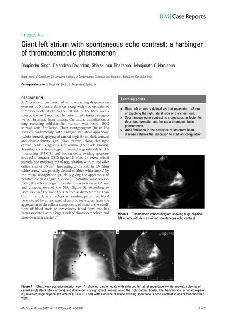
Images In…<strong>Giant</strong> <strong>left</strong> <strong>atrium</strong> <strong>with</strong> <strong>spontaneous</strong> <strong>echo</strong> <strong>contrast</strong>: a <strong>harbinger</strong><strong>of</strong> thromboembolic phenomenonBhupinder Singh, Rajendran Ravindran, Shivakumar Bhairappa, Manjunath C NanjappaDepartment <strong>of</strong> Cardiology, Sri Jayadeva Institute <strong>of</strong> Cardiovascular Sciences and Research, Bangalore, Karnataka, IndiaCorrespondence to Dr Bhupinder Singh, dr_bhupinders@yahoo.inDESCRIPTIONA 25-year-old man presented <strong>with</strong> worsening dyspnoea onexertion <strong>of</strong> 5 months duration along <strong>with</strong> two episodes <strong>of</strong>thromboembolic stroke to the <strong>left</strong> side <strong>of</strong> the body over aspan <strong>of</strong> the last 2 months. The patient had a history suggestive<strong>of</strong> rheumatic heart disease. On cardiac auscultation, along rumbling mid-diastolic murmur was heard. ECGshowed atrial fibrillation. Chest roentgenogram (figure 1A)revealed cardiomegaly <strong>with</strong> enlarged <strong>left</strong> atrial appendage(white arrows), splaying <strong>of</strong> carinal angle (thick black arrows)and double-density sign (black arrows) along the rightcardiac border suggesting <strong>left</strong> <strong>atrium</strong> (LA; black arrows).Transthoracic <strong>echo</strong>cardiogram revealed a grossly dilated LA(measuring 18.4×11.1 cm) having dense swirling <strong>spontaneous</strong><strong>echo</strong> <strong>contrast</strong> (SEC; figure 1B, video 1), severe mitralstenosis and moderate mitral regurgitation <strong>with</strong> mitral valveorifice area <strong>of</strong> 0.9 cm 2 . Interestingly, the SEC in LA (thinwhite arrow) was partially cleared <strong>of</strong> (thick white arrow) bythe mitral regurgitation jet, thus giving the appearance <strong>of</strong>negative <strong>contrast</strong> (figure 2; video 2). Postmitral valve replacement,the <strong>echo</strong>cardiogram revealed the regression <strong>of</strong> LA sizeand disappearance <strong>of</strong> the SEC (figure 3). According toIsomura et al, 1 the giant LA is defined as diameter more than8 cm. The SEC is an <strong>echo</strong>genic swirling pattern <strong>of</strong> bloodflow, caused by an increased ultrasonic backscatter from theaggregation <strong>of</strong> the cellular components <strong>of</strong> blood in the conditions<strong>of</strong> blood stasis or low-velocity blood flow 2 and hasbeen associated <strong>with</strong> a higher risk <strong>of</strong> thromboembolism andcerebrovascular accident. 3Learning points▸ <strong>Giant</strong> <strong>left</strong> <strong>atrium</strong> is defined as that measuring >8 cmor touching the right lateral side <strong>of</strong> the chest wall.▸ Spontaneous <strong>echo</strong> <strong>contrast</strong> is a predisposing factor forthrombus formation and hence a thromboembolicphenomenon.▸ Arial fibrillation in the presence <strong>of</strong> structural heartdisease satisfies the indication to start anticoagulation.Video 1 Transthoracic <strong>echo</strong>cardiogram showing huge elliptical<strong>left</strong> <strong>atrium</strong> <strong>with</strong> dense swirling <strong>spontaneous</strong> <strong>echo</strong> <strong>contrast</strong>.Figure 1 Chest x-ray posterior-anterior view (A) showing cardiomegaly <strong>with</strong> enlarged <strong>left</strong> atrial appendage (white arrows), splaying <strong>of</strong>carinal angle (thick black arrows) and double-density sign (black arrows) along the right cardiac border. The transthoracic <strong>echo</strong>cardiogram(B) revealed huge elliptical <strong>left</strong> <strong>atrium</strong> (18.4×11.1 cm) <strong>with</strong> evidence <strong>of</strong> dense swirling <strong>spontaneous</strong> <strong>echo</strong> <strong>contrast</strong> in apical four-chamberview.BMJ Case Reports 2012; doi:10.1136/bcr-2012-0068801<strong>of</strong>3
Figure 2 The colour compare in apical four-chamber view demonstrating the partial clearance <strong>of</strong> the <strong>spontaneous</strong> <strong>echo</strong> <strong>contrast</strong> in <strong>left</strong>artrium (LA; thick white arrows) by the mitral regurgitation jet, giving the appearance <strong>of</strong> negative <strong>contrast</strong>.Competing interests None.Patient consent ObtainedREFERENCES1. Isomura T, Hisatomi K, Hirano A, et al. Left atrial plication and mitral valvereplacement for giant <strong>left</strong> <strong>atrium</strong> accompanying mitral lesion. J Card Surg1993;8:365–70.2. Merino A, Hauptman P, Badimon L, et al. Echocardiographic “smoke” isproduced by an interaction <strong>of</strong> erythrocytes and plasma proteins modulated byshear forces. J Am Coll Cardiol 1992;20:1661–8.3. Daniel WG, Nellessen U, Schroder E, et al. Left atrial <strong>spontaneous</strong> <strong>echo</strong><strong>contrast</strong> in mitral valve disease: an indicator for an increased thromboembolicrisk. J Am Coll Cardiol 1988;11:1204–11.Video 2 Transthoracic <strong>echo</strong>cardiogram (apical four-chamberview) showing clearance <strong>of</strong> the <strong>spontaneous</strong> <strong>echo</strong> <strong>contrast</strong> in the<strong>left</strong> <strong>atrium</strong> by the mitral regurgitation jet on colour Dopplerinterrogation.Figure 3 Postmitral valve replacement, the transthoracic <strong>echo</strong>cardiogram showing regressed <strong>left</strong> <strong>atrium</strong> size, laminar flow (white arrow)across the mitral prosthetic valve (yellow arrow) and disappearance <strong>of</strong> <strong>spontaneous</strong> <strong>echo</strong> <strong>contrast</strong> in the <strong>left</strong> <strong>atrium</strong>.2<strong>of</strong>3BMJ Case Reports 2012; doi:10.1136/bcr-2012-006880
This pdf has been created automatically from the final edited text and images.Copyright 2012 BMJ Publishing Group. All rights reserved. For permission to reuse any <strong>of</strong> this content visithttp://group.bmj.com/group/rights-licensing/permissions.BMJ Case Report Fellows may re-use this article for personal use and teaching <strong>with</strong>out any further permission.Please cite this article as follows (you will need to access the article online to obtain the date <strong>of</strong> publication).Singh B, Ravindran R, Bhairappa S, Nanjappa MC. <strong>Giant</strong> <strong>left</strong> <strong>atrium</strong> <strong>with</strong> <strong>spontaneous</strong> <strong>echo</strong> <strong>contrast</strong>: a <strong>harbinger</strong> <strong>of</strong> thromboembolic phenomenon.BMJ Case Reports 2012;10.1136/bcr-2012-006880, Published XXXBecome a Fellow <strong>of</strong> BMJ Case Reports today and you can:▸ Submit as many cases as you like▸ Enjoy fast sympathetic peer review and rapid publication <strong>of</strong> accepted articles▸ Access all the published articles▸ Re-use any <strong>of</strong> the published material for personal use and teaching <strong>with</strong>out further permissionFor information on Institutional Fellowships contact consortiasales@bmjgroup.comVisit casereports.bmj.com for more articles like this and to become a FellowBMJ Case Reports 2012; doi:10.1136/bcr-2012-0068803<strong>of</strong>3