

Alexander Disease: Report of Two Unrelated Infantile Form Cases ...
Alexander Disease: Report of Two Unrelated Infantile Form Cases ...
Alexander Disease: Report of Two Unrelated Infantile Form Cases ...

Create successful ePaper yourself
Turn your PDF publications into a flip-book with our unique Google optimized e-Paper software.
Case <strong>Report</strong>Iran J PediatrAug 2013; Vol 23 (No 4), Pp: 481-484<strong>Alexander</strong> <strong>Disease</strong>: <strong>Report</strong> <strong>of</strong> <strong>Two</strong> <strong>Unrelated</strong> <strong>Infantile</strong> <strong>Form</strong> <strong>Cases</strong>, Identified byGFAP Mutation Analysis and Review <strong>of</strong> Literature; The First <strong>Report</strong> from IranMahmoud-Reza Ashrafi 1,2,3 , MD; Alireza Tavasoli 2,3 *, MD; Omid Aryani 4 , MD; Hooman Alizadeh 3,5 , MD;Massoud Houshmand 4,6 , PhD1. Department <strong>of</strong> Pediatrics, Tehran University <strong>of</strong> Medical Sciences, Tehran, Iran2. Growth and Development Research Center, Tehran University <strong>of</strong> Medical Sciences, Tehran, Iran3. Children’s Medical Center, Pediatric Center <strong>of</strong> Excellence, Tehran, Iran4. Department <strong>of</strong> Medical Genetics, Special Medical Center, Tehran, Iran5. Department <strong>of</strong> Radiology, Tehran University <strong>of</strong> Medical Sciences, Tehran, Iran6. Genetic Department, National Institute for Genetic Engineering and Biotechnology, Tehran, IranReceived: May 13, 2012; Accepted: Jan 19, 2013; First Online Available: Jul 18, 2013AbstractBackground: <strong>Alexander</strong> disease (AD) is a sporadic leukodystrophy that predominantly affects infants andchildren and usually results in death within ten years after onset. The infantile form comprises the most <strong>of</strong>affected individuals. It presents in the first two years <strong>of</strong> life, typically with progressive psychomotorretardation with loss <strong>of</strong> developmental milestones, megalencephaly and frontal bossing, seizures, pyramidalsigns and ataxia. The diagnosis is based on magnetic resonance imaging (MRI) findings and confirmed byGFAP gene molecular testing. GFAP gene encodes glial fibrillary acidic protein, is the only gene in whichmutation is currently known to cause AD which is inherited in autosomal dominant manner.Case Presentation: In this article we report the first two Iranian cases <strong>of</strong> infantile AD and their clinical, brainMRI and molecular findings. We report two novel mutations too in the GFAP gene that are associated withinfantile form <strong>of</strong> AD.Conclusion: GFAP gene mutations are a reliable marker for infantile AD diagnosed according to clinical andMRI defined criteria. A genotype-phenotype correlation had been discerned for the two most frequentlyreported GFAP gene mutations in infantile type <strong>of</strong> AD (R79 and R239), with the phenotype <strong>of</strong> the R79mutations appearing much less severe than that <strong>of</strong> the R239 mutations. Our findings confirm this theory.Iranian Journal <strong>of</strong> Pediatrics, Volume 23 (Number 4), August 2013, Pages: 481-484Key Words: <strong>Alexander</strong> <strong>Disease</strong>; Leukoencephalopathy; <strong>Alexander</strong>’s leukodystrophy; Megalencephaly; MacrocephalyIntroduction<strong>Alexander</strong> disease (AD) is a sporadicleukodystrophy that predominantly affects infantsand children and usually results in death withinten years after onset. It was first described byStewart <strong>Alexander</strong> in 1949 as a ”progressivedegeneration <strong>of</strong> fibrillary astrocytes” [1-3] . AD ischaracterized by progressive failure <strong>of</strong> centralmyelination and the accumulation <strong>of</strong> Rosenthalfibers in astrocytes and on magnetic resonanceimaging (MRI) by leukodystrophy. According tothe age <strong>of</strong> clinical presentations, it can be dividedinto three subtypes: infantile (between birth and 2years <strong>of</strong> age), juvenile (between 2 and 12 years <strong>of</strong>age) and adult (from the second to seventh* Corresponding Author;Address: Division <strong>of</strong> Pediatric Neurology, Children’s Medical Center, No 62, Dr Gharib St., Tehran, IranE-mail: dralit73@yahoo.com© 2012 by Pediatrics Center <strong>of</strong> Excellence, Children’s Medical Center, Tehran University <strong>of</strong> Medical Sciences, All rights reserved.Iran J Pediatr; Vol 23 (No 4), Aug 2013Published by: Tehran University <strong>of</strong> Medical Sciences (http://ijp.tums.ac.ir)
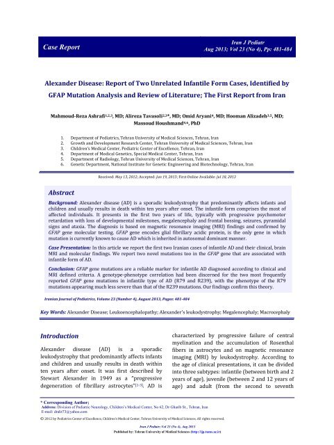
482<strong>Alexander</strong> <strong>Disease</strong>; <strong>Report</strong> <strong>of</strong> <strong>Two</strong> <strong>Cases</strong>decade) [4,5] . The infantile form comprises mostaffected individuals (about 63% <strong>of</strong> reportedcases). It presents in the first two years <strong>of</strong> life,typically with progressive psychomotorretardation with loss <strong>of</strong> developmental milestones,megalencephaly and frontal bossing, seizures,pyramidal signs and ataxia. The diagnosis is basedon MRI findings and confirmed by GFAP genemolecular testing. GFAP gene encodes glialfibrillary acidic protein, is the only gene in whichmutation is currently known to cause AD which isinherited in autosomal dominant manner. In thisarticle we report the first two Iranian cases <strong>of</strong>infantile AD and their clinical, brain MRI andmolecular findings. We report two novelmutations too in the GFAP gene that are associatedwith infantile form <strong>of</strong> AD. The first geneticallyproved cases <strong>of</strong> AD were reported by Brenner’sgroup in 2001 [6] . Nearly all cases <strong>of</strong> AD areassociated with heterozygous point mutation <strong>of</strong>the Glial Fibrillary Acidic Protein (GFAP) genelocated on chromosome 17q21, but missense andde novo mutations also have been found [3,6,7] .Case PresentationCase 1: A 15 month-old boy was referred to ourhospital for evaluation <strong>of</strong> seizure disorder anddevelopmental regression. He was the secondchild <strong>of</strong> non-consanguineous parents, born at termby elective cesarean section after an uneventfulpregnancy. His birth weight and headcircumference (HC) were 3.1Kg and 34cm,respectively. Family history was unremarkable. Heattained head holding at 7 months <strong>of</strong> age. Visualfixation and following were not good. First episode<strong>of</strong> seizure occurred with fever at the age <strong>of</strong> 5months. The second episode <strong>of</strong> seizure occurredwithout fever at the age <strong>of</strong> 7 months, and thendevelopmental regression started. Macrocephaly(HC 51 cm), generalized spasticity and searchingnystagmus were the main neurological findings.Metabolic tests including High performance liquidchromatography (HPLC) <strong>of</strong> serum aminoacids,urine organic acid pr<strong>of</strong>ile, serum ammonia andlactate level and thyroid function tests werewithin normal limit. Brain MRI showed four <strong>of</strong> thefive diagnostic criteria <strong>of</strong> <strong>Alexander</strong> diseasedescribed by van der Knapp et al in 2001 [1,8] (Fig1).The diagnosis <strong>of</strong> AD was confirmed by geneticanalysis, which revealed a heterozygous mutation<strong>of</strong> p.Arg239His in the exon 4 <strong>of</strong> GFAP gene.Case 2: A 5.5 year-old boy was referred to ourhospital for evaluation <strong>of</strong> developmentalregression and seizures. He was the third child <strong>of</strong>healthy and consanguineous parents, born at termby elective cesarean section after an uneventfulFig. 1: Axial T1 and T2-weighted MRI images <strong>of</strong> case-one, showing a periventricular rim (hyper intense in T1 and hypointense in T2) and predominant frontal white matter involvement with subependymal cystsIran J Pediatr; Vol 23 (No 4), Aug 2013Published by: Tehran University <strong>of</strong> Medical Sciences (http://ijp.tums.ac.ir)
Ashrafi MR, et al 483Fig. 2: Axial T1 and T2-weighted MRI images <strong>of</strong> case-two, showing a periventricular rim (hyper intense in T1 and hypointense in T2) and predominant frontal white matter involvementpregnancy. His birth weight was 3 Kg, HC at birthwas not determined. Family history wasunremarkable. At the age <strong>of</strong> 6 months he haddevelopmental delay and was not able to partialweight bearing. Following the DPT vaccinationseizures started and thereafter developmentalregression occurred. Significant findings inphysical examination were macrocephly (HC51cm) and spasticity. Laboratory tests similar tothe first patient, were all within normal limit. As incase 1, brain MRI showed four <strong>of</strong> the fivediagnostic criteria <strong>of</strong> AD described by van derKnapp et al in year 2001 (Fi. 2). Genetic studyrevealed a heterozygous mutation <strong>of</strong> p.Arg79His inthe exon 1 <strong>of</strong> GFAP gene.According to the age <strong>of</strong> onset our two caseswere the infantile type. MRI findings werecompatible with four <strong>of</strong> the five criteria <strong>of</strong> van derKnapp et al (2001) [1,8] for the diagnosis <strong>of</strong> AD.Based on following criteria, AD diagnosis in ourpatients was wstablished.1. Periventricular rim (high signal on T1 and lowsignal on T2-weighted images.2. Extensive white matter abnormalities predominantly<strong>of</strong> frontal lobe and cystic degeneration<strong>of</strong> anterior deep periventricular white matter(Figs 1 and 2).3. Abnormalities <strong>of</strong> the basal ganglia and thalami.4. Brain stem abnormalities especially involvingthe mid brain area.DiscussionPrior to availability <strong>of</strong> the molecular geneticanalysis <strong>of</strong> AD, only genetic demonstration <strong>of</strong>Rosenthal fibers in astrocytes <strong>of</strong> brain specimenwas able to confirm the studies [3,8] . We used GFAPgene mutation analysis for confirmation <strong>of</strong> ourclinical diagnosis. Genetic tests revealedheterozygous mutation in exons 4 and 1 <strong>of</strong> GFAPgene. In two large studies on the infantile type <strong>of</strong>AD by Brenner et al, 2007 and Rodriguez et al,2001 [7] (collectively 28 patients), GFAP mutationswere detected in 93% <strong>of</strong> patients by amplification<strong>of</strong> only exons 1, 4, and 8.In our patients the detected mutations werealso in exons 1 and 4 and carried argininemutations (p.Arg239His and p.Arg79His)(Table 1).Both the p.Arg239His and p.Arg79His codingchanges have been previously associated with ADpatients. The p.Arg79His mutation is consideredcausative for AxD due to its de novo appearance,whereas the role <strong>of</strong> p.Glu223Gln is unclear [6] . Todate, <strong>of</strong> the 72 different mutations that have beenidentified, 68 are missense mutations andmutations at three amino acid residues (Arg79,Arg88, and Arg239) account for 42% (80/189) <strong>of</strong>all molecularly confirmed cases. In two beforementioned studies a genotype-phenotypecorrelation had been discerned for the two mostfrequently mutated arginine residues (R79 andIran J Pediatr; Vol 23 (No 4), Aug 2013Published by: Tehran University <strong>of</strong> Medical Sciences (http://ijp.tums.ac.ir)
484<strong>Alexander</strong> <strong>Disease</strong>; <strong>Report</strong> <strong>of</strong> <strong>Two</strong> <strong>Cases</strong>Table 1: Clinical features and GFAP mutations <strong>of</strong> patientsPatient Degradation HC ExonNucleotide Amino acidchange changeStatus1 7mo 51 4 716G>A p.Arg239His Alive2 6mo 51 1 236G>A p.Arg79His AliveR239), with the phenotype <strong>of</strong> the R79 mutationsappearing much less severe than that <strong>of</strong> the R239mutations [6,7] . The number <strong>of</strong> patients in our studyfor this analysis is not enough. It seems thatfurther studies are needed to confirm the theoryproposed by two previous large studies [6,7] .ConclusionIt seems that further studies are needed toconfirm the theory proposed by two previouslarge studies. In conclusion, GFAP gene mutationsare a reliable marker for infantile AD diagnosedaccording to clinical and MRI defined criteria. Alsoit is a strong rationale for the analysis <strong>of</strong> the GFAPgene, even in the absence <strong>of</strong> macrocephaly orneurological deterioration, when MRIabnormalities are characteristic for AD and othercauses <strong>of</strong> leukodystrophy have been ruled out.References1. Claudia B. Poloni. <strong>Alexander</strong> disease: Early presence<strong>of</strong> cerebral MRI criteria. Eur J Pediatr Neurol2009;13(6):556-8.2. Balbi P. The clinical spectrum <strong>of</strong> late-onset <strong>Alexander</strong>disease: a systematic literature review. J Neurol2010; 257(12):1955-62.3. Murakami N. Novel Deletion Mutation inGFAP Genein an infantile form <strong>of</strong> <strong>Alexander</strong> <strong>Disease</strong>. PediatricNeurol 2008; 38(1):50-2.4. Sawaishi Y. Review <strong>of</strong> <strong>Alexander</strong> disease: Beyond theclassical concept <strong>of</strong> leukodystrophy. Brain Dev 2009;31(7):493-8.5. Gordon N. <strong>Alexander</strong> disease. Eur j pediatr neurolo2003; 7(6):395-9.6. Brenner M. Mutation in GFAP, encoding glialfibrillary acidic protein,are associated with<strong>Alexander</strong> disease. Nature Genetic 2001; 27(1): 117-20.7. Rodriguez D. <strong>Infantile</strong> <strong>Alexander</strong> <strong>Disease</strong>: Spectrum<strong>of</strong> GFAP Mutations and Genotype-phenotypecorrelation. Am J. Hum Genet 2001; 69(5):1134-40.8. Christine A. Magnetic Resonance Imaging Findings in<strong>Alexander</strong> <strong>Disease</strong>. Pediatric Neurol 2008; 38(5):373-4.9. Jacob J. The clinicopathological spectrum <strong>of</strong>Rosenthal fibre enceohalopathy and <strong>Alexander</strong>’sdisease: a case report and review <strong>of</strong> thr literature. JNeurol Neurosurg Psychiatry 2003; 74(6):807-10.Iran J Pediatr; Vol 23 (No 4), Aug 2013Published by: Tehran University <strong>of</strong> Medical Sciences (http://ijp.tums.ac.ir)