Chitosan-4-mercaptobenzoic acid: synthesis and characterization of ...
Chitosan-4-mercaptobenzoic acid: synthesis and characterization of ...
Chitosan-4-mercaptobenzoic acid: synthesis and characterization of ...

You also want an ePaper? Increase the reach of your titles
YUMPU automatically turns print PDFs into web optimized ePapers that Google loves.
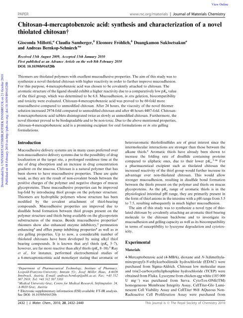
View OnlinePAPERwww.rsc.org/materials | Journal <strong>of</strong> Materials Chemistry<strong>Chitosan</strong>-4-<strong>mercaptobenzoic</strong> <strong>acid</strong>: <strong>synthesis</strong> <strong>and</strong> <strong>characterization</strong> <strong>of</strong> a novelthiolated chitosan†Gioconda Millotti, a Claudia Samberger, b Eleonore Fr€ohlich, b Duangkamon Sakloetsakun a<strong>and</strong> Andreas Bernkop-Schn€urch* aReceived 13th August 2009, Accepted 13th January 2010First published as an Advance Article on the web 8th February 2010DOI: 10.1039/b916528bDownloaded by UNIVERSITAETSBIBLIOTHEK INNSBRUCK on 21 October 2010Published on 08 February 2010 on http://pubs.rsc.org | doi:10.1039/B916528BThiomers are thiolated polymers with excellent mucoadhesive properties. The aim <strong>of</strong> this study was tosynthesize a novel thiolated chitosan with higher reactivity in order to further improve mucoadhesion.For this purpose, 4-<strong>mercaptobenzoic</strong> <strong>acid</strong> was chosen to be covalently attached to chitosan. Thearomatic structure <strong>of</strong> the lig<strong>and</strong> should exhibit a higher reactivity due to a comparatively low pK a value<strong>of</strong> the thiol group, which was determined to be 6.8. Mucoadhesion, in situ gelation, biocompatibility<strong>and</strong> toxicity were evaluated. <strong>Chitosan</strong>-4-<strong>mercaptobenzoic</strong> <strong>acid</strong> was proved to be 60-fold moremucoadhesive compared to unmodified chitosan. After 24 hours, the viscosity <strong>of</strong> the novel thiomersolution increased 2974-fold compared to unmodified chitosan <strong>and</strong> after 48 hours 4487-fold. <strong>Chitosan</strong>-4-<strong>mercaptobenzoic</strong> <strong>acid</strong> tablets disintegrated twice as slowly as unmodified chitosan. Furthermore, thenovel thiomer proved to be biodegradable <strong>and</strong> to be non-toxic. Due to the above mentioned properties,chitosan-4-<strong>mercaptobenzoic</strong> <strong>acid</strong> is a promising excipient for oral formulations or in situ gellingformulations.IntroductionMucoadhesive delivery systems are in many cases preferred overnon-mucoadhesive delivery systems due to the possibility <strong>of</strong> druglocalization at the target site, a prolonged residence time at thesite <strong>of</strong> drug absorption <strong>and</strong> an increase in drug concentrationgradient on the mucosa. <strong>Chitosan</strong> is a natural polymer that hasbeen shown to have mucoadhesive properties. These are quiteweak, as they are the result <strong>of</strong> non-covalent bonds between thepositive charges <strong>of</strong> the polymer <strong>and</strong> negative charges <strong>of</strong> mucusglycoproteins. These mucoadhesive properties can be improvedlog-fold by introducing thiol groups on the polymer structure.Thiomers are hydrophilic polymers whose structure has beenmodified by the covalent attachment <strong>of</strong> thiol-bearingcompounds. Mucoadhesive properties are improved due todisulfide bond formation between thiol groups present on thepolymer structure <strong>and</strong> thiols being available on the glycoproteinsubstructures <strong>of</strong> the mucus. Beside mucoadhesive properties 1thiomers show also enhanced enzyme inhibitory, 2 permeationenhancing 3 <strong>and</strong> efflux pump inhibiting properties 4 as well as insitu gelling properties. Up to now, a considerable number <strong>of</strong>thiolated chitosans have been developed by using alkyl thiolbearing compounds. It is known that aryl thiols (pK a 5–7),however, are far more reactive than alkyl thiols (pK a 8–10). 5 Rayet al., for instance, performed electrochemical studies <strong>of</strong>a 6-mercaptonicotinic <strong>acid</strong> monolayer stating that aromatic oraDepartment <strong>of</strong> Pharmaceutical Technology, Institute <strong>of</strong> Pharmacy,Leopold-Franzens-University, Innrain 52c, Josef M€oller Haus, A-6020Innsbruck, Austria. E-mail: <strong>and</strong>reas.bernkop@uibk.ac.at; Fax: +43 512507 2933; Tel: +43 512 507 5383bMedical University Graz, Centre for Medical Research, Stiftingtalstr. 24,A-8010 Graz, Austria† Electronic supplementary information (ESI) available: FT-IR analysis.See DOI: 10.1039/b916528bheteroaromatic thiols/disulfides are <strong>of</strong> great interest since theintermolecular interactions are stronger than those between thealkane thiols. 6 Aromatic thiols have already been shown toincrease the folding rate <strong>of</strong> disulfide containing proteinscompared to aliphatic ones, due to their lower pK a . 7–10 Fora pharmaceutical excipient such as thiolated chitosan theincreased reactivity <strong>of</strong> the thiol group would further increase itsadvantage over non-thiolated chitosan. This would allowstronger mucoadhesion, resulting in disulfide bond formationbetween the thiols present on the polymer <strong>and</strong> thiols on mucusglycoproteins. As the pK a range <strong>of</strong> aromatic thiols is in thephysiological intestinal pH range, they are primarily present inthe form <strong>of</strong> thiol anions in the intestine with a pH range from 5.5to 7.5, resulting subsequently in much higher mucoadhesion.The aim <strong>of</strong> this study was to synthesize a novel type <strong>of</strong> thiolatedchitosan by covalently attaching an aromatic thiol bearingmolecule to the chitosan backbone <strong>and</strong> to investigate itsmucoadhesion <strong>and</strong> gelling capacity as well as its biocompatibilityin terms <strong>of</strong> susceptibility to lysozyme degradation <strong>and</strong> cytotoxicity.ExperimentalMaterials4-Mercaptobenzoic <strong>acid</strong> (4-MBA), dioxane <strong>and</strong> N-3(dimethylaminopropyl)-N-ethylcarbodiimidehydrochloride (EDAC) werepurchased from Sigma-Aldrich. <strong>Chitosan</strong> low molecular mass<strong>and</strong> tris(2-carboxyethyl)phosphine hydrochloride (TCEP) wereobtained from Fluka. Lysozyme from chicken egg white (185 000U mg 1 ) was purchased from Serva. CytoTox-ONE(TM)homogeneous Membrane Integrity Assay, CellTiter-Glo LuminescentCell Viability Assay <strong>and</strong> CellTiter 96Ò AQueous Non-Radioactive Cell Proliferation Assay were purchased from2432 | J. Mater. Chem., 2010, 20, 2432–2440 This journal is ª The Royal Society <strong>of</strong> Chemistry 2010
View OnlineDownloaded by UNIVERSITAETSBIBLIOTHEK INNSBRUCK on 21 October 2010Published on 08 February 2010 on http://pubs.rsc.org | doi:10.1039/B916528Bdisintegration apparatus (Erweka G.m.b.H., Germany) containing1 L <strong>of</strong> 100 mM phosphate buffer pH 6.8 at 37 C. Theoscillating frequency was set to 0.5 s 1 . Time was recorded whenthe tablets disintegrated completely.Increase in viscosity <strong>and</strong> decrease in free thiol groups content<strong>Chitosan</strong> <strong>and</strong> chitosan-4-MBA were dissolved in 100 mM acetatebuffer pH 5.5 obtaining a solution <strong>of</strong> 1% (m/v). The samples werekept at 37 C. At predetermined time points the viscosity <strong>of</strong> 600ml aliquots was measured at 37 C with a plate-plate viscosimeter(Haake MARS) connected to a personal computer for setting theanalysis parameters <strong>and</strong> for processing <strong>and</strong> recording the datawith the Haake Rheowin program. In parallel, at each predeterminedtime point, 200 ml aliquots were withdrawn <strong>and</strong> 50 mL<strong>of</strong> 1 M HCl was added to stop any further oxidation. The thiolcontent was measured by iodometric titration. Briefly, 1 mg <strong>of</strong>the freeze-dried polymer was dissolved in 1 mL <strong>of</strong> demineralisedwater, adjusting the pH to 1–2 with one drop <strong>of</strong> 1 M HCl. Then,150 mL <strong>of</strong> a 2% (m/v) starch solution were added as indicator.The solution was titrated with 1 mM iodine solution untila permanent blue-violet color appeared.Degradation <strong>of</strong> chitosan <strong>and</strong> chitosan-4-MBA by lysozymeFirst, 300 mg <strong>of</strong> the polymers were hydrated in 8 ml <strong>of</strong> demineralisedwater. The pH was adjusted to 5.0 by the addition <strong>of</strong> 1 ml <strong>of</strong>0.1 M acetic buffer pH 5.0. Afterwards lysozyme previously dissolvedin demineralised water, was added in a final concentration<strong>of</strong> 1% (m/v). The final polymer concentration was 3% (m/v). Thesamples were kept at 37 C. At predetermined time points theviscosity <strong>of</strong> 600 ml aliquots was measured at 37 C with a plateplateviscosimeter (Haake MARS) connected to a personalcomputer for setting the analysis parameters <strong>and</strong> for processing<strong>and</strong> recording the data with the Haake Rheowin program. Polymersolutions (3% m/v in demineralised water), without addition<strong>of</strong> lysozyme, were treated in the same way <strong>and</strong> served as controls.Cell culture conditionsFor the Caco-2 cell line, the medium was composed <strong>of</strong> 80%minimum essential medium (MEM) (with Earle’s salts), 20%10 mM phosphate buffered saline (PBS) pH 7.2, 1x non-essentialamino <strong>acid</strong>s, 1% penicillin-streptomycin liquid. The media usedfor the EAhy926 cells consisted <strong>of</strong> 90% Dulbecco’s modifiedEagle’s medium, 10% 10 mM phosphate buffered saline (PBS),2mML-glutamate <strong>and</strong> 1% penicillin-streptomycin liquid.LDH release assayA st<strong>and</strong>ardized number <strong>of</strong> cells was seeded in a 96 wells plate(100 ml per well) <strong>and</strong> grown for 24 h in an incubator (37 C, 5%CO 2 , 95% relative humidity) prior to stimulation. The mediumwas removed <strong>and</strong> 100 ml <strong>of</strong> the polymer (0–100 mg ml 1 inmedium) were added. Experiments were performed in triplicate.The plate was incubated at 37 C, 5% CO 2 , 95% relative humidityfor 4 to 24 hours. The st<strong>and</strong>ard assay set-up included: blank(culture medium alone), growth control (culture medium withthe cells), lysis control (untreated cells in medium where 100%lysis was performed), particulate positive <strong>and</strong> negative controls(26 nm carboxyl White Polystyrene latex as positive control <strong>and</strong>160 nm Surfactant-Free Carboxyl white polystyrene Latex asnegative control). The CytoTox-ONE assay kit was performed asindicated by the producer. The average value <strong>of</strong> the blank wassubtracted from every fluorescence value. The percent toxicitywas calculated according to the formula:Percent cytotoxicity 100xðExperimental - Culture Medium BackgroundÞ¼ðMaximum LDH Release - Culture Medium BackgroundÞMTT testCell seeding <strong>and</strong> exposure to the polymer was the same as for theLDH-release but no lysis control was included in the set-up.For the assay, MTS solution from the kit (tetrazolium compound[3-(4,5-dimethylthiazol-2-yl)-5-(3-carboxymethoxyphenyl)-2-(4-sulpophenyl)-2H-tetrazolium] <strong>and</strong> PMS solution from the kit(phenazine methosulfate) were thawed. Afterwards, 100 ml <strong>of</strong> thePMS solution were added to 2 ml <strong>of</strong> MTS solution <strong>and</strong> gentlyswirled before addition to the wells. Then, 20 ml <strong>of</strong> the MTS/PMSsolution were pipetted into each well <strong>of</strong> the 96 well assay platecontaining 100 ml, <strong>and</strong> the plate was incubated for 2 hours at 37 C. The absorbance was recorded at 490 nm with a plate reader.The blank (background from the culture medium) was subtractedfrom all other absorbance values.Quantification <strong>of</strong> ATPCell seeding <strong>and</strong> exposure to the polymer was the same as for theLDH-release but no lysis control was included in the set-up.Assay was performed as indicated by the producer. The blankvalues were subtracted from all values.Quantification <strong>of</strong> endotoxinsLAL (Limulus Amebocyte Lysate) was performed using thePyrogent Ultra kit. This test is a qualitative test for Gram-negativebacterial endotoxin. Gram-negative bacterial endotoxin catalysesthe activation <strong>of</strong> a proenzyme in the Limulus Amebocyte Lysate.To prepare the Limulus Amebocyte Lysate, the lyophilized lysatewas reconstituted by adding 5.2 ml LAL Reagent Water (from thekit) to 50 test vials. The solution was gently swirled. For the liquidendotoxin st<strong>and</strong>ards, vials containing a liquid preparation <strong>of</strong>purified endotoxin from E. coli strain (055:B5) were inverted fivetimes to allow mixing <strong>of</strong> the contents. Afterwards, 100 ml <strong>of</strong>st<strong>and</strong>ard (sample or water) were transferred into the reactiontube. Then, 100 ml <strong>of</strong> the reconstituted LAL were added to eachtube beginning with the highest concentration <strong>of</strong> endotoxin <strong>and</strong>mixed thoroughly. This procedure was followed for each dilution<strong>of</strong> the endotoxin. The sample was run in parallel with the endotoxinst<strong>and</strong>ards. After the incubation time, each tube was examinedfor gellation. A positive reaction is characterized by theformation <strong>of</strong> a firm gel that remains intact when the tube isinverted by a vertical rotation <strong>of</strong> 180 .Statistical data analysisStatistical data analyses were performed using Student’s t-testwith p < 0.05 as the minimum level <strong>of</strong> significance.2434 | J. Mater. Chem., 2010, 20, 2432–2440 This journal is ª The Royal Society <strong>of</strong> Chemistry 2010
View OnlineDownloaded by UNIVERSITAETSBIBLIOTHEK INNSBRUCK on 21 October 2010Published on 08 February 2010 on http://pubs.rsc.org | doi:10.1039/B916528BResults <strong>and</strong> discussionSynthesis <strong>and</strong> <strong>characterization</strong> <strong>of</strong> chitosan-4-<strong>mercaptobenzoic</strong><strong>acid</strong>The covalent attachment <strong>of</strong> 4-MBA to chitosan was achieved viathe formation <strong>of</strong> amide bonds between the carboxylic <strong>acid</strong> group<strong>of</strong> the 4-MBA <strong>and</strong> amine groups <strong>of</strong> chitosan, as illustrated inFig. 1. The enhanced reactivity <strong>of</strong> 4-<strong>mercaptobenzoic</strong> <strong>acid</strong> in theintestinal pH range should favour the adhesive interactions withthe mucus, allowing the thiomer incorporated drug to stay incontact with the absorption site for a prolonged period <strong>of</strong> time,therefore favouring its uptake. Furthermore, the enhanced thiolreactivity is advantageous for in situ gelation. The lyophilizedchitosan-4-MBA conjugates appeared as a white powder <strong>of</strong>fibrous structure.The efficacy <strong>of</strong> the purification method described could beverified by the corresponding controls which were prepared inexactly the same way but omitting EDAC. The amount <strong>of</strong> totalthiols present in the control was negligible. The amount <strong>of</strong>reduced thiol groups present on the polymer was determined tobe 98.49 mmol g 1 polymer, while the total amount <strong>of</strong> thiolgroups were 176.27 mmol g 1 polymer. This results in a percent <strong>of</strong>substitution <strong>of</strong> the chitosan backbone <strong>of</strong> 3.8%. After dissolvingthe final product in a 5% NaCl solution <strong>and</strong> precipitating it inisopropanol, the iodometric titration gave a value <strong>of</strong> reducedthiol groups <strong>of</strong> 94.57 mmol g 1 polymer. Therefore, the conjugation<strong>of</strong> 4-<strong>mercaptobenzoic</strong> <strong>acid</strong> to the chitosan backbone haseffectively taken place. The FT-IR spectrum <strong>of</strong> chitosan-4-<strong>mercaptobenzoic</strong><strong>acid</strong> (ESI†) shows the appearance <strong>of</strong> new peakswhich are characteristic for an aromatic system in comparison tothe spectra for unmodified chitosan. These are mainly the peak at3100 cm 1 , which is assigned to the C–H bond in aromaticsystems, <strong>and</strong> the peak at 1650, which is assigned to C]C bond inaromatic systems as well as to C]O in R–CO–NR 2 aromaticsystems. This proves the attachment <strong>of</strong> 4-<strong>mercaptobenzoic</strong> <strong>acid</strong>to chitosan.Theoretical <strong>and</strong> experimental pK a calculationpK a values are used to express the pH at which <strong>acid</strong>ic <strong>and</strong> basicgroups are half-ionized in aqueous solutions. Based on a determinedpK a value it is possible to predict the molecules interactionwith charged species or its reactivity.Accordingly, the pK a value was calculated both for the freemolecule <strong>and</strong> simulated as attached to the chitosan chain, bychanging the free carboxylic group to an amide with a methylending. The pK a value for the thiol group 4-<strong>mercaptobenzoic</strong><strong>acid</strong> non-conjugated to chitosan was calculated to be 6.21 whilethe pK a value for the thiol group simulated as attached to thechitosan chain was calculated to be 6.04. The experimentalresults are presented in Fig. 2, where the distribution <strong>of</strong> thespecies at different pH values is presented. The curves refer to4-<strong>mercaptobenzoic</strong> <strong>acid</strong> non-conjugated to chitosan.The pK a value is determined where the curve representing thedistribution <strong>of</strong> the species when only one group is deprotonated(AH) <strong>and</strong> the curve where both groups are deprotonated (A)cross the distribution curve <strong>of</strong> the protonated molecule (AH2).The experimental value determined for the thiol group is 6.832.This pK a value falls in the range <strong>of</strong> intestinal pH. Therefore, atintestinal pH values, the thiol group <strong>of</strong> 4-<strong>mercaptobenzoic</strong> <strong>acid</strong>bound to chitosan would be mostly present in the form <strong>of</strong> a thiolateanion, which is the active form for disulfide bond formation.Mucoadhesive properties would be maximized in this way.This represents a great improvement <strong>of</strong> aliphatic thiolated chitosanswith a higher pK a , since at intestinal pH the thiol groupwould be only partially present as the thiolate anion. Furthermore,a low pK a value <strong>of</strong> the thiol group would also have a bigimpact on the in situ gelling properties <strong>of</strong> thiomers for applicationson targets where the pH is lower, such the vagina. If the pK aFig. 1Schematic structure <strong>of</strong> the newly synthesized polymer.Fig. 2 Distribution <strong>of</strong> the species at different pH values <strong>and</strong> determination<strong>of</strong> the pK a value for the thiol group <strong>of</strong> 4-MBA not bound to thepolymer. AH2 (A) indicates the molecule in its protonated state; AH(-) indicates the molecule in the state where only the carboxylic group isdeprotonated <strong>and</strong> A (:) indicates the molecule in the state where boththe carboxylic <strong>and</strong> thiol group are deprotonated.This journal is ª The Royal Society <strong>of</strong> Chemistry 2010 J. Mater. Chem., 2010, 20, 2432–2440 | 2435
View OnlineDownloaded by UNIVERSITAETSBIBLIOTHEK INNSBRUCK on 21 October 2010Published on 08 February 2010 on http://pubs.rsc.org | doi:10.1039/B916528B<strong>of</strong> thiols is high, at physiological pH, the gelling process caneither be too slow or not be significant without the addition <strong>of</strong>oxidation agents. Therefore aromatic thiols, by being readilyavailable to react at lower pH in comparison to aliphatic thiols,could improve the in situ gelling properties <strong>of</strong> various formulations.Swelling behaviourThe swelling behaviour <strong>of</strong> mucoadhesive polymers is a veryimportant parameter to consider as it determines the adhesive<strong>and</strong> cohesive properties. For mucoadhesion to occur, mucoadhesivepolymers need to take up water from the underlyingmucosal tissues by absorbing, swelling <strong>and</strong> capillary effects. 12 Ifthe delivery system is addressed to the small intestine, however, itwill reach the target in an at least partially hydrated form. Anexcessive water uptake will lead to overhydration with loss <strong>of</strong>adhesiveness. Therefore, moderate swelling is necessary to avoidoverhydration <strong>and</strong> to avoid the loss <strong>of</strong> adhesive properties beforethe delivery system reaches the target. 13 Water uptake studies, asshown in Fig. 3, demonstrated that the covalent attachment <strong>of</strong>4-<strong>mercaptobenzoic</strong> <strong>acid</strong> to chitosan reduces the swelling behaviour<strong>of</strong> the polymer.After 360 minutes, water uptake by chitosan-4-MBA wasaround 1.5-fold lower compared to unmodified chitosan. Thisobservation is in contrast to almost all other thiolated chitosansthat did not significantly change their swelling properties incomparison to unmodified chitosan 14,15 or that even increased thewater uptake compared to unmodified chitosan. 13,16 Thisbehaviour is likely based on the lipophilic nature <strong>of</strong> the lig<strong>and</strong>.Mucoadhesive propertiesMucoadhesion studies were performed using the rotatingcylinder method. This method is supposed to simulate the in vivosituation better than simple tensile studies, as it imitates theadhesion <strong>and</strong> cohesiveness <strong>of</strong> the polymer in physiologicalmedium. 15 The results are shown in Fig. 4. Unmodified chitos<strong>and</strong>etached from the intestinal mucosa after approximately 3 h,while chitosan-4-MBA remained attached for about 170 hours.Mucoadhesive properties were increased by about 60-fold.Compared with other alkyl-thiolated chitosans exhibitinga similar amount <strong>of</strong> thiol groups like for instance chitosan-thioethylamidine,exhibiting 139.68 mmol <strong>of</strong> thiol groups per gram<strong>of</strong> polymer, chitosan-4-MBA was about 7-fold more mucoadhesive.17 This comparison confirms the higher reactivity <strong>of</strong> arylthiols. The mechanism <strong>of</strong> mucoadhesion is explained by theformation <strong>of</strong> covalent bonds between thiol groups <strong>of</strong> the polymer<strong>and</strong> cysteine-rich subdomains <strong>of</strong> glycoproteins in the mucuslayer. 18 As aromatic thiolated lig<strong>and</strong>s are supposed to be morereactive compared to aliphatic thiols, chitosan-4-MBA shouldexhibit stronger mucoadhesive properties. As the pK a <strong>of</strong> 4-MBAwas determined to be around 6.8 <strong>and</strong> the pH <strong>of</strong> the intestine isaround 6.8, when the thiolated polymer is present in the intestine,its thiol groups will be readily present in the thiol anion form.Therefore, disulfide bond formation with the thiolated glycoproteins<strong>of</strong> the mucus would be favoured.Furthermore, due to its structure, 4-MBA should increase thelipophilic character <strong>of</strong> the polymer. Besides the higher reactivity<strong>of</strong> the thiol group, mucoadhesion could also be enhanced bylowering the water uptake by the polymer. It is well documentedthat slower swelling rates favor longer duration <strong>of</strong> adhesion. 19,20Moisture is important as it will plasticize the system allowingmucoadhesive molecules to become more flexible, conform to theshape <strong>of</strong> the surface, <strong>and</strong> subsequently to be bound to it.However, the mucoadhesive polymer in an aqueous environmentcan overhydrate to form a slippery mucilage, which is readilyremoved. 21 Controlling the rate <strong>and</strong> extent <strong>of</strong> hydration isrequired to produce prolonged adhesion, <strong>and</strong> strategies such asintroducing hydrophobic entities have been tried to achieve thisgoal. 22 As previously described, the uptake <strong>of</strong> water <strong>of</strong> chitosan-4-MBA was 1.5-fold lower compared to unmodified chitosan.Moreover, the attachment <strong>of</strong> mucin molecules to hydrophobicsurfaces resulted in strong <strong>and</strong> stable complexes. 23 The inclusion<strong>of</strong> hydrophobic entities in mucoadhesive systems is thereforea promising strategy to prolong mucoadhesion. 24 Hence, thishydrophobic substructure combined with the disulfide bondFig. 3 Uptake <strong>of</strong> water by unmodified chitosan (-) <strong>and</strong> chitosan-4-MBA (A). Tablets <strong>of</strong> the polymers were fixed on needles <strong>and</strong> immersedin 100 mM phosphate buffer pH 6.8 <strong>and</strong> kept at 37 C. The values are theresults <strong>of</strong> at least 3 experiments SD.Fig. 4 Mucoadhesive properties <strong>of</strong> unmodified chitosan <strong>and</strong> chitosan-4-MBA determined using the rotating cylinder method on freshly excisedporcine mucosa in 100 mM phosphate buffer pH 6.8 at 37 C. The resultsare means <strong>of</strong> at least 3 experiments SD.2436 | J. Mater. Chem., 2010, 20, 2432–2440 This journal is ª The Royal Society <strong>of</strong> Chemistry 2010
View Onlineformation with mucus glycoproteins seems to be a good strategy<strong>and</strong> could improve the delivery <strong>of</strong> many drugs by <strong>of</strong>fering highmucoadhesion.Downloaded by UNIVERSITAETSBIBLIOTHEK INNSBRUCK on 21 October 2010Published on 08 February 2010 on http://pubs.rsc.org | doi:10.1039/B916528BCohesive propertiesUnmodified chitosan tablets were stable for about 6 hours whilechitosan-4-MBA tablets maintained their integrity twice as long(Fig. 5). The increased stability <strong>of</strong> chitosan-4-MBA is assigned tothe formation <strong>of</strong> stabilizing disulfide bonds within the polymericnetwork. The cohesiveness <strong>of</strong> thiomer tablets increases immediatelyin aqueous media due to water interpenetration in thepolymer matrix enabling the crosslinking formation within thepolymer chains. Therefore, disulfide bonds, as an integral part <strong>of</strong>the structure <strong>of</strong> thiolated polymers, contribute to the cohesiveness<strong>of</strong> the tablets.Rheological behaviourThe free thiol groups <strong>of</strong> thiolated polymers are oxidized todisulfide bonds. The extent <strong>of</strong> the reaction depends on the pK avalue <strong>of</strong> the thiol group <strong>and</strong> the pH value <strong>of</strong> the thiomer solution.Thiomers whose thiol group has a low pK a value will be able t<strong>of</strong>orm disulfide bonds at lower pH values, whereas polymerswhose thiol group has a high pK a value will form disulfide bondsto an effective extent only at higher pH values. There is a significantdecrease in thiol groups for chitosan-4-<strong>mercaptobenzoic</strong><strong>acid</strong> although the pH <strong>of</strong> the experiment was 5.5 (Fig. 6).Other thiomers were quite inefficient to form disulfide bonds atthis pH range. For instance, chitosan-thioglycolic <strong>acid</strong> showeda substantial decrease <strong>of</strong> thiol groups only at pH 6.5. At pH 5 theoxidation process was limited. The authors reported that the pK athiol group was 10.6. 16 Moreover, chitosan-2-iminothiolaneshowed very limited disulfide formation at pH 4, 5 <strong>and</strong> 6. Theauthors estimated the pK a <strong>of</strong> the thiol group <strong>of</strong> 2-iminothiolaneattached to the polymer to be 9.94. 25 This comparison allows usto hypothesize that due to the structure <strong>of</strong> 4-<strong>mercaptobenzoic</strong><strong>acid</strong> <strong>and</strong> its lower pK a value, the newly <strong>synthesis</strong>ed thiomer seemsto have a higher reactivity. Indeed, it was already suggested thatFig. 6 Decrease in reduced thiol groups on chitosan-4-<strong>mercaptobenzoic</strong><strong>acid</strong> (A) <strong>of</strong> a 1% (m/v) polymer solution in 100 mM acetate buffer pH 5.5incubated at 37 C. Data are means <strong>of</strong> at least three experiments SD(n ¼ 3).the formation <strong>of</strong> disulfide bonds could be accelerated bydecreasing the pK a <strong>of</strong> the thiol groups via electrostatic interactionsor by addition <strong>of</strong> traces <strong>of</strong> Cu 2+ . 26 <strong>Chitosan</strong>-4-MBA <strong>of</strong>fersthe advantage <strong>of</strong> a lower pK a without the need <strong>of</strong> further additionalsubstances.The decrease in thiol groups is in good agreement with theincrease in viscosity. There is a very high increase in viscosity <strong>of</strong>the polymer over time, although the coupling rate is rather low(Fig. 7).Beside the high reactivity <strong>of</strong> the thiol group, such high increasein viscosity could be favoured by the hydrophobic nature <strong>of</strong> the4-<strong>mercaptobenzoic</strong> <strong>acid</strong>. Hydrophobic forces may play animportant role in the gelation process. 27 It has been reported thatthe thickening power <strong>of</strong> poloxamers in water increased withFig. 5 Cohesive properties <strong>of</strong> unmodified chitosan <strong>and</strong> chitosan-4-MBAperformed using the disintegration apparatus in 100 mM phosphatebuffer pH 6.8 at an oscillation frequency <strong>of</strong> 0.5 s 1 at 37 C. The resultsare means <strong>of</strong> at least 3 experiments SD.Fig. 7 Increase in viscosity <strong>of</strong> a 1% (m/v) solutions <strong>of</strong> chitosan-4-<strong>mercaptobenzoic</strong><strong>acid</strong> (-) <strong>and</strong> unmodified chitosan (A) in 100 mM acetatebuffer pH 5.5 incubated at 37 C. Data are means <strong>of</strong> at least threeexperiments SD (n ¼ 3).This journal is ª The Royal Society <strong>of</strong> Chemistry 2010 J. Mater. Chem., 2010, 20, 2432–2440 | 2437
View OnlineDownloaded by UNIVERSITAETSBIBLIOTHEK INNSBRUCK on 21 October 2010Published on 08 February 2010 on http://pubs.rsc.org | doi:10.1039/B916528Bincreasing hydrophobic molecular mass. 28 Therefore, due to thehigh reactivity <strong>and</strong> increased hydrophobicity <strong>of</strong> the lig<strong>and</strong>, thegelling properties can be greatly improved. The combination <strong>of</strong>gelling <strong>and</strong> mucoadhesive properties renders this novel thiolatedchitosan a useful excipient for various drug delivery systems.Vaginal drug delivery, for instance, has to deal with rapidremoval <strong>of</strong> inserted systems. 29 Due to its mucoadhesive <strong>and</strong> insitu gelling properties, chitosan-4-MBA could be a promisingvaginal drug delivery system. Ocular delivery <strong>of</strong> drugs results inpoor bioavailability. In order to increase the bioavailability <strong>of</strong>ocular applied drugs, the residence time should be increased. 30Once again, mucoadhesive <strong>and</strong> in situ gelling properties suchthose exhibited by chitosan-4-MBA could be a solution.Furthermore, another potential application would be the nasaldelivery <strong>of</strong> drugs as the major drawback is the removal <strong>of</strong>substances by mucociliary activity. 31Susceptibility towards lysozyme degradation<strong>Chitosan</strong> is a biodegradable polymer which does not accumulatein the body. It is therefore important to evaluate how thiolationwith this new class <strong>of</strong> thiolated lig<strong>and</strong>s affects biodegradability <strong>of</strong>the original polymer. The loss in viscosity determined by theaddition <strong>of</strong> lysozyme to the polymer is the result <strong>of</strong> the degradation<strong>of</strong> the polymer network in smaller fragments. Some alkylthiolatedchitosans have already been tested regarding lysozymedegradation. For example, chitosan-thioglycolic <strong>acid</strong> has beenreported to show a slower degree <strong>of</strong> degradation when comparedto unmodified chitosan. 16 It was reported that chitosan-N-acetylcysteine shows a lower susceptibility towards lysozyme withrespect to unmodified chitosan. Results obtained by studyingchitosan-4-MBA are shown in Fig. 8.<strong>Chitosan</strong>-4-MBA was degraded to a slightly greater extentcompared to unmodified chitosan. It is probable that thehydrophobic nature <strong>of</strong> the lig<strong>and</strong> increases the affinity for theenzyme at the cleavage site. It was observed that by the introduction<strong>of</strong> hydrophobic entities on the chitosan structure, theblood compatibility properties were improved. 32 Furthermore,aromatic thiols significantly enhanced the folding rate <strong>of</strong> lysozymerelative to glutathione. The enhanced degradation rateconstant was proposed to be due to the enhanced leaving groupability <strong>of</strong> aromatic thiols relative to glutathione <strong>and</strong> theirenhanced nucleophilicity relative to aliphatic thiols with similarthiol pK a values. 10,33Safety aspectsLDH test. This assay permits the investigation <strong>of</strong> substancesthat may induce alterations in cell integrity <strong>and</strong> therefore quantifiesthe amount <strong>of</strong> non-viable cells. LDH is a stable enzymepresent in the cytosol that is released upon cell lysis. 34The release <strong>of</strong> lactate dehydrogenase into the culture mediumis measured via a coupled enzymatic assay that results in theconversion <strong>of</strong> resazurin into a fluorescent res<strong>of</strong>urin product. Theamount <strong>of</strong> fluorescence is proportional to the number <strong>of</strong> lysedcells. The test was performed on human colorectal carcinoma cellFig. 8 Rheological studies <strong>of</strong> unmodified chitosan <strong>and</strong> chitosan-4-MBA. The viscosity expressed as a percentage <strong>of</strong> the initial value wasmeasured for both polymers in presence as well as in absence <strong>of</strong> 1% (m/v)lysozyme over a period <strong>of</strong> 60 minutes. Legend: (>) unmodified chitosan,(,) chitosan-4-MBA, (A) unmodified chitosan + lysozyme, (-) chitosan-4-MBA+ lysozyme. The results are means <strong>of</strong> at least 3 experiments SD.Fig. 9 (a) LDH test performed on Caco-2 cell line after 4 hour <strong>of</strong>incubation with chitosan-4-MBA (black bars) <strong>and</strong> unmodified chitosan(grey bars) at indicated polymer concentrations. The results are expressedas % <strong>of</strong> cell death. The results are means <strong>of</strong> at least 3 experiments SD.(b) LDH test performed on Caco-2 cell line after 24 hour <strong>of</strong> incubationwith chitosan-4-MBA (black bars) <strong>and</strong> unmodified chitosan (grey bars) atindicated polymer concentrations. The results are expressed as % <strong>of</strong> celldeath. The results are means <strong>of</strong> at least 3 experiments SD.2438 | J. Mater. Chem., 2010, 20, 2432–2440 This journal is ª The Royal Society <strong>of</strong> Chemistry 2010
View OnlineDownloaded by UNIVERSITAETSBIBLIOTHEK INNSBRUCK on 21 October 2010Published on 08 February 2010 on http://pubs.rsc.org | doi:10.1039/B916528BFig. 10 (a) Cell viability test performed on Caco-2 cell line for chitosan-4-MBA (black bars) <strong>and</strong> unmodified chitosan (grey bars) after 4 hours <strong>of</strong>incubation. The results are means <strong>of</strong> at least 3 experiments SD. (b) Cellviability test performed on Caco-2 cell line for chitosan-4-MBA (blackbars) <strong>and</strong> unmodified chitosan (grey bars) after 24 hours <strong>of</strong> incubation.The results are means <strong>of</strong> at least 3 experiments SD.lines (Caco-2). The tests were performed at an incubation time <strong>of</strong>4 <strong>and</strong> 24 hours in different polymer concentrations. The test(Fig. 9a,b) indicates that chitosan-4-MBA has the same toxicityas chitosan, which is very low. The values stay constant after24 indicating the lack <strong>of</strong> toxicity. Therefore, the polymer can beconsidered as relatively safe for in vivo applications.MTT test. This test is measuring the number <strong>of</strong> viable cells.Although formazan bioreduction is the classical cytotoxicityscreening assay for conventional substances, The MTS (tetrazoliumcompound [3-(4,5-dimethylthiazol-2-yl)-5-(3-carboxymethoxyphenyl)-2-(4-sulfophenyl)-2H-tetrazolium]isbioreduced by cells into a formazan product that is soluble intissue culture medium by dehydrogenase enzymes found inmetabolically active cells. The tests performed on Caco-2 celllines showed a cell viability <strong>of</strong> around 100% for all the concentrationstested after 4 <strong>and</strong> 24 hours indicating that the polymersare not harmful for the cells. The viability was around 100%.There was no trend in results depending on the concentrationused (data not shown).Quantification <strong>of</strong> ATP. This test measures the number <strong>of</strong> viablecells by luminescent quantification <strong>of</strong> the ATP present, an indication<strong>of</strong> metabolically active cells. The tests showed (Fig. 10a,b)a very high cell viability for all the concentration tested after4 <strong>and</strong> 24 hours, indicating that cells maintain their metabolicactivity, <strong>and</strong> therefore the polymers can be considered as nontoxic.Endotoxin quantification. Endotoxins are natural compoundsfound inside pathogens such as bacteria. These are not secretedin soluble form by live bacteria but are a structural component <strong>of</strong>the bacteria which is released mainly when bacteria are lysed. Thesamples in stock solution contained less than 0.06 EU ml 1(Endotoxic Unit ml 1 ).ConclusionWithin this study, chitosan-4-mercaptnicotinic <strong>acid</strong> has beensynthesized <strong>and</strong> characterized for the first time. The new strategyfor optimizing thiolated chitosans led to a unique type <strong>of</strong> thiolatedchitosan which exhibits its full reactivity at intestinal pH.The newly synthesized polymer exhibited excellent in situ gellingproperties <strong>and</strong> improved mucoadhesive properties compared toaliphatic thiolated chitosans with similar coupling rates. Due tothese properties, the newly <strong>synthesis</strong>ed polymer is an excellentc<strong>and</strong>idate as excipient for oral formulations <strong>and</strong> in situ gellingformulations for various applications, such as vaginal delivery.AcknowledgementsThis work has been supported by the EC. Nano-BioPharmaceutics is an Integrated Project funded within the 6thFramework Programme <strong>of</strong> the European Commission. Authorsare thankful to Jonny Easmond for help with the pK a calculations.Authors are thankful to Lukas Bittner from the AnalyticalChemistry Department at the University <strong>of</strong> Innsbruck for theFT-IR analysis.References1 A. Bernkop-Schnurch, V. Schwarz <strong>and</strong> S. Steininger, Pharm. Res.,1999, 16, 876–881.2 A. Bernkop-Schnurch, H. Zarti <strong>and</strong> G. F. Walker, J. Pharm. Sci.,2001, 90, 1907–1914.3 A. E. Clausen, C. E. Kast <strong>and</strong> A. Bernkop-Schnurch, Pharm. Res.,2002, 19, 602–608.4 F. Foger, T. Schmitz <strong>and</strong> A. Bernkop-Schnurch, Biomaterials, 2006,27, 4250–4255.5 J. M. Wilson, R. J. Bayer <strong>and</strong> H.D.J., J. Am. Chem. Soc., 1977, 99(24),7922–7926.6 C. R. a. B. Ray <strong>and</strong> S, J. Electroanal. Chem., 2005, 581, 61–69.7 J. D. Gough, J. M. Gargano, A. E. Don<strong>of</strong>rio <strong>and</strong> W. J. Lees,Biochemistry, 2003, 42, 11787–11797.8 J. D. Gough <strong>and</strong> W. J. Lees, Bioorg. Med. Chem. Lett., 2005, 15, 777–781.9 J. D. Gough <strong>and</strong> W. J. Lees, J. Biotechnol., 2005, 115, 279–290.10 M. C. Gurbhele-Tupkar, L. R. Perez, Y. Silva <strong>and</strong> W. J. Lees, Bioorg.Med. Chem., 2008, 16, 2579–2590.11 I. Bravo-Osuna, D. Teutonico, S. Arpicco, C. Vauthier <strong>and</strong>G. Ponchel, Int. J. Pharm., 2007, 340, 173–181.12 D. Duchene <strong>and</strong> G. Ponchel, Biomaterials, 1992, 13, 709–714.13 M. Roldo, M. Horn<strong>of</strong>, P. Caliceti <strong>and</strong> A. Bernkop-Schnurch, Eur.J. Pharm. Biopharm., 2004, 57, 115–121.14 T. Schmitz, V. Grabovac, T. F. Palmberg, M. H. H<strong>of</strong>fer <strong>and</strong>A. Bernkop-Schnurch, Int. J. Pharm., 2007.This journal is ª The Royal Society <strong>of</strong> Chemistry 2010 J. Mater. Chem., 2010, 20, 2432–2440 | 2439
View OnlineDownloaded by UNIVERSITAETSBIBLIOTHEK INNSBRUCK on 21 October 2010Published on 08 February 2010 on http://pubs.rsc.org | doi:10.1039/B916528B15 K. Kafedjiiski, F. Foger, M. Werle <strong>and</strong> A. Bernkop-Schnurch,Pharm. Res., 2005, 22, 1480–1488.16 C. E. Kast <strong>and</strong> A. Bernkop-Schnurch, Biomaterials, 2001, 22, 2345–2352.17 K. Kafedjiiski, M. H<strong>of</strong>fer, M. Werle <strong>and</strong> A. Bernkop-Schnurch,Biomaterials, 2006, 27, 127–135.18 V. M. Leitner, G. F. Walker <strong>and</strong> A. Bernkop-Schnurch, Eur.J. Pharm. Biopharm., 2003, 56, 207–214.19 S. A. Mortazavi <strong>and</strong> J. D. Smart, J. Controlled Release, 1994, 26, 207–212.20 S. A. Mortazavi <strong>and</strong> J. D. Smart, J. Controlled Release, 1993, 25, 197–203.21 J. D. Smart, Adv. Drug Delivery Rev., 2005, 57, 1556–1568.22 T. Inoue, G. Chen <strong>and</strong> A. S. H<strong>of</strong>fman, J. Bioact. Compat. Polym.,1998, 13, 50–64.23 L. Shi <strong>and</strong> K. D. Caldwell, J. Colloid Interface Sci., 2000, 224, 372–381.24 N. A. Peppas <strong>and</strong> Y. Huang, Adv. Drug Delivery Rev., 2004, 56, 1675–1687.25 A. Bernkop-Schnurch, M. Horn<strong>of</strong> <strong>and</strong> T. Zoidl, Int. J. Pharm., 2003,260, 229–237.26 G. H. Snyder, R.M.K., M. J. Cennerazzo <strong>and</strong> D. Field, Biochimica etBiophisica Acta, 1983, 749, 219–224.27 A. Chenite, C. Chaput, D. Wang, C. Combes, M. D. Buschmann,C. D. Hoemann, J. C. Leroux, B. L. Atkinson, F. Binette <strong>and</strong>A. Selmani, Biomaterials, 2000, 21, 2155–2161.28 I. R. Schmolka, J. Am. Oil Chem. Soc., 1977, 54, 110–116.29 G. C. Ceschel, P. Maffei, S. Lombardi Borgia, C. Ronchi <strong>and</strong>S. Rossi, Drug Dev. Ind. Pharm., 2001, 27, 541–547.30 O. Sechoy, G. Tissie, C. Sebastian, F. Maurin, J. Y. Driot <strong>and</strong>C. Trinquard, Int. J. Pharm., 2000, 207, 109–116.31 J. Woodley, Clin. Pharmacokinet., 2001, 40, 77–84.32 S. Hirano <strong>and</strong> Y. Yagi, Carbohydr. Res., 1980, 83, 103–108.33 R. Martien, B. Loretz, M. Thaler, S. Majzoob <strong>and</strong> A. Bernkop-Schnurch, J. Biomed. Mater. Res., Part A, 2007, 82a, 1–9.34 S. Mao, X. Shuai, F. Unger, M. Wittmar, X. Xie <strong>and</strong> T. Kissel,Biomaterials, 2005, 26, 6343–6356.2440 | J. Mater. Chem., 2010, 20, 2432–2440 This journal is ª The Royal Society <strong>of</strong> Chemistry 2010