

Mapping the Mechanical Stiffness of Live Cells ... - Soft Matter World
Mapping the Mechanical Stiffness of Live Cells ... - Soft Matter World
Mapping the Mechanical Stiffness of Live Cells ... - Soft Matter World

Create successful ePaper yourself
Turn your PDF publications into a flip-book with our unique Google optimized e-Paper software.
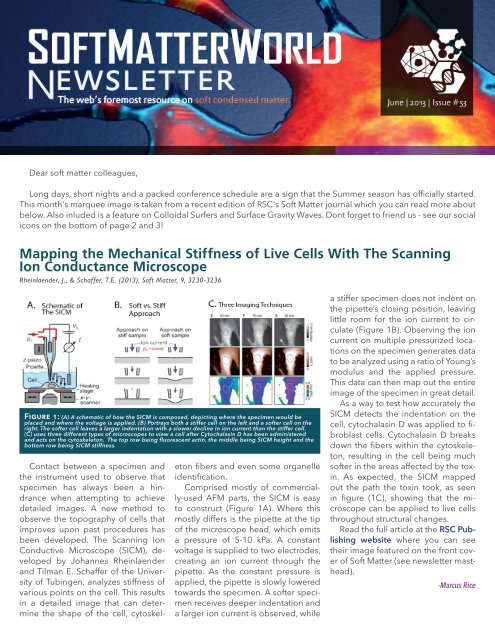
Marquee goes hereDear s<strong>of</strong>t matter colleagues,Long days, short nights and a packed conference schedule are a sign that <strong>the</strong> Summer season has <strong>of</strong>ficially started.This month's marquee image is taken from a recent edition <strong>of</strong> RSC's S<strong>of</strong>t <strong>Matter</strong> journal which you can read more aboutbelow. Also inluded is a feature on Colloidal Surfers and Surface Gravity Waves. Dont forget to friend us - see our socialicons on <strong>the</strong> bottom <strong>of</strong> page 2 and 3!<strong>Mapping</strong> <strong>the</strong> <strong>Mechanical</strong> <strong>Stiffness</strong> <strong>of</strong> <strong>Live</strong> <strong>Cells</strong> With The ScanningIon Conductance MicroscopeRheinlaender, J., & Schaffer, T.E. (2013). S<strong>of</strong>t <strong>Matter</strong>, 9, 3230-3236Figure 1: (A) A schematic <strong>of</strong> how <strong>the</strong> SICM is composed, depicting where <strong>the</strong> specimen would beplaced and where <strong>the</strong> voltage is applied. (B) Portrays both a stiffer cell on <strong>the</strong> left and a s<strong>of</strong>ter cell on <strong>the</strong>right. The s<strong>of</strong>ter cell leaves a larger indentation with a slower decline in ion current than <strong>the</strong> stiffer cell.(C) uses three different types <strong>of</strong> microscopes to view a cell after Cytochalasin D has been administeredand acts on <strong>the</strong> cytoskeleton. The top row being fluorescent actin, <strong>the</strong> middle being SICM height and <strong>the</strong>bottom row being SICM stiffness.Contact between a specimen and<strong>the</strong> instrument used to observe thatspecimen has always been a hindrancewhen attempting to achievedetailed images. A new method toobserve <strong>the</strong> topography <strong>of</strong> cells thatimproves upon past procedures hasbeen developed. The Scanning IonConductive Microscope (SICM), developedby Johannes Rheinlaenderand Tilman E. Schaffer <strong>of</strong> <strong>the</strong> University<strong>of</strong> Tubingen, analyzes stiffness <strong>of</strong>various points on <strong>the</strong> cell. This resultsin a detailed image that can determine<strong>the</strong> shape <strong>of</strong> <strong>the</strong> cell, cytoskeletonfibers and even some organelleidentification.Comprised mostly <strong>of</strong> commercially-usedAFM parts, <strong>the</strong> SICM is easyto construct (Figure 1A). Where thismostly differs is <strong>the</strong> pipette at <strong>the</strong> tip<strong>of</strong> <strong>the</strong> microscope head, which emitsa pressure <strong>of</strong> 5-10 kPa. A constantvoltage is supplied to two electrodes,creating an ion current through <strong>the</strong>pipette. As <strong>the</strong> constant pressure isapplied, <strong>the</strong> pipette is slowly loweredtowards <strong>the</strong> specimen. A s<strong>of</strong>ter specimenreceives deeper indentation anda larger ion current is observed, whilea stiffer specimen does not indent on<strong>the</strong> pipette’s closing position, leavinglittle room for <strong>the</strong> ion current to circulate(Figure 1B). Observing <strong>the</strong> ioncurrent on multiple pressurized locationson <strong>the</strong> specimen generates datato be analyzed using a ratio <strong>of</strong> Young’smodulus and <strong>the</strong> applied pressure.This data can <strong>the</strong>n map out <strong>the</strong> entireimage <strong>of</strong> <strong>the</strong> specimen in great detail.As a way to test how accurately <strong>the</strong>SICM detects <strong>the</strong> indentation on <strong>the</strong>cell, cytochalasin D was applied to fibroblastcells. Cytochalasin D breaksdown <strong>the</strong> fibers within <strong>the</strong> cytoskeleton,resulting in <strong>the</strong> cell being muchs<strong>of</strong>ter in <strong>the</strong> areas affected by <strong>the</strong> toxin.As expected, <strong>the</strong> SICM mappedout <strong>the</strong> path <strong>the</strong> toxin took, as seenin figure (1C), showing that <strong>the</strong> microscopecan be applied to live cellsthroughout structural changes.Read <strong>the</strong> full article at <strong>the</strong> RSC Publishingwebsite where you can see<strong>the</strong>ir image featured on <strong>the</strong> front cover<strong>of</strong> S<strong>of</strong>t <strong>Matter</strong> (see newsletter mas<strong>the</strong>ad).-Marcus Rice
Observation <strong>of</strong> Star-ShapedSurface Gravity WavesRajchenbach, J., Clamond, D., & Leroux, A. (2013). Physical Review Letters,110(9), 094502-1 Ð 094502-5.Figure 2: A shows <strong>the</strong> standing wave for a vibrational amplitude <strong>of</strong> 1.95mm alternating between a star (A) and a pentagon (B). Figures 2C and 2Dshow that <strong>the</strong> wave pattern is independent <strong>of</strong> container size and shapeMany types <strong>of</strong> waves occur in different physical systems.One particularly interesting example, standing gravitywaves, occur due to nonlinearity and dispersive effects inliquids. A collaborative research team from <strong>the</strong> Universitede Nice has discovered a new type <strong>of</strong> standing gravitywave formed in silicon oil that transitions between star andpolygon shapes.The experiment involved placing oil filled containers <strong>of</strong>varying size and shape onto a shaking apparatus. Oncemounted, <strong>the</strong> containers experienced a vertical sinusoidalmotion resulting in surface waves that were dependenton frequency and vibrational amplitude. After reaching aspecific vibrational amplitude, <strong>the</strong> oil waves alternated betweena star shape and polygon shape.When a cylindrical container vibrated at a frequency <strong>of</strong>8 Hz and vibrational amplitude <strong>of</strong> 1.55 mm, <strong>the</strong> oil wouldcreate two axisymmetric gravity waves that moved toward<strong>the</strong> center <strong>of</strong> <strong>the</strong> container. Once <strong>the</strong> wave reached <strong>the</strong>center, <strong>the</strong> silicon oil would jet up and fall down back into<strong>the</strong> container as a droplet. Increasing <strong>the</strong> vibrational amplitudeto 1.85 mm caused <strong>the</strong> crest line <strong>of</strong> <strong>the</strong> waves t<strong>of</strong>orm five corners, <strong>the</strong> tips <strong>of</strong> <strong>the</strong> corners representing <strong>the</strong>break <strong>of</strong> rotational symmetry. At an amplitude <strong>of</strong> 1.95 mm<strong>the</strong> wave geometry alternated between a star shape andpolygon shape (Figure 2 A-B). The shape alteration wasdue to a phase shift between <strong>the</strong> waves.The forming gravity waves were independent <strong>of</strong> containersize and shape. Alternating star and polygon shapeswere observed when a cylindrical container was used; <strong>the</strong>square container resulted in similar shapes adjacent toone ano<strong>the</strong>r (Figure 2 C-D).The research group attempted to derive a <strong>the</strong>oreticalexplanation for <strong>the</strong> results based on previous <strong>the</strong>ories inquasicrystals and <strong>the</strong> formation <strong>of</strong> quasipatterns in capillarywaves. The proposed model explained <strong>the</strong> formation<strong>of</strong> <strong>the</strong> gravity waves, but could not predict <strong>the</strong> final symmetries<strong>of</strong> <strong>the</strong> waves as <strong>the</strong>y relate to <strong>the</strong> forcing parameters<strong>of</strong> <strong>the</strong> experiment. Future studies may focus on <strong>the</strong>design <strong>of</strong> a <strong>the</strong>ory to explain steep cnoidal standing wavesin shallow water.Visit PRL to read <strong>the</strong> full text and to see some <strong>of</strong> <strong>the</strong>supplemental material included with <strong>the</strong> text.-Amanda BaijnauthLiving Crystals <strong>of</strong> Light-ActivatedColloidal SurfersJeremie Palacci, Stefano Sacanna, Asher Preska Steinberg, David J. Pine, Paul M. Chaikin. 2013.Science 339, 936 DOI: 10.1126/science.1230020In order to study self-organization<strong>of</strong> colloids, U.S. researchers have createda planar system <strong>of</strong> self-propelledparticles, <strong>the</strong> motility <strong>of</strong> which canbe turned on and <strong>of</strong>f with blue light.The colloidal particles consisted <strong>of</strong> acubic hematite core mostly enclosedin a polymer sphere, and <strong>the</strong>y can beJune | 2013 | Issue #53steered by a magnetic field within <strong>the</strong>containing basic solution.When subjected to blue light, <strong>the</strong>particles moved randomly, collidingand creating dynamic crystallinestructures while continuously breakingapart and reforming (Figure 3A-C). When a magnetic field was applied,<strong>the</strong> particles aligned with <strong>the</strong>field and <strong>the</strong> break up <strong>of</strong> <strong>the</strong> crystalswas suppressed. In effect, <strong>the</strong>y moveden masse in a single direction. Alternatively,when <strong>the</strong> magnetic field wasapplied before <strong>the</strong> blue light was activated,<strong>the</strong> particles again aligned.When <strong>the</strong> blue light was applied, <strong>the</strong>ymoved en masse with no collisions,and <strong>the</strong>refore no crystal formation occurred.The researchers tested each part <strong>of</strong><strong>the</strong> system individually to better un-2
derstand <strong>the</strong> mechanism behind <strong>the</strong> motility. When subjectedto blue light, an uncovered hematite cube catalyzeddecomposition <strong>of</strong> hydrogen peroxide present in <strong>the</strong> surroundingbasic solution, producing <strong>the</strong>rmal and chemicalgradients. Through <strong>the</strong> chemical diffusion gradient, <strong>the</strong> silicatracers were attracted to <strong>the</strong> cube via a process calleddiffusiophoresis. The converse was also true – hematitecubes were attracted to a fixed silica surface.The effect <strong>of</strong> surface area fraction ϕ Swas also analyzed.Above 7%, crystallites began forming after 25 seconds <strong>of</strong>exposure to blue light. Cluster size, averaging 35 particles,was not dependent on ϕ Swhen above 10%. Fluctuationsin <strong>the</strong> number <strong>of</strong> local particles followed a power law ΔN~ N α .The researchers suggest this demonstration <strong>of</strong> self-assemblythrough non-equilibrium driving forces may beFigure 3: The false colors show <strong>the</strong> time evolution <strong>of</strong> different clusters.The clusters rearrange, exchange particles, merge (A), break apart (B),becomeunstable and explode (blue cluster, C).applied to directed self assembly, opening a new area fordesign and <strong>the</strong> creation <strong>of</strong> novel motile structures.Visit ScienceMag to read <strong>the</strong> full article.- Michael LaneSITE UPDATESConferencesWe have released a seperate bulletin pertaining to all<strong>the</strong> upcoming conference deadlines and features. It isnever too late to have your conference, featured free<strong>of</strong> charge.postings@s<strong>of</strong>tmatterworld.orgGalleryFor any <strong>of</strong> our readers who missed out on a chance toreceive a hard copy <strong>of</strong> <strong>the</strong> 2013 S<strong>of</strong>t <strong>Matter</strong> <strong>World</strong> Calendarit is now available for download in <strong>the</strong> Gallerysection in high resolution PDF format.Feel free to print <strong>the</strong>m out for yourselfand remember that <strong>the</strong> S<strong>of</strong>t <strong>Matter</strong><strong>World</strong> 2014 Calendar is not far away.We hope you enjoy browsing and come back soonLinda S. Hirst & Adam P. OssowskiJoin <strong>the</strong> Mailing List!June | 2013 | Issue #533