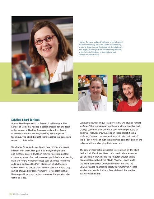
Heather Canavan, assistant pr<strong>of</strong>essor <strong>of</strong> chemical andnuclear <strong>engineering</strong>, (left) and chemical <strong>engineering</strong>graduate student Jamie Reed (below left), collaboratewith Angela Wandinger-Ness, pr<strong>of</strong>essor <strong>of</strong> pathologyat the <strong>School</strong> <strong>of</strong> Medicine in developing smartsurfaces for cell analysis.Solution: Smart SurfacesAngela Wandinger-Ness, pr<strong>of</strong>essor <strong>of</strong> pathology at the<strong>School</strong> <strong>of</strong> Medicine, needed a better process for one facet<strong>of</strong> her research. Heather Canavan, assistant pr<strong>of</strong>essor<strong>of</strong> chemical and nuclear <strong>engineering</strong>, had the perfecttechnique. The CBME brought them together in a successfulresearch collaboration.Wandinger-Ness studies cells and how therapeutic drugsinteract with them. Her goal is to analyze single cellsand measure protein levels on their surface using a flowcytometer, a machine that measures particles in a streamingfluid. Currently, Wandinger-Ness uses enzymes to removecells from surfaces like Petri dishes, on which they aregrown. Then she places them into suspension, where theycan be analyzed by flow cytometry. Her concern is thatthe enzymatic process destroys some <strong>of</strong> the proteins shewants to study.Canavan’s new technique is a perfect fit. She studies “smartsurfaces,” thermoresponsive polymers with properties thatchange based on environmental cues like temperature orelectrical field. By growing cells on these smart, flexiblesurfaces, Canavan can create clumps <strong>of</strong> cells that peel <strong>of</strong>flike a Post-It note, or even isolate single cells that pop <strong>of</strong>f thepolymer without changing their structure.The researchers’ ultimate goal is to create an <strong>of</strong>f-the-shelfdevice that Wandinger-Ness could use to allow accuratecell analysis. Canavan says the research wouldn’t havebeen possible without the CBME. “Gabriel Lopez madethe initial connection between the two sides and theCBME provided financial support,” says Canavan. “Therewas both an intellectual and financial contribution thatwas very significant.”08 <strong>UNM</strong> <strong>Engineering</strong>
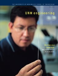
One Bead, Two InnovationsTwo sides <strong>of</strong> campus, two disciplines, two goals, one sharedinterest: microscopic beads. Gabriel Lopez and Larry Sklar,pr<strong>of</strong>essor <strong>of</strong> pathology at <strong>UNM</strong>’s <strong>School</strong> <strong>of</strong> Medicine, arecollaborating on fluorescent biosensors that use tiny, proteincoatedbeads the size <strong>of</strong> a human cell. While their researchgoals are different, it’s a strategic collaboration because theresearchers share an interest in the microbiosensors andin biochemistry. “It’s technically a natural fit and our workis nicely complementary,” says Lopez. The two researchers,along with Tione Buranda, research assistant pr<strong>of</strong>essor inthe Pathology Department, have shared a series <strong>of</strong> five-yeargrants during their twelve-year collaboration.Sklar uses the beads for drug discovery, coating them with aspecific protein and suspending them in a fluid that he wantsto test. By running the suspension through a flow cytometer,Sklar can evaluate the reaction <strong>of</strong> the proteins on the surface<strong>of</strong> the beads with those in the solution. Using this process,Sklar and Buranda can screen molecules involving more thana dozen molecular interactions that contribute to cancer,infectious diseases, and inflammatory diseases. Sklar sayscollaborating with engineers at the CBME moves the teamcloser to its goals. “An engineer’s skill set is complementaryto those <strong>of</strong> a biomedical scientist,” he explains. “One <strong>of</strong> thebiggest factors is the different types <strong>of</strong> materials that weuse for building sensors or instrumentation. <strong>Engineering</strong>students have different interests in technology thanbiologists, including materials, fluidics, automation,electronics, and computers.”Lopez and his team use the beads to create diagnosticassays. They make chip-based, micr<strong>of</strong>luidic devices bypacking thousands <strong>of</strong> the protein-coated beads into tiny glasscapillaries. Lopez injects a fluid, for example a blood sample,into the bead-packed channels. When he exposes the channelsto a laser, the beads that are attached to specific proteinsfluoresce, indicating the presence <strong>of</strong> a toxin or diseasemarker in the liquid. Lopez and his team are currently testingsuch devices, using samples from patients with respiratoryinfections. The devices have already successfully detectedseveral different viruses in one small sample. dTione Buranda, research assistant pr<strong>of</strong>essor in thePathology Department and Gabriel Lopez, director<strong>of</strong> the CBME and pr<strong>of</strong>essor <strong>of</strong> chemical and nuclear<strong>engineering</strong> and chemistry, have shared a series <strong>of</strong> fiveyeargrants during their twelve-year collaboration.Above: Confocal scanning laser fluorescencemicroscopy image <strong>of</strong> mesoporous silica particlesfilled with rhodamine dye (red) and encapsulatedby fluorescent lipid bilayer (green). Bottomimage shows one particle (arrow) which hasbeen partially bleached for measurements <strong>of</strong>diffusive fluorescence recovery.<strong>UNM</strong> <strong>Engineering</strong> 09