

Is the protein folding an aim-oriented process ... - IngentaConnect
Is the protein folding an aim-oriented process ... - IngentaConnect
Is the protein folding an aim-oriented process ... - IngentaConnect

You also want an ePaper? Increase the reach of your titles
YUMPU automatically turns print PDFs into web optimized ePapers that Google loves.
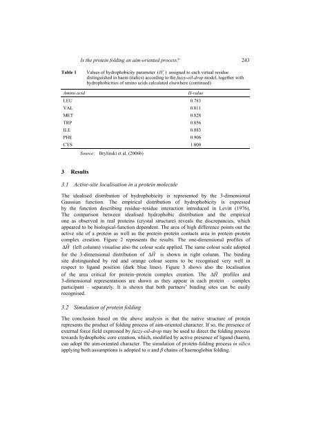
<strong>Is</strong> <strong>the</strong> <strong>protein</strong> <strong>folding</strong> <strong>an</strong> <strong>aim</strong>-<strong>oriented</strong> <strong>process</strong>? 243rTable 1 Values of hydrophobicity parameter ( Hi) assigned to each virtual residuedistinguished in haem (italics) according to <strong>the</strong> fuzzy-oil-drop model, toge<strong>the</strong>r withhydrophobicities of amino acids calculated elsewhere (continued)Amino acidH-valueLEU 0.783VAL 0.811MET 0.828TRP 0.856ILE 0.883PHE 0.906CYS 1.000Source: Brylinski et al. (2006b)3 Results3.1 Active-site localisation in a <strong>protein</strong> moleculeThe idealised distribution of hydrophobicity is represented by <strong>the</strong> 3-dimensionalGaussi<strong>an</strong> function. The empirical distribution of hydrophobicity is expressedby <strong>the</strong> function describing residue–residue interaction introduced in Levitt (1976).The comparison between idealised hydrophobic distribution <strong>an</strong>d <strong>the</strong> empiricalone as observed in real <strong>protein</strong>s (crystal structure) reveals <strong>the</strong> discrep<strong>an</strong>cies, whichappeared to be biological-function dependent. The area of high difference points out <strong>the</strong>active site of a <strong>protein</strong> as well as <strong>the</strong> <strong>protein</strong>–<strong>protein</strong> contacts area in <strong>protein</strong>–<strong>protein</strong>complex creation. Figure 2 represents <strong>the</strong> results. The one-dimensional profiles of∆H (left column) visualise also <strong>the</strong> colour scale applied. The same colour scale adoptedfor <strong>the</strong> 3-dimensional distribution of ∆H is shown in right column. The bindingsite distinguished by red <strong>an</strong>d or<strong>an</strong>ge colour seems to be recognised very well inrespect to lig<strong>an</strong>d position (dark blue lines). Figure 3 shows also <strong>the</strong> localisationof <strong>the</strong> area critical for <strong>protein</strong>–<strong>protein</strong> complex creation. The ∆H profiles <strong>an</strong>d3-dimensional representations are shown as <strong>the</strong>y appear in each <strong>protein</strong> – complexparticip<strong>an</strong>t – separately. It is shown that both partners’ binding sites c<strong>an</strong> be easilyrecognised.3.2 Simulation of <strong>protein</strong> <strong>folding</strong>The conclusion based on <strong>the</strong> above <strong>an</strong>alysis is that <strong>the</strong> native structure of <strong>protein</strong>represents <strong>the</strong> product of <strong>folding</strong> <strong>process</strong> of <strong>aim</strong>-<strong>oriented</strong> character. If so, <strong>the</strong> presence ofexternal force field expressed by fuzzy-oil-drop may be used to direct <strong>the</strong> <strong>folding</strong> <strong>process</strong>towards hydrophobic core creation, which, modified by active presence of lig<strong>an</strong>d (haem),c<strong>an</strong> adopt <strong>the</strong> <strong>aim</strong>-<strong>oriented</strong> character. The simulation of <strong>protein</strong>-<strong>folding</strong> <strong>process</strong> in silicoapplying both assumptions is adopted to <strong>an</strong>d chains of haemoglobin <strong>folding</strong>.