

Isolation Of Human pancreatic Acinar cells Pdf - The Pancreapedia
Isolation Of Human pancreatic Acinar cells Pdf - The Pancreapedia
Isolation Of Human pancreatic Acinar cells Pdf - The Pancreapedia

You also want an ePaper? Increase the reach of your titles
YUMPU automatically turns print PDFs into web optimized ePapers that Google loves.
METHODS PAGE<strong>Isolation</strong> of human <strong>pancreatic</strong> acinar <strong>cells</strong> fromrat and human pancreasMatthew C. Cane 1 , Robert Sutton 2 and David N. Criddle 11 Department of Cellular and Molecular Physiology, Institute of Translational Medicine, University ofLiverpool, and 2 Liverpool NIHR Pancreas Biomedical Research Unit, Royal Liverpool UniversityHospital, Liverpool, UKe-mail: m.cane@liv.ac.uk, sutton@liv.ac.uk, criddle@liv.ac.ukVersion 1.0, June 13, 2011 [DOI: 10.3998/panc.2011.20]<strong>The</strong> <strong>pancreatic</strong> acinar cell is a specialisedsecretory unit, the physiology of which has beenextensively studied over a number of decadesprincipally using rodents, although recentlysignificant progress has been made with respect toinvestigation of human samples. Due to significantdifficulties in developing culture methods for thiscell type, primary isolation procedures are essentialin order to study function. <strong>The</strong> high quantity ofdense-core vesicles and the clustered nature ofacini, mean that low-speed centrifugation isextremely effective at isolating this cell type fromless dense material such as cellular debris andother component <strong>cells</strong> of the pancreas. An isolationprocedure was developed for murine tissue (1, 2)which was based on a series of purification stepsfollowing enzymatic dispersal of freshly dissectedpancreas to yield isolated <strong>cells</strong> and small acinarclusters. We have recently modified this procedurefor the isolation of human <strong>pancreatic</strong> acinar <strong>cells</strong>,taking into account differences between adulthuman and murine pancreas samples, mostnotably contamination with blood products and fat,and presence of fibrosis in the former (3). Here wedescribe a simple and effective procedure for theisolation of human <strong>pancreatic</strong> acinar <strong>cells</strong> usingcollagenase digestion, mechanical dispersion andlow-speed centrifugation.1. Materials1. Extracellular solution (mM): 140 NaCl, 4.7 KCl,1.13 MgCl 2 , 1 CaCl 2 , 10 D-glucose, 10 HEPES(adjusted to pH 7.35 with NaOH)2. Collagenase (CLSPA, 200 U/ml) (Worthington)to be dissolved in extracellular solution (above)3. Polystyrene 15 ml tubes (Sarstedt) (see Note1)4. Tabletop centrifuge5. Water bath (shaking if possible)6. 70 μm nylon Cell Strainer (BD Biosciences),7. <strong>Human</strong> pancreas (see 2.1 <strong>Human</strong> PancreaticTissue Sample Retrieval)8. *Protease inhibitor tablets 1 per 10 ml (RocheDiagnostics)9. *Sodium Pyruvate 100 μM (100 mM stocksolution, Sigma-Aldrich)1
10. Soybean trypsin inhibitor (lyophilized powder,0.01% w/v, Sigma-Aldrich)*<strong>The</strong>se extra compounds are used to augment theextracellular solution for use during transport fromtheatre to the laboratory (see 2.1 point 3), but areomitted from the basic extracellular solution for allsubsequent steps.2. Methods2.1 <strong>Human</strong> Pancreatic Tissue Sample RetrievalSamples may only be taken from consentingpatients. <strong>Human</strong> <strong>pancreatic</strong> tissue should be ofhigh quality, which will normally only be availablefrom surgery for left-sided pancreatectomy,resections for duodenal tumours, or nonobstructiveright sided cancer resections in patientswith no history of jaundice or chronic pancreatitis(3). Samples from patients with chronic pancreatitisor ductal obstruction are unsuitable due to the highlevels of activated trypsin in this tissue (Fig. 1).1. During surgery (<strong>pancreatic</strong> resection), a smallpiece (~1 - 3 cm 3 ) of pancreas should be cutfrom the transection margin of the remainingpancreas. This procedure should be carried outusing a new scalpel blade to limit grossmacroscopic cell damage from diathermy burns(see Note 2).2. <strong>The</strong> piece of pancreas should be immediatelywashed by transfer between two 50 ml tubes ofice-cold extracellular solution to remove debrisand blood products. <strong>The</strong>se may containneutrophils, macrophages etc. which have thepotential to induce additional oxidative stressand tissue damage.3. After washing, the sample should be added toa third, pre-prepared tube containing ~ 50 mlice-cold extracellular solution plus soya beantrypsin inhibitor (see Note 3), proteaseinhibitors, and sodium pyruvate.4. <strong>The</strong> sample is then immediately transported inthis solution on ice to the laboratory. Crucially,the time from sampling to the start of cellisolation should be as brief as possible (ideallyless than 10 minutes) to ensure good qualitycell preparations. Failure to do so may result inpoorly responsive <strong>cells</strong> that are eitherinsensitive to agonists (cholecystokinin,acetylcholine) or require supramaximalstimulation to elicit responses e.g. oscillatoryCa 2+ elevations.2.2 <strong>Human</strong> <strong>pancreatic</strong> acinar cell isolation1. Tissue with a high proportion of fat should beavoided. This is extremely important whenloading with lipophilic dyes. Fatty tissue iselastic, pinkish and translucent, and will float inthe extracellular solution. Using theseidentifying criteria, fatty tissue can beimmediately recognised and removed toprevent lipid droplets from building up in theexternal solution / collagenase solution. Use afresh surgical blade (size 15) or sharp scissorsto remove all unwanted sections of the tissue.2. Inject the resultant section of pancreas atseveral points with collagenase solution. Inmost cases, 1 ml of collagenase solution isused (see Note 4). Unlike the mouse pancreas,delivery to every portion of the <strong>pancreatic</strong>sample is impossible as the <strong>pancreatic</strong> ductalsystem is not intact following the initial surgicalresection. To attempt to overcome this, thesample should be cut into fine pieces using afresh surgical blade or sharp scissors so thatthe surface area exposed to the collagenase isas large as possible. <strong>The</strong> sample is thentransferred into fresh collagenase solution toremove damaged cellular material and fatdroplets.3. Incubate the sample in a shaking water bath for30 minutes at 37ºC.4. Following digestion, add the pancreas sampleto 5 ml standard extracellular solution.Manually agitate and disperse the suspensionthrough pipette tips of progressivelydiminishing diameter. This is achieved bymanually slicing the tip off 1 ml plastic pipette2
tips, cut at a slight angle with a fresh surgicalblade to give a sharp clean edge.5. When the supernatant is cloudy, collect andtransfer it to a fresh tube. <strong>The</strong> speed of thisprocess is determined by many variations intissue quality, collagenase digestion andambient conditions but should be repeated untilthe supernatant stays clear upon agitation ofthe sample. <strong>The</strong> remaining bulk of the tissueshould then remain white and fibrous. <strong>The</strong>collected supernatant now contains the finalpopulation of <strong>pancreatic</strong> acinar <strong>cells</strong>. <strong>Isolation</strong>of acini from other <strong>pancreatic</strong> cell types isachieved by two rounds of low-speedcentrifugation.6. Centrifuge the sample for 1 minute at 260 gand remove the supernatant by rapid inversionof the tube. 5 ml of fresh extracellular solutionis then added and the pellet resuspended.Repeat this centrifugation step.7. Resuspend the pellet in ~2 ml extracellularsolution, filter using a 70 µm cell strainer toremove larger clumps of tissue, and centrifugeas before (see Note 5).8. Finally, resuspend this pellet in ~2 ml ofextracellular solution.9. Cells can then be loaded with fluorescent dyesas required. Generally, loading conditions aresimilar to that of murine <strong>pancreatic</strong> acinar <strong>cells</strong>,but often require 35ºC.2.3 Confirmation of human <strong>pancreatic</strong> acinarcell viabilityMicroscopic confirmation of the <strong>cells</strong> should alwaysbe performed in the first instance, as the quality ofthe <strong>cells</strong> will vary between patients (see all figuresfor examples of light transmitted images of human<strong>pancreatic</strong> acinar <strong>cells</strong>).Cell viability was originally confirmed by therecording of transient cytosolic Ca 2+ increases inresponse to cholecystokinin (10 pM), which werecoupled to mitochondrial NAD(P)H increases(detected as autofluorescence) and amylasesecretion (Figs. 2, 3); responses typical of freshlyisolated murine <strong>pancreatic</strong> acinar <strong>cells</strong>. Ca 2+signals were shown to occur by direct stimulationof acinar <strong>cells</strong> by cholecystokinin and not via theindirect release of acetylcholine from neuronaltissue (that might have potentially adhered to theacinar <strong>cells</strong> during separation) by the addition ofatropine and tetrodotoxin (3).3. Notes1. Avoid the use of polypropylene tubes for theisolation procedure. Polystyrene tubesincreased the yield of the isolation significantly,possibly due to single <strong>cells</strong> and small clustersreadily adhering to the polypropylene.2. Wherever possible, the amount of time duringwhich the pancreas is clamped should be keptto a minimum to prevent ischaemic damage.3. Original protocols included the addition of 1mM Benzamidine (Worthington), a cellmembrane permeable trypsin inhibitor, to try toavoid any premature intracellular trypsinogenactivation and features of acute pancreatitis.However, for simplicity this is no longer used inisolation procedures since if the tissue is ofsuperior quality (as defined in 2.1), the <strong>cells</strong>should not show intracellular trypsin activation(Fig. 1).4. During the injection, fat droplets can build upon the surface of the collagenase solution. Inthis case, the sample should be washedthrough external solution to remove any fatdroplets and returned to a fresh aliquot ofcollagenase solution. <strong>The</strong>refore it is advised tohave surplus collagenase solution available.5. Filtration will necessarily cause a reduction inthe number of <strong>cells</strong>. If the final cell suspensiontends to have little or no acinar <strong>cells</strong>, this stepmay be omitted to increase yield. In the case ofconfocal microscopy experiments, anyremaining large clumps should not interferewith single cell recordings from acinar <strong>cells</strong>which can be easily identified visually.3
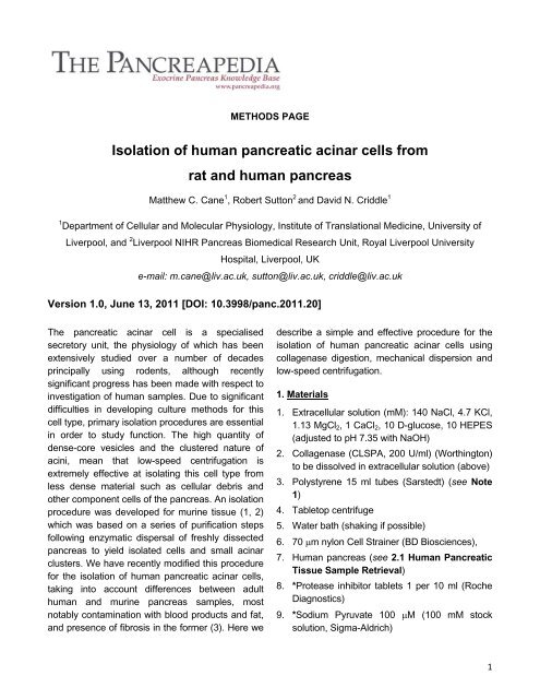
Figure 1. Morphology and integrity of isolated human <strong>pancreatic</strong> acinar cell clusters loaded with BZiPAR, a probewhich fluoresces after cleavage by trypsin. Baseline BZiPAR trace (black) and accompanying inset shows nodetectable trypsin activity and typical, well-preserved morphology of <strong>cells</strong> isolated from a sample of anunobstructed pancreas. Increased BZiPAR trace and inset display trypsin activity in <strong>cells</strong> with poorly definedmorphology, typical of an acinar cell cluster isolated from an obstructed pancreas (Reproduced from ref 3).Figure 2. Direct cholecystokinin (CCK)-mediated cytosolic Ca2+ signals in isolated human <strong>pancreatic</strong> acinar <strong>cells</strong>.Application of a physiological concentration (10 pM) of CCK-8 evoked oscillatory increases of cytosolic Ca2+within human <strong>pancreatic</strong> acinar <strong>cells</strong>. Experiments were performed in the presence of both atropine andtetrodotoxin (added to block any potential neural component to the response). Caffeine (5 mM), an IP3 receptorblocker, reversibly inhibited these signals (Reproduced from ref 3).4
Figure 3. Secretory responses to cholecystokinin in isolated human <strong>pancreatic</strong> acinar <strong>cells</strong>. (A) Exocytosis wasobserved upon stimulation of isolated human <strong>pancreatic</strong> acinar <strong>cells</strong> with a representative physiologicalconcentration of CCK-8 (10 pM), seen as a prompt decrease of quinacrine fluorescence from a steady baseline(blue), reflecting loss of quinacrine-containing zymogen granules; such decreases were not observed withoutstimulation. Inset shows typical quinacrine staining of zymogen granules. A coordinated increase in NAD(P)Hautofluorescence (red) occurred at the start of stimulation, indicative of increased ATP production to fuelsecretion. <strong>The</strong>se experiments were performed in the presence of atropine and tetrodotoxin to preventneurotransmitter release or effects that might theoretically occur from possible adherent nerve endings. (B)Concentration-dependent amylase secretion, expressed as a percentage of total amylase, evoked from freshlyisolated human <strong>pancreatic</strong> acinar <strong>cells</strong> by stimulation with CCK-8 (10 pM – 10 nM) (Reproduced from ref 3).5
4. References1. Osipchuk YV, Wakui M, Yule DI, Gallacher DV, and Petersen OH. Cytoplasmic Ca 2+ oscillations evoked byreceptor stimulation, G-protein activation, internal application of inositol trisphosphate or Ca 2+ : simultaneousmicrofluorimetry and Ca 2+ dependent Cl - current recording in single <strong>pancreatic</strong> acinar <strong>cells</strong>. EMBO J 9: 697–704,1990. PMID: 16901232. Petersen OH, Wakui M, Osipchuk Y, Yule D and Gallacher DV. Electrophysiology of <strong>pancreatic</strong> acinar <strong>cells</strong>.Methods in Enzymology 192: 300-308, 1990. PMID: 17060553. Murphy JA, Criddle DN, Sherwood M, Chvanov M, Mukherjee R, McLaughlin E, Booth D, GerasimenkoJV, Raraty MG, Ghaneh P, Neoptolemos JP, Gerasimenko OV, Tepikin AV, Green GM, Reeve JR Jr,Petersen OH, Sutton R. Direct activation of cytosolic Ca 2+ signaling and enzyme secretion by cholecystokinin inhuman <strong>pancreatic</strong> acinar <strong>cells</strong>. Gastroenterology, 135: 632-41, 2008. PMID: 185558026


