
Create successful ePaper yourself
Turn your PDF publications into a flip-book with our unique Google optimized e-Paper software.
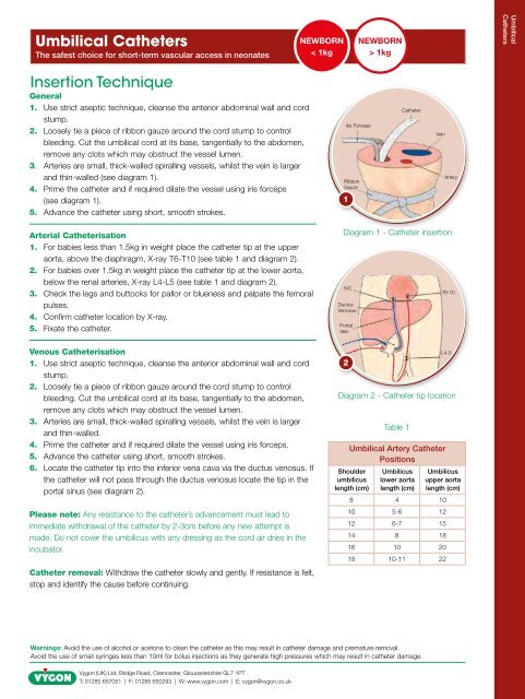
<strong>Umbilical</strong> <strong>Catheters</strong>The safest choice for short-term vascular access in neonatesNEWBORN< 1kgNEWBORN> 1kg<strong>Umbilical</strong><strong>Catheters</strong>Insertion TechniqueGeneral1. Use strict aseptic technique, cleanse the anterior abdominal wall and cordstump.2. Loosely tie a piece of ribbon gauze around the cord stump to controlbleeding. Cut the umbilical cord at its base, tangentially to the abdomen,remove any clots which may obstruct the vessel lumen.3. Arteries are small, thick-walled spiralling vessels, whilst the vein is largerand thin-walled (see diagram 1).4. Prime the catheter and if required dilate the vessel using iris forceps(see diagram 1).5. Advance the catheter using short, smooth strokes.Iris ForcepsRibbonGauze1CatheterVeinArteryArterial Catheterisation1. For babies less than 1.5kg in weight place the catheter tip at the upperaorta, above the diaphragm, X-ray T6-T10 (see table 1 and diagram 2).2. For babies over 1.5kg in weight place the catheter tip at the lower aorta,below the renal arteries, X-ray L4-L5 (see table 1 and diagram 2).3. Check the legs and buttocks for pallor or blueness and palpate the femoralpulses.4. Confirm catheter location by X-ray.5. Fixate the catheter.Venous Catheterisation1. Use strict aseptic technique, cleanse the anterior abdominal wall and cordstump.2. Loosely tie a piece of ribbon gauze around the cord stump to controlbleeding. Cut the umbilical cord at its base, tangentially to the abdomen,remove any clots which may obstruct the vessel lumen.3. Arteries are small, thick-walled spiralling vessels, whilst the vein is largerand thin-walled.4. Prime the catheter and if required dilate the vessel using iris forceps.5. Advance the catheter using short, smooth strokes.6. Locate the catheter tip into the inferior vena cava via the ductus venosus. Ifthe catheter will not pass through the ductus venosus locate the tip in theportal sinus (see diagram 2).Please note: Any resistance to the catheter’s advancement must lead toimmediate withdrawal of the catheter by 2-3cm before any new attempt ismade. Do not cover the umbilicus with any dressing as the cord air dries in theincubator.Catheter removal: Withdraw the catheter slowly and gently. If resistance is felt,stop and identify the cause before continuing.Diagram 1 - Catheter insertionIVCDuctusVenosusPortalVein2Diagram 2 - Catheter tip location<strong>Umbilical</strong> Artery CatheterPositionsShoulderumbilicuslength (cm)Table 1Umbilicuslower aortalength (cm)Umbilicusupper aortalength (cm)8 4 1010 5-6 1212 6-7 1514 8 1816 10 2018 10-11 22T6-10L4-5Warnings: Avoid the use of alcohol or acetone to clean the catheter as this may result in catheter damage and premature removal.Avoid the use of small syringes less than 10ml for bolus injections as they generate high pressures which may result in catheter damage.<strong>Vygon</strong> (<strong>UK</strong>) Ltd, Bridge Road, Cirencester, Gloucestershire GL7 1PTT: 01285 657051 | F: 01285 650293 | W: www.vygon.com | E: vygon@vygon.co.uk