ingenios™βha synthetic Bone Particles technical Data ... - Implacom
ingenios™βha synthetic Bone Particles technical Data ... - Implacom
ingenios™βha synthetic Bone Particles technical Data ... - Implacom

You also want an ePaper? Increase the reach of your titles
YUMPU automatically turns print PDFs into web optimized ePapers that Google loves.
ingenios βha <strong>synthetic</strong> <strong>Bone</strong> <strong>Particles</strong><br />
<strong>technical</strong> <strong>Data</strong> sheet 1<br />
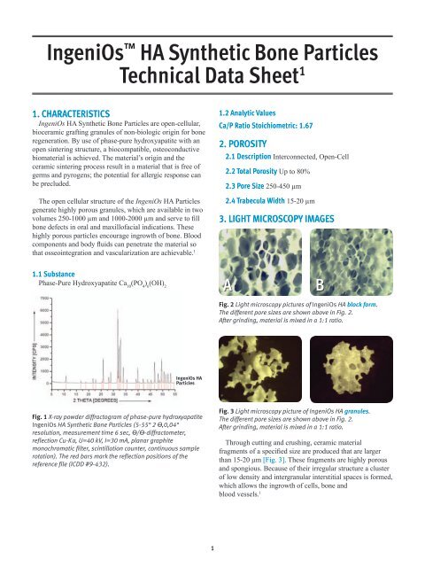
1. CharaCteristiCs<br />
IngeniOs HA Synthetic <strong>Bone</strong> <strong>Particles</strong> are open-cellular,<br />
bioceramic grafting granules of non-biologic origin for bone<br />
regeneration. By use of phase-pure hydroxyapatite with an<br />
open sintering structure, a biocompatible, osteoconductive<br />
biomaterial is achieved. The material’s origin and the<br />
ceramic sintering process result in a material that is free of<br />
germs and pyrogens; the potential for allergic response can<br />
be precluded.<br />
The open cellular structure of the IngeniOs HA <strong>Particles</strong><br />
generate highly porous granules, which are available in two<br />
volumes 250-1000 µm and 1000-2000 µm and serve to fill<br />
bone defects in oral and maxillofacial indications. These<br />
highly porous particles encourage ingrowth of bone. Blood<br />
components and body fluids can penetrate the material so<br />
that osseointegration and vascularization are achievable. 1<br />
1.1 substance<br />
Phase-Pure Hydroxyapatite Ca 10 (PO 4 ) 6 (OH) 2<br />
IngeniOs HA<br />
<strong>Particles</strong><br />
Fig. 1 X-ray powder diffractogram of phase-pure hydroxyapatite<br />
IngeniOs HA Synthetic <strong>Bone</strong> <strong>Particles</strong> (5-55° 2 ϴ,0,04°<br />
resolution, measurement time 6 sec, ϴ/ϴ-diffractometer,<br />
reflection Cu-Kα, U=40 kV, I=30 mA, planar graphite<br />
monochromatic filter, scintillation counter, continuous sample<br />
rotation). The red bars mark the reflection positions of the<br />
reference file (ICDD #9-432).<br />
1<br />
1.2 analytic Values<br />
Ca/P ratio stoichiometric: 1.67<br />
2. Porosity<br />
2.1 Description Interconnected, Open-Cell<br />
2.2 total Porosity Up to 80%<br />
2.3 Pore size 250-450 µm<br />
2.4 trabecula Width 15-20 µm<br />
3. Light MiCrosCoPy iMages<br />
a B<br />
Fig. 2 Light microscopy pictures of IngeniOs HA block form.<br />
The different pore sizes are shown above in Fig. 2.<br />
After grinding, material is mixed in a 1:1 ratio.<br />
Fig. 3 Light microscopy picture of IngeniOs HA granules.<br />
The different pore sizes are shown above in Fig. 2.<br />
After grinding, material is mixed in a 1:1 ratio.<br />
Through cutting and crushing, ceramic material<br />
fragments of a specified size are produced that are larger<br />
than 15-20 µm [Fig. 3]. These fragments are highly porous<br />
and spongious. Because of their irregular structure a cluster<br />
of low density and intergranular interstitial spaces is formed,<br />
which allows the ingrowth of cells, bone and<br />
blood vessels. 1
Fig. 4 Scanning electron microscopy (SEM) pictures: IngeniOs HA<br />
<strong>Particles</strong> produced following the Schwartzwalder process.<br />
The SEM pictures [Fig. 4] show a highly porous material<br />
with web widths of 15-20 µm and an interconnecting pore<br />
system. The macro structure (i.e. the former foam structure)<br />
shows a completely spongious product. The micro structure<br />
shows small primary particles that are fused with each other.<br />
The particles do not crumble because of the regularity of the<br />
structure. All subparticles (i.e. fragments of the web) have<br />
dimensions larger than 10 µm.<br />
4. µ-Ct iMages<br />
The µ-CT pictures in Fig. 5 show a highly porous pile<br />
of particles where pores as well as intergranular interstitial<br />
spaces can be seen. <strong>Bone</strong> and blood vessels have the<br />
opportunity to enter and grow into these areas to support<br />
vascularized bone growth.<br />
Fig. 5 µ-CT images of a granule pile of IngeniOs HA <strong>Particles</strong><br />
Fig. 5A Three dimensional view. Fig. 5B Horizontal section through the<br />
middle of the sample.<br />
2<br />
In Fig. 6 the web of IngeniOs HA <strong>Particles</strong> are<br />
highlighted. Blue areas show voids, green areas show thin<br />
webs and X-ray areas are opaque (i.e. successive webs or<br />
accumulation of material) are marked yellow to red. Pictures<br />
B and C show structures of similar density in all areas of<br />
the pile, which indicates only slight irregularities in the pore<br />
size distribution and web widths.<br />
The pore representation in Fig. 7 (above) shows a<br />
horizontal section through the middle of the sample. The<br />
open-cellular structure of the material is obvious and is<br />
consistent with the structure observed in the SEM pictures.<br />
5. ManufaCturing<br />
Reticulated sponges (a sponge with a net like pattern)<br />
with consistent, open-cellular porosity serve as a sintering<br />
matrix. The pore size of open-cell sponges is calculated in<br />
“pore per inch” (ppi).<br />
The HA slurry is kneaded into the regular open-cell<br />
sponges. During a thermal sintering process the opencellular<br />
polyurethane sponge burn off residue-free, the<br />
liquid of the slurry evaporates and the ceramic particles<br />
sinter with each other. It remains a pure hydroxyapatite<br />
ceramic in form of an open-cellular sponge. With a cutting<br />
mill the open-cellular block forms are crushed to the desired<br />
particle size and materials of the different pore sizes are mixed.<br />
Fig. 5C Vertical section through the<br />
middle of the sample.
Fig. 6 False color representation of µ-CT images of IngeniOs HA <strong>Particles</strong>.<br />
Fig. 6A Vertical section through the<br />
middle of the sample (50 layers per 16 µm)<br />
Fig. 7 Pore representation in a granule heap.<br />
1. <strong>Data</strong> on file with curasan AG.<br />
Fig. 6B Horizontal section through the<br />
upper part of the sample (50 layers<br />
per 16 µm)<br />
3<br />
Fig. 6C Horizontal section through the<br />
lower part of the sample (50 layers<br />
per 16 µm)