Neural Control and Clinical Disorders of Supranuclear Eye ...
Neural Control and Clinical Disorders of Supranuclear Eye ...
Neural Control and Clinical Disorders of Supranuclear Eye ...
Create successful ePaper yourself
Turn your PDF publications into a flip-book with our unique Google optimized e-Paper software.
N E U RO-OPHTHALMOLO GY<br />
Janet C Rucker MD<br />
is an Associate Pr<strong>of</strong>essor in the<br />
Departments <strong>of</strong> Neurology <strong>and</strong><br />
Ophthalmology at the Mount Sinai<br />
School <strong>of</strong> Medicine in New York.<br />
Dr Rucker’s area <strong>of</strong> clinical practice,<br />
research, <strong>and</strong> academic teaching is<br />
Neuro-Ophthalmology. Dr Rucker’s<br />
primary research interest is<br />
utilisation <strong>of</strong> eye movements for<br />
the advancement <strong>of</strong> the<br />
underst<strong>and</strong>ing <strong>of</strong> brain neural<br />
connections <strong>and</strong> facilitation <strong>of</strong><br />
clinical diagnosis.<br />
Correspondence to:<br />
Associate Pr<strong>of</strong>essor,<br />
Departments <strong>of</strong> Neurology <strong>and</strong><br />
Ophthalmology,<br />
The Mount Sinai Medical Center,<br />
One Gustave L Levy Place Box<br />
1052, New York,<br />
New York 10128, USA.<br />
Email: janet.rucker@mssm.edu<br />
Tel: +1 (646) 537-9217.<br />
12 > ACNR > VOLUME 12 NUMBER 3 > JULY/AUGUST 2012<br />
<strong>Neural</strong> <strong>Control</strong> <strong>and</strong> <strong>Clinical</strong><br />
<strong>Disorders</strong> <strong>of</strong> <strong>Supranuclear</strong><br />
<strong>Eye</strong> Movements<br />
Extensive knowledge exists about anatomic<br />
<strong>and</strong> pathophysiologic mechanisms<br />
governing eye movements. 1 The shared goal<br />
<strong>of</strong> all eye movements is stable, clear vision via<br />
placement <strong>of</strong> an object <strong>of</strong> visual interest on the<br />
fovea, the retinal region with the best visual acuity.<br />
Several types <strong>of</strong> eye movements exist to achieve<br />
this shared goal, including smooth pursuit,<br />
vergence, vestibulo-ocular reflexes, optokinetic<br />
nystagmus, <strong>and</strong> saccades. Separate anatomic<br />
supranuclear neural networks exist for each eye<br />
movement type <strong>and</strong> converge upon a ‘final<br />
common pathway’ that includes the motoneuron<br />
originating in cranial nerve nuclei, the neuromuscular<br />
junction, <strong>and</strong> the extraocular muscle.<br />
Systematic exam <strong>of</strong> each type <strong>of</strong> eye movement,<br />
including range <strong>and</strong> dynamic aspects <strong>of</strong> motion,<br />
is essential for accurate localisation <strong>of</strong> supranuclear<br />
eye movement abnormalities.<br />
<strong>Eye</strong> movement types <strong>and</strong> brainstem<br />
anatomy<br />
Smooth pursuit maintains the image <strong>of</strong> a small,<br />
slowly moving target on the fovea. Vergence is a<br />
disconjugate eye movement by which a single<br />
foveal image is maintained with gaze shifts from<br />
near to far (divergence) or from far to near<br />
(convergence). Vestibulo-ocular reflexes generate<br />
compensatory eye movements during brief head<br />
movements that are essential for seeing clearly<br />
while walking or when the head is in motion.<br />
Optokinetic responses (OKN) are reflexive <strong>and</strong><br />
generated by movement <strong>of</strong> a large visual scene<br />
<strong>and</strong> during sustained head rotation. OKN<br />
consists <strong>of</strong> slow eye movements in the direction<br />
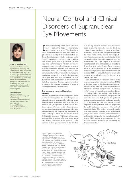
Figure 1. Sagittal monkey brainstem<br />
diagram showing ocular motor-related<br />
nuclei. The shaded region in the pons<br />
represents the paramedian pontine reticular<br />
formation (PPRF), containing excitatory<br />
burst neurons (EBN) for horizontal<br />
saccades (black oval in lower PPRF). The<br />
asterisk just caudal to the CN VI rootlets<br />
represents the location <strong>of</strong> the omnipause<br />
neurons in the raphe interpositus.<br />
Abbreviations: PC = posterior commisure;<br />
riMLF = rostral interstitial medial longitudinal<br />
fasciculus; INC = interstitial nucleus<br />
<strong>of</strong> Cajal; CN III = oculomotor nerve<br />
fascicle; III = oculomotor nucleus; IV =<br />
trochlear nucleus; MLF = medial longitudinal<br />
fasciculus; VI = abducens nucleus; CN<br />
VI = abducens nerve rootlets; NRTP =<br />
nucleus reticularis tegmenti pontis.<br />
Courtesy <strong>of</strong> Jean Büttner-Ennever.<br />
<strong>of</strong> a moving stimulus, followed by quick movements<br />
to reset the eyes in the opposite direction.<br />
Saccades are conjugate, extremely rapid eye<br />
movements with which we shift gaze <strong>and</strong> explore<br />
the visual world. Several factors, including sufficient<br />
force to overcome the elastic inertia <strong>of</strong> the<br />
extraocular orbital tissues, high saccadic velocity,<br />
<strong>and</strong> the need for a high degree <strong>of</strong> accuracy to<br />
place the small fovea on target, make saccades a<br />
dem<strong>and</strong>ing task for the brain. 2 These dem<strong>and</strong>s<br />
result in the requirement <strong>of</strong> a high-frequency<br />
neural discharge from brainstem excitatory burst<br />
neurons (EBN) to stimulate the motoneuron to<br />
generate a saccade <strong>of</strong> a specific size <strong>and</strong> in a<br />
specific direction.<br />
EBN for horizontal saccades are located in the<br />
paramedian pontine reticular formation (PPRF)<br />
in the pons rostral to the abducens nucleus <strong>and</strong>,<br />
for vertical <strong>and</strong> torsional saccades, in the rostral<br />
interstitial medial longtitudinal fasciculus<br />
(riMLF) rostral to the oculomotor nucleus (Figure<br />
1). 3,4 A few EBN for vertical saccades lie in the<br />
interstitial nucleus <strong>of</strong> Cajal (INC) (Figure 1). For<br />
horizontal saccades, EBN project to ipsilateral<br />
motoneurons to generate an ipsilateral saccade<br />
(for a rightward saccade, the premotor signal<br />
originates in the right PPRF EBN <strong>and</strong> projects to<br />
the right abducens nucleus). 5 For vertical<br />
saccades, single EBN project to yoked muscle<br />
pairs (for example, superior rectus <strong>and</strong> inferior<br />
oblique for upward saccades <strong>and</strong> inferior rectus<br />
<strong>and</strong> superior oblique for downward saccades). 6<br />
Vertical EBN project to motoneurons for the<br />
elevator muscles bilaterally, but unilaterally to<br />
depressor muscles. 6,7
Figure 2. A downgaze supranuclear gaze palsy. A. The maximum extent <strong>of</strong> downward movement<br />
<strong>of</strong> the eyes with following <strong>of</strong> a smoothly moving target (smooth pursuit) is to the horizontal<br />
midline. B. Downward saccades are completely eliminated. This picture shows the<br />
eyes “stuck” in upgaze following an upward saccade. C. Vestibulo-ocular reflexes overcome<br />
the downgaze palsy.<br />
Inhibition <strong>of</strong> EBN, required at all times other than during a saccade, is<br />
mediated by tonically discharging omnipause neurons (OPN) in the<br />
nucleus raphe interpositus (RIP) in the PPRF (Figure 1). 8 OPN firing<br />
ceases just before EBN firing <strong>and</strong> resumes at saccade end, however it is<br />
unclear if the OPN or the cerebellar caudal fastigial nucleus terminates<br />
the saccade. 9-11<br />
<strong>Clinical</strong> supranuclear <strong>and</strong> internuclear disorders<br />
<strong>Supranuclear</strong> eye movement abnormalities may result from dysfunction<br />
<strong>of</strong> cerebral, cerebellar, <strong>and</strong> brainstem connections to the ocular motor<br />
nuclei. The focus here is on brainstem supranuclear disorders (Table).<br />
<strong>Clinical</strong> hallmarks <strong>of</strong> a brainstem supranuclear gaze palsy include disproportionate<br />
impairment in the range or velocity <strong>of</strong> saccades <strong>and</strong> impairment<br />
<strong>of</strong> OKN, with VOR retention (Figure 2). Smooth pursuit may be<br />
affected, but usually to a lesser extent than saccades. In contrast, nuclear<br />
<strong>and</strong> infranuclear (cranial nerve, neuromuscular junction, <strong>and</strong> extraocular<br />
muscle) lesions tend to affect all eye movement types equally.<br />
Table. Localisation <strong>of</strong> supranuclear, nuclear, <strong>and</strong> internuclear saccadic gaze disorders.<br />
N E U RO-OPHTHALMOLO GY<br />
Many vertical brainstem supranuclear gaze palsies affect the range <strong>of</strong><br />
each eye movement symmetrically. As a result, visual symptoms may be<br />
minimised by the symmetry <strong>of</strong> the process. <strong>Supranuclear</strong> gaze palsies<br />
may be incidentally noted <strong>and</strong> diagnostically helpful in a visually asymptomatic<br />
patient with multifocal neurological disease. On the other h<strong>and</strong>,<br />
vague visual complaints such as visual blurring may occur, but are nonlocalising.<br />
Binocular diplopia will occur only when the two eyes are<br />
affected differently, causing an ocular misalignment. Diplopia may also<br />
be more common when the deficits have an acute catastrophic onset,<br />
such as with brainstem stroke.<br />
The eye movement abnormalities discussed may be caused by any<br />
lesion affecting the structure specified. The eye movements themselves<br />
are exquisitely localising, but not indicative <strong>of</strong> underlying etiology. In the<br />
acute setting, brainstem ischaemia, hemorrhage, <strong>and</strong> demyelination are<br />
the most common causes. In the chronic setting, neurodegenerative <strong>and</strong><br />
metabolic disease are most common. The eye movement disorders<br />
discussed may occur in isolation or in combination with other neurological<br />
findings, such as hemiparesis, ataxia, or extrapyramidal signs.<br />
When in isolation, it is possible for the lesion to be radiographically<br />
occult on MRI.<br />
Vertical gaze palsies<br />
Lesions <strong>of</strong> EBN in the riMLF result in slowing <strong>of</strong> vertical saccades <strong>and</strong>/or<br />
limitation in the range <strong>of</strong> vertical saccades. Vertical OKN may be absent<br />
or only slow phases generated, with no resetting fast phases. Smooth<br />
pursuit may be affected, but usually to a lesser extent than saccades. If<br />
limitation in the range <strong>of</strong> vertical eye movement is present, passive<br />
vertical VOR should overcome the limitation, as the patient fixates on a<br />
target while the examiner moves the head vertically (Figure 2). Because<br />
vertical EBN projecting to motoneurons for the elevator muscles project<br />
bilaterally <strong>and</strong> to motoneurons for depressor muscles unilaterally, unilateral<br />
riMLF lesions may preferentially impair downward saccades.<br />
Bilateral riMLF lesions may abolish all vertical saccades. Individual case<br />
reports in humans do not always match these anatomic expectations, but<br />
it is probable that the lesions extend beyond the riMLF to other structures<br />
involved in vertical eye movement control.<br />
An acute onset vertical gaze palsy is most <strong>of</strong>ten due to midbrain<br />
infarction. If in isolation, the infarct is typically due to microvascular<br />
ischaemia in the territory <strong>of</strong> the thalamic-subthalamic paramedian<br />
artery, which originates from the posterior cerebral artery. Bilateral riMLF<br />
lesions may occur from a single vessel occlusion because a single thalamic-subthalamic<br />
paramedian artery, the artery <strong>of</strong> Percheron, supplies<br />
both riMLF in 20% <strong>of</strong> patients. 12 An acute onset vertical supranuclear<br />
gaze palsy in combination with other neurological symptoms such as<br />
somnolence, delirium, homonymous hemianopia, <strong>and</strong> cortical blindness<br />
may represent a ‘top <strong>of</strong> the basilar’ stroke with riMLF, thalamic, occipital<br />
lobe, <strong>and</strong> temporal lobe involvement. An acute onset supranuclear<br />
upgaze palsy in combination with eyelid retraction (Collier’s sign),<br />
convergence-retraction nystagmus, <strong>and</strong> pupillary light-near dissociation<br />
is the dorsal midbrain syndrome (also called Parinaud’s syndrome). The<br />
riMLF is not the location <strong>of</strong> the lesion, but rather the upgaze paresis is<br />
LESION / SYNDROME GAZE DISORDER AETIOLOGIC EXAMPLES<br />
riMLF* – midbrain <strong>Supranuclear</strong> vertical gaze palsy Acute – stroke<br />
Chronic – progressive supranuclear palsy<br />
Dorsal midbrain syndrome <strong>Supranuclear</strong> upgaze paresis, convergence-retraction nystagmus Stroke, hydrocephalus, pineal pathology<br />
PPRF** If unilateral – ipsilateral supranuclear horizontal gaze palsy Acute – stroke, demyelination, Wernicke’s encephalopathy<br />
If bilateral – bilateral supranuclear horizontal gaze palsy Chronic – Spinocerebellar ataxia type 2<br />
Abducens nucleus Ipsilateral horizontal gaze palsy with saccades, pursuit,<br />
vestibulo-ocular reflexes affected<br />
Stroke, Wernicke’s encephalopathy<br />
MLF*** Internuclear ophthalmoplegia Demyelination, stroke<br />
PPRF or abducens nucleus<br />
<strong>and</strong> MLF<br />
One-<strong>and</strong>-a-half syndrome Stroke<br />
* riMLF – rostral interstitial medial longitudinal fasciculus ** PPRF – paramedian pontine reticular formation ***MLF - medial longitudinal fasciculus<br />
ACNR > VOLUME 12 NUMBER 3 > JULY/AUGUST 2012 > 13
N E U RO-OPHTHALMOLO GY<br />
Figure 3. One-<strong>and</strong>-a-half-syndrome. A. The resting position <strong>of</strong> the eyes. B. Attempts to<br />
elicit rightward eye movements reveal a complete right horizontal gaze palsy from involvement<br />
<strong>of</strong> the right paramedian pontine reticular formation or abducens nucleus. C. Upon left<br />
gaze, there is impaired adduction <strong>of</strong> the right eye with intact abduction <strong>of</strong> the left eye from<br />
a right internuclear ophthalmoplegia.<br />
due to projecting fibres from the vertical supranuclear control centres to<br />
the rostral dorsal midbrain. It is most commonly due to infarct, hydrocephalus,<br />
or pineal pathology, given the proximity <strong>of</strong> the pineal gl<strong>and</strong> to<br />
the rostral dorsal midbrain. Wernicke’s encephalopathy (WE), due to<br />
thiamine deficiency, consists <strong>of</strong> the classic triad <strong>of</strong> ophthalmoplegia,<br />
confusion, <strong>and</strong> ataxia. Characteristic MRI findings in acute WE are T2<br />
hyperintensity in the periacqueductal gray <strong>and</strong> diencephalic periacqueductal<br />
regions. WE is more likely to cause prominent horizontal gaze<br />
paresis than vertical gaze paresis.<br />
The most common chronic brainstem supranuclear vertical gaze<br />
palsy is the neurodegenerative condition progressive supranuclear palsy.<br />
The gaze palsy may be one <strong>of</strong> elevation, depression, or both.<br />
Accompanying features are parkinsonism with excessive early falls, a<br />
frontal lobe syndrome, axial rigidity, <strong>and</strong> dysphagia. A characteristic additional<br />
eye movement finding is excessive square wave jerks (small involuntary<br />
saccades that intrude upon fixation, taking the eye quickly away<br />
from centre followed after a brief interval by a small saccade that returns<br />
the eye to central fixation). Whipple’s disease, due to Tropheryma whippelii<br />
infection, may cause a syndrome that mimics PSP with a vertical<br />
supranuclear gaze palsy <strong>and</strong> parkinsonism. The pathognomonic eye<br />
movement abnormality in Whipple’s disease is oculomasticatory myorrhythmia<br />
(OMM), although it may not always be present. OMM consists <strong>of</strong><br />
acquired pendular nystagmus (e.g. there are no nystagmus quick phases,<br />
only oscillating slow phases) with a convergent-divergent trajectory with<br />
accompanying rhythmic movements <strong>of</strong> masticatory structures. The metabolic<br />
disorder Niemann-Pick Type C characteristically causes vertical<br />
brainstem supranuclear gaze palsy, in addition to dystonia, dementia,<br />
seizures, ataxia, <strong>and</strong> hepatosplenomegaly.<br />
Horizontal gaze palsies<br />
Lesions <strong>of</strong> EBN in the PPRF result in slowing <strong>of</strong> horizontal saccades <strong>and</strong>/or<br />
limitation in the range <strong>of</strong> horizontal saccades in the direction ipsilateral to<br />
the lesion. For example, a right PPRF lesion affecting EBN will result in<br />
slowing <strong>and</strong>/or range limitation <strong>of</strong> rightward saccades. Horizontal OKN<br />
may be absent or only the slow phases generated, with no resetting fast<br />
14 > ACNR > VOLUME 12 NUMBER 3 > JULY/AUGUST 2012<br />
phases. Smooth pursuit may be affected, but usually to a lesser extent than<br />
saccades. If limitation in the range <strong>of</strong> horizontal eye movement is present,<br />
passive horizontal VOR should overcome the limitation as the patient<br />
fixates on a target while the examiner moves the head horizontally.<br />
Bilateral PPRF lesions affecting bilateral EBN will result in a complete<br />
absence <strong>of</strong> all horizontal saccades <strong>and</strong> slowing <strong>of</strong> vertical saccades. 13<br />
Although not supranuclear gaze disorders, a discussion <strong>of</strong> supranuclear<br />
EBN PPRF is not complete without mention <strong>of</strong> abducens nuclear lesions<br />
<strong>and</strong> internuclear ophthalmoplegia (INO). Paired abducens nuclei lie in the<br />
floor <strong>of</strong> the fourth ventricle in the dorsal pons. Each nucleus is comprised<br />
<strong>of</strong> two intermixed neuronal populations: abducens motoneurons that<br />
project to the ipsilateral lateral rectus via the abducens nerve <strong>and</strong> interneurons<br />
that decussate in the pons <strong>and</strong> project to the contralateral medial<br />
rectus oculomotor subnucleus via the medial longitudinal fasciculus<br />
(MLF) (Figure 1). An abducens nuclear lesion will result in an ipsilateral<br />
horizontal gaze palsy, however saccades, smooth pursuit, <strong>and</strong> vestibuloocular<br />
reflexes will all be affected with the nuclear lesion. Abducens<br />
nuclear lesions are <strong>of</strong>ten accompanied by ipsilateral facial weakness, since<br />
the facial nerve fascicle wraps around the abducens nucleus. A lesion <strong>of</strong> the<br />
MLF in the pons or in the midbrain will result in an INO. The lesion most<br />
<strong>of</strong>ten occurs in the fibres projecting to the medial rectus subnucleus after<br />
their pontine decussation. The hallmark features <strong>of</strong> INO are impaired<br />
adduction in the eye ipsilateral to the MLF lesion <strong>and</strong> abducting nystagmus<br />
in the contralateral eye. When an INO occurs in combination with a PPRF<br />
EBN or abducens nuclear lesion, the one-<strong>and</strong>-a-half syndrome results. As an<br />
example, a right PPRF EBN or abducens nuclear lesion also affecting the<br />
MLF that originated on the left <strong>and</strong> decussated already will cause a right<br />
horizontal gaze palsy (limited abduction <strong>of</strong> the right eye <strong>and</strong> adduction <strong>of</strong><br />
the left eye) <strong>and</strong> a right INO (limited adduction <strong>of</strong> the right eye with<br />
abducting nystagmus <strong>of</strong> the left eye) (Figure 3).<br />
An acute onset horizontal gaze palsy or one-<strong>and</strong>-a-half syndrome is<br />
most <strong>of</strong>ten due to pontine ischaemic or hemorrhagic stroke, although<br />
haemorrhage into a vascular lesion or demyelination may also be<br />
causes. In addition to the impairment <strong>of</strong> saccades in the ipsilateral direction,<br />
gaze may be acutely deviated contralaterally past the midline. INO<br />
is most <strong>of</strong>ten demyelinating, but may occur acutely due to stroke.<br />
Horizontal gaze deficits in combination with nystagmus (upbeating or<br />
gaze-evoked most <strong>of</strong>ten) are the hallmark eye findings <strong>of</strong> Wernicke’s<br />
encephalopathy. The finding <strong>of</strong> slow horizontal saccades in chronic<br />
progressive ataxia may suggest spinocerebellar ataxia type 2. l<br />
REFERENCES<br />
1. Leigh RJ, Zee DS. The Neurology <strong>of</strong> <strong>Eye</strong> Movements. 4 ed. New York: Oxford University<br />
Press; 2006.<br />
2. Horn AK, Buttner-Ennever JA, Suzuki Y, Henn V. Histological identification <strong>of</strong> premotor<br />
neurons for horizontal saccades in monkey <strong>and</strong> man by parvalbumin immunostaining. J<br />
Comp Neurol 1995;359:350-63.<br />
3. Buttner-Ennever JA, Buttner U, Cohen B, Baumgartner G. Vertical glaze paralysis <strong>and</strong> the<br />
rostral interstitial nucleus <strong>of</strong> the medial longitudinal fasciculus. Brain 1982;105:125-49.<br />
4. Horn AK, Buttner-Ennever JA. Premotor neurons for vertical eye movements in the rostral<br />
mesencephalon <strong>of</strong> monkey <strong>and</strong> human: histologic identification by parvalbumin immunostaining.<br />
J Comp Neurol 1998;392:413-27.<br />
5. Strassman A, Highstein SM, McCrea RA. Anatomy <strong>and</strong> physiology <strong>of</strong> saccadic burst<br />
neurons in the alert squirrel monkey. I. Excitatory burst neurons. J Comp Neurol<br />
1986;249:337-57.<br />
6. Moschovakis AK, Scudder CA, Highstein SM. A structural basis for Hering's law: projections<br />
to extraocular motoneurons. Science 1990;248:1118-9.<br />
7. Bhidayasiri R, Plant GT, Leigh RJ. A hypothetical scheme for the brainstem control <strong>of</strong> vertical<br />
gaze. Neurology 2000;54:1985-93.<br />
8. Buttner-Ennever JA, Cohen B, Pause M, Fries W. Raphe nucleus <strong>of</strong> the pons containing<br />
omnipause neurons <strong>of</strong> the oculomotor system in the monkey, <strong>and</strong> its homologue in man. J<br />
Comp Neurol 1988;267:307-21.<br />
9. Kaneko CR. Effect <strong>of</strong> ibotenic acid lesions <strong>of</strong> the omnipause neurons on saccadic eye movements<br />
in rhesus macaques. J Neurophysiol 1996;75:2229-42.<br />
10. Optican LM, Quaia C. Distributed model <strong>of</strong> collicular <strong>and</strong> cerebellar function during<br />
saccades. Annals <strong>of</strong> the New York Academy <strong>of</strong> Sciences 2002;956:164-77.<br />
11. Rucker JC, Ying SH, Moore W, et al. Do brainstem omnipause neurons terminate saccades?<br />
Annals <strong>of</strong> the New York Academy <strong>of</strong> Sciences 2011;1233:48-57.<br />
12. Percheron G. The anatomy <strong>of</strong> the arterial supply <strong>of</strong> the human thalamus <strong>and</strong> its use for the<br />
interpretation <strong>of</strong> the thalamic vascular pathology. Z Neurol 1973;205:1-13.<br />
13. Hanson MR, Hamid MA, Tomsak RL, Chou SS, Leigh RJ. Selective saccadic palsy caused by<br />
pontine lesions: clinical, physiological, <strong>and</strong> pathological correlations. Ann Neurol<br />
1986;20:209-17.


