SeeLens MF Intraocular Lens
SeeLens MF Intraocular Lens 4 August 2011 - Hanita Lenses
SeeLens MF Intraocular Lens 4 August 2011 - Hanita Lenses
- No tags were found...

You also want an ePaper? Increase the reach of your titles
YUMPU automatically turns print PDFs into web optimized ePapers that Google loves.
1. HANITALENSESClinical EvaluationPhase 1 Report(9-12 month follow up)<strong>See<strong>Lens</strong></strong> <strong>MF</strong><strong>Intraocular</strong> <strong>Lens</strong>4 August 2011Version 2
Page 1 of 21CONTENTS1. OBJECTIVES 22. EFFICACY AND SAFETY ASSESSMENTS 23. MEDICAL DEVICE SPECIFICATION AND ADMINISTRATION 34. METHODS 65. STATISTICAL METHODS 76. RESULTS (3 MONTHS) 77. RESULTS (9 TO 12 MONTHS) 148. INTRA-OPERATIVE COMPLICATIONS 169. POSTOPERATIVE COMPLICATIONS 1610. ADVERSE EVENTS 1811. CONCLUSIONS: 19
Page 2 of 211. OBJECTIVESThis report describes clinical experience with the <strong>See<strong>Lens</strong></strong> <strong>MF</strong>, Hanita <strong>Lens</strong>es’ novel multifocallens for cataract patients. It is a post CE mark authorization, prospective, non-comparativestudy.The clinical evaluation aims were: to evaluate visual acuity, Establish the A- constant of the IOLThis report summarizes the results of the 3 month investigation and the results of a 9 to 12month follow up of 8 bilateral implanted eyes (4 patients) and 10 unilateral implanted eyes (10patients) implanted with <strong>See<strong>Lens</strong></strong> <strong>MF</strong>.The key safety and efficacy parameters are:Best Corrected Visual Acuity (BCVA)2. EFFICACY AND SAFETY ASSESSMENTSThe efficacy and safety assessments were determined as defined by and according to the ISO11979 directive. The following are the demands required by the directive:1. Post Operative BCVA of at least 6/12 (20/40) within 88% of patients' population. For the"best cases" patients, BCVA 6/12 (20/40) or better, for at least 94% of the patients.(Requirements defined by ISO 11979-7 2006 for a sample size of 100 patients).2. IOL related Post Operative complication and Adverse Events equal to or less then theallowed rate defined by ISO 11979-7 2006.
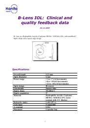
Page 3 of 213. MEDICAL DEVICE SPECIFICATION AND ADMINISTRATIONSeelens <strong>MF</strong> is a multifocal apodized diffractive aspheric, foldable, one piece lens.The intraocular lens is a CE-marked medical device. Table 1 summarizes the lens specifications<strong>See<strong>Lens</strong></strong> <strong>MF</strong> SpecificationsOptic DiameterPower range6.0 mm+15 to +35 (15-30D in 0.5Dincrements, 30-35D in 1Dincrements)Addition Power +3DOptic design<strong>Lens</strong> designHapticangulationMaterialLighttransmissionRefractiveindexNd-YAG laserEstimated AconstantPlacementApodized diffractive asphericMultifocal IOL360° Double square edge5°Hydrophilic Acrylic 25%water contentUV blocker and violet lightfilter2% transmission @400nm90% transmission @460nm1.462 (35°C)Compatible118.6Capsular bagTABLE 1: SEELENS <strong>MF</strong> SPECIFICATIONS
Page 4 of 21An increasingly important goal of modern cataract and implant surgery is to obtain the mostdesirable outcome for the patients, thus contributing to spectacle-free vision and highest qualityof life.The ideal state of the human phakic eye without any refractive error is known as emmetropia;Rays of light perfectly focused from an infinitely distant object onto the fovea withoutaccommodation 1 .Refractive power of the eye is determined by three main parameters: power(s) of the cornea,power of the crystalline lens and axial length of the eye. Incompatibility between theseparameters leads to various types of refractive errors known as myopia and hyperopia.The natural crystalline lens has the ability to accommodate in order to maintain a clear image(focus) of an object whatever the distance from the eye. At about the age of 40, the lens becomesless flexible and accommodation is lost gradually 2 , making close-range activities increasinglydifficult. This is called presbyopia. Once presbyopia has been diagnosed reading glasses orcorrective contact lenses are necessary to maintain near vision.With age, a normal crystalline lens opacifies (cataract) disabling the eye in generating a clear,well contrasted image. The only therapeutic solution to this problem is surgical replacement ofthe crystalline lens with an intraocular lens (cataract surgery).New technologies in IOLs optic designs provide for better options for cataract patients to correcttheir visual deficits and to live their lives without visual aids.The clinical demand for a solution to presbyopia is very high, as presbyopia afflicts the majorityof the world’s adult population.Implantation of a bifocal IOL, like <strong>See<strong>Lens</strong></strong> <strong>MF</strong>, with a bifocal aspheric refractive/diffractivestructure, pupil size dependence and asymmetrical light distribution provides for a satisfactory1 Diepes H. 2004, Refraktionsbestimmung2 Lang G.K., Spraul C.W. (1998) Augenheilkunde
Page 5 of 21full range of vision, a high level of uncorrected and corrected distance, intermediate and nearacuity and improved contrast sensitivity. Furthermore the <strong>See<strong>Lens</strong></strong> <strong>MF</strong> should allow forindependence of spectacles, thus enhancing patients’ satisfaction.The optical performance of bifocal IOLs restores near, distance and intermediate vision for highpatient satisfaction. With such optical performances, patients may benefit from independencefrom visual aids.The <strong>See<strong>Lens</strong></strong> <strong>MF</strong> is designed for micro-incision cataract surgery (MICS), through sub-2mmincisions.In order to confirm these statements, a post-marketing study is initiated: Clinical experience onthe implantation of the <strong>See<strong>Lens</strong></strong> <strong>MF</strong> IOL. The main purpose of this study is to evaluate visualacuity and contrast sensitivity of patients receiving the new bifocal IOL.<strong>See<strong>Lens</strong></strong> <strong>MF</strong> is an apodized diffractive bifocal lens; so that the IOL is dependent on pupil size,different proportions of the light energy are directed to each focus of the lens. Figure 1 belowshows the relative energy distribution at different pupil diameters.FIGURE 1 SEELENS <strong>MF</strong> HAS A FAR FOCUS, AND A NEAR FOCUS AT +3D ADDITION POWER.The optic of the IOL is designed for the highest possible MTF in the well established Arizona eyemodel, which takes into account the negative spherical aberration required by the IOL in orderto lower the positive spherical aberration of the human cornea.The Hanita <strong>Lens</strong>es intraocular lens hydrophilic material has been in use at Hanita <strong>Lens</strong>es formore than 11 years and effectively verified its outstanding long-term behaviour in the market in
Page 6 of 21terms of biocompatibility, transparency and stability of the visual function and centration. Afoldable and highly adaptable implant for all bag conformations, the <strong>See<strong>Lens</strong></strong> <strong>MF</strong> displaysoutstanding tensile strength for maximum resistance during insertion, and offer controlledunfolding for rapid visual recovery.Surgical ProcedureThe phacoemulsification procedure and the lens implantation was performed following theinstructions of use from the device’s manufacturers, and surgeon’s technique.The <strong>See<strong>Lens</strong></strong> <strong>MF</strong> has directionality; such that the leading haptic must point left. The lens is dialedclockwise.4. METHODS1. Vision and refractive performance:Refraction for distance was measured in order to assess the A constant of the IOL.2. Visual acuity will be measured using an ETDRS chart for distance at room illumination.All results will be expressed in decimal (distance) or Jaeger (near) values.3. Objective investigator’s opinion of the lens and the patient’s satisfaction.2 centers were included in this evaluation, each one included total of 10 eyes of 10 patients.A bilateral implantation was optional, as considered by the surgeon, as long as there were 10patients included.SurgeonLeast number of eyesProf. Roberto Belluci, Italy. 10Prof. Jan Novak, Czech Republic. 10
Page 7 of 21Dr. Luis Paparo 8The study was started in December 2010.5. STATISTICAL METHODSThe following analyses were used to describe the data in this report:Descriptive statistics: continuous variables are described with mean ± standard deviation (SD),median, minimum and maximum. Nominal scale variables are described with absolute andrelative (percents) frequencies. Ordinal variables are described with means ± SD andfrequencies of the ordinal grades.All analyses were done using Excel 2007 statistics tool package.6. RESULTS (3 MONTHS)Patients that were enrolled in the study were not consistently targeted for emmetropia andwere not necessarily capable of achieving a flawless retinal image. This was because the purposeof this study was to evaluate the safety and the A-constant of the <strong>See<strong>Lens</strong></strong> <strong>MF</strong>.6.1. POSTOPERATIVE BE ST CORRECTED VISUAL ACUITY (BCVA)Best Corrected Visual Acuity (BCVA) was reported in the pre-operative and the three monthfollow-up visit. Pre-operative BCVA results are shown on Graph 2, and Post-operative BCVAresults are shown on Graph 3.Figure 2: Pre-operative BCVA distribution (Novak data, n=22)
Page 8 of 21FIGURE 2 NOVAK RESULTSPre op data was unavailable from Prof. Belluci.FIGURE 3 NOVAK AND BELLUCI RESULTSFigure 3: 3-month Post-operative BCVA distribution (Novak: n=12, Belluci: n=17)Two patients had macular problems (0.2 and 0.6 BCVA).As shown in graph 3, postoperative (3 months) BCVA of 6/9 or better was reported in 86% ofthe eyes.BCVA of 6/12 or better was achieved by 96.5% of the eyes (n=29) after 3 months.
Page 9 of 21Dr. Paparo measured distance vision in 4 bilateral patients (8 eyes)FIGURE 4 PAPARO RESULTS6.2. EVALUATION OF A-CONSTPostoperative refractive deviation, calculated by Spherical Equivalent (SE), was reported in thefollow- up visits, and optimized with the Novak and Belluci data. The results have shown that theA-const should be updated to 118.6.6.3. INTERMEDIATE VISUAL ACUITYVisual acuity was measured at 80cm, after 3 months (Novak). The average uncorrectedintermediate visual acuity (UIVA) was 0.815.
Page 10 of 21FIGURE 5 NOVAK RESULTSThe full range of near (40cm), intermediate (63cm and 100cm) was measured by Dr. Paparo.This was performed using Colenbrander mixed contrast visual acuity charts (ETDRS), whichhave optotypes matched for the true distance from the patient’s eyes. This is different from adefocus curve which uses only the usual distance ETDRS optotypes.FIGURE 6 PAPARO RESULTSThe following chart (figure 7) shows the logMAR visual acuity at different distances, usingETDRS charts with suitable optotypes for each distance (40cm, 63cm, 100cm, and distancevision).
Page 11 of 21FIGURE 7 – PAPARORESULTSBelow is the poster presented by Dr. Paparo at the PAAO 2011 conference.
Page 12 of 21
Page 13 of 216.4. NEAR VISUAL ACUITYVisual acuity was measured for near vision, after 3 months (Novak). Binocular near visual acuitywas J1.5.FIGURE 8 - NOVAK RESULTSFIGURE 9 PAPARO RESULTS (VALUES IN DECIMAL NOTATION BECAUSE THE RESULTS ARE OFF THEJAEGER NUMBER SCALE)
Page 14 of 217. RESULTS (9 TO 12 MONTHS)Patients that were enrolled in the study were not consistently targeted for emmetropia andwere not necessarily capable of achieving a flawless retinal image. This was because the purposeof this study was to evaluate the safety and the A-constant of the <strong>See<strong>Lens</strong></strong> <strong>MF</strong>.The following graphs show the distribution of monocular (eyes) and binocular (patients) visualacuity. The results are presented in decimal values and Jaeger numbers. All data for these followup visits from Prof. Novak.1 patient (Novak) with J6 had ARMD.
Page 15 of 21Bilateral patients (n=4, 8 eyes) Far UCVA binocular Far BCVA monocular with <strong>MF</strong> IOL DCNVA Jaeger Noaveragestd0.90 0.92 10.12 0.15 0Unilateral patients (n=10, 10eyes)averagestdFar UCVAbinocularFar BCVA monocular with <strong>MF</strong>IOLDCNVA JaegerNo0.90 0.88 2.250.14 0.14 2.50FIGURE 10 -NOVAK RESULTSResults of competitor lenses (bilateral only) taken from:[1] Alfonso J, Fernández-Vega L, Señaris A, Montés-Micó R. Prospective study of theAcri.LISA bifocal intraocular lens. J Cataract Refract Surg 2007; 33:1930-5[2] Apodized diffractive versus refractive multifocal intraocular lenses: Optical and visualevaluation Beata Z_ elichowska, MD, Marek Re˛kas, PhD, Andrzej Stankiewicz, PhD, AlejandroCervin˜o, PhD,Robert Monte´s-Mico´ , PhD J Cataract Refract Surg 2008; 34:2036–2042[3] Optical analysis, reading performance, and quality-of-life evaluation after implantation ofa diffractive multifocal intraocular lens. Jorge L. Ali_o, MD, PhD, Ana B. Plaza-Puche, MSc, DavidP. Pi~nero, PhD, Francisco Amparo, MD, Ram_on Jim_enez, MSc, Jose L. Rodríguez-Prats, MD,Jaime Javaloy, MD, Vanessa Pongo, MD; J Cataract Refract Surg 2011; 37:27–37
Page 16 of 218. INTRA-OPERATIVE COMPLICATIONSNo intra-operative complications were reported. No IOL-related adverse events were observed.Two patients had macular problems prior to surgery. One patient suffered from amblyopia priorto surgery.9. POSTOPERATIVE COMPLICATIONSNo evidence of post operative infection or excessive inflammation was reported in any of thepatients. Patients were asked as to perception of halos, glare and their subjective feeling abouttheir vision. The grading of visual phenomena was 0 for no halos or glare, 1 for existence oftolerable or non-severe halos and/or glare, and 2 for problems with halos or glare.
Page 17 of 21Most patients reported some halos and a few reported glare. One eye of one patient out of 12eyes had problems with visual phenomena. All patients asked expressed satisfaction with theirvision, 6/8 (75%) patients asked felt their subjective vision was “excellent”.The <strong>See<strong>Lens</strong></strong> <strong>MF</strong> IOL position as seen through a slit lamp is presented in Image 1.IMAGE 3: THE SEELENS <strong>MF</strong> IOL AS SEEN THROUGH A SLIT LAMP; 6 DAYS POSTOPERATIVELY (NOVAK)
Page 18 of 21Image 3 shows that the <strong>See<strong>Lens</strong></strong> <strong>MF</strong> is well centered. A typical clear cornea and healthyconjunctiva can be observed, as was seen in all <strong>See<strong>Lens</strong></strong> <strong>MF</strong> implants 3 months & 1 month postoperatively. Implantation was performed by Dr. J. Novak, Regional Hospital, Pardubice, CzechRepublic.10. ADVERSE EVENTSAdverse EventSPErate 3(%)Number of subjects = 100Maximalallowablerate (%)Max. numberof casesallowedRate of adverseeffect occurredat this studyNumber ofadverseeventscasesPass / FailCumulative (total number of occurrences at any time postoperative):Cystoid macular oedema 3.0% 8.9% 6 0 % 0 PassHypopyon 0.3% 3.0% 1 0% 0 PassEndophthalmitis 4 0.1% 3.0% 1 0% 0 Pass<strong>Lens</strong> dislocation fromposterior chamber0.1% 3.0% 1 0% 0 PassPupillary block 0.1% 3.0% 1 0% 0 PassRetinal detachment 0.1% 3.0% 1 0% 0 Pass3 SPE (safety and performance endpoint) rate is the target rate for each event.4 Endophthalmitis is defined as inflammatory reaction (sterile or infectious) involving the vitreous body.
Page 19 of 21Secondary surgicalintervention 50.1% 4.2% 2 0% 0 PassPersistent (total number of occurrences at 3 months postoperative):Corneal stroma oedema 0.1% 3.0% 1 0% 0 PassCystoid macular oedema 0.1% 4.2% 2 2.5% 0 PassIritis 0.1% 3.0% 1 0% 0 PassRaised IOP req. treatment 0.1% 4.2% 2 0% 0 PassTABLE 7: ADVERSE EVENTS AS DEFINED BY ISO 11979-7 2006:As shown in table 7, 100% of 37 implanted eyes were not reported with any adverse events.Thus, it can be concluded that safety of <strong>See<strong>Lens</strong></strong> <strong>MF</strong> is in accordance to the ISO 11979-7 2006.11. CONCLUSIONS:3 months resultsThe detailed data from the current study on 37 eyes shows the following benefits of the <strong>See<strong>Lens</strong></strong><strong>MF</strong> IOL:Optical performance is in accordance to ISO 11979-7 2006 requirements. The IOL A constant using IOL MASTER and SRK/T is set at 118.6.Very good safety profile as reflected by a very low rate of intra- and post-operativecomplications at 3 months.5 Excludes posterior capsulotomies.
Page 20 of 21Dr. Paparo patients were enrolled according to the Phase 2 recommended patient selectioncriteria, and following the optimization of the A constant. This resulted in excellentuncorrected visual acuity and emmetropic refractive predictability, an excellent defocuscurve and high patient satisfaction.9-12 months resultsAs the A constant was not optimized at the time of the first implantations, correction wasnecessary in order to evaluate near vision. When corrected for distance all patientsimplanted bilaterally achieved J1.Binocular uncorrected vision for distance was excellent (0.9±0.12) for bilaterally implantedpatients.Contrast sensitivity in photopic conditions (bilateral patients only) was comparable to othermultifocal IOLs.