Summary Carbon Monoxide Lurking within: The Danger of ...
Summary Carbon Monoxide Lurking within: The Danger of ...
Summary Carbon Monoxide Lurking within: The Danger of ...

Create successful ePaper yourself
Turn your PDF publications into a flip-book with our unique Google optimized e-Paper software.
m a s i m o c o r p o r a t i o n 4 0 p a r k e r i r v i n e c a 9 2 6 1 8 w w w. m a s i m o . c o m<br />
Carboxyhemoglobinemia in Acute Care<br />
<strong>Carbon</strong> <strong>Monoxide</strong> <strong>Lurking</strong> <strong>within</strong>: <strong>The</strong> <strong>Danger</strong> <strong>of</strong><br />
Carboxyhemoglobinemia in Acute Care<br />
<strong>Summary</strong><br />
Much is known about the causes, symptoms and detection <strong>of</strong> exogenous carbon monoxide (CO)<br />
poisoning and the treatment <strong>of</strong> the resultant carboxyhemoglobinemia seen in patients after CO<br />
exposure. However, it may be surprising to learn that CO poisoning can occur in the hospital<br />
and not just outside the hospital. In most cases, these patients present in the emergency<br />
department (ED) with equivocal, flu-like symptoms. Because the clearance <strong>of</strong> CO from the<br />
blood begins to occur immediately after the etiologic agent is removed, patients may begin to<br />
feel better in the ED and suffer a missed diagnosis as the symptoms abate. In many reported<br />
cases, these poisoned patients are sent back to the source <strong>of</strong> the odorless, colorless toxin, and<br />
poisoning continues, sometimes with lethal consequences. Others will find themselves admitted<br />
for acute care as the symptoms <strong>of</strong> CO poisoning mimic cardiac, pulmonary, and neurologic<br />
disorders that demand emergent care. Whether in the ED or other places in the hospital such as<br />
the OR, if and when carboxyhemoglobinema detection occurs, it is <strong>of</strong>ten after costly diagnostic<br />
procedures and protocols have been attempted with negative results.<br />
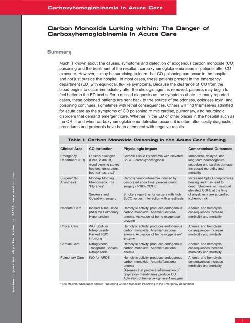
Table I: <strong>Carbon</strong> <strong>Monoxide</strong> Poisoning in the Acute Care Setting<br />
Clinical Area CO Induction Physiologic Impact Compromised Outcomes<br />
Emergency<br />
Department (ED)<br />
Surgery/OR/<br />
Anesthesia<br />
Outside etiologies<br />
(Fires, exhaust,<br />
wood burning stoves,<br />
heaters, generators,<br />
boat ramps, etc.)*<br />
Monday Morning<br />
Phenomena. <strong>The</strong><br />
“Fluranes”<br />
Smokers and<br />
Outpatient surgery<br />
Neonatal Care Inhaled Nitric Oxide<br />
(iNO) for Pulmonary<br />
Hypertension<br />
Critical Care iNO, Sodium<br />
Nitroprusside,<br />
Packed RBC<br />
infusions<br />
Cardiac Care Nitroglycerin,<br />
Transplant, Sodium<br />
Nitroprisside<br />
Chronic Tissue Hypoxemia with elevated<br />
SpCO - carboxyhemoglobin<br />
Carboxyhemoglobinemia induced by<br />
desiccated soda lime, poisons during<br />
surgery (7-36% COHb)<br />
Smokers reporting for surgery with high<br />
SpCO values. Interaction with anesthesia<br />
Hemolytic activity produces endogenous<br />
carbon monoxide. Anemia/functional<br />
anemia. Activation <strong>of</strong> heme oxygenase-1<br />
enzyme<br />
Hemolytic activity produces endogenous<br />
carbon monoxide. Anemia/functional<br />
anemia. Activation <strong>of</strong> heme oxygenase-1<br />
enzyme<br />
Hemolytic activity produces endogenous<br />
carbon monoxide. Anemia/functional<br />
anemia<br />
Pulmonary Care iNO for ARDS Hemolytic activity produces endogenous<br />
carbon monoxide. Anemia/functional<br />
anemia<br />
Diseases that produce inflammation <strong>of</strong><br />
respiratory membranes produce CO<br />
Activation <strong>of</strong> heme oxygenase-1 enzyme<br />
* See Masimo Whitepaper entitled: “Detecting <strong>Carbon</strong> <strong>Monoxide</strong> Poisoning in the Emergency Department.”<br />
Immediate, delayed, and<br />
long term neurocognitive<br />
sequelae and cardiac damage<br />
Increased morbidity and<br />
mortality<br />
Increased SpCO compromises<br />
healing and may lead to<br />
death. Smokers with residual<br />
elevated COHb at the time<br />
<strong>of</strong> anesthesia are at cardiac<br />
ischemic risk<br />
Anemia and hemolysis<br />
consequences increase<br />
morbidity and mortality<br />
Anemia and hemolysis<br />
consequences increase<br />
morbidity and mortality<br />
Anemia and hemolysis<br />
consequences increase<br />
morbidity and mortality<br />
Anemia and hemolysis<br />
consequences increase<br />
morbidity and mortality<br />
1
m a s i m o c o r p o r a t i o n 4 0 p a r k e r i r v i n e c a 9 2 6 1 8 w w w. m a s i m o . c o m<br />
Carboxyhemoglobinemia in Acute Care<br />
However, CO poisoning may be quickly caught <strong>within</strong> the acute care setting using a state-<strong>of</strong>the-art<br />
Masimo Rainbow SET Pulse CO-Oximetry device. Expeditious diagnosis ensures proper<br />
treatment may ensue to minimize the known long-term cardiac and neuropsychiatric damage <strong>of</strong><br />
CO exposure. In the case <strong>of</strong> a CO poisoned patient that presents in the ED and then transitions<br />
into the acute care environment, Pulse CO-Oximetry provides clinicians with immediate detection<br />
and subsequent continuous monitoring <strong>of</strong> carboxyhemoglobin (COHb) levels as feedback to the<br />
efficacy <strong>of</strong> treatment decisions. However, CO poisoning also occurs <strong>within</strong> the acute care setting<br />
– both endogenously and exogenously - contributing to severe tissue hypoxemia, ischemia, and<br />
death. <strong>The</strong>refore, Pulse CO-Oximetry serves an additional role in detection and monitoring <strong>of</strong><br />
nosocomial CO poisoning.<br />
This paper explores a sampling <strong>of</strong> several areas in which nosocomial CO exposure is possible.<br />
Several disease processes produce endogenous CO through their natural progression as in<br />
the development <strong>of</strong> systemic inflammatory response syndrome (SIRS), sepsis, pulmonary<br />
inflammation, and hemolysis. In these events, the detection and measurement <strong>of</strong> endogenously<br />
produced CO may prove to be a valuable marker <strong>of</strong> disease severity. <strong>The</strong>re are also reports <strong>of</strong><br />
CO poisoning by the Monday Morning Phenomena where carboxyhemoglobinemia is induced<br />
<strong>within</strong> the closed circuit anesthesia system during surgery, inhaled nitric oxide therapy, and<br />
anti-hypertension treatment with sodium nitroprusside. Though these acute care sources <strong>of</strong> CO<br />
may not raise blood COHb to life-threatening levels for many patients, even slight increases in<br />
CO concentrations can be life-threatening to those patients compromised by cardiac disease,<br />
anemia, loss <strong>of</strong> pulmonary reserve and a host <strong>of</strong> other diseases.<br />
In all cases, the clinical importance <strong>of</strong> continually and accurately measuring COHb <strong>within</strong> the<br />
hospital is established in the medical literature. New Pulse CO-Oximetry technology platforms,<br />
available as handheld and bedside devices, provide immediate and continuous results through<br />
non-invasive monitoring and enhance the clinical workflow by eliminating the need for a physician<br />
order, painful blood sampling, and the attendant high costs and likely delays associated with a<br />
laboratory CO-Oximeter analysis. In addition, they allow trended presentations <strong>of</strong> COHb which<br />
quickly illustrate significant changes that occur over time, changes that might be lost in visual or<br />
tabular displays <strong>of</strong> invasive COHb measurements.<br />
I. Emergency Department/ER<br />
Study shows prevalence <strong>of</strong> carbon monoxide toxicity in the ED may be higher<br />
than previously recognized<br />
In a study led by Dr. Robert Partridge and Dr. Gregory Jay <strong>of</strong> Rhode Island Hospital at Brown<br />
University Medical School, a team <strong>of</strong> researchers performed a study to assess baseline CO<br />
levels <strong>of</strong> nearly 5,000 patients presenting to the emergency room. To accomplish this, all pulse<br />
oximeters in the ED were replaced with Masimo Rainbow SET Pulse CO-Oximeters and the ED<br />
staff began assessing baseline COHb levels <strong>of</strong> all adult patients as part <strong>of</strong> the standard triage<br />
process. In addition to confirming suspected cases <strong>of</strong> CO toxicity (COT) from smoke inhalation,<br />
there were nine unsuspected cases <strong>of</strong> COT discovered, in just three months, in patients who<br />
presented with non-specific symptoms or unrelated complaints. Toxic COHb levels ranged from<br />
16-33% and were confirmed with an invasive laboratory blood test. If this rate were indicative<br />
<strong>of</strong> all US hospitals, it would equate to as many as 50,000 cases <strong>of</strong> unsuspected CO toxicity<br />
annually.
<strong>The</strong> study concluded that the use <strong>of</strong> Masimo Rainbow SET as a noninvasive test for COT can effectively and<br />
efficiently be performed at ED triage, and that “unsuspected COT may be identified using noninvasive COHb<br />
screening and the prevalence <strong>of</strong> COT may be higher than previously recognized.” 1<br />
<strong>The</strong> team from Brown University also presented a case report <strong>of</strong> a previously healthy 52-year old nonsmoking<br />
female who was brought to the ED complaining <strong>of</strong> nausea, headache, dizziness, and feeling cold.<br />
<strong>The</strong> patient had no history <strong>of</strong> carbon monoxide exposure. <strong>The</strong> Masimo Rainbow SET device recorded an<br />
SpCO level <strong>of</strong> 33%, which was later confirmed with an invasive laboratory measurement. After interviewing<br />
the woman, clinicians learned that her utilities had been shut <strong>of</strong>f and she was running a gas-powered<br />
generator in her basement.<br />
In the report, researchers said that since early CO toxicity shares symptoms with other more common<br />
illnesses, “physicians must maintain a high index <strong>of</strong> suspicion to avoid incorrect diagnosis, management and<br />
disposition. Unrecognized CO poisoned patients returned to the site <strong>of</strong> exposure may develop more serious<br />
CO toxicity.” <strong>The</strong>y added that the noninvasive testing provided by Masimo Rainbow SET technology “is a<br />
rapid, inexpensive method for screening large numbers <strong>of</strong> patients for CO toxicity and identifying unsuspected<br />
cases that might otherwise be missed.” 2<br />
II. Surgery/Operating Room/Anesthesia<br />
Monday Morning Phenomena and Anesthesia-Related COHb<br />
whitepaper<br />
<strong>The</strong>re is increasing evidence that exposure <strong>of</strong> volatile anesthetics, i.e., desflurane, enflurane, and is<strong>of</strong>lurane<br />
(in descending order <strong>of</strong> magnitude) to desiccated carbon dioxide (CO 2) absorbents may result in reactions<br />
in anesthetic breathing circuits and production <strong>of</strong> toxic products (e.g., CO, methanol, formaldehyde). 3-6 CO 2<br />
absorbents such as soda lime are mixtures <strong>of</strong> chemicals, used in closed breathing environments, such as<br />
general anesthesia to remove CO 2 from breathing gases to prevent CO 2 retention and poisoning. <strong>The</strong>re is<br />
significant evidence that potentially toxic products can be produced upon exposure <strong>of</strong> volatile anesthetics<br />
to other desiccated absorbents containing strong bases, particularly potassium and sodium hydroxide. <strong>The</strong><br />
clinical scenario has been called the “Monday Morning Phenomenon,” as anesthesia breathing circuits may<br />
be left on and cycling through the weekend in preparation for the early morning procedures on Monday.<br />
Unfortunately, during the delivery <strong>of</strong> the anesthetic the desiccant becomes exhausted and loses its ability<br />
to properly “scrub” the anesthetic gases. CO may be produced in significant quantities to poison the patient<br />
under anesthesia (see Figure 2).<br />
3
m a s i m o c o r p o r a t i o n 4 0 p a r k e r i r v i n e c a 9 2 6 1 8 w w w. m a s i m o . c o m<br />
Carboxyhemoglobinemia in Acute Care<br />
<strong>The</strong> desiccant does not transition from fully functional to suddenly consumed. <strong>The</strong> transition<br />
occurs over time, and thus, some patients may be exposed to low, but clinically significant levels<br />
<strong>of</strong> CO during a time when they can ill-afford a compromise to oxygen delivery, especially to<br />
the heart and brain. Case studies demonstrate this, and in spite <strong>of</strong> the general awareness and<br />
the adjustment <strong>of</strong> guidelines to thwart the possibility <strong>of</strong> this unfortunate preventable adverse<br />
event, it occurs even today. As concern has grown, the Anesthesia Patient Safety Foundation<br />
held a conference entitled <strong>Carbon</strong> Dioxide Absorbent Desiccation: APSF Conference on<br />
Safety Considerations on April 27, 2005. From the proceedings: “<strong>The</strong>re is increasing evidence<br />
that exposure <strong>of</strong> volatile anesthetics to desiccated carbon dioxide absorbents may result in<br />
exothermic reactions leading to fires in anesthetic breathing circuits and production <strong>of</strong> toxic<br />
products (e.g., carbon monoxide, compound A, methanol, formaldehyde)… In some cases this<br />
may lead to sub-clinical carbon monoxide exposure.”<br />
<strong>The</strong> exact incidence <strong>of</strong> patient exposure to CO through CO 2 absorbent desiccation is unknown.<br />
<strong>The</strong> American Society <strong>of</strong> Anesthesia (ASA) estimates that 25 million anesthetic procedures are<br />
performed each year in the US. If as little as 33 percent <strong>of</strong> these anesthetics involve is<strong>of</strong>lurane,<br />
enflurane, or desflurane, and if four cases are performed in the average operating room each<br />
day so that 25 percent <strong>of</strong> cases will be first cases, i.e., the most likely to be impacted by<br />
desiccated absorbent, then up to 2 million patients may be at risk each year for intraoperative<br />
CO exposure. 7 If the published incidence <strong>of</strong> CO exposures can be extrapolated to other<br />
institutions and remains between 0.0005 and 0.005 first cases, 8 then approximately 1,000-10,000<br />
patients may be exposed to CO annually in the US as a result <strong>of</strong> anesthetic breakdown. If these<br />
CO poisonings go undetected they can't be treated and injury and even death can occur.<br />
Reports <strong>of</strong> elevated COHb concentrations detected intraoperatively in humans have ranged from<br />
7 to 32 percent. 9-11 Berry et al. reported a patient who attained 36 percent COHb. 12 As a subject<br />
in a clinical study, the patient was a healthy female who did not appear to be adversely affected<br />
by her CO exposure. However, it may be possible that far lower CO exposure in the presence <strong>of</strong><br />
concurrent disease may predispose patients to far greater risks. In patients with coronary artery<br />
disease, COHb levels as low as 2.9-4.5 percent can exacerbate myocardial ischemia. 13-14 Similarly,<br />
smoke inhalation with relatively mild CO exposure (COHb levels
III. Neonatal/Critical Care<br />
1. Hemolysis<br />
It is possible that clinicians may suspect a case <strong>of</strong> CO poisoning due to desiccated CO absorbent when, in fact,<br />
the etiologic culprit may be an entirely different cause. As one clinical case illustrates hemolysis, although not<br />
usually a clinically significant event in routine delivery <strong>of</strong> anesthesia, has the potential to result in significant CO<br />
exposure.<br />
Hemolysis is the breaking open <strong>of</strong> red blood cells and the release <strong>of</strong> hemoglobin into the surrounding fluid. A<br />
39-year-old female with a history <strong>of</strong> hemolytic episodes was scheduled as the first surgical case on a Friday<br />
morning. Because <strong>of</strong> her continued hemolysis, intraoperative laboratory studies were obtained 20 minutes after<br />
induction <strong>of</strong> anesthesia. <strong>The</strong> test revealed a COHb <strong>of</strong> 7.3 percent. Desflurane breakdown was suspected and<br />
the absorbent was changed to fresh, unused normally hydrated absorbent. However, subsequent analysis <strong>of</strong><br />
the initial absorbent revealed that it was not the source <strong>of</strong> CO production.<br />
In this case, the CO exposure <strong>of</strong> the hemolytic patient imitated CO production from anesthetic breakdown. In<br />
reality, analysis <strong>of</strong> the patient’s blood estimated CO production <strong>of</strong> 257 ml per 24 hours. Normal endogenous<br />
CO production is approximately 10 ml per day. 18 A mathematical model <strong>of</strong> CO uptake 19-21 predicts a COHb<br />
concentration between 5.6 percent and 7.3 percent using this rate <strong>of</strong> hemolysis. If this patient had received an<br />
anesthetic through a closed breathing circuit, the oxygen binding capacity <strong>of</strong> hemoglobin could have become<br />
an additional 23 percent saturated with CO during the 6-hour procedure because none <strong>of</strong> the endogenously<br />
produced CO would be removed. <strong>The</strong> model predicts that closed-circuit anesthesia during an episode <strong>of</strong><br />
hemolysis may dangerously increase COHb concentrations. 22 As such, anesthesiologists should be aware <strong>of</strong> all<br />
sources <strong>of</strong> CO in the perioperative period and maintain constant awareness <strong>of</strong> the patient’s COHb status.<br />
2. Inhaled Nitric Oxide<br />
Inhaled nitric oxide (iNO) is occasionally used to improve arterial oxygenation in patients with the acute<br />
respiratory distress syndrome (ARDS). 23 Inhaled NO induces selective vasodilation in pulmonary vessels to<br />
relieve hypertension. <strong>The</strong> use in ARDS cases has brought to light a potential pathophysiologic mechanism<br />
linking iNO, methemoglobin (MetHb), and carboxyhemoglobin (COHb). <strong>The</strong> withdrawal <strong>of</strong> iNO in this study<br />
resulted in a parallel decline in MetHb and COHb levels. Due to the negative influence <strong>of</strong> COHb on the oxygencarrying<br />
capacity <strong>of</strong> the blood, its iNO-induced increase (through stimulants <strong>of</strong> hemoxygenase inductions)<br />
cancelled out the slight benefit <strong>of</strong> iNO on arterial oxygenation. A case report published in 2004 demonstrated<br />
a correlation between iNO and COHb. 24 <strong>The</strong> authors do propose that not only MetHb but also COHb levels be<br />
monitored if iNO is administered during the course <strong>of</strong> ARDS, since even low levels <strong>of</strong> COHb may potentially<br />
<strong>of</strong>fset any benefit <strong>of</strong> iNO.<br />
3. Sodium Nitroprusside<br />
whitepaper<br />
Sodium nitroprusside is the most widely used vasodilator drug in critically ill patients. 25-28 <strong>The</strong> drug is <strong>of</strong>ten<br />
administered intravenously to patients who are experiencing a hypertensive emergency and to produce<br />
controlled hypotension (low blood pressure) in anesthetized patients during surgery. Sodium nitroprusside<br />
breaks down in the blood and releases nitric oxide (NO) which enters the muscle cells in the walls <strong>of</strong> the blood<br />
vessels and causes them to relax. When the muscles relax, the vessels become wider and the blood pressure<br />
decreases. <strong>The</strong> most important toxic effects <strong>of</strong> sodium nitroprusside are cyanide poisoning, thiocyanate toxicity,<br />
and methemoglobinemia. 29 Like the reaction triggered by sepsis and pulmonary inflammation, research suggest<br />
that NO donors, such as sodium nitroprusside, can induce heme oxygenase-1, and produce CO by breakdown<br />
<strong>of</strong> heme molecules. 30-32<br />
5
m a s i m o c o r p o r a t i o n 4 0 p a r k e r i r v i n e c a 9 2 6 1 8 w w w. m a s i m o . c o m<br />
Carboxyhemoglobinemia in Acute Care<br />
One study examines the cases <strong>of</strong> four pediatric heart transplant cases. 33 <strong>The</strong> patients showed a<br />
moderate increase in COHb level after nitroprusside administration, and in three <strong>of</strong> these cases<br />
the withdrawal <strong>of</strong> the drug led to the normalization <strong>of</strong> COHb level. If in fact prolonged treatment<br />
with moderate or high doses <strong>of</strong> sodium nitroprusside can produce carboxyhemoglobinemia in<br />
children after heart transplant, specific medical management after pediatric heart transplant<br />
should include frequent measurement <strong>of</strong> COHb. Even low levels <strong>of</strong> carbon monoxide bound to<br />
hemoglobin in cardiac compromised patients can be lethal, starving the tissues <strong>of</strong> oxygen due to<br />
functional anemia, poor perfusion, cardiac output compromise, and suboptimal oxygen delivery<br />
mechanisms.<br />
Accepted standards <strong>of</strong> patient monitoring associated with nitroprusside administration include<br />
analysis <strong>of</strong> MetHb concentrations. 34 Research suggests that COHb levels should be evaluated<br />
as well. Current blood analysis devices that measure CO-Oximetry in each blood gas sample<br />
permit diagnosis <strong>of</strong> moderate COHb elevations that probably would not have been discovered<br />
in the past. With noninvasive and continuous Pulse CO-Oximetry, results are faster and less<br />
resource-intensive than ever before. Of significant importance is the ability to trend the changes<br />
in COHb over time (see Figure 1), and view the trend at will. Days <strong>of</strong> trend data are saved for<br />
retrospective analysis <strong>of</strong> subtle changes in COHb (and when necessary, MetHb). Without the<br />
continuous assessment, the task <strong>of</strong> associating subtle changes in the dyshemoglobins is daunting,<br />
and impossible using traditional laboratory CO-Oximetry. As well, the trend value allows the<br />
patient to serve as their own baseline. At the beginning <strong>of</strong> the trend period, carboxyhemoglobin by<br />
Pusle CO-Oximetry (SpCO) may measure 1.0 percent, but with a course <strong>of</strong> sodium nitroprusside<br />
treatment, or following the transitions into SIRS, the clinician may note trends in elevation <strong>of</strong><br />
COHb, and interact accordingly with the patient to achieve the desired outcome.<br />
IV. COHb as a Marker<br />
Endogenous production <strong>of</strong> CO was first reported in the mid 20th century, but it has been a<br />
known poison since Claude Bernard first noted its high affinity for hemoglobin a century earlier. 35<br />
Moderate endogenous increases in COHb levels (0.8–2 percent) have been reported in critically<br />
ill patients 36 and clinical interest has grown rapidly as CO production has been proposed to induce<br />
excessive vascular relaxation, and hence a fall in blood pressure. 37 <strong>The</strong> mechanism behind this<br />
reaction is heme oxygenase (HO), the initial enzyme in heme metabolism. 38 HO produces CO<br />
during breakdown <strong>of</strong> heme molecules primarily in the liver and spleen. It is well established that<br />
metabolism <strong>of</strong> heme via heme oxidase results in production <strong>of</strong> one molecule <strong>of</strong> CO for each<br />
molecule <strong>of</strong> heme destroyed. 37 Recent data suggest that CO is also produced in the lungs. A<br />
number <strong>of</strong> stress-associated agents induce the expression <strong>of</strong> heme oxygenase, including heavy<br />
metals, hyperthermia, hyperoxia, hypoxia, heat shock, endotoxin, hydrogen peroxide, cytokines,<br />
ultraviolet radiation and nitric oxide, producing CO. 39-42<br />
To investigate whether critical illness results in increased CO production researchers have<br />
measured the CO concentration in exhaled air in critically ill patients and in healthy controls. 43<br />
Sampling exhaled CO is only an approximation <strong>of</strong> COHb levels in the blood. In patients with<br />
pulmonary compromise, high dead space to tidal volume ratios, or ventilation to perfusion<br />
mismatch, exhaled CO will correlate poorly with CO bound to hemoglobin and induce tissue<br />
hypoxemia. In a study <strong>of</strong> 95 mechanically ventilated, critically ill patients, CO production was<br />
correlated with multiple organ dysfunction score. Patients suffering from cardiac disease and<br />
critically ill patients undergoing dialysis produced significantly higher amounts <strong>of</strong> CO compared to<br />
other critically ill controls. <strong>The</strong> findings suggest that endogenous CO production might reflect the<br />
severity <strong>of</strong> acute organ dysfunction and therefore may <strong>of</strong>fer clinicians an effective, non-invasive<br />
gauge <strong>of</strong> patient condition. Two examples <strong>of</strong> this correlation exist in patients with sepsis and<br />
pulmonary inflammation.
1. Sepsis<br />
Sepsis is among the top causes <strong>of</strong> death in the world today. It kills 210,000 people in the U.S. each year -<br />
more than lung and breast cancer combined. Nationally, sepsis is a complication in about 3.0 cases per 1,000<br />
population, or 751,000 cases annually, 44 where related intravenous (IV) lines, surgical wounds or drains, and<br />
bedsores can be entry points for bacteria. Sepsis is caused most commonly by bacteria in the bloodstream,<br />
and is thought to be preceded by Systemic Inflammatory Response Syndrome (SIRS) with attendant hemolysis<br />
<strong>of</strong> red blood cells, producing CO. In adults, sepsis is most <strong>of</strong>ten a nosocomial infection seen after surgery or<br />
another invasive medical procedure in the hospital. Experts predict that sepsis will increase by 1.5 percent per<br />
year due to the high incidence <strong>of</strong> sepsis in the elderly and the overall aging <strong>of</strong> the population. <strong>The</strong>y estimate<br />
that there will be 934,000 cases in the United States in the year 2010 and 1,110,000 cases in 2020. 45-46<br />
During the 1990s, CO was recognized as a new participant in the pathogenesis <strong>of</strong> sepsis syndrome. Products<br />
<strong>of</strong> the HO enzyme include COHb and bilirubin, which have protective effects in stressed states. <strong>The</strong> HO<br />
enzyme up-regulates during states <strong>of</strong> oxidative stress. <strong>The</strong> marked increase in HO activity stimulated by<br />
endotoxin suggests that overproduction <strong>of</strong> CO may contribute to the reduction in vascular tone during endotoxic<br />
shock. In support <strong>of</strong> this theory, research has demonstrated increased CO concentrations during stress, sepsis,<br />
and shock. 47-48<br />
Because early detection and intervention <strong>of</strong> patients who are sepsis/septic shock candidates has significant<br />
impact on morbidity and mortality, the clinical importance <strong>of</strong> measuring and trending CO concentrations as an<br />
ancillary marker <strong>of</strong> sepsis may prove highly valuable in treating this condition. To date, an evidence-base is<br />
being compiled to determine if monitoring subtle changes in CO production may prove to be a robust marker <strong>of</strong><br />
sepsis or septic shock onset.<br />
2. Pulmonary Disease<br />
whitepaper<br />
Exhaled CO is increased in patients with inflammatory pulmonary diseases such as bronchial asthma,<br />
bronchiectasis, upper respiratory tract infections, and seasonal allergic rhinitis. 49-53 Treatment with inhaled<br />
and oral corticosteroids, which have been shown to reduce airway inflammation, is associated with a<br />
reduction in the exhaled levels <strong>of</strong> CO in asthma. 54 Furthermore, exhaled CO is increased in exacerbations <strong>of</strong><br />
bronchial asthma induced by respiratory virus infections. 55 Based on these findings, it has been proposed that<br />
measurements <strong>of</strong> exhaled CO may serve as an indirect marker <strong>of</strong> airway inflammation. 56-61<br />
Exhaled CO concentration is reported to correlate closely with blood COHb in smokers and non-smokers, 62<br />
which suggests that the COHb levels may increase in patients with inflammatory pulmonary diseases. A<br />
study that was undertaken to determine whether arterial blood COHb increases in patients with inflammatory<br />
pulmonary diseases confirmed that COHb concentrations are increased in patients with bronchial asthma,<br />
pneumonia, and idiopathic pulmonary fibrosis (IPF). 63 Increased blood levels <strong>of</strong> COHb in patients with<br />
inflammatory pulmonary diseases may reflect lung inflammation. This finding was seen as a benefit for<br />
ventilatory limited patients, especially children, who cannot perform the vital capacity maneuver to measure<br />
exhaled CO. Also, patients with lung disease demonstrate poor correlations between COHb in the blood<br />
and exhaled values. <strong>The</strong> continuous measurement <strong>of</strong> blood levels <strong>of</strong> carboxyhemoglobin allows a trending<br />
presentation that graphically depicts subtle yet clinically significant elevations in COHb, providing a simple and<br />
valuable marker to indicate pulmonary inflammation.<br />
Cystic fibrosis treatment stands to benefit in particular. Inflammation, oxidative stress, and recurrent<br />
pulmonary infections are major aggravating factors in cystic fibrosis. NO, a common marker <strong>of</strong> inflammation,<br />
is not increased in cystic fibrosis patients probably because it is metabolized to peroxynitrite, 64-65 making this<br />
measurement <strong>of</strong> little use for monitoring lung inflammation in cystic fibrosis. However, exhaled CO which is<br />
induced by inflammatory cytokines and oxidants, has been established as an effective noninvasive marker <strong>of</strong><br />
airway inflammation and oxidative stress. 66 If CO measurement were simple and non-invasive it could be used<br />
to continuously monitor all patients with severe disease.<br />
7
m a s i m o c o r p o r a t i o n 4 0 p a r k e r i r v i n e c a 9 2 6 1 8 w w w. m a s i m o . c o m<br />
Carboxyhemoglobinemia in Acute Care<br />
V. Detection Systems: Expired CO, CO-Oximetry and Pulse CO-Oximetry<br />
Two methods have been widely studied for assessing CO concentrations in clinical practice:<br />
exhaled CO and COHb levels measured via CO-Oximetry. <strong>The</strong> differences between the readings<br />
obtained from the two methods have deemed exhaled CO to be clinically acceptable for the<br />
purposes <strong>of</strong> epidemiological studies, but only in those few patients who can perform a robust,<br />
repeatable vital capacity maneuver and in those patients without cardiopulmonary compromise.<br />
While the end-expired method can be used to measure moderate and low COHb levels in<br />
individuals, patients admitted with CO poisoning or who are otherwise critically ill are not in<br />
a state to blow a sample <strong>of</strong> expired air into an analyzer sample reservoir. <strong>The</strong>refore, taking a<br />
sample <strong>of</strong> blood is the primary method by which COHb level is measured in cases with high acuity.<br />
In hospitals, the most common means <strong>of</strong> measuring CO exposure is a CO-Oximeter. A blood<br />
sample, under a physician order, is drawn from either venous or arterial vessel and injected<br />
into a laboratory CO-Oximeter. <strong>The</strong> laboratory device measures the invasive blood sample<br />
using a method called spectrophotometric blood gas analysis. 67 Because the CO-Oximeter can<br />
only yield a single, discrete reading for each aliquot <strong>of</strong> blood sampled, the reported value is<br />
a noncontinuous snapshot <strong>of</strong> the patient’s condition at the particular moment that the sample<br />
was collected. Another issue that pr<strong>of</strong>oundly affects the clinical usefulness <strong>of</strong> invasive CO-<br />
Oximetry relates to the relative paucity <strong>of</strong> devices currently purchased by and installed in acute<br />
care hospitals. One recent study indicates that fewer than half <strong>of</strong> hospitals in the U.S. have the<br />
necessary equipment on site to diagnose CO poisoning. 68 For those that did not have the testing<br />
equipment, the average time to receive results <strong>of</strong> a blood sample sent to another facility was<br />
over 15 hours.<br />
Conventional two-wavelength pulse oximeters are incapable <strong>of</strong> isolating the carbon monoxide<br />
contaminated hemoglobin from oxyhemoglobin. 69 Of greater potential confusion and negative<br />
consequence, two-wavelength oximeters will report carboxyhemoglobin as oxygenated<br />
hemoglobin, a false negative with potentially fatal results.<br />
<strong>The</strong> latest technology in CO poisoning detection in the acute care setting is Masimo Rainbow<br />
SET Pulse CO-Oximetry [Masimo Corporation, Irvine, CA]. This is the first technology that allows<br />
clinicians to non-invasively detect and continuously monitor CO levels in the bloodstream. Using<br />
one sensor with more than 7 wavelengths <strong>of</strong> light to distinguish the various forms <strong>of</strong> hemoglobin<br />
(oxy-, deoxy-, carboxy- and met-) the device is capable <strong>of</strong> measuring blood SpCO levels, in<br />
addition to pulse rate, arterial hemoglobin oxygen saturation during motion and low perfusion,<br />
perfusion index, plethysmograph variation index (PVI) and SpMet. <strong>The</strong> device’s accuracy has<br />
been demonstrated to 40 percent SpCO, with a range <strong>of</strong> ±3 percent (at 1 Standard Deviation)<br />
around the measurement. 70 <strong>The</strong> trending feature benefit <strong>of</strong> the Rainbow technology platform<br />
allows for the real-time monitoring <strong>of</strong> the critical dyshemoglobins COHb and MetHb, permitting<br />
prophylactic and/or early interventions to elevations <strong>of</strong> the critical dyshemoglobins. Since these<br />
dyshemoglobins can change their pr<strong>of</strong>ile and effect dynamically during the course <strong>of</strong> therapy,<br />
trend monitoring through continuous evaluations is considered a significant breakthrough.<br />
Non-invasive monitoring reduces the opportunity for hospital acquired infection, sepsis and<br />
overall patient discomfort. Needle-free testing means a safer environment for patients and<br />
caregivers alike. In addition, the immediacy <strong>of</strong> results available at the point <strong>of</strong> care represents<br />
a less resource intensive, streamlined workflow. As opposed to conventional CO-Oximetry<br />
which requires a new blood sample for each time a status <strong>of</strong> dyshemoglobins is required, the<br />
continuous nature <strong>of</strong> the Masimo Rainbow SET Pulse CO-Oximeter platform enables the ability<br />
to non-invasively trend data over time.
Conclusions<br />
<strong>The</strong>re are several conditions that cause dangerous elevations<br />
<strong>of</strong> carbon monoxide in the blood, and thus, CO poisoning, in<br />
the acute care environment. <strong>The</strong>re are also many disease<br />
states that are accompanied by a more subtle rise <strong>of</strong> COHb<br />
in the blood. <strong>The</strong>se subtle elevations may be significant<br />
or insignificant. However the ability to trend analyze these<br />
subtle increases is an important breakthrough to capture the<br />
dyshemoglobins potentially as disease markers. As these<br />
are recent breakthroughs, there may be more <strong>of</strong> which we<br />
are unaware. We are likely on the rise <strong>of</strong> a steep learning<br />
curve when it comes to fully understanding heme metabolism<br />
and its affect on COHb levels in the hospital. However, the<br />
medical literature does suggest that even low levels <strong>of</strong> COHb<br />
can have serious deleterious health effects on patients with<br />
pre-existing disease states including cardiac disease, anemia,<br />
and respiratory impairment. With 71 million American adults<br />
afflicted with one or more types <strong>of</strong> cardiovascular disease,<br />
with sepsis cases growing rapidly in our aging population, and<br />
considering the other disease states that induce hemolysis<br />
and endogenously produce CO, the accurate noninvasive<br />
detection <strong>of</strong> carboxyhemoglobin concentrations as well as<br />
methemogobin concentrations will become an increasing vital<br />
clinical tool for the diagnosis and treatment <strong>of</strong> hospitalized<br />
patients.<br />
Due to the lack <strong>of</strong> onsite laboratory CO-Oximetry equipment<br />
at many hospitals, timely detection via blood draw and<br />
analysis is not practical given the severity <strong>of</strong> the conditions<br />
described in this paper. Periodic “spot-checks” do not<br />
provide enough useful clinical data to intervene. Real time<br />
measurements are important to track the COHb levels and<br />
insure that they are being adequately managed to low,<br />
innocuous levels. Access to an immediate and continuous<br />
gauge <strong>of</strong> COHb levels from the ED to the inpatient care unit<br />
and in the surgical suite is essential for optimal patient care.<br />
Acute care is now able to realize the untapped potential <strong>of</strong> non-invasive Pulse CO-Oximetry, a technology<br />
cleared for market by the FDA, readily available, fully validated and easy to use. 71 <strong>The</strong>re is good reason to<br />
believe that this technology will have a positive impact on mortality and morbidity statistics in the hospital.<br />
With patient safety awareness issues elevated to unprecedented levels, the case for noninvasive and<br />
continuous monitoring <strong>of</strong> the critical dyshemoglobins COHb and MetHb has never been more compelling.<br />
Without monitoring COHb, patients remain vulnerable to known but preventable toxic episodes involving carbon<br />
monoxide.<br />
References<br />
“How Many People Are We Missing?”<br />
Ann left New York in 2005 to retire in<br />
the warm Florida weather. She rented<br />
a condominium in a newly renovated<br />
building in the Tampa area. Within<br />
months she became increasingly ill,<br />
collapsed, and was rushed to a local<br />
hospital for a battery <strong>of</strong> tests over several<br />
days, all negative. Her condition<br />
improved, and she was sent home<br />
where her symptoms returned. After<br />
her headaches, fatigue, and flu-like<br />
symptoms progressed to convulsions,<br />
she was transported to a different<br />
hospital where she was properly diagnosed<br />
with carbon monoxide poisoning.<br />
Ann died from the exposure to<br />
the toxic CO gas that seeped into her<br />
condo via holes in her chimney flue.<br />
Because new technology is now available<br />
to instantly detect the blood levels<br />
<strong>of</strong> carbon monoxide without a blood<br />
sample, her mourning daughter wonders<br />
why every hospital does not have<br />
access to this test that could save so<br />
many lives. Mary Russell, EdD MSN,<br />
and a Research & Organizational Preparedness<br />
Specialist at Boca Raton<br />
Community Hospital, uses and trains<br />
on the new detection technology. She<br />
asks the question: “How many people<br />
are we missing?”<br />
1 Layne T, Snyder C, Brooks D, Enjeti. Evaluation <strong>of</strong> a New Pulse CO-Oximeter: Noninvasive Measurement <strong>of</strong> Carboxyhemoglobin in the<br />
Outpatient Pulmonary Lab and Emergency Departments. Pulmonary Physiology Department, Erlanger Health System, Chattanooga, TN.<br />
2 Partridge R, Chee KJ, Suner S, Sucov A, Jay G. Non-Invasive Carboxyhemoglobin Monitoring: Screening Emergency Department<br />
Patients for <strong>Carbon</strong> <strong>Monoxide</strong> Exposure. Department <strong>of</strong> Emergency Medicine, Rhode Island Hospital, Brown Medical School,
m a s i m o c o r p o r a t i o n 4 0 p a r k e r i r v i n e c a 9 2 6 1 8 w w w. m a s i m o . c o m<br />
Carboxyhemoglobinemia in Acute Care<br />
Providence, RI.<br />
3. Janshon GP, Dudziak R: Interactions <strong>of</strong> dry soda lime with enflurane and sev<strong>of</strong>lurane. clinical report on two unusual<br />
anesthesias. Anaesthesist. 1997;46:1050-3.<br />
4. Woehlck HJ, Dunning MB III, Connolly L: Reduction in the incidence <strong>of</strong> carbon monoxide exposures in humans<br />
undergoing general anesthesia. Anesthesiology. 1997;87:228-34.<br />
5. Woehlck HJ, Dunning M III, Gandhi S, Chang D, Milosavljevic D: Indirect detection <strong>of</strong> intraoperative carbon monoxide<br />
exposure by mass spectrometry during is<strong>of</strong>lurane anesthesia. Anesthesiology. 1995;83:213-7.<br />
6. Fang ZX, Eger EI II, Laster MJ, Chortk<strong>of</strong>f BS, Kandel L, Ionescu P: <strong>Carbon</strong> monoxide production from degradation<br />
<strong>of</strong> desflurane, enflurane, is<strong>of</strong>lurane, halothane, and sev<strong>of</strong>lurane by soda lime and Baralyme. Anesth Analg<br />
1995;80:1187-93.<br />
7. Woehlck, HJ. [Editorial Views] Severe Intraoperative CO poisoning: should apathy prevail? Anesthesiology: Volume<br />
90(2) February 1999 pp 353-354.<br />
8. Woehlck HJ, Dunning MB III, Connolly L: Reduction in the incidence <strong>of</strong> carbon monoxide exposures in humans<br />
undergoing general anesthesia. Anesthesiology. 1997;87:228-34.<br />
9. Janshon GP, Dudziak R: Interactions <strong>of</strong> dry soda lime with enflurane and sev<strong>of</strong>lurane. clinical report on two unusual<br />
anesthesias. Anaesthesist. 1997;46:1050-3.<br />
10. Woehlck HJ, Dunning MB III, Connolly L: Reduction in the incidence <strong>of</strong> carbon monoxide exposures in humans<br />
undergoing general anesthesia. Anesthesiology. 1997;87:228-34.<br />
11. Woehlck HJ, Dunning M III, Gandhi S, Chang D, Milosavljevic D: Indirect detection <strong>of</strong> intraoperative carbon monoxide<br />
exposure by mass spectrometry during is<strong>of</strong>lurane anesthesia. Anesthesiology. 1995;83:213-7.<br />
12. Berry PD, Sessler DI, Larson MD: Severe carbon monoxide poisoning during desflurane anesthesia. Anesthesiology.<br />
1999;90:613-6.<br />
13. Allred EN, Bleecker ER, Chaitman BR, Dahms TE, Gottlieb SO, Hackney JD, Pagano M, Selvester RH, Walden SM,<br />
Warren J: Short-term effects <strong>of</strong> carbon monoxide exposure on the exercise performance <strong>of</strong> subjects with coronary<br />
artery disease. N Engl J Med. 1989;321:1426-32.<br />
14. Anderson EW, Andelman RJ, Strauch JM, Fortuin NJ, Knelson JH: Effect <strong>of</strong> low-level carbon monoxide exposure<br />
on onset and duration <strong>of</strong> angina pectoris. a study in ten patients with ischemic heart disease. Ann Intern Med.<br />
1973;79:46-50.<br />
15. Seger D, Welch L: <strong>Carbon</strong> monoxide controversies: neuropsychologic testing, mechanism <strong>of</strong> toxicity, and hyperbaric<br />
oxygen. Ann Emerg Med. 1994;24:242-8.<br />
16. ECRI Editorial Staff: <strong>Carbon</strong> monoxide exposure during inhalation anesthesia: the interaction between halogenated<br />
anesthetics agents and carbon dioxide absorbents (Hazard Report). Health Devices. 1998;27(11):402-4).<br />
17. Woehlck, HJ. [Editorial Views] severe intraoperative CO Poisoning: should apathy prevail? Anesthesiology: Volume<br />
90(2) February 1999 pp 353-354.<br />
18. Sethi, JM. <strong>Carbon</strong> monoxide. Crit Care Med. 2005 Vol. 33, No. 12 (Suppl.).<br />
19. Peterson JE, Stewart RD. Absorption and elimination <strong>of</strong> carbon monoxide by inactive young men. Arch Environ<br />
Health. 1970;21:165–71.<br />
20. Peterson JE, Stewart RD. Predicting the carboxyhemoglobin levels resulting from carbon monoxide exposures. J<br />
Appl Physiol. 1975;39:633– 8.<br />
21. Coburn RF, Forster RE, Kane PB. Considerations <strong>of</strong> the physiological variables that determine the blood<br />
carboxyhemoglobin concentration in man. J Clin Invest. 1965;44:1899 –910.<br />
22. Wohlfeil ER, Woehlck HJ, Gottschall JL and Poole W. CRNA. Increased carboxyhemoglobin from hemolysis<br />
mistaken as intraoperative desflurane breakdown. Anesth Analg. 2001;92:1609–10.<br />
23. Klinger JR: Inhaled nitric oxide in ARDS. Crit Care Clin. 2002;18:45– 68, vi.<br />
24. Rusca M, Oddo M, Schaller MD, Liaudet L. Carboxyhemoglobin formation as an unexpected side effect <strong>of</strong> inhaled<br />
nitric oxide therapy in severe acute respiratory distress syndrome. Crit Care Med. 2004 32;12:2537-2539.<br />
25. Friederich JA, Butterworth JF (1995) Sodium nitroprusside: twenty years and counting. Anesth Analg. 81:152–162.<br />
26. Taketomo CK, Hodding JH, Kraus DM (2003) Pediatric dosage handbook, 10th edn. Lexi-Comp, Ohio, pp 818–819.<br />
27. Benitz WE, Malachowski N, Cohen RS, Stevenson DK, Ariagno RL, Sunshine P. Use <strong>of</strong> sodium nitroprusside in<br />
neonates: efficacy and safety. J Pediatr. (1985) 106:102–110.<br />
28. Curry SC, Arnold-Capell P. Toxic effects <strong>of</strong> drugs used in the ICU. Nitroprusside, nitroglycerin, and angiotensinconverting<br />
enzyme inhibitors. Crit Care Clin. (1991) 7:555–581.<br />
29. Curry SC, Arnold-Capell P. Toxic effects <strong>of</strong> drugs used in the ICU. Nitroprusside, nitroglycerin, and angiotensinconverting<br />
enzyme inhibitors. Crit Care Clin. (1991) 7:555–581.<br />
30. Durante W, Kroll MH, Christodoulides N, Peyton KJ, Schafer AI. Nitric oxide induces heme oxygenase-1 gene<br />
expression and carbon monoxide production in vascular smooth muscle cells. Circ Res. (1997) 80:557–564.<br />
31. Vesely MJ, Exon DJ, Clark JE, Foresti R, Green CJ, Motterlini R. Heme oxygenase-1 induction in skeletal muscle<br />
cells: hemin and sodium nitroprusside are regulators in vitro. Am J Physiol. (1998) 275:C1087–1094.<br />
32. Hara E, Takahashi K, Takeda K, Nakayama M, Yoshizawa M, Fujita H, Shirato K, Shibahara S. Induction <strong>of</strong> heme<br />
oxygenase-1 as a response in sensing the signals evoked by distinct nitric oxide donors. Biochem Pharmacol (1999)<br />
58:227–236.<br />
33. Lopez-Herce J, Borrego R, Bustinza A, Carrillo A. Elevated carboxyhemoglobin associated with sodium nitroprusside<br />
treatment. Intensive Care Med. (2005) 31:1235–1238.
34. Nitropress package insert (Abbott—US), 9/90.<br />
35. Sethi, JM. <strong>Carbon</strong> monoxide.Crit Care Med. 2005 Vol. 33, No. 12 (Suppl.)<br />
36. Scharte M, Bone HG, Van Aken H, et al: Increased carbon monoxide in exhaled air <strong>of</strong> critically ill patients. Biochem Biophys Res<br />
Commun. 2000;267:423–426.<br />
37. Marks GS, Brien JF, Nakatsu K, et al. Does carbon monoxide have a physiological function? Trends Pharmacol Rev. 1991;12:185–8.<br />
38. Maines MD. <strong>The</strong> heme oxygenase system: a regulator <strong>of</strong> second messenger gases. Annu Rev Pharmacol Toxicol. 1997;37:517–54.<br />
39. Coburn RF. Endogenous carbon monoxide metabolism. Annu Rev Med.1973;24:241-50.<br />
40. Morse D, Sethi J, Choi AM. <strong>Carbon</strong> monoxide-dependent signaling. Crit Care Med. (2002) 30 [Suppl]:S12–S17<br />
41. Wagener FA, Volk HD, Willis D, Abraham NG, Soares MP, Adema GJ, Figdor CG. Different faces <strong>of</strong> the heme-heme oxygenase system<br />
in inflammation. Pharmacol Rev. (2003) 55:551–571.<br />
42. Durante W, Kroll MH, Christodoulides N, Peyton KJ, Schafer AI. Nitric oxide induces heme oxygenase-1 gene expression and carbon<br />
monoxide production in vascular smooth muscle cells. Circ Res. (1997) 80:557–564.<br />
43. Scharte M, Bone H, Van Aken H and Meyer J. Increased carbon monoxide in exhaled air <strong>of</strong> critically Ill patients. Klinik und Poliklinik für<br />
Anästhesiologie und operative Intensivmedizin, Westfälische Wilhelms-Universität, Münster, D-48149, Germany.<br />
44. Angus DC, Linde-Zwirble WT, Lidicker J, et al. Epidemiology <strong>of</strong> severe sepsis in the United States: analysis <strong>of</strong> incidence, outcome, and<br />
associated costs <strong>of</strong> care. Crit Care Med. 2001;29:1303-1310.<br />
45. Moncure, M; Brathwaite, C, Samaha, E, Marburger, R, Ross, SE. Carboxyhemoglobin elevation in trauma victims. Journal <strong>of</strong> Trauma-<br />
Injury Infection & Critical Care. 46(3):424-427, March 1999.<br />
46. Y Shi, F Pan, H Li, J Pan, S Qin, D Jiang, C Shen. <strong>Carbon</strong> monoxide concentrations in paediatric sepsis syndrome. Arch Dis Child<br />
2003;88:889–890.<br />
47. Wenzel RP, Edmond MB. Severe sepsis—national estimates. Crit Care Med. 2001;29:1472-1473.<br />
48. Sands KE, Bates DW, Lanken PN, et al. Epidemiology <strong>of</strong> sepsis syndrome in eight academic medical centers. JAMA. 1997;278:234-<br />
240.<br />
49. Zayasu K, Sekizawa K, Okinaga S, et al. Increased carbon monoxide in exhaled air <strong>of</strong> asthmatic patients. Am J Respir Crit Care Med.<br />
1997;156:1140–3.<br />
50. Horváth I, Donnelly LE, Kiss A, et al. Raised levels <strong>of</strong> exhaled carbon monoxide are associated with an increased expression <strong>of</strong> heme<br />
oxygenase-1 in airway macrophages in asthma: a new marker <strong>of</strong> oxidative stress. Thorax. 1998;53:668–72.<br />
51. Horváth I, Loukides S, Wodehouse T, et al. Increased levels <strong>of</strong> exhaled carbon monoxide in bronchiectasis: a new marker <strong>of</strong> oxidative<br />
stress.Thorax. 1998;53:867–70.<br />
52. Yamaya M, Sekizawa K, Ishizuka S, et al. Increased carbon monoxide in exhaled air <strong>of</strong> subjects with upper respiratory tract infections.<br />
Am J Respir Crit Care Med. 1998;158:311–4.<br />
53. Monma M, Yamaya M, Sekizawa K, et al. Increased carbon monoxide in exhaled air <strong>of</strong> patients with seasonal allergic rhinitis. Clin Exp<br />
Allergy. 1999;29:1537–41.<br />
54. Zayasu K, Sekizawa K, Okinaga S, et al. Increased carbon monoxide in exhaled air <strong>of</strong> asthmatic patients. Am J Respir Crit Care Med.<br />
1997;156:1140–3.<br />
55. Yamaya M, Sekizawa K, Ishizuka S, et al. Exhaled carbon monoxide levels during treatment <strong>of</strong> acute asthma. Eur Respir J.<br />
1999;13:757–60.<br />
56. Zayasu K, Sekizawa K, Okinaga S, et al. Increased carbon monoxide in exhaled air <strong>of</strong> asthmatic patients. Am J Respir Crit Care Med.<br />
1997;156:1140–3.<br />
57. Horváth I, Donnelly LE, Kiss A, et al. Raised levels <strong>of</strong> exhaled carbon monoxide are associated with an increased expression <strong>of</strong> heme<br />
oxygenase-1 in airway macrophages in asthma: a new marker <strong>of</strong> oxidative stress. Thorax. 1998;53:668–72.<br />
58. Horváth I, Loukides S, Wodehouse T, et al. Increased levels <strong>of</strong> exhaled carbon monoxide in bronchiectasis: a new marker <strong>of</strong> oxidative<br />
stress. Thorax. 1998;53:867–70.<br />
59. Yamaya M, Sekizawa K, Ishizuka S, et al. Increased carbon monoxide in exhaled air <strong>of</strong> subjects with upper respiratory tract infections.<br />
Am J Respir Crit Care Med. 1998;158:311–4.<br />
60. Monma M, Yamaya M, Sekizawa K, et al. Increased carbon monoxide in exhaled air <strong>of</strong> patients with seasonal allergic rhinitis. Clin Exp<br />
Allergy. 1999;29: 1537–41.<br />
61. Yamaya M, Sekizawa K, Ishizuka S, et al. Exhaled carbon monoxide levels during treatment <strong>of</strong> acute asthma. Eur Respir J.<br />
1999;13:757–60.<br />
62. Jarvis MJ, Russell MAH, Saloojee Y. Expired air carbon monoxide: a simple breath test <strong>of</strong> tobacco smoke intake. BMJ. 1980;281:484–5.<br />
63. Yasuda H, Yamaya M, Yanai M, Ohrui T, Sasaki H. Increased blood carboxyhaemoglobin concentrations in inflammatory pulmonary<br />
diseases. Thorax. 2002 Sep;57(9):779-83.<br />
64. Vreman HJ, Wong RJ, Stevenson DK. Exhaled carbon monoxide in asthma. J Pediatr. 2000;137:889–91.<br />
65. Antuni JD, Kharitonov SA, Hughes D, et al. Increases in exhaled carbon monoxide during exacerbations <strong>of</strong> cystic fibrosis. Thorax.<br />
2000;55:138–42.<br />
66. Paredi P, Shah P L, Montuschi P, Sullivan P, Hodson M E, Kharitonov S A, Barnes P J. Increased carbon monoxide in exhaled air <strong>of</strong><br />
patients with cystic fibrosis. Thorax. 1999;54:917–920.<br />
67. Cunnington AJ, Hormbrey P. Breath analysis to detect recent exposure to carbon monoxide. Postgraduate Medical Journal.<br />
78(918):233–237, 2002.<br />
68. Hampson NB, Scott KL, Zmaeff JL. Carboxyhemoglobin measurement by hospitals: implications for the diagnosis <strong>of</strong> carbon monoxide<br />
poisoning. J Emerg Med. 2006 Jul;31(1):13-6.<br />
69. Hampson NB. Pulse oximetry in severe carbon monoxide poisoning. Chest. 114(4):1036–1041, 1998.<br />
70. Masimo Corp. Rad-57 Pulse CO-oximeter. www.masimo. com/rad-57/index.htm. [Accessed Sept. 26, 2005].<br />
71. Annas GJ. A patients right to safety. NEJM. 2006 Volume 354(19):2063-2066.
Instruments and sensors containing Masimo SET technology are identified with the<br />
Masimo SET logo. Look for the Masimo SET designation on both the<br />
sensors and monitors to ensure accurate pulse oximetry when needed most.<br />
Masimo Corporation 40 Parker Irvine, California 92618 Tel 949-297-7000 Fax 949-297-7001 www.masimo.com<br />
© 2007 Masimo Corporation. All rights reserved. Masimo, SET, and are federally registered trademarks <strong>of</strong> Masimo Corporation.<br />
Rainbow, Pulse CO-Oximeter, SpMet, and SpCO are trademarks or registered trademarks <strong>of</strong> Masimo Labs. All rights reserved.<br />
7554-4427A-0207
masimo corporation corporation 40 parker irvine irvine ca 92618 www.masimo.com<br />
Demystifying Carboxyhemoglobinemia - ED<br />
Detecting <strong>Carbon</strong> <strong>Monoxide</strong> Posioning in the<br />
Emergency Department<br />
<strong>Summary</strong><br />
<strong>Carbon</strong> <strong>Monoxide</strong> (CO) is a gas produced by the combustion <strong>of</strong> carbon-containing fuels or<br />
the inadequate ventilation <strong>of</strong> natural gas. Once in the bloodstream, CO prevents oxygen from<br />
reaching tissues. Undetectable by humans, exposure to CO is the leading cause <strong>of</strong> death by<br />
poisoning in industrialized countries. Still, the condition presents a constellation <strong>of</strong> symptoms<br />
that mimic other illnesses. As a result as many as half <strong>of</strong> all CO-poisoned patients may be<br />
misdiagnosed when presenting to emergency departments, delaying treatment and even<br />
returning vulnerable patients and their families to potentially lethal environments.<br />
CO exposure may be detected by measuring the carboxyhemoglobin (COHb) levels in a<br />
person’s blood. In hospitals, the most common means <strong>of</strong> measuring COHb is through the<br />
analysis <strong>of</strong> an invasive blood sample using a laboratory CO-Oximeter. However, according<br />
to one recently published study, only about half <strong>of</strong> all hospitals have the devices onsite. For<br />
hospitals with a CO-Oximeter, results may be obtained in about 10 minutes but in hospitals that<br />
must send the samples elsewhere for testing, results require an average <strong>of</strong> 15 hours. Each<br />
additional reading requires another blood draw and analysis. Conventional pulse oximeters are<br />
unable to detect COHb but a new device, the Masimo SET with Rainbow Technology monitor<br />
allows clinicians to detect and continuously monitor CO levels in the bloodstream noninvasively.<br />
Using multiple wavelengths <strong>of</strong> light to distinguish the various forms <strong>of</strong> hemoglobin (oxy-, deoxy-,<br />
carboxy- and met-) the device is capable <strong>of</strong> measuring blood CO saturation (SpCO TM ) levels and<br />
methemoglobin saturation (SpMet TM ) levels, in addition to the conventional variables <strong>of</strong> pulse<br />
rate, perfusion index and arterial oxygen saturation.<br />
<strong>The</strong> stakes for properly diagnosing and treating CO poisoning are high. Assessment <strong>of</strong> a<br />
patient’s COHb level first provides an accurate diagnosis <strong>of</strong> CO poisoning and then guides<br />
treatment especially in cases elevated to the range <strong>of</strong> 10 percent or greater. Some mistakenly<br />
believe that if a patient recovers from the initial CO poisoning, they have made a complete<br />
recovery. However, multiple studies show that patients with prolonged and untreated CO<br />
exposure have long-term side effects and increased risk. If untreated, CO exposure may<br />
damage the neurological, cardiac, metabolic, pulmonary and renal systems <strong>of</strong> the body. Organs<br />
with a high metabolic requirement for oxygen, such as the heart and brain, are most susceptible<br />
to injury from CO. Even at relatively low COHb levels, patients with underlying cardiovascular<br />
disease are especially at serious risk for cardiac complications including myocardial ischemia or<br />
infarction, and even cardiac arrest.<br />
Fortunately, the effects <strong>of</strong> CO poisoning can be reversed if caught in time. <strong>The</strong> immediacy<br />
<strong>of</strong> results and ability to trend the results over time expedite efficacious treatment and may<br />
contribute to improved clinical outcomes. With lives and significant resources at stake, the speed<br />
at which suspicion evolves to diagnosis is critical. A quick noninvasive measurement <strong>of</strong> COHb<br />
using a new Rainbow Pulse CO-Oximeter TM device may contribute to better informed treatment<br />
decisions.<br />
1
masimo corporation 40 parker irvine ca 92618 www.masimo.com<br />
Demystifying Carboxyhemoglobinemia - ED<br />
<strong>The</strong> Guessing Game<br />
CO poisoning is the leading cause <strong>of</strong> death by poisoning in industrialized countries 1 and<br />
may be responsible for more than half <strong>of</strong> all fatal poisonings worldwide. 2 It is estimated that<br />
approximately 43,000 emergency room visits are attributed to CO poisoning in the United States<br />
each year. 3 At least 3,800 people die annually in the U.S. from the effects <strong>of</strong> CO poisoning,<br />
and 1,400 <strong>of</strong> these deaths are accidental. 4-5 Unfortunately, for most patients poisoned by the<br />
colorless, odorless, tasteless gas, CO poisoning is not the immediate and obvious diagnosis.<br />
Variable symptoms, a wide range <strong>of</strong> patient sensitivity and unsophisticated detection systems<br />
<strong>of</strong>ten result in misdiagnosis and treatment delays.<br />
Rapid Determination <strong>of</strong> <strong>Carbon</strong> <strong>Monoxide</strong> Poisoning (COHb)<br />
Emergency Departments Acute Care Hospitals<br />
Urgent Care Facilities Point <strong>of</strong> Care – Natural Disaster Zones<br />
Physicians Offi ces Toll Booths / Parking Garages<br />
Inpatient / Outpatient Surgery Centers Airplanes<br />
First – Responders/ Emergency Medical<br />
Services and Fire / Homeland Security<br />
Construction zone<br />
Even in the case <strong>of</strong> Randal McCloy, the sole survivor <strong>of</strong> the Sago Mine tragedy, in which CO<br />
poisoning was the probable cause <strong>of</strong> illness (according to multiple published accounts <strong>of</strong> the<br />
incident), the first physician to attend to the miner reported that McCloy's carbon monoxide levels<br />
were negative. “That means that as best as we can tell with somewhat primitive equipment that<br />
we have here for measuring those, his carboxyhemoglobin levels were negative, indicating no<br />
carbon monoxide in his system, as far as we could tell,” she told reporters.<br />
When pressed for more information, the clinician told the media, “When you put the oxygen<br />
saturation monitor on their finger, it's false [but] it doesn't give you a true reading in somebody<br />
with carbon monoxide poisoning. So you really have to be able to run the blood and check for<br />
carboxyhemoglobin.” In fact, tests run in the subsequent days show McCloy to be suffering from<br />
brain hemorrhaging and edema, muscle injury, liver failure and faulty heart function due to severe<br />
CO poisoning.<br />
Later, physicians at Allegheny General Hospital stated that after receiving three hyperbaric<br />
oxygen (HBO) treatments, McCloy was showing signs <strong>of</strong> improved brain stem and organ<br />
function. MRI scans illustrated the evidence <strong>of</strong> neurological damage but the clinical<br />
consequences remain to be seen.<br />
Some people are more susceptible to longterm<br />
harm from CO exposure than others.<br />
It is possible that the same physiology that<br />
enabled McCloy to survive generally lethal<br />
CO levels for more than forty hours may<br />
also afford him a better clinical outcome<br />
than would be expected. While there are<br />
populations known to be highly susceptible<br />
to the negative effects <strong>of</strong> CO: children,<br />
pregnant women, adults with cardiac<br />
disease, individuals with increased oxygen<br />
demand and patients with chronic respiratory<br />
problems; it is not possible to assess a<br />
person’s CO resilience.<br />
Elevated COHb: Patients at<br />
High Risk for Negative Outcome<br />
Children; elderly<br />
Adults with cardiac disease;<br />
Pregnant women<br />
Patients with increased oxygen<br />
demand or decreased oxygencarrying<br />
capacity;<br />
Patients with chronic respiratory<br />
insuffi ciency.
Symptoms<br />
CO poisoning is the single most common source <strong>of</strong> poisoning injury as treated in hospital emergency<br />
departments. While its presentation is not uncommon, the constellation <strong>of</strong> symptoms that manifest when a<br />
patient is poisoned with carbon monoxide do not prompt most clinicians to consider carboxyhemoglobinemia<br />
when attempting a diagnosis. <strong>The</strong> vague symptoms can be mistaken for those <strong>of</strong> many other illnesses<br />
including food poisoning, influenza, migraine headache, or substance abuse. In the attempt to find the<br />
causative agent for the symptoms, many unnecessary, potentially costly and sometime resource-intensive<br />
diagnostics may be ordered, to no avail. Because the symptoms <strong>of</strong> CO poisoning may mimic an intracranial<br />
bleed, time and cost for a negative result may precede proper diagnosis, unnecessarily increasing healthcare<br />
costs. During the delay associated with running unnecessary diagnostics, patients may find that their<br />
symptoms abate and their health improves as the hidden culprit, CO, is flushed from the blood during the<br />
normal ventilation patterns over time. Multiple reports have shown that the patients may be discharged and<br />
returned back to the environment where the poisoning occurred, only to once again be exposed to the silent<br />
killer, carbon monoxide.<br />
<strong>The</strong>re are two main types <strong>of</strong> CO poisoning: acute, which is caused by short exposure to a high level <strong>of</strong><br />
carbon monoxide, and chronic or subacute, which results from long exposure to a low level <strong>of</strong> CO. Which<br />
symptoms appear depend on the level <strong>of</strong> CO in the environment and the length <strong>of</strong> exposure, as well as the<br />
patient’s state <strong>of</strong> health.<br />
<strong>The</strong> general symptoms <strong>of</strong> CO poisoning, including headache, dizziness, nausea, fatigue, and weakness,<br />
are vague. See Table 1. Patients with acute CO poisoning are more likely to present with more serious<br />
symptoms, such as cardiopulmonary problems, confusion, syncope, coma, and seizure. Chronic poisoning<br />
is generally associated with the less severe symptoms. 14 Low-level exposure can exacerbate angina and<br />
chronic obstructive pulmonary disease, and patients with coronary artery disease are at risk for ischemia and<br />
myocardial infarction even at low levels <strong>of</strong> CO. 15-16<br />
Patients that present with low COHb levels correlate well with mild symptoms described in Table 2 as do<br />
cases that register levels <strong>of</strong> 50-70%, 17 which are generally fatal. However, intermediate levels show little<br />
correlation with symptoms or with prognosis. It seems that the severity <strong>of</strong> clinical condition is not only related<br />
to CO concentration but also the duration <strong>of</strong> exposure and the prevailing clinical disposition <strong>of</strong> the patient.<br />
Some patients presenting with a carboxyhemoglobin level <strong>of</strong> 20% may be remarkably symptomatic, while<br />
others experiencing the same level <strong>of</strong> COHb% may exhibit only mild, equivocal symptoms. A patient exposed<br />
to high concentrations for a short time may be less symptomatic than a patient who reaches the same COHb<br />
level after a prolonged exposure.<br />
Table 1: Clinical Signs & Symptoms associated with CO<br />
Poisoning and correlated COHb levels 18-19<br />
whitepaper<br />
Severity COHb Level Signs & Symptoms<br />
Mild
masimo corporation 40 parker irvine ca 92618 www.masimo.com<br />
Demystifying Carboxyhemoglobinemia - ED<br />
Detection<br />
Table 2: COHb Levels in Persons 3 - 74 Years <strong>of</strong> Age 20<br />
Smoking Status<br />
Percent COHb<br />
(mean + SD)<br />
Percent COHb<br />
(98th percentile)<br />
Nonsmokers 0.83 + 0.67 < 2.50<br />
Current smokers 4.30 + 2.55 < 10.00<br />
All statuses combined 1.94 + 2.24 < 9.00<br />
One thing that is certain about COHb levels is that smokers present with higher levels than do<br />
non-smokers. As can be seen in Table 2, 20 the COHb level in non-smokers is approximately<br />
one to two percent. In patients who smoke, a baseline level <strong>of</strong> nearly five percent is considered<br />
normal, although it can be as high as 13 percent. Although COHb concentrations between 11<br />
percent and 30 percent can produce symptoms, it is important to consider the patient’s smoking<br />
status.<br />
CO poisoning is known as the great imitator for its ability to present with equivocal signs and<br />
symptoms, many <strong>of</strong> which closely resemble other diseases. In particular, patients may be<br />
misdiagnosed with viral illness, acute myocardial infarction, and migraine. It is estimated that CO<br />
poisoning misdiagnosis may occur in up to 30-50 percent <strong>of</strong> CO-exposed patients presenting to<br />
emergency departments. 21-23 As described below, failing to assess COHb levels early may return<br />
vulnerable patients and their families to potentially lethal environments.<br />
Missing the Signs<br />
A 67-year-old man sought medical help after three days <strong>of</strong> light-headedness,<br />
vertigo, stabbing chest pain, cough, chills and headache. His wife had experienced<br />
similar ailments over the past week. He was admitted, evaluated and discharged<br />
with a diagnosis <strong>of</strong> viral syndrome. Ten days later he returned to the ER with vertigo,<br />
palpitations and nausea but was sent home for outpatient follow-up. Four days<br />
later he again returned to the ER with diarrhea and severe chest pain, collapsing<br />
to the fl oor. He was admitted to the Coronary Care Unit with acute myocardial<br />
infarction. Among the results <strong>of</strong> a routine arterial blood gas analysis, it was found<br />
that his COHb levels were 15.6%. A COHb level then obtained on his wife was<br />
18.1%. A rusted furnace was found to be the source. 15<br />
A 69-year-old man came to the ER after days <strong>of</strong> confusion, nausea, vomiting,<br />
intermittent syncope, hallucinations and shortness <strong>of</strong> breath. An arterial blood<br />
gas measurement found an oxygen saturation <strong>of</strong> 89%. He was admitted to the<br />
coronary care unit with a diagnosis <strong>of</strong> acute myocardial infarction. <strong>The</strong> next day<br />
a COHb level was measured and normal. While the patient was hospitalized he<br />
invited his sister and daughter-in-law to stay in his home. <strong>The</strong>y both arrived at the<br />
ER the next morning with headaches, vomiting, and vertigo. <strong>The</strong>ir COHb levels on<br />
initial observation were 28% and 32%. <strong>The</strong> man’s gas water heater was faulty. 15<br />
A 47-year-old male urologist and his wife attended a medical conference in Jackson,<br />
Wyoming. Both reported to a local emergency department with symptoms<br />
including headache, malaise, and metabolic acidosis. <strong>The</strong> husband and wife were<br />
sent back to the conference resort hotel with a diagnosis <strong>of</strong> gastroenteritis. <strong>The</strong><br />
following day, they were both found unresponsive in their hotel. He died 3 hours<br />
later, and his wife has severe long-term neurocognitive sequelae. <strong>The</strong> cause <strong>of</strong><br />
death and long-term morbidity was carbon monoxide poisoning due to a faulty<br />
boiler. A $17,000,000 verdict was awarded to the affl icted family against the<br />
resort hotel owners.
Regardless <strong>of</strong> the means <strong>of</strong> detection used in emergency department care, several factors make assessing the<br />
severity <strong>of</strong> the CO poisoning difficult. <strong>The</strong> length <strong>of</strong> time since CO exposure is one such factor. <strong>The</strong> half-life <strong>of</strong><br />
CO is four to six hours when the patient is breathing room air, and 40–60 minutes when the patient is breathing<br />
100 percent oxygen. If a patient is given oxygen during their transport to the emergency department, it will be<br />
difficult to know when the COHb level peaked. 15<br />
In addition, COHb levels may not fully correlate with the clinical condition <strong>of</strong> CO-poisoned patients because<br />
the COHb level in the blood is not an absolute index <strong>of</strong> compromised oxygen delivery at the tissue level.<br />
Furthermore, levels may not match up to specific signs and symptoms; patients with moderate levels will not<br />
necessarily appear sicker than patients with lower levels. 31<br />
In hospitals, the most common means <strong>of</strong> measuring CO exposure is through the use <strong>of</strong> a laboratory CO-<br />
Oximeter. A blood sample, under a physician order, is drawn from either venous or arterial vessel and injected<br />
into a lab CO-Oximeter. <strong>The</strong> laboratory device measures the invasive blood sample using a method called<br />
spectrophotometric blood gas analysis. 24 Because the CO-Oximeter can only yield a single, discrete reading<br />
for each aliquot <strong>of</strong> blood sampled, the reported value is a noncontinuous snapshot <strong>of</strong> the patient’s condition<br />
at the particular moment that the sample was collected. To compound the difficulty <strong>of</strong> detecting CO exposure,<br />
when the laboratory calculates the patient’s oxygen saturation levels from the oxygen partial pressure (PO 2),<br />
the arterial SaO 2 may appear normal. <strong>The</strong> clinical usefulness <strong>of</strong> CO-Oximetry is inhibited further by the relative<br />
deficiency <strong>of</strong> devices currently installed in acute care hospitals. One recent study found that fewer than half<br />
<strong>of</strong> hospitals in the U.S. have the necessary equipment on site to diagnose CO poisoning. 25 For those that did<br />
not have the testing equipment, the average time to receive results <strong>of</strong> a blood sample sent to another facility<br />
was over 15 hours. In hospitals that have CO-Oximetry equipment, results may be returned in an average <strong>of</strong> 10<br />
minutes (see Table 4.)<br />
Unfortunately, standard pulse oximeters are incapable <strong>of</strong> isolating the carbon monoxide contaminated<br />
hemoglobin from the oxyhemoglobin. 26 Thus, pulse oximeters artificially overestimate arterial oxygen saturation<br />
in the presence <strong>of</strong> elevated blood carbon monoxide. <strong>The</strong>refore, the readings will be falsely high when carbon<br />
monoxide is occupying binding sites on the heme molecule.<br />
Table 3: Comparison <strong>of</strong> Testing Methods Time to Results 25<br />
Testing Method Average Time to Result<br />
Pulse CO-Oximeter Seconds<br />
Onsite CO-Oximeter 10 Minutes<br />
Off-Site CO-Oximeter 15 Hours<br />
whitepaper<br />
<strong>The</strong> latest technology in CO poisoning detection employs a noninvasive and continuous platform. <strong>The</strong> Masimo<br />
SET ® with Rainbow Technology Pulse CO-Oximeter Monitor [Masimo, Irvine, CA] is the first device that allows<br />
clinicians to detect and continuously monitor CO levels in the bloodstream noninvasively. Using 7+ wavelengths<br />
<strong>of</strong> light to distinguish the various forms <strong>of</strong> hemoglobin (oxy-, deoxy-,carboxy- and met-) the device is capable <strong>of</strong><br />
measuring blood CO saturation (SpCO TM ) levels, methemoglobin saturation (SpMet TM ) levels, in addition to pulse<br />
rate, arterial oxygen saturation, and perfusion index. <strong>The</strong> device’s accuracy has been demonstrated accurate to<br />
40 percent SpCO, with a range <strong>of</strong> ± 3 percent around the measurement. 27<br />
Noninvasive monitoring reduces the opportunity for hospital acquired infection and overall patient discomfort.<br />
Needle-free testing means a safer environment for patients and caregivers alike. In addition, the immediacy <strong>of</strong><br />
results available at the point-<strong>of</strong>-care results in less drain on resources while expediting efficacious treatment<br />
and better outcomes. <strong>The</strong> continuous nature <strong>of</strong> the noninvasive Rainbow Pulse CO-Oximeter device enables<br />
the ability to trend data over time while conventional CO-Oximetry requires a new blood sample each time the<br />
status <strong>of</strong> the dyshemoglobins is required.<br />
5
masimo corporation 40 parker irvine ca 92618 www.masimo.com<br />
Demystifying Carboxyhemoglobinemia - ED<br />
Because clinicians traditionally order blood measurement <strong>of</strong> COHb only when the condition is<br />
suspected, there has been a tendency to diagnose only the most symptomatic patient whose<br />
exposure history is known. With noninvasive Pulse CO-Oximeter technology now available, one<br />
might expect that many instances <strong>of</strong> elevated COHb will be discovered among patients without a<br />
classic history <strong>of</strong> recognized exposure to CO. 28<br />
Treatment<br />
Due to the challenges <strong>of</strong> traditional COHb detection and the lack <strong>of</strong> correlation between<br />
levels and symptoms, most experts recommend using COHb level only as confirmation <strong>of</strong><br />
the diagnosis in a patient with suspected CO exposure. Treatment is then based on the<br />
patient’s history and the severity <strong>of</strong> symptoms. Still, COHb levels are recommended to guide<br />
management especially in cases elevated to the range <strong>of</strong> 25 percent or greater. 29 Because<br />
noninvasive and continuous Pulse CO-Oximetry allows real-time trend evaluation <strong>of</strong> the COpoisoned<br />
patient, efficacious treatment protocols can be quickly identified and implemented.<br />
<strong>Carbon</strong> monoxide toxicity is traditionally treated with either 100% oxygen therapy by mask or<br />
high flow device, or by hyperbaric medicine (HBO). A significant relationship exists between the<br />
delay between efficacious treatment, the severity <strong>of</strong> the CO toxicity, and the potential for delayed<br />
neuropsychiatric and/or abnormal cardiac sequelae.<br />
Table 4: <strong>Carbon</strong> <strong>Monoxide</strong>: Half-life Elimination from Blood<br />
Room Air 240 - 360 minutes<br />
Oxygen (100%) 80 minutes<br />
Hyperbaric Oxygen (HBO) 22 minutes<br />
With the half-life <strong>of</strong> COHb at four to six hours, a COHb level should be obtained soon after<br />
exposure is suspected. Noninvasive and continuous COHb measurements employing Rainbow<br />
technology provide an increasingly valued diagnostic methodology without delays and potentially<br />
costly missed diagnosis.<br />
Noninvasive Pulse CO-Oximeters capable <strong>of</strong> immediately and accurately detecting COHb in<br />
patients presenting with a host <strong>of</strong> symptoms are likely to drastically reduce misdiagnosis and aid<br />
rapid treatment. However, emergency department clinicians will need a guidance protocol for<br />
patient management when they uncover patients suffering from CO poisoning. One such triage<br />
protocol was developed for first-responders 28 but applies well for emergency department care.<br />
See Figure 1 (next page).
0 - 3%<br />
No further medical<br />
evaluation <strong>of</strong> SpCO needed.<br />
Measure SpCO<br />
Figure 1: Hampson SpCO Triage Algorithm 28<br />
Loss <strong>of</strong> consciousness or<br />
neurological<br />
impairmentation for<br />
SpCO > 25%<br />
Yes No<br />
Transport on 100% oxygen<br />
for ED evaluation. Consider<br />
transport to hospital with<br />
hyperbaric chamber.<br />
> 3%<br />
SpCO > 12% SpCO < 12%<br />
Transport on 100% oxygen<br />
for ED evaluation.<br />
Symptoms <strong>of</strong> CO exposure?<br />
Yes No<br />
Transport on 100% oxygen<br />
for ED evaluation.<br />
No further evaluation <strong>of</strong> SpCO<br />
needed. Determine source<br />
<strong>of</strong> CO if nonsmoker.<br />
whitepaper<br />
If the SpCO level is 3–12 percent, the elevation could be due either to smoking or another source. If the<br />
patient is experiencing such symptoms as headache, nausea or vomiting, they should receive 100 percent<br />
oxygen and undergo further evaluation and treatment as needed. If the SpCO level is 3–12 percent and the<br />
individual is asymptomatic, no further medical evaluation <strong>of</strong> the SpCO level is necessary (although the source<br />
<strong>of</strong> the exposure should be identified and fully understood such that it can be eliminated as a future etiologic<br />
agent <strong>of</strong> CO poisoning). Without this understanding a patient may be inadvertantly sent from the ED back to<br />
the environment where the poisoning likely occurred.<br />
Hyperbaric Oxygen <strong>The</strong>rapy (HBO) can decrease the half-life <strong>of</strong> CO to 22 minutes, induce cerebral<br />
vasoconstriction to reducing intracranial pressure and cerebral edema, and reduce the risk <strong>of</strong> long-term<br />
disability. In particular, HBO treatment is appropriate for patients who experience unconsciousness,<br />
neurological signs, cardiovascular dysfunction or severe metabolic acidosis, irrespective <strong>of</strong> their COHb levels. 30<br />
7
masimo corporation 40 parker irvine ca 92618 www.masimo.com<br />
Demystifying Carboxyhemoglobinemia - ED<br />
Clinical Effects<br />
<strong>The</strong> stakes for diagnosing and treating CO poisoning are high. Fast, effective treatment can do<br />
much to improve clinical outcomes and contain damage to the neurologic, cardiac, metabolic,<br />
pulmonary and renal systems <strong>of</strong> the body as described in Table 5.<br />
Table 5: Impact <strong>of</strong> CO Poisoning on the Body Systems<br />
Neurologic CO poisoning causes central nervous system depression<br />
presenting in a host <strong>of</strong> impairments. In mild cases, patients<br />
report headaches, dizziness and confusion. In severe cases,<br />
patients may be comatose or develop seizures. Long-term<br />
neurocognitive and neuropsychiatric sequelae are reported<br />
even after moderate to severe single exposures.<br />
Cardiac CO poisoning causes decreased myocardial function and vasodilatation<br />
and a decreased oxygen delivery to, and utilization<br />
<strong>of</strong>, oxygen by the myocardium. As a result, the patients may<br />
present hypotensive or with tachycardia, chest pain, arrhythmias<br />
or myocardial ischemia. Most deaths from CO poisoning<br />
ultimately result from ventricular dysrhythmias. 7 Long-term<br />
cardiac sequelae are reported even after moderate to severe<br />
single exposures, increasing the odds ratio <strong>of</strong> premature<br />
cardiac death.<br />
Metabolic Respiratory alkalosis (hyperventilation) is possible in mild<br />
cases. With severe exposure, metabolic acidosis may result in<br />
elevated levels <strong>of</strong> acid throughout the body.<br />
Pulmonary Pulmonary edema occurs in 10 – 30 percent <strong>of</strong> acute CO<br />
exposures. 7 This may be due to a direct effect on the alveolar<br />
membrane, left ventricular failure, aspiration or neurogenic<br />
pulmonary edema.<br />
Multiple Organ Failure At high levels, multiple organ failures are expected, with a<br />
lethal outcome likely without immediate treatment to remove<br />
the CO.
<strong>The</strong> effects <strong>of</strong> CO are not confined to the period<br />
immediately after exposure. Persistent or delayed<br />
effects have been reported. In particular, a syndrome <strong>of</strong><br />
delayed neurological effects, <strong>of</strong>ten referred to as DNS<br />
may manifest in a myriad <strong>of</strong> forms. DNS is experienced<br />
by 11 percent to 30 percent <strong>of</strong> patients who have had<br />
CO poisoning. 12,19 <strong>The</strong> resultant sequelae—confusion,<br />
seizures, hallucinations, persistent vegetative state,<br />
parkinsonism, short-term memory loss, psychosis,<br />
and behavioral changes— may appear anywhere from<br />
three to 240 days after carbon monoxide exposure,<br />
even in patients in whom neurologic impairment isn’t<br />
initially recognized, and may be chronic. 31 <strong>The</strong>re is<br />
no way <strong>of</strong> predicting which patients will suffer such<br />
sequelae. In general, those with more severe initial<br />
symptoms are at highest risk. Most mild cases resolve<br />
<strong>within</strong> two months, although patients with severe<br />
exposure may never make a full recovery from delayed<br />
neuropsychiatric sequelae. 20<br />
Lasting Effects <strong>of</strong> CO<br />
whitepaper<br />
Catherine Mormile was competing in<br />
her third Iditarod race in Alaska when<br />
she stopped at a tent along the route<br />
to change her wet socks. Minutes<br />
later, she felt nauseous. Hours later, she<br />
would be unconscious from the carbon<br />
monoxide from a propane heater in an<br />
unventilated tent. With no medical evaluation<br />
or oxygen treatment, she was put<br />
back on her sled to continue for another<br />
4 days by the Iditarod race <strong>of</strong>fi cials.<br />
<strong>The</strong> 51-year-old physical therapist<br />
breathed the odorless gas for three<br />
hours. She said it took her years to<br />
recover. Her IQ plunged from 140 to 76.<br />
She had to relearn skills such as reading<br />
and writing.<br />
Patients with underlying cardiovascular disease are at risk for cardiac complications. Risk <strong>of</strong> sudden cardiac<br />
death increases with CO poisoning. Hypotension and inadequate oxygenation can cause myocardial ischemia<br />
or infarction, and even cardiac arrest. 15,16,31 Even years after being treated for moderate to severe CO poisoning,<br />
patients who sustained myocardial injury as a result <strong>of</strong> exposure had an increased risk <strong>of</strong> death. 32<br />
Metabolic disturbances such as respiratory alkalosis and metabolic acidosis, as well as pulmonary and renal<br />
maladies may also arise from CO exposure. Pulmonary edema which occurs in 10–30 percent <strong>of</strong> acute CO<br />
exposures. <strong>The</strong> build up <strong>of</strong> COHb in the blood stream may also cause rhabdomyolysis - the breakdown <strong>of</strong><br />
muscle fibers resulting in the release <strong>of</strong> muscle fiber contents into the circulation. Some <strong>of</strong> these are toxic to the<br />
kidney and frequently result in kidney damage and renal failure.<br />
9
masimo corporation 40 parker irvine ca 92618 www.masimo.com<br />
Demystifying Carboxyhemoglobinemia - ED<br />
Causes <strong>of</strong> CO Poisoning<br />
<strong>Carbon</strong> monoxide is a gas produced by the combustion <strong>of</strong> carbon-containing fuels (oil, kerosene,<br />
gasoline, coal, wood) or the inadequate ventilation <strong>of</strong> natural gas. It is undetectable by humans.<br />
Faulty furnaces, motor vehicles, motor boat docks with swimming platforms, portable generators,<br />
stoves, gas ranges, and gas heaters are the most common sources <strong>of</strong> carbon monoxide<br />
poisoning. At one time, it was estimated that 29 percent <strong>of</strong> unintentional CO-related deaths were<br />
due to motor vehicles exhaust. 4 This rate has declined significantly since 1979, likely owing to<br />
improved emissions standards. Before catalytic converters, closed environment exposure to car<br />
exhaust could produce death <strong>within</strong> 30 minutes. 6 CO poisoning occurs most <strong>of</strong>ten in the fall and<br />
winter months, when the use <strong>of</strong> gas furnaces and alternative heat sources increases. 1 It also<br />
occurs when generators, which provide power to residences and businesses, are used in poorly<br />
ventilated areas. Portable generators are commonly used in situations where power interruptions<br />
are expected (hurricane zones, ice storms, flood regions). A lesser known source <strong>of</strong> carbon<br />
monoxide is the vapors from methylene chloride 7 , a compound commonly found in paint strippers.<br />
When the fumes are inhaled, it is converted in vivo to carbon monoxide.<br />
Pathophysiology<br />
Once in the bloodstream, CO has a multi-prong deleterious effect on the body. But despite the<br />
many adverse mechanisms outlined in Table 6 8-11 each produces the same result: preventing<br />
oxygen from reaching tissues thus causing tissue hypoxia:<br />
Table 6: Oxygen-limiting mechanisms <strong>of</strong> CO<br />
Limits oxygen transport <strong>The</strong> affi nity between carbon monoxide and hemoglobin is more<br />
than 200 times greater than that between oxygen and hemoglobin.<br />
As a result, CO more readily binds to hemoglobin,<br />
forming carboxyhemoglobin (COHb). 12<br />
Inhibits oxygen transfer CO further changes the structure <strong>of</strong> the hemoglobin molecule,<br />
which inhibits the already limited oxygen that has attached to<br />
be prematurely released into tissues.<br />
Tissue infl ammation <strong>The</strong> tissue damage caused by poor perfusion and lack <strong>of</strong><br />
oxygen attracts leukocytes to the damaged area. This initiates<br />
and sustains an infl ammatory response, causing further tissue<br />
damage with increased capillary leakage and edema. 12 This<br />
process results in a tissue reperfusion injury, similar to what is<br />
seen in patients who have suffered a myocardial infarction.<br />
Poor cardiac function Decreased oxygen delivery to, and utilization <strong>of</strong>, oxygen by the<br />
myocardium may cause tachycardia, arrhythmias or myocardial<br />
ischemia. 7 Long-term damage to the heart has been demonstrated<br />
even after a single moderate to severe CO exposure.<br />
Increased activation <strong>of</strong><br />
nitric oxide<br />
CO increases the production <strong>of</strong> nitric oxide, an important component<br />
in peripheral vasodilatation. In systemic infl ammatory<br />
response syndrome (SIRS), nitric oxide can increase secondary<br />
to CO, or induce CO secondary to hemolysis.<br />
Vasodilatation CO increases the production <strong>of</strong> nitric oxide. Nitric oxide causes<br />
vasodilation. Vasodilatation is responsible for decreased<br />
cerebral blood fl ow and systemic hypotension. 13 Nitric oxide is<br />
largely converted to methemoglobin in the body.<br />
Free radical formation Nitric oxide also accelerates free radical formation. Free<br />
radical formation causes endothelial damage and oxidative<br />
damage to the brain. 13
Conclusion<br />
<strong>The</strong> effects <strong>of</strong> CO poisoning can be reversed if caught in time. Detection and diagnosis <strong>of</strong> CO poisoning is<br />
currently based upon clinical suspicion and confirmed by invasive blood sampling for COHb analyzed by CO-<br />
Oximetry. While many hospitals have blood gas machines with CO-Oximetry, many smaller hospitals do not,<br />
which makes timely confirmed diagnosis <strong>of</strong> CO poisoning in these situations impossible. Organs with a high<br />
metabolic requirement for oxygen, such as the heart and brain, are particularly susceptible to injury from CO. <strong>The</strong><br />
resulting tissue ischemia can lead to organ failure, permanent changes in cognition, or death. Those that survive<br />
the initial poisoning may experience serious long-term neurological, cardiac, metabolic, pulmonary and renal<br />
impairment as a result <strong>of</strong> their CO exposure.<br />
With lives and significant resources at stake, the speed at which suspicion evolves to diagnosis is critical. A quick<br />
noninvasive measurement <strong>of</strong> COHb using the new Masimo Pulse CO-Oximeter device may contribute to better<br />
informed treatment decisions ending the guessing game.<br />
References<br />
whitepaper<br />
1. Unintentional non-fire-related carbon monoxide exposures—United States, 2001-2003. MMWR Morb Mortal Wkly Rep. 2005;54(2):36-9.<br />
2. Raub JA, Mathieu-Nolf M, Hampson NB, Thom SR. <strong>Carbon</strong> <strong>Monoxide</strong> Poisoning – a public health perspective. Toxicology. 2000;145:1-14.<br />
3. Hampson HB. Emergency Department visits for carbon monoxide poisoning in the Pacific Northwest. <strong>The</strong> Journal <strong>of</strong> Emergency<br />
Medicine, Vol. 16, No. 5, pp. 695–698, 1998.<br />
4. Mott JA, Wolfe MI, Alverson CJ, et al: “National vehicle emissions policies and practices and declining US carbon monoxiderelated<br />
mortality.” JAMA. 288:988–995, 2002.<br />
5. Hampson NB, Stock AL. Storm-Related <strong>Carbon</strong> <strong>Monoxide</strong> Poisoning: Lessons learned from recent epidemics. Undersea Hyperbaric<br />
Medicine. 2006;33(4):257-263.<br />
6. Vossberg B, Skolnick J. <strong>The</strong> role <strong>of</strong> catalytic converters in automobile carbon monoxide poisoning: A case report. Chest. 1999;115:580-1.<br />
7. Bartlett D. <strong>The</strong> Great imitator: Understanding & treating carbon monoxide poisoning. Lethal Exposure. Elsevier Public Safety, Spring<br />
2006.<br />
8. Piantadosi CA: “<strong>Carbon</strong> monoxide intoxication.” In: Vincent JL, ed.: Update in Intensive Care and Emergency Medicine. 10:460–471,<br />
1990.<br />
9. Zhang J, Piantadosi CA: “Mitochondrial oxidative stress after carbon monoxide hypoxia in the rat brain.” <strong>The</strong> Journal <strong>of</strong> Clinical<br />
Investigation. 90:1193–1199, 1991.<br />
10. Thom SR: “Leukocytes in carbon monoxide mediated brain oxidative injury.” Toxicology and Applied Pharmacology. 123:234–247, 1993.<br />
11. Thom SR, Bhopale VM, Fisher D, et al: “Delayed neuropathology after carbon monoxide poisoning is immune-mediated.” PNAS USA.<br />
101(37):13,660–665, 2004.<br />
12. Harper A, Cr<strong>of</strong>t-Baker J. <strong>Carbon</strong> monoxide poisoning: undetected by both patients and their doctors. Age and Ageing. 2004;33(2):105-9.<br />
13. Kao LW, Nanagas KA. <strong>Carbon</strong> monoxide poisoning. Emerg Med Clin North Am. 2004;22(4):985-1018.<br />
14. Varon J, et al. <strong>Carbon</strong> monoxide poisoning: a review for clinicians. J Emerg Med. 1999;17(1):87-93.<br />
15. Wright J. Chronic and occult carbon monoxide poisoning: we don’t know what we’re missing. Emerg Med J. 2002;19(5):386-90.<br />
16. Mokhlesi B, et al. Adult toxicology in critical care: Part II: specific poisonings. Chest. 2003;123(3):897-922.<br />
17. Olsen KR. <strong>Carbon</strong> <strong>Monoxide</strong> Poisoning: mechanisms, presentation, and controversies in management. J Emerg Med. 1984;1:233-43<br />
18. Tomaszewski C. <strong>Carbon</strong> monoxide. In: Goldfrank LR, Flomenbaum NE, Lewis NA, Howland MA, H<strong>of</strong>fman RS, Nelson LS, Eds.<br />
Goldfrank’s Toxicologic emergencies. 7th edition. New York: McGraw-Hill; 2002.<br />
19. Ernst A, Zibrak JD. <strong>Carbon</strong> monoxide poisoning. N Engl J Med. 1998;339(22):1603-8.<br />
20. Radford EP, Drizd TA: “Blood carbon monoxide levels in persons 3–74 Years <strong>of</strong> Age: United States, 1976–80.” US Dept <strong>of</strong> Health and<br />
Human Services PHS. 82–1250; March 17, 1982.<br />
21. Baker MD, Henretig FM, Ludwig S. Carboxyhemoglobin levels in children with nonspecific flu-like symptoms. J Pediatr. 1988;113:501–4.<br />
22. Barret L, Danel V, Faure J. <strong>Carbon</strong> monoxide poisoning: A diagnosis frequently overlooked. Clin Toxicol. 1985;23:309 –13.<br />
23. Grace TW, Platt FW. Subacute carbon monoxide poisoning: Another great imitator. JAMA. 1981;246:1698 –700.<br />
24. Cunnington AJ, Hormbrey P: “Breath analysis to detect recent exposure to carbon monoxide.” Postgraduate Medical Journal.<br />
78(918):233–237, 2002.<br />
25. Hampson NB, Scott KL, Zmaeff JL. Carboxyhemoglobin measurement by hospitals: implications for the diagnosis <strong>of</strong> carbon monoxide<br />
poisoning. J Emerg Med. 2006 Jul;31(1):13-6.<br />
26. Hampson NB: “Pulse oximetry in severe carbon monoxide poisoning.” Chest. 114(4):1036–1041, 1998.<br />
27. Masimo Corp.: “Rad-57 Pulse CO-Oximeter.” www.masimo. com/rad-57/index.htm. Accessed Sept. 26, 2005.<br />
28. Hampson NB, Weaver LK. Non-invasive CO Measurement by First Responders: A suggested Management Algorithm. Lethal Exposure.<br />
Elsevier Public Safety, Spring 2006.<br />
29. Hampson NB, Mathieu D, Piantadosi CA, et al: “<strong>Carbon</strong> monoxide poisoning: Interpretation <strong>of</strong> randomized clinical trials and unresolved<br />
treatment issues.” Undersea Hyperbaric Medicine. 28:157–164, 2001.<br />
30. Thom SR, Weaver LK: “<strong>Carbon</strong> monoxide poisoning.” In: Hyperbaric Oxygen 2003, Indications and Results, <strong>The</strong> Hyperbaric Oxygen<br />
<strong>The</strong>rapy Committee Report. Undersea and Hyperbaric Medical Society. 11–18, 2003.<br />
31. Abelsohn A, et al. Identifying and managing adverse environmental health effects: 6. <strong>Carbon</strong> monoxide poisoning. CMAJ.<br />
2002;166(13):1685-90.<br />
32. Henry CR, et al. Myocardial injury and long-term mortality following moderate to severe carbon monoxide poisoning. JAMA.<br />
2006;295(4):398-402.
Instruments and sensors containing Masimo SET technology are identified with the<br />
Masimo SET logo. Look for the Masimo SET designation on both the<br />
sensors and monitors to ensure accurate pulse oximetry when needed most.<br />
Masimo Corporation 40 Parker Irvine, California 92618 Tel 949-297-7000 Fax 949-297-7001 www.masimo.com<br />
© 2006 Masimo Corporation. All rights reserved. Masimo, SET, and are federally registered trademarks <strong>of</strong> Masimo Corporation.<br />
Rainbow, Rainbow Pulse CO-Oximeter, SpMet, and SpCO are trademarks or registered trademarks <strong>of</strong> Masimo Labs. All rights reserved.<br />
7553-4425A-1006