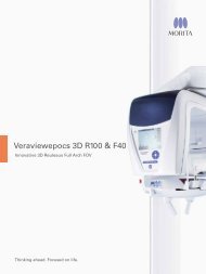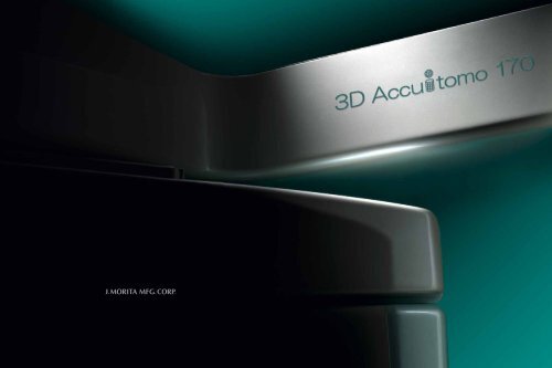
Thinking ahead Focused on life
zoom reconstruction - METCO Dental
zoom reconstruction - METCO Dental
- No tags were found...

You also want an ePaper? Increase the reach of your titles
YUMPU automatically turns print PDFs into web optimized ePapers that Google loves.
<str<strong>on</strong>g>Thinking</str<strong>on</strong>g> <str<strong>on</strong>g>ahead</str<strong>on</strong>g>.<str<strong>on</strong>g>Focused</str<strong>on</strong>g> <strong>on</strong> <strong>life</strong>.
THE NEW WORLD OF THE 80 µm VOXELSUPER RESOLUTION: 80 µm VOXELLow Patient Effective DoseHigh Quality Images withLow X-Ray Radiati<strong>on</strong>Four Imaging ModesA mode to serve every purpose. High Resoluti<strong>on</strong> Modeand High Fidelity Mode can be used for even higher qualityimages. High Speed Mode reduces moti<strong>on</strong> artifacts. UseStandard Mode for both limited and broad fields of view.Nine Sizes for Field of View (FOV)Choose from nine sizes for the FOV with diameters rangingfrom 170 mm to 40 mm to minimize X-ray dosage.Five Resoluti<strong>on</strong> LevelsSelect the voxel size 80 µm, 125 µm, 160 µm, 200 µm,or 250 µm, that best suits your diagnostic needs.Zoom Rec<strong>on</strong>structi<strong>on</strong>Use the original exposure data to zoom in <strong>on</strong> criticalareas using voxel sizes as small as 80 µm.CompactSpace-saving dimensi<strong>on</strong>s:W 1,620 mm x D 1,250 mm (63-3/4” x 49-1/4”)Recommended minimum room size:W 2,000 mm x D 1,800 mm (6-3/4’ x 6’)DICOM CompatibleViewing SoftwareWith these native Morita software packages, you canview and manipulate the 3D-CT image data even <strong>on</strong> acomputer that does not have i-Dixel software.
80 µm SUPER HIGH RESOLUTIONFOUR IMAGING MODESSELECT A REGION OF INTEREST SUCH AS THE TEMPORALBONE, PARANASAL SINUS, JAWBONE OR INDIVIDUAL TEETHAND OBSERVE IT WITH 80 µm VOXEL RESOLUTION FORGREATER DETAIL.HIGH RESOLUTION MODE ANDHIGH FIDELITY MODE CAN BE USEDfor even higher quality images. High speed modereduces moti<strong>on</strong> artifacts. Use Standard Mode for bothlimited and broad fields of view.Five Resoluti<strong>on</strong> LevelsSelect voxel size, 80 µm, 125 µm, 160 µm, 200 µm, or 250 µm,that best suits your diagnostic needs.*Zoom Rec<strong>on</strong>structi<strong>on</strong>Use the original exposure data to zoom in <strong>on</strong> critical areas usinga voxel size as small as 80 µmFor a higher resoluti<strong>on</strong> image, a specified area can be recalculatedand rec<strong>on</strong>structed using a smaller voxel size.*Depending <strong>on</strong> the size of the Field of View, some voxel sizes may not be possible.High Resoluti<strong>on</strong> Mode (Hi-Res)This is the highest resoluti<strong>on</strong>. Exposures are made at <strong>on</strong>e-fourth the sizeof the detector pixels for the greatest spatial resoluti<strong>on</strong>. Ideal for observati<strong>on</strong>of delicate b<strong>on</strong>e structures such as the ossicular chain.High Fidelity Mode (Hi-Fi)This mode has high data density data to make clearer and sharper images.This is especially good for performing zoom rec<strong>on</strong>structi<strong>on</strong>s.High Speed Mode (Hi-Speed)Full scan: 10.5 sec. Half scan: 5.4 sec.Reduces moti<strong>on</strong> artifacts. Good for children or others with difficulty remaining moti<strong>on</strong>less.Standard Mode (Std)Suitable for limited and wide views of temporal b<strong>on</strong>e, paranasal sinus, maxilla andmandible, individual teeth etc.ø 170 × H 120 mm, Voxel size: 250 µm ø 50 × H 50 mm, Voxel size: 80 µm Std Mode: ø 60 mmHi-Res Mode: ø 60 mmHi-Fi Mode: ø 60 Zoom rec<strong>on</strong>structi<strong>on</strong>Hi-Fi Mode: ø 170 × 120 mm
HIGH QUALITY 3D-CT IMAGESWITH LOW X-RAY RADIATIONCsI scintillatorUSING A HIGH-SENSITIVITY,HIGH-RESOLUTION FLAT PANEL DETECTOR,high quality and extremely detailed images of themany regi<strong>on</strong>s of the head and neck such as thetemporal b<strong>on</strong>e, paranasal sinuses, eye sockets,mandible, and cranial base can be obtained for awide range of multi-purpose diagnostic scanning.Pixela-Si (amorphoussilic<strong>on</strong>) arrayDigital SignalSpatial Resoluti<strong>on</strong>*MTF: Modulati<strong>on</strong> Transfer Functi<strong>on</strong>MTF [%]1009080706050403020Tube Voltage: 60 kVTube Current: 1.0 mAMTF at 2 lp/mm = 13.2 %1000 1 2 3 4This functi<strong>on</strong> is based <strong>on</strong> data from a typical product.Spatial frequency [lp/mm]Flat Panel Detector (FPD)FPD c<strong>on</strong>versi<strong>on</strong> of X-ray exposure into a digitalsignal results in a dramatic improvement inimage quality and a reducti<strong>on</strong> in X-ray dosage.The FPD is not affected by magnetic fields andhas superb sensitivity and resoluti<strong>on</strong> to producesuperior 3D-CT images with a minimum ofdistorti<strong>on</strong> and a wide dynamic range expressedwith a rich distributi<strong>on</strong> of the gray scale.X-rays are c<strong>on</strong>verted into visible light by thedirectly deposited Csl scintillator and then thelight is c<strong>on</strong>verted into an electrical signal by aphoto diode. The FPD is quite thin and has a l<strong>on</strong>gworking <strong>life</strong>.High Resoluti<strong>on</strong>Detailed images have a resoluti<strong>on</strong> of at least2.0 lp/mm (MTF 10%) with a voxel size of 80 µm.Highly detailed imagingMinimal Distorti<strong>on</strong>The flatness of the detector minimizes distorti<strong>on</strong>.This eliminates the necessity of making correcti<strong>on</strong>sfor distorti<strong>on</strong> before rec<strong>on</strong>structing imagesas is the case for analog systems.Wide Dynamic Range **The Flat Panel Detector (FPD) has a wide dynamicrange of 14 bit data (64 times 8 bit data).This produces a richer and truer gray scale.* Spatial resoluti<strong>on</strong> refers to how distinct an image appears the smallerit becomes; it measures the fineness of an image. Spatial frequency isthe unit of measurement of line pairs per distance (mm). As the mapscale decreases, the patterns of c<strong>on</strong>trast become harder to see. This isknown as MTF (Modulati<strong>on</strong> Transfer Functi<strong>on</strong>). It represents the numberof line pairs per 1 mm that can be distinguished based <strong>on</strong> c<strong>on</strong>trast.It is said that humans can <strong>on</strong>ly differentiate about 10%.** Dynamic range: Numerical values express the reproducibility of thesignal and the ratio of the largest and smallest input values in dBs.The dynamic range of the digital signal is also sometimes expressedin bits. The highest signal level is taken to be the level remaining aftersubtracting the noise level. The value of the dynamic range indicateshow weak of a signal can be reproduced – or, in other words, how highthe c<strong>on</strong>trast resoluti<strong>on</strong> will actually turn out to be.
i-Dixel DIAGNOSTIC SOFTWAREi-Dixel DIAGNOSTIC SOFTWARE can be used as a database to archivea wide variety of image informati<strong>on</strong>. Its multiple image processingfuncti<strong>on</strong>s can easily access and manipulate many types of informati<strong>on</strong>for 2D and 3D images.Volume RenderingVolume rendering of CT data produces threedimensi<strong>on</strong>al images.Select the area of interest and adjust thec<strong>on</strong>trols for the histogram to create a detailedimage of very fine stractures.Real Time Re-SliceSlices and volume rendered images can belinked and easily manipulated in real time.Curved MPR (cMPR)This way of image processing allows you toobserve an orthog<strong>on</strong>al representati<strong>on</strong> of thedental arch or any arbitrary curve.Report CommentsIt is easy to enter comments for any image.These comments can be printed with a c<strong>on</strong>venti<strong>on</strong>alWindows printer or a DICOM printer.Other Key Features• XYZ view windows• Re-slice• Zoom• Rotate• Histogram• Edge Enhancement• Distance and AngleMeasurement• Negative Image• Mirror image• Slice DistanceMeasurement• Surface Rendering• DICOM 3.0 Compatible• Brightness C<strong>on</strong>versi<strong>on</strong>• Spatial Frequency Filter• Patient Orientati<strong>on</strong> Display• Density Measurement• Implant Database• Nerv Chanel Marking• 3D Zoom• VDDS
SHARING IMAGE DATAINSTALLING I-DIXEL SOFTWARE<strong>on</strong> all intra-clinic computers enables sharing of image data <strong>on</strong>each linked client computer. Observati<strong>on</strong> of images <strong>on</strong> n<strong>on</strong>-networkcomputers can be achieved with the One Data Viewer, and theOne Volume Viewer without installing i-Dixel.3D Accuitomo 170i-Dixel software i-Dixel software i-Dixel softwareIntra-clinic networkOut of network computerIn external clinic networks without 3D Accuitomo 170,3D-CT images can be viewed <strong>on</strong> a PC with both ofthe following methods:i-Dixel Viewer SoftwareOne Data ViewerOne Volume ViewerOne Data Viewer &One Volume Viewer SoftwareThese unique Morita applicati<strong>on</strong>s letyou view three dimenti<strong>on</strong>al images andvolume rendered images even if thecomputer does not have i-Dixel softwareinstalled.CT data can be exported from the i-Dixelapplicati<strong>on</strong> and later stored <strong>on</strong> a DVD.This DVD can then be used <strong>on</strong> a computeroutside the clinic to view CT images,volume rendered images and patientinformati<strong>on</strong>.i-Dixel c<strong>on</strong>forms to the followingDICOM standards:1. Modality worklist managementservice class (opti<strong>on</strong>al)2. Storage service class3. Modality performed procedure stepservice class4. Print management service classOne Volume ViewerAdditi<strong>on</strong>al functi<strong>on</strong>s include zoom,black and white reverse, brightness,and c<strong>on</strong>trast adjustment as well asopti<strong>on</strong>al length and angle measurementcapabilities.
SIMPLE, ACCURATE POSITIONINGTHE SCOUT POSITIONING SYSTEM IS EASY AND ACCURATE.USE THE TRIPLE BEAM POSITIONING SYSTEM FOR EVENGREATER PRECISION.X CursorZ CursorY CursorTwo-Directi<strong>on</strong> ScoutThe regi<strong>on</strong> of interest can be easily targeted by making images from two directi<strong>on</strong>s.Then you can simply click <strong>on</strong> the images to specify the center of the regi<strong>on</strong> of interest.This informati<strong>on</strong> is transmitted to the X-ray unit, and the chair automatically moves intopositi<strong>on</strong>.Easy High Precisi<strong>on</strong>The regi<strong>on</strong> of interest can be easily targeted using the three positi<strong>on</strong>ing laser beams.The patient’s head is safely and securely stabilized by the chinrest and headrest.* The Scout exposure (80 kV and 2.0 mA) will increase the total X-ray dosage of a Standard Mode CT exposure(90 kV and 5.0 mA) by about 2%.3D-CT imageRegi<strong>on</strong> of interest iswell centered.
LIMITED CT IMAGE AREA FOR REDUCED X-RAY DOSAGELIMIT THE X-RAY DOSAGEUse scout to accurately determine the minimal regi<strong>on</strong>of interest before exposing the patient to the highterdosage CT scanTemporal B<strong>on</strong>e: ø 60 x 60 mm; Voxel size: 125 µmIMAGING AREA: Diameter x Height (mm)ø 170 x 120 mm ø 170 x 50 mmø 140 x 100 mm ø 140 x 50 mmø 100 x 100 mm ø 100 x 50 mmø 80 x 80 mmø 60 x 60 mmø 40 x 40 mmScout ImageParanasal sinuses: ø 170 x 120 mm; Voxel size: 250 µm
ZOOM RECONSTRUCTIONWITH 80 µm VOXEL RESOLUTIONSelect a regi<strong>on</strong> of interest suchas the temporal b<strong>on</strong>e or paranasalsinus and zoom in with 80 µm voxelresoluti<strong>on</strong> for a more detailedobservati<strong>on</strong>.Volume rendering produces adetailed 3D view of internal structures.Auditory ossicular chainAuditory ossicular chain zoom rec<strong>on</strong>structi<strong>on</strong>Ethmoid sinus zoom rec<strong>on</strong>structi<strong>on</strong>Lamina Cribrosa
HIGH RESOLUTION EVEN FOR WIDE AREASTECHNICAL SPECIFICATIONSHIGH RESOLUTION IMAGESwith a size of 170 mm (diameter) x 120 mm (height) enablescomprehensive examinati<strong>on</strong> and visualizati<strong>on</strong> of the entirefacial and maxillofacial and mandibular regi<strong>on</strong>s.Dimensi<strong>on</strong>sC<strong>on</strong>trol BoxOutlet of computer cable and operati<strong>on</strong> cableOutlet of power supplyOutlet of operati<strong>on</strong> cableOutlet of computer cableunit: mmX-ray roomInternal size: W x D x H2,000 mm x 1,800 mm x 2,400 mm(6-9/16’ x 5-7/8’ x 7’)Dedicated PCsSpecificati<strong>on</strong>sTrade NameModelTypeInput VoltagePower C<strong>on</strong>sumpti<strong>on</strong>X-ray HeadTube VoltageTube CurrentFocal Spot SizeExposure Time(360° / 180°)Field of View(Diameter × Height)3D AccuitomoXYZ Slice View TomographMCT-1EX1/2 F17100/110/120VAC220/230/240VACmax. 2.0 kVA60 - 90 kV1-10 mA (Max. 8 mA: Hi-Fi, Hi-Res Modes)0.5 mmStd Mode: 17.5 / 9.0 sec.Hi-Fi Mode: 30.8 / 15.8 sec.Hi-Res Mode: 30.8 / 15.8 sec.Hi-Speed Mode: 10.5 / 5.4 sec.ø 170 x 120 mm ø 170 x 50 mmø 140 x 100 mm ø 140 x 50 mmø 100 x 100 mm ø 100 x 50 mmø 80 x 80 mmø 60 x 60 mmø 40 x 40 mmVoxel Size 80 µm, 125 µm, 160 µm, 200 µm, 250 µmOuter Dimensi<strong>on</strong>sMain Unit 1,620 mm × 1,250 mm × 2,080 mm(W x D x H) (63-3/4” x 49-1/4” x 82”)C<strong>on</strong>trol Box 96 mm × 40 mm × 115 mm(W x D x H) (3-3/4” x 1-5/8” x 4-1/2”)WeightApprox. 400 kg. (882 lbs)Images provided by Fukushima Medical UniversityTechnical Assistance: NUBIC (Nih<strong>on</strong> University Business, Research,and Intellectual Property Center)Collaborative Development: J. Morita Corp. & Nih<strong>on</strong> University* X-ray protecti<strong>on</strong> should be provided for the patient when X-rays are emitted.* Design and specificati<strong>on</strong>s are subject to change without notificati<strong>on</strong>.ø 170 x H 120 mm, Voxel size: 250 µm (90 kV, 6 mA)Developed and Manufactured byJ. MORITA Mfg. Corp.680 Higashihama Minami-cho, Fushimi-ku, Kyoto,612-8533 JapanT +81. 75. 611 2141, F +81. 75. 622 4595www.morita.comJ. Morita Corporati<strong>on</strong>33-18, 3-Chome, Tarumi-cho Suita City, Osaka, 564-8650 JapanT +81. 6. 6380 1521, F +81. 6. 6380 0585, www.morita.com/asiaJ. Morita USA, inc.9 Mas<strong>on</strong> lrvine, CA 92618 U.S.A.T +1. 949. 581 9600, F +1. 949. 465 1095, www.morita.com/usaJ. Morita Europe GmbHJustus-v<strong>on</strong>-Liebig-Str. 27a, 63128 Dietzenbach, GermanyT +49. 6074. 836 0, F +49. 6074. 836 299, www.morita.com/europeSiamdent Co., Ltd.71/10 Bangpak<strong>on</strong>g Industrial Park 1, Bangna-Trad KM 52,Bangpak<strong>on</strong>g, Chachuengsao 24130, ThailandT +66. 3857. 3042, F +66. 3857. 3043, www.siamdent.comSubject to technical changes and errors. Printed in Germany, JMMC EN PUB 43046 0213 *3.