附加檔案下載 - 國立臺灣大學獸醫專業學院
附加檔案下載 - 國立臺灣大學獸醫專業學院
附加檔案下載 - 國立臺灣大學獸醫專業學院
- No tags were found...

Create successful ePaper yourself
Turn your PDF publications into a flip-book with our unique Google optimized e-Paper software.
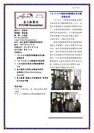
各 位 比 較 病 理 學 會 會 員 大 家 好 :<br />
中 華 民 國 比 較 病 理 學 會 第 45 次 比 較 病 理 學 研 討 會 將 於 98 年 3 月 13 日 ( 五 ) 及<br />
14 日 ( 六 ), 與 國 立 臺 灣 大 學 獸 醫 專 業 學 院 及 動 物 癌 症 醫 學 研 究 中 心 聯 合 舉 辦 「 比<br />
較 病 理 學 暨 動 物 癌 症 聯 合 學 術 研 討 會 」, 敬 請 各 單 位 惠 予 提 供 病 理 診 斷 病 例 ( 特<br />
別 是 典 型 、 新 興 與 重 大 法 定 疾 病 之 病 例 ), 並 請 各 會 員 踴 躍 報 名 參 加 ( 詳 情 如 附<br />
件 )。<br />
重 要 日 期 :<br />
2 月 16 日 ( 一 ) 以 前 :<br />
1) 請 欲 提 供 病 例 之 會 員 將 報 名 表 (Registration Form) 以 及 相 關 病 例 文 件 (Case<br />
History & Case Result sheets) 以 電 子 郵 件 寄 回 shsiao1@ntu.edu.tw。<br />
並 請 將 30 片 組 織 切 片 寄 達 :<br />
蕭 世 烜 助 理 教 授<br />
國 立 臺 灣 大 學 獸 醫 專 業 學 院<br />
10617 台 北 市 大 安 區 羅 斯 福 路 四 段 一 號<br />
2) 請 其 他 與 會 人 員 將 報 名 表 寄 回 shsiao1@ntu.edu.tw<br />
3 月 14 日 ( 六 ): 開 會 日 期<br />
祝<br />
研 安<br />
秘 書 長 : 蕭 世 烜 助 理 教 授<br />
理 事 長 : 劉 振 軒 教 授<br />
國 立 臺 灣 大 學 獸 醫 專 業 學 院<br />
地 址 :10617 台 北 市 大 安 區 羅 斯 福 路 四 段 一 號<br />
電 話 : 02-33663858<br />
傳 真 : 02-23682423<br />
電 子 郵 件 : shsiao1@ntu.edu.tw<br />
中 華 民 國 97 年 2 月 2 日<br />
1
Call for Papers and Registration<br />
45th Meetings of the Chinese Society of Comparative Pathology (CSCP) hosted by the School of<br />
Veterinary Medicine, National Taiwan University<br />
You are invited to submit a case for presentation at the 45th CSCP Meeting jointed with the<br />
2009 Animal Cancer Symposium. This meeting is a forum for continuing education and<br />
professional development attracting pathologists from hospitals, academia, government,<br />
industry and diagnostic laboratories including pathology residents and graduate students.<br />
Each presenter will submit 30 microscope slides along with single copies of a one-page case<br />
history sheet and a one-page case result sheet. Brief (15 minute) Powerpoint presentations of<br />
an interesting diagnostic pathology case (emerging diseases, classic diseases, reportable<br />
diseases, cases with uncertain diagnosis or new information regarding pathophysiology) will be<br />
given at the meeting. Prior to the meeting, all presenters will receive a set of slides for<br />
review along with the case histories. For diagnostic cases with limited materials, such as<br />
cytology or gross cases, digital photos may be substituted for the microscope slides.<br />
Meeting Date: March 14, 2009 (Saturday)<br />
Host: School of Veterinary Medicine, National Taiwan University ( 國 立 臺 灣 大 學 獸 醫 專 業 學 院 )<br />
Location: Room B01, Veterinary Medicine 3rd Building ( 獸 醫 三 館 ), No. 1, Sec. 4, Roosevelt<br />
Road, Taipei, 10617 Taiwan<br />
Contact:<br />
General Secretary: Dr. S.H. Vincent Hsiao ( 蕭 世 烜 秘 書 長 )<br />
Phone: 02-33663858<br />
Fax: 02-23682423<br />
Email: shsiao1@ntu.edu.tw<br />
Registration:<br />
Presenters: Deadline is February 16, 2009. Complete the registration form attached and<br />
email to shsiao1@ntu.edu.tw. Please include your presentation title or diagnosis at the<br />
bottom of the form.<br />
Attendees: Deadline is February 16, 2009, if not making a case presentation. Complete the<br />
registration form and return by email to Dr. Hsiao.<br />
Case Materials: Submission deadline of case materials is February 16, 2009, for 30 microscope<br />
slides, and single copies of a one-page case history sheet and a one-page case result sheet.<br />
Format for case history and case results can be found below.<br />
2
Please submit microscope slides to:<br />
蕭 世 烜 助 理 教 授<br />
國 立 臺 灣 大 學 獸 醫 專 業 學 院<br />
10617 台 北 市 大 安 區 羅 斯 福 路 四 段 1 號<br />
Please submit case history and result sheets to:<br />
e-mail: shsiao1@ntu.edu.tw<br />
3
Registration Form<br />
45th Meeting of the Chinese Society of Comparative Pathology<br />
March 14, 2009 (Saturday)<br />
Full Name:<br />
Title:<br />
Institution:<br />
Address:<br />
Telephone:<br />
E-mail:<br />
For presenters:<br />
Presentation title or diagnosis:<br />
Signalment:<br />
P.S. Please submit your registration form by email to shsiao1@ntu.edu.tw. Registration<br />
deadline is February 16, 2009 for both presenters and attendees.<br />
4
Case Number: 15 Chinese Society of Comparative Pathology, July 2008<br />
Camilo Bulla and Mike Scott<br />
Michigan State University<br />
Case History<br />
Signalment: 5.5-year-old mixed breed, castrated, dog<br />
Clinical History: The dog was presented to the MSU VTH because of a 1 wk history of fever of<br />
unknown origin (104-106 ∘F) that was not responsive to antibiotics. Physical exam revealed<br />
pyrexia (103.6 o F), a grade II/VI systolic heart murmur, and an enlarged left popliteal lymph<br />
node.<br />
Clinical pathology:<br />
EXAMPLE<br />
CBC Abnormalities<br />
<br />
<br />
Neutropenia (1,270/μL [ref: 3,800-7,800]) with a left shift (bands:1,060/μL) and<br />
moderate toxic change<br />
Nonregenerative normocytic normochromic anemia (PCV = 26% [ref: 41-55]) with no<br />
evidence of polychromasia<br />
2+ eccentrocytosis and pyknocytosis<br />
Possible thrombocytopenia; platelets density was low; microclots in the smear's<br />
feathered edge contained few platelets; there was moderate thrombocytopenia 2 days<br />
later<br />
Clinical Chemistry and Urinalysis Abnormalities<br />
Hypocalcemia (8.5 mg/dL [ref: 9.3-10.8]) with hypoalbuminemia (2.1 g/dL [ref: 2.8-4.0])<br />
and mild hyperglobulinemia (4.3 g/dL [ref: 2.2-4.1])<br />
4x increase in alkaline phosphatase (480 U/L [ref: 14-104])<br />
Other Tests<br />
Bone marrow aspirate and core biopsy were collected to investigate the cytopenias.<br />
Sections of the core are provided.<br />
Serology: mild positive for Borrelia (1:160) and follow-up Western blot analysis<br />
suggested vaccination-induced; mild positive for Bartonella; negative for B. canis, E.<br />
canis, and R. rickettsiae; negative for Leptospira.<br />
Bartonella PCR (after antimicrobial therapy): negative<br />
Blood cultures (3 samples over 6 hr): negative<br />
Left popliteal lymph node aerobic and anaerobic cultures: negative<br />
Echocardiogram: mild mitral and tricuspid valve insufficiency<br />
Fluorescent antinuclear antibody assay (FANA): negative<br />
5
Case Number: 15 Chinese Society of Comparative Pathology, July 2008<br />
Camilo Bulla and Mike Scott<br />
Michigan State University<br />
Case Results<br />
Histopathologic description: A moderate length of trabecular bone was examined. The<br />
hematopoietic cellularity averaged about 70%. Hemosiderin was present in moderate to<br />
abundant amounts. Megakaryocytes were in low-normal to adequate numbers, and the series<br />
appeared left shifted with a few mature megakaryocytes appearing to have single large nuclei.<br />
The myeloid to erythroid ratio was markedly increased with a predominance of segmented<br />
neutrophils. Erythroid precursors were present but appeared decreased. There was a mild<br />
increase in normal appearing plasma cells.<br />
Morphologic diagnosis: Marked myeloid hyperplasia; erythroid hypoplasia<br />
Bone marrow aspirate: Cytologic findings were similar to the core findings. Additionally, no<br />
dysplasia was noted in hemic cells, and there were no other significant cell populations.<br />
EXAMPLE<br />
Erythroid maturation was orderly, but there were few polychromatophils. Neutrophils had mild<br />
to moderate toxic change.<br />
Clinical progression: The animal was treated with ampicillin and enrofloxacin from the day of<br />
admission, and doxycycline was added 2 days later for possible bartonellosis. This was<br />
continued for 3 wk during which the neutrophil concentrations varied from about 2,000/μL to<br />
4,500 /μL [ref: 3,800-7,800], and the dog remained bright and active. At that time, bone<br />
marrow aspirate and core again showed marked myeloid hyperplasia concurrent with<br />
neutropenia. Cytologically, a mild increase in macrophages was noted, and many contained<br />
phagocytized material including nuclear remnants; 1 cell contained an intact neutrophil.<br />
Immunosuppressive therapy was instituted and the neutrophil concentration rose quickly:<br />
14,700/µL on day 4, and 30,600/µL on day 11.<br />
Comments: It is very uncommon to find a marked myeloid hyperplasia concurrent with<br />
persistent neutropenia. Given the findings in this case, the major differentials were<br />
myelodysplasia and primary and secondary immune-mediated neutropenia. In this case,<br />
diagnostic testing and therapeutic trials with antimicrobials failed to identify an infectious<br />
process. Immunosuppressive therapy rapidly resolved the neutropenia after documenting<br />
neutropenia for 3 wk. Therefore, this was a steroid responsive neutropenia, likely<br />
immune-mediated. Immune-mediated neutropenia is an uncommon disorder that is usually<br />
diagnosed by excluding other causes of neutropenia and finding a positive response to<br />
immunosuppressive therapy. Bone marrow examination is expected to reveal maturation arrest<br />
6
or ineffective neutropoiesis, and the granulocyte series may range from hypoplastic to<br />
hyperplastic depending on which maturation stage is targeted. Unfortunately, there is not a<br />
well established commercial test to detect anti-neutrophil antibodies.<br />
References:<br />
1. Perkins MC, Canfield P, Churcher RK, Malik R. Immune-mediated neutropenia suspected in<br />
five dogs. Aust Vet J. 2004. 82, 52-7.<br />
2. McManus PM, Litwin C, Barber L. Immune-mediated neutropenia in 2 dogs. J Vet Intern<br />
Med. 1999. 13, 372-4.<br />
EXAMPLE<br />
7
Case Number: 26 Chinese Society of Comparative Pathology, July 2008<br />
LB Borst and CA Lichtensteiger<br />
Veterinary Diagnostic Laboratory, University of Illinois<br />
Case History<br />
Signalment: 6-day-old American Quarter Horse colt<br />
Clinical history: The foal was born 16 days premature and weak but was able to suckle and<br />
received adequate colostrum (serum IgG >800 mg/dl). At 3 days of age the foal was<br />
observed on three separate occasions to seizure. The foal's blood glucose was measured at<br />
9 mg/dl. The foal was given intravenous dextrose, fed milk through a nasogastric tube and<br />
immediately referred to the University of Illinois Large Animal Clinic. On presentation the<br />
foal was recumbent but responsive and the foal's blood glucose was measured at 30 mg/dl.<br />
Intravenous (IV) dextrose and fluids and feeding via the nasogastric tube were continued.<br />
Antimicrobial therapy consisting of amikacin and ampicillin was started and serum chemistry,<br />
complete blood count, joint taps and blood cultures were performed. The foal improved<br />
slightly over the next 2 days and the foal's blood glucose stabilized with IV supplementation.<br />
EXAMPLE<br />
On the third day of treatment the foal rose to suckle but collapsed suddenly with cyanotic<br />
mucous membranes and irregular and faint heart sounds.<br />
unsuccessful.<br />
Resuscitation attempts were<br />
Clinical pathology: The CBC reveled a normal white blood cell count (7.13x10 3 ; reference range:<br />
5.50 to 12.00x10 3 cells/ul) with a moderate lymphopenia (0.427x10 3 ; 1.50 to 5.00x10 3 cells/ul).<br />
Scattered neutrophils exhibited mild (1+) toxic changes (cytoplasmic vacuoles and Dohle<br />
bodies. Fibrinogen was moderately elevated at 600 mg/dl (100 to 400 mg/dl). Serum<br />
chemistry revealed a slightly low blood glucose (66; 71 to 100 mg/dl) moderate to marked<br />
elevations in all liver enzymes including alkaline phosphatase (1333; 45 to 239 U/L), AST<br />
(1094;160 to 300 U/L), SDH (7.8; 0 to 7.0) and GGT (23; 4 to 20 U/L). Total serum bilirubin<br />
was also elevated (6.5; 0.60 to 2.60 mg/dl). Serum creatine phosphokinase was also<br />
moderately elevated (1352; 120 to 350 U/L). Cytologic examination of the joint fluid was<br />
unremarkable and the blood cultures were negative.<br />
Gross lesions: Scattered randomly throughout all lung fields were approximately 25 firm<br />
raised spherical approximately 0.25 to 2.5 cm in diameter nodules that had a tan center<br />
surrounded by a bright red rim. The foramen ovale and ductus arteriosis were probe patent.<br />
No additional gross abnormalities were observed in any organ system.<br />
8
Case Number: 26 Chinese Society of Comparative Pathology, July 2008<br />
LB Borst and CA Lichtensteiger<br />
Veterinary Diagnostic Laboratory, University of Illinois<br />
Case Results<br />
Histopathologic description: In most cardiomyocytes and particularly prominent in the Purkinje<br />
fibers were variably large intrasarcoplasmic amphophilic to basophilic vaguely laminated waxy<br />
inclusions. The inclusions stained bright magenta with periodic acid Schiff (PAS). Positive<br />
staining did not fade when treated with diastase (diastase resistant carbohydrate). Scattered<br />
myocytes and Purkinje cells are degenerative with shrunken or fragmented and disorganized<br />
sarcoplasm that lacks cross striations.<br />
Morphologic diagnosis: Heart: Cardiomyocytes and Purkinje fibers: Amphophilic to basophilic<br />
intrasarcoplasmic inclusions, diffuse, numerous, with mild degeneration, quarterhorse, equine<br />
Etiology: Glycogen storage disease type IV due to a point mutation in the GBE1 gene coding for<br />
glycogen branching enzyme.<br />
EXAMPLE<br />
Comments: An autosomal recessive fatal fetal and neonatal glycogen storage disease (GSD IV)<br />
due to a defect in glycogen branching enzyme (GBE) has recently been identified in American<br />
Quarter Horses 1,2,3,4 . GBE is responsible for the transfer of existing blocks of glucosyl residues<br />
from a growing chain to another chain to produce α-1,6 linkages. Affected foals are<br />
homozygous for a Y34X nonsense mutation in exon 1 of the GBE1 gene which knocks out<br />
enzyme activity 4 . Decreased GBE activity results in poorly branched glycogen which<br />
accumulates in the cytoplasm of heart, liver and nervous tissue disrupting cellular functions.<br />
While clinical signs in affected foals overlap with common neonatal diseases like septicemia or<br />
neonatal asphyxia syndrome, the distinguishing feature of cytoplasmic accumulation of<br />
diastase resistant polysaccharide is consistent 4 . In this case abnormal carbohydrate<br />
accumulations were visualized in the neurons of the central nervous system as well as in the<br />
heart. Genomic DNA was isolated from formalin fixed heart and liver tissue and a portion of<br />
the GBE1 gene flanking the mutation site was amplified and sequenced. The foal was found<br />
to be homozygous for the Y34X mutation confirming the diagnosis of GSD IV in this case.<br />
The changes in the lungs were likely of little clinical importance and histologically were<br />
discrete granulomas. No fungal or bacterial organisms were observed on oversight or silver<br />
stained sections and bacterial cultures were negative.<br />
References:<br />
1. Render JA, Common RS, Kennedy FA, Jones MZ, Fyfe JC (1999) Amylopectinosis in fetal<br />
and neonatal quarter horses. Vet Pathol 36, 157–160<br />
9
2. Sponseller BT, Valberg SJ, Ward T, Williams AJ, Mickelson JR (2003) Muscular weakness<br />
and recumbency in a Quarter Horse colt due to glycogen branching enzyme deficiency.<br />
Equine Vet Ed 15, 182–188<br />
3. Valberg SJ, Ward TL, Rush B, Kinde H, Hiraragi H, et al. (2001) Glycogen branching enzyme<br />
deficiency in Quarter Horse foals. J Vet Intern Med 15, 572–580<br />
4. Ward TL, Valberg SJ, Adelson DL, Abbey CA, Binns MM, Mickelson JR (2004) Glycogen<br />
branching enzyme (GBE1) causing equine glycogen storage disease IV. Mammalian<br />
Genome 15. 570-577.<br />
EXAMPLE<br />
10