中 華 民 國 比 較 病 理 學 會
第54屆比較病理研討會 - 國立臺灣大學獸醫專業學院
第54屆比較病理研討會 - 國立臺灣大學獸醫專業學院
- No tags were found...

You also want an ePaper? Increase the reach of your titles
YUMPU automatically turns print PDFs into web optimized ePapers that Google loves.
<strong>中</strong> <strong>華</strong> <strong>民</strong> <strong>國</strong> <strong>比</strong> <strong>較</strong> <strong>病</strong> <strong>理</strong> <strong>學</strong> <strong>會</strong><br />
Chinese Society of Comparative Pathology<br />
第 54 次 <strong>比</strong> <strong>較</strong> <strong>病</strong> <strong>理</strong> <strong>學</strong> 研 討 <strong>會</strong><br />
School of Veterinary Medicine, National Taiwan University and Taipei Zoo<br />
<strong>國</strong> 立 臺 灣 大 <strong>學</strong> 獸 醫 專 業 <strong>學</strong> 院 與 台 北 市 立 動 物 園<br />
March 10, 2012 ( <strong>中</strong> <strong>華</strong> <strong>民</strong> <strong>國</strong> 101 年 3 月 10 日 )<br />
Chinese Society of Comparative Pathology<br />
<strong>中</strong> <strong>華</strong> <strong>民</strong> <strong>國</strong> <strong>比</strong> <strong>較</strong> <strong>病</strong> <strong>理</strong> <strong>學</strong> <strong>會</strong>
<strong>會</strong> 員 資 料 更 新 服 務<br />
各 位 <strong>會</strong> 員 :<br />
您 好 ! 如 果 您 的 <strong>會</strong> 員 資 料 有 更 新 或 誤 刊 情 形 , 麻 煩 您 填 妥 表 格 後 寄 回 <strong>學</strong> <strong>會</strong> 秘 書 處 或<br />
電 話 連 絡 :<br />
<strong>中</strong> <strong>華</strong> <strong>民</strong> <strong>國</strong> <strong>比</strong> <strong>較</strong> <strong>病</strong> 理 <strong>學</strong> <strong>會</strong> 秘 書 處<br />
10617 臺 北 市 大 安 區 羅 斯 福 路 四 段 1 號<br />
<strong>國</strong> 立 臺 灣 大 <strong>學</strong> 獸 醫 系 三 館 106 室 鄭 謙 仁 秘 書 長 收<br />
Tel: (02) 33663868<br />
Fax: (02) 23621965<br />
e-mail address: crjeng@ntu.edu.tw<br />
------------------------------ <strong>中</strong> <strong>華</strong> <strong>民</strong> <strong>國</strong> <strong>比</strong> <strong>較</strong> <strong>病</strong> 理 <strong>學</strong> <strong>會</strong> ----------------------------<br />
<strong>會</strong> 員 資 料 更 改 卡<br />
姓 名 : <strong>會</strong> 員 類 別 : 一 般 <strong>會</strong> 員<br />
<strong>學</strong> 生 <strong>會</strong> 員<br />
贊 助 <strong>會</strong> 員<br />
最 高 <strong>學</strong> 歷 :<br />
服 務 單 位 : 職 稱 :<br />
永 久 地 址 :<br />
通 訊 地 址 :<br />
電 話 : 傳 真 :<br />
E-Mail Address:<br />
71
<strong>中</strong> <strong>華</strong> <strong>民</strong> <strong>國</strong> <strong>比</strong> <strong>較</strong> <strong>病</strong> 理 <strong>學</strong> <strong>會</strong><br />
誠 摯 邀 請 您 加 入<br />
入 <strong>會</strong> 辦 法<br />
一 、 本 <strong>會</strong> <strong>會</strong> 員 申 請 資 格 為 :<br />
( 一 ) 一 般 <strong>會</strong> 員 : 贊 同 本 <strong>會</strong> 宗 旨 , 年 滿 二 十 歲 , 具 有 <strong>國</strong> 內 外 大 專 院 校 ( 或 同 等 <strong>學</strong><br />
歷 ) 生 命 科 <strong>學</strong> 及 其 它 相 關 科 系 畢 業 資 格 或 高 職 畢 業 從 事 生 命 科<br />
<strong>學</strong> 相 關 工 作 滿 兩 年 者 。<br />
( 二 ) <strong>學</strong> 生 <strong>會</strong> 員 : 贊 同 本 <strong>會</strong> 宗 旨 , 在 <strong>國</strong> 內 、 外 大 專 院 校 生 命 科 <strong>學</strong> 或 其 他 相 關 科 系<br />
肄 業 者 ( 請 檢 附 <strong>學</strong> 生 身 份 證 明 )。<br />
( 三 ) 贊 助 <strong>會</strong> 員 : 贊 助 本 <strong>會</strong> 工 作 之 團 體 或 個 人 。<br />
( 四 ) 榮 譽 <strong>會</strong> 員 : 凡 對 <strong>比</strong> <strong>較</strong> <strong>病</strong> 理 <strong>學</strong> 術 或 <strong>會</strong> 務 之 推 廣 有 特 殊 貢 獻 , 經 理 事 <strong>會</strong> 提 名 並<br />
經 <strong>會</strong> 員 大 <strong>會</strong> 通 過 者 。<br />
二 、 <strong>會</strong> 員 :<br />
( 一 ) 入 <strong>會</strong> 費 : 一 般 <strong>會</strong> 員 新 台 幣 一 仟 元 , <strong>學</strong> 生 <strong>會</strong> 員 一 佰 元 , 贊 助 <strong>會</strong> 員 伍 仟 元 ,<br />
於 入 <strong>會</strong> 時 繳 納 。<br />
( 二 ) 常 年 <strong>會</strong> 費 : 一 般 <strong>會</strong> 員 新 台 幣 伍 佰 元 , <strong>學</strong> 生 <strong>會</strong> 員 一 佰 元 。<br />
【 註 : <strong>學</strong> 生 <strong>會</strong> 員 身 份 變 更 為 一 般 <strong>會</strong> 員 時 , 只 需 繳 交 一 般 <strong>會</strong> 員 之 常 年 <strong>會</strong> 費 】<br />
三 、 入 <strong>會</strong> 費 及 常 年 <strong>會</strong> 費 繳 交 方 式 : 以 銀 行 轉 帳 或 匯 款 (006 合 作 金 庫 銀 行 、 帳 號 :<br />
0190-717-052017、 戶 名 : <strong>中</strong> <strong>華</strong> <strong>民</strong> <strong>國</strong> <strong>比</strong> <strong>較</strong> <strong>病</strong> 理 <strong>學</strong> <strong>會</strong> ); 並 請 填 妥 入 <strong>會</strong> 申 請 表 連 同 銀<br />
行 轉 帳 交 易 明 細 表 或 匯 款 單 以 郵 寄 或 傳 真 方 式 寄 回 <strong>中</strong> <strong>華</strong> <strong>民</strong> <strong>國</strong> <strong>比</strong> <strong>較</strong> <strong>病</strong> 理 <strong>學</strong> <strong>會</strong> 秘 書 處<br />
收 。 地 址 :116 臺 北 市 羅 斯 福 路 四 段 一 號 <strong>國</strong> 立 臺 灣 大 <strong>學</strong> 獸 醫 專 業 <strong>學</strong> 院 三 館 106、<br />
電 話 :02-33663858、 傳 真 02-23682423。<br />
72
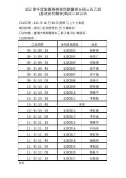
SCHEDULE<br />
54TH MEETING OF COMPARATIVE PATHOLOGY<br />
<strong>中</strong> <strong>華</strong> <strong>民</strong> <strong>國</strong> <strong>比</strong> <strong>較</strong> <strong>病</strong> <strong>理</strong> <strong>學</strong> <strong>會</strong> 第 54 次 <strong>比</strong> <strong>較</strong> <strong>病</strong> <strong>理</strong> <strong>學</strong> 研 討 <strong>會</strong><br />
Date: March 10, 2012 (Sat) 08:30~17:00 時 間 :101 年 3 月 10 日 ( 星 期 六 ) 08:30~17:00<br />
Location: Lecture Hall of Education Center, Taipei Zoo 地 點 : 台 北 市 立 動 物 園 行 政 大 樓 ( 教 育 <strong>中</strong> 心 ) 演 講 廳<br />
Address: No.30 Sec.2 Xinguang Rd., Taipei City 11656, Taiwan (R.O.C.) 地 址 :11656 臺 北 市 新 光 路 二 段 30 號<br />
Telephone: 02-33663868 電 話 :02-33663868<br />
Time( 時 間 ) Schedule( 議 程 ) Moderator( 主 持 )<br />
08:30~09:00 Registration ( 報 到 )<br />
09:00~09:10 Opening Ceremony ( 致 詞 )<br />
09:10~10:10 專 題 演 講<br />
Dr. Feng-Yee Chang ( 張 峰 義 疾 <strong>病</strong> 管 制 局 局 長 )<br />
新 興 傳 染 <strong>病</strong> : 從 人 畜 共 通 傳 染 <strong>病</strong> 談 起<br />
10:10~10:30 Coffee Break<br />
Chia-Wen Shih ( 施 洽 雯 醫 師 )<br />
10:30~11:00 Case 381<br />
Department of Pathology, Lotung Poh-Ai Hospital ( 羅 東 博 愛 醫 院 )<br />
L.F. Wang ( 王 玲 芳 獸 醫 師 )<br />
11:00~11:30 Case 382<br />
Department of veterinary pathology, NPUST<br />
( <strong>國</strong> 立 屏 東 科 技 大 <strong>學</strong> 獸 醫 教 <strong>學</strong> 醫 院 <strong>病</strong> 理 科 )<br />
Jing-Lan Liu ( 劉 淨 蘭 醫 師 )<br />
11:30~12:00 Case 383<br />
St. Martin De Porres Hospital ( 聖 馬 爾 定 醫 院 <strong>病</strong> 理 科 )<br />
Lunch, and Board Meeting<br />
12:00~13:30<br />
( <strong>中</strong> <strong>華</strong> <strong>民</strong> <strong>國</strong> <strong>比</strong> <strong>較</strong> <strong>病</strong> 理 <strong>學</strong> <strong>會</strong> <strong>會</strong> 員 大 <strong>會</strong> 暨 理 監 事 <strong>會</strong> 議 )<br />
S.J. Wu ( 吳 詩 柔 獸 醫 師 )<br />
13:30~14:00 Case 384 Department of Veterinary Medicine, National Chung Hsing<br />
University ( <strong>中</strong> 興 大 <strong>學</strong> 獸 醫 <strong>學</strong> 系 )<br />
Too-Yuan Tsai ( 蔡 斗 元 醫 師 )<br />
14:00~14:30 Case 385 Buddhist Tzu Chi General Hospital and University, Taiwan<br />
( 佛 教 慈 濟 綜 合 醫 院 暨 慈 濟 大 <strong>學</strong> <strong>病</strong> 理 科 )<br />
14:30~14:50 Coffee Break<br />
陳 憲 全 獸 醫 師<br />
14:50~15:20 Case 386<br />
Animal Technology Institute Taiwan ( 台 灣 動 物 科 技 研 究 所 )<br />
Mu-Tsung Tsai ( 蔡 睦 宗 獸 醫 師 )<br />
15:20~15:50 Case 387 Pingtung County Livestock Disease Control Center<br />
( 屏 東 縣 家 畜 疾 <strong>病</strong> 防 治 所 )<br />
Y.C. Jian ( 簡 耀 君 獸 醫 師 )<br />
Graduated Institute of Molecular and Comparative Pathology<br />
15:50~16:20 Case 388<br />
School of Veterinary Medicine, NTU<br />
( <strong>國</strong> 立 台 灣 大 <strong>學</strong> 分 子 暨 <strong>比</strong> <strong>較</strong> <strong>病</strong> 理 研 究 所 )<br />
16:20~17:00 General Discussion ( 綜 合 討 論 )<br />
Dr. C. W. Shih<br />
施 洽 雯 主 任<br />
Dr. F. J. Leu<br />
呂 福 江 主 任<br />
Dr. Y. H. Hsu<br />
許 永 祥 主 任<br />
Dr. C. H. Liu<br />
劉 振 軒 院 長
目<br />
錄<br />
一 、 Schedule( 議 程 表 )…………………………………………………. 1<br />
二 、 目 錄 ……………………………………………………………………. 2<br />
三 、 Case Signalment ……………………………………………………. 3<br />
四 、 Case Diagnosis………………………………………………………. 5<br />
Comparative Pathology Case 381…………………………………. 5<br />
Comparative Pathology Case 382………………………………… 9<br />
Comparative Pathology Case 383…………………………………. 12<br />
Comparative Pathology Case 384…………………………………. 16<br />
Comparative Pathology Case 385…………………………………. 23<br />
Comparative Pathology Case 386…………………………………. 27<br />
Comparative Pathology Case 387…………………………………. 32<br />
Comparative Pathology Case 388…………………………………. 36<br />
五 、 <strong>中</strong> <strong>華</strong> <strong>民</strong> <strong>國</strong> <strong>比</strong> <strong>較</strong> <strong>病</strong> 理 <strong>學</strong> <strong>會</strong> 章 程 …………………………………………. 41<br />
六 、 第 六 屆 理 監 事 名 單 簡 歷 冊 ……………………………………………. 46<br />
七 、 100 年 度 資 產 負 債 表 ………………………………………………… 47<br />
100 年 度 收 支 決 算 表 ………………………………………………… 48<br />
100 年 度 基 金 收 支 表 ………………………………………………… 49<br />
100 年 度 現 金 出 納 表 ………………………………………………… 50<br />
101 年 度 收 支 預 算 表 ………………………………………………….. 51<br />
八 、 數 位 組 織 切 片 資 料 庫 ………………………………………............... 52<br />
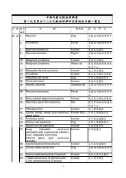
九 、 <strong>比</strong> <strong>較</strong> <strong>病</strong> 理 研 討 <strong>會</strong> <strong>病</strong> 例 分 類 一 覽 表 ……………………………………. 53<br />
十 、 <strong>會</strong> 員 資 料 更 新 服 務 ……………………………………………………. 71<br />
十 一 、 入 <strong>會</strong> 辦 法 ………………………………………………………………. 72<br />
2
CASE SIGNALMENT<br />
54TH MEETING OF COMPARATIVE PATHOLOGY<br />
March 10, 2012<br />
( <strong>中</strong> <strong>華</strong> <strong>民</strong> <strong>國</strong> <strong>比</strong> <strong>較</strong> <strong>病</strong> <strong>理</strong> <strong>學</strong> <strong>會</strong> 第 54 次 <strong>比</strong> <strong>較</strong> <strong>病</strong> <strong>理</strong> <strong>學</strong> 研 討 <strong>會</strong> )<br />
Case No. Presenter Institution Slide No. Signalment<br />
Case 381<br />
施 洽 雯<br />
Lo-Hsu Foundation, Inc., Lotung Poh-Ai<br />
Hospital ( 羅 東 博 愛 醫 院 )<br />
65-year-old woman.<br />
Department of veterinary pathology,<br />
Case 382<br />
王 玲 芳<br />
National Pingtung University of Science<br />
and Technology<br />
0T-10696<br />
Kidney tissues,<br />
slaughter pigs<br />
( <strong>國</strong> 立 屏 東 科 技 大 <strong>學</strong> 獸 醫 教 <strong>學</strong> 醫 院 <strong>病</strong> 理 科 )<br />
Case 383<br />
劉 淨 蘭<br />
St. Martin De Porres Hospital<br />
( 聖 馬 爾 定 醫 院 <strong>病</strong> 理 科 )<br />
S00-6602Y3<br />
48-year-old woman<br />
Case 384<br />
吳 詩 柔<br />
Department of Veterinary Medicine,<br />
National Chung Hsing University<br />
( <strong>中</strong> 興 大 <strong>學</strong> 獸 醫 <strong>學</strong> 系 )<br />
CW11-014C<br />
Leopard Cat, male,<br />
2-3 year-old<br />
Buddhist Tzu Chi General Hospital and<br />
Case 385<br />
蔡 斗 元<br />
University, Taiwan<br />
S2011-9609 57-year-old woman<br />
( 佛 教 慈 濟 綜 合 醫 院 暨 慈 濟 大 <strong>學</strong> <strong>病</strong> 理 科 )<br />
Case 386<br />
陳 憲 全<br />
Animal Technology Institute Taiwan<br />
( 台 灣 動 物 科 技 研 究 所 )<br />
S12-597b<br />
19-year-old<br />
Chinchila male cat<br />
Case 387<br />
蔡 睦 宗<br />
Pingtung County Livestock Disease<br />
Control Center ( 屏 東 縣 家 畜 疾 <strong>病</strong> 防 治 所 )<br />
Q100-297<br />
7-month-old,<br />
finishing pig, LYD<br />
type, Swine<br />
Graduated Institute of Molecular and<br />
Case 388<br />
簡 耀 君<br />
Comparative Pathology School of<br />
Veterinary Medicine, NTU<br />
NP-376F<br />
5-year-old, intact<br />
male Maltese<br />
( <strong>國</strong> 立 台 灣 大 <strong>學</strong> 分 子 暨 <strong>比</strong> <strong>較</strong> <strong>病</strong> 理 研 究 所 )<br />
3
CASE DIAGNOSIS<br />
54TH MEETING OF COMPARATIVE PATHOLOGY<br />
March 10, 2012<br />
( <strong>中</strong> <strong>華</strong> <strong>民</strong> <strong>國</strong> <strong>比</strong> <strong>較</strong> <strong>病</strong> <strong>理</strong> <strong>學</strong> <strong>會</strong> 第 54 次 <strong>比</strong> <strong>較</strong> <strong>病</strong> <strong>理</strong> <strong>學</strong> 研 討 <strong>會</strong> )<br />
Case No. Presenter Institution Slide No. Diagnosis<br />
Case 381<br />
施 洽 雯<br />
Lo-Hsu Foundation, Inc., Lotung<br />
Poh-Ai Hospital ( 羅 東 博 愛 醫 院 )<br />
Polyomavirus<br />
infection of urinary<br />
tract<br />
Department of veterinary pathology,<br />
Case 382<br />
王 玲 芳<br />
National Pingtung University of<br />
Science and Technology<br />
0T-10696 Leptospirosis<br />
( <strong>國</strong> 立 屏 東 科 技 大 <strong>學</strong> 獸 醫 教 <strong>學</strong> 醫 院 <strong>病</strong> 理 科 )<br />
Case 383<br />
劉 淨 蘭<br />
St. Martin De Porres Hospital<br />
( 聖 馬 爾 定 醫 院 <strong>病</strong> 理 科 )<br />
S00-6602Y3<br />
Langerhans cell<br />
histiocytosis<br />
Case 384<br />
吳 詩 柔<br />
Department of Veterinary Medicine,<br />
National Chung Hsing University<br />
( <strong>中</strong> 興 大 <strong>學</strong> 獸 醫 <strong>學</strong> 系 )<br />
CW11-014C<br />
Neisseria Infected<br />
Pneumonitis in a<br />
Housed Leopard<br />
Cat<br />
Buddhist Tzu Chi General Hospital and<br />
Mycobacteria avian<br />
Case 385<br />
蔡 斗 元<br />
University, Taiwan<br />
S2011-9609<br />
complex<br />
( 佛 教 慈 濟 綜 合 醫 院 暨 慈 濟 大 <strong>學</strong> <strong>病</strong> 理 科 )<br />
dacryocyctitis<br />
Case 386<br />
陳 憲 全<br />
Animal Technology Institute Taiwan<br />
( 台 灣 動 物 科 技 研 究 所 )<br />
S12-597b<br />
Dermatophytic<br />
pseudomycetoma<br />
Case 387<br />
蔡 睦 宗<br />
Pingtung County Livestock Disease<br />
Control Center ( 屏 東 縣 家 畜 疾 <strong>病</strong> 防 治 所 )<br />
Q100-297 Swine Erysipelas<br />
Graduated Institute of Molecular and<br />
Case 388<br />
簡 耀 君<br />
Comparative Pathology School of<br />
Veterinary Medicine, NTU<br />
NP-376F<br />
Canine<br />
protothecosis<br />
( <strong>國</strong> 立 台 灣 大 <strong>學</strong> 分 子 暨 <strong>比</strong> <strong>較</strong> <strong>病</strong> 理 研 究 所 )<br />
4
Case Number: 381 54th Meeting of Comparative Pathology, March 2012<br />
Shih, C.W ( 施 洽 雯 ), M.D., M.S<br />
Chen, C.T. ( 陳 朱 德 ), M.D.<br />
Department of Pathology, Lotung Poh-Ai Hospital ( 羅 東 博 愛 醫 院 <strong>病</strong> 理 科 )<br />
CASE HISTORY:<br />
Signalment: 65-year-old woman.<br />
Clinical History:<br />
A 65 y/o female called Uro OPD with the problem of persistent microhematuria.<br />
General examination did not show any other abnormality. Abdominal echo, KUB and urine routine<br />
were performed. Abdomen echo of the right kidney shows mild<br />
thickening of renal cortex, mild<br />
increased echogenicity. It is compatible with renal parenchymal disease. Abdomen echo of the left<br />
kidney also shows mild thickening of renal cortex, mild increased echogenicity. It is compatible with<br />
renal parenchymal disease.<br />
Gross Findings:<br />
Creatinine: 0.7 mg/dL (0.6-1.3 mg/dL), ALT: 18 U/L (5-40 U/L), Cholesterol: 224 mg/dL (125-199<br />
mg/dL). Urine routine shows Color: yellow, PH: 6 (4.5-8.0),<br />
Glu: -, Bil: -, KET: -, Pro: -, Ery: +, RBC: 0-2 (0-2), WBC: 0-2 (0-5).<br />
5
Case Number: 381 54th Meeting of Comparative Pathology, March 2012<br />
Shih, C.W ( 施 洽 雯 ), M.D., M.S<br />
Chen, C.T. ( 陳 朱 德 ), M.D.<br />
Department of Pathology, Lotung Poh-Ai Hospital ( 羅 東 博 愛 醫 院 <strong>病</strong> 理 科 )<br />
CASE RESULT:<br />
Cytopathologic Findings:<br />
Some benign transitional epithelial cells with some typical cells were noted. Some of the atypical<br />
cells with large homogenous, ground-glass intranuclear bodies. A condensed rim of chromatin is<br />
visible under the nuclear membranes may be seen. Some of the atypical cells with vesicular nuclei<br />
and a distinct network of coarsely granular and clumped chromatin.<br />
Differential diagnosis:<br />
1. Adenovirus<br />
2. Herpes simplex virus<br />
3. Cytomegalovirus<br />
4. Carcinoma<br />
5. Polyomavirus<br />
Diagnosis: Polyomavirus infection.<br />
Discussion:<br />
Murine polyomavirus was the first polyomavirus discovered by Ludwik Gross in 1953.<br />
Subsequently, many polyomaviruses have been found to infect birds and mammals. The first strain<br />
of human polyomaviruses was isolated from the urine in 1971 and named “BK-polyomavirus” strain.<br />
Polyomaviruses are DNA-based (double-stranded DNA), small (40-50 nanometers in diameter). Nine<br />
polyomaviruses have been found in humans as BK virus (1971), JC virus (1971), KI virus (2007), WU<br />
virus (2007), Merckel cell polyomavirus (2008), TSV (2010), HPyV6 (2010), HPyV7 (2010), HPyV9<br />
(2011).<br />
The name polyoma refers to the viruses' ability to produce multiple (poly-) tumors (-oma). It<br />
has been report that BKV was found in fibrosarcomas, papillary ependymomas, insulinomas of the<br />
pancreas, choroids plexus papillomas and osteosarcomas. The JCV was found in neruoblasoma,<br />
retinoblastoma, colon cancer, malignant gliomas and breast cancer. JC virus can infect the<br />
respiratory sysyem, kidneys, or brain (sometimes causing the progressive multifocal<br />
leukoencephalopathy, PML). BK virus produces a mild respiratory infection and can affect the<br />
kidneys of immunosuppressed transplant patients. Polyomaviruses of the BK- and JC-strains often<br />
remain latent within the transitional cell layer of the bladder, ureters and the renal pelvis as well as<br />
6
in tubular epithelial cells of the kidney.<br />
All the polyomaviruses are highly common childhood and young adult infections.<br />
BK and JC viruses are very widespread: approximately 80% of the adult population in the<br />
United States has antibodies to BK and JC. Most of these infections appear to persist as latent<br />
infections and cause little or no symptoms. They are potentially oncogenic and produce tumors in a<br />
host of a different species. In 2008, Merkel cell polyomavirus, was described and shown to cause<br />
most Merkel skin cancer. Diseases caused by human polyomavirus infections are most common<br />
among persons who become immunosuppressed by AIDS, old age or after transplantation.<br />
There are three main diagnostic techniques used for the diagnosis of polyomavirus infection of<br />
urinary tract: 1. Urine cytology. 2. Quantification of the viral load in both urine and blood. 3.<br />
Renal biopsy.<br />
In1992, Koss and colleagues described polyomavirus inclusion bearing cells for the first time in<br />
urine cytology specimens. They coined the term “decoy cells”.<br />
The reactivation of polyomavirus in the kidneys and urinary tract causes the shedding of<br />
infected cells (decoy cells) in the urine. The name “decoy cell” is a descriptive term for epithelial<br />
cells with intranuclear viral inclusion bodies that can have different phenotypes (types 1-4)<br />
depending upon the state of viral replication and maturation as well as the state of cellular<br />
preservation. Type 1: Large homogenous, ground-glass intranuclear inclusion bodies. A condensed<br />
rim of chromatin is visible under the nuclear membranes and /or with eccentric “comet-like”<br />
cytoplasm. Type 2 : “CMV-like”decoy cells showing central, intranuclear viral inclusion bodies<br />
surrounded by nuclear halo. The nuclear membranes are easily discernible and/or with a<br />
“comet-like”cytoplasm. Type 3: decoy cells showing a granular chromatin pattern and<br />
multinucleation. Type 4: decoy cells with vesicular nuclei and a distinct network of coarsely granular<br />
and clumped chromatin.<br />
Based on the detection of decoy cells in the urine, transient and asymptomatic reactivation of<br />
polyomaviruses can be seen in 0.5-0.6% of all urine cytology specimens. The sensitivity and<br />
specificity of decoy cells for diagnosing BKN is 99% and 95% respectively. A high prevalence of decoy<br />
cell shedding is found in pregnant women (3%), patients suffering from cancer (13%), and diabetes<br />
mellitus (3%), as well as in healthy renal allograft (23%) and pancreas transplant (11%) recipients.<br />
Renal biopsy can also be used in the diagnosis of polyomavirus infection. The polyomavirus inclusion<br />
in the nucleus of the renal cells can be seen under light microscopy. Immunohistochemically, a<br />
monoclonal antibody against polyomavirus antigen shows utility in the diagnosis of polyomavirus.<br />
PCR and EM analyses of urine samples have also been used to evaluate the activation of<br />
polyomaviruses.<br />
References.<br />
1. Berutti A, Saini A, Leonardo E, Cappia S, Borasio P, Dogliotti L. Management of neuroendocrine<br />
differentiated breast carcinoma: a case report. The Breast 2004; 13: 527-9.<br />
7
2. Tavassoli FA, Devilee P. Pathology & Genetics. Tumours of the Breast and Female Genital Organs.<br />
World Health Organization (WHO) Classification of Tumours. Lyon, 32-34, 2003.<br />
3. Fujimoto Y, Yagyu R, Murase K, Kawajiri H, Ohtani H, Arimoto Y, Yamamura T, et al . A case of<br />
solid neuroendocrine carcinoma of the breast in a 40 year old woman. Breast Cancer 2007; 14:<br />
250-3<br />
4. Sawaki M, Yokoi K, Watanabe R, Kagawa C. Prognostic importance of neuroendocrine<br />
differentiation in Japanes breast cancer patients. Surg Today. 2010 Sep, 40(9):831-5<br />
5. Ajisaka H, Maeda K, Miwa A and all. Breast cancer with endocrine differentiation: report of two<br />
cases showing different histologic patterns. Surg Today. 2003, 33, 909-12<br />
8
Case Number: 382 54th Meeting of Comparative Pathology, March 2012<br />
Wang, L.F.( 王 玲 芳 ), Chang, T.C.( 張 聰 洲 ), Chang, C.D.( 張 清 棟 ), Wang, H.C.( 汪 鴻 展 ), Chiou, M.T. ( 邱<br />
明 堂 )<br />
Department of veterinary pathology, National Pingtung University of Science and Technology ( <strong>國</strong> 立<br />
屏 東 科 技 大 <strong>學</strong> 獸 醫 教 <strong>學</strong> 醫 院 <strong>病</strong> 理 科 )<br />
CASE HISTORY:<br />
Signalment: Kidney tissues, slaughter pigs.<br />
Clinical history:<br />
Seven condemned kidney tissues from 7 different pigs were sent to the pathology laboratory of<br />
National Pingtung University of Science and Technology for examinations and necropsy.<br />
Gross findings:<br />
The grading for gross examination were based on the macroscopic criteria proposed by Baker et<br />
al.(1989). Kidneys were pathologically classified from 0 to 3 grades. The grading criteria were as<br />
follows: grade 0 (no gross lesions), grade 1 (less than 10 whitish foci between 2–5 mm in diameter),<br />
grade 2 (more than 10 whitish foci, or the presence of one white stain, or more, measuring less than<br />
1 cm in diameter), grade 3 (renal cortical tissue completely covered by whitish foci or stains).<br />
Among the 7 kidneys with macroscopic white foci, 3 were classified as grade 1(No.693-694), 3 were<br />
classified as grade 2(No.692, 696), and 1 was classified as grade 3(No.698).<br />
9
Case Number: 382 54th Meeting of Comparative Pathology, March 2012<br />
Wang, L.F.( 王 玲 芳 ), Chang, T.C.( 張 聰 洲 ), Chang, C.D.( 張 清 棟 ), Wang, H.C.( 汪 鴻 展 ), Chiou, M.T. ( 邱<br />
明 堂 )<br />
Department of veterinary pathology, National Pingtung University of Science and Technology ( <strong>國</strong> 立<br />
屏 東 科 技 大 <strong>學</strong> 獸 醫 教 <strong>學</strong> 醫 院 <strong>病</strong> 理 科 )<br />
CASE RESULT:<br />
Histopathological finding:<br />
Kidneys showed interstitial nephritis characterized by several peritubular and perivascular<br />
lymphoplasmocitic inflammatory foci associated with few neutrophils. Part of lumen of the<br />
collecting ducts of the medulla had coagulative necrosis.<br />
Laboratory results:<br />
Silver Staining:5 of seven kidney tissues were Silver Staining positive. The black, spiral leptospirae<br />
were found in the lymphoid follicles lesion in cortex.<br />
Differential diagnosis:<br />
1. Porcine circovirus type 2<br />
2. Porcine Reproductive and Respiratory Syndrome<br />
Diagnosis: Leptospirosis<br />
Discussion:<br />
Leptospirosis has been reported all around the world and it usually takes place after rainy<br />
season or flood. According to the data from Center for Disease Control, there were several cases<br />
happening in Taiwan recently. In September 2001, Typhoon Nali hit northern Taiwan and caused<br />
severe flooding. The flood was followed by an outbreak of leptospirosis in October 2001 with 16<br />
cases confirmed. The onset of this outbreak was compatible with the flooding for incubation time<br />
and contact history. Leptospirosis is a significant public health concern because of its global<br />
distribution, the risk of epidemics, and the potentially high case-fatality rates when left untreated.<br />
(WHO, No. 6, 2011). In Taiwan, reported cases of leptospirosis have been investigated by the<br />
Centers for Disease Control since 2001. During 2001–2006, of 7,733 suspected human cases of<br />
leptospirosis, 291 cases were confirmed. The major serotype identified was L. santarosai serovar<br />
Shermani. (Guy Boivin, Vol. 14, No. 5, May 2008). Although leptospirosis is not a major public health<br />
problem in many countries, endemic and epidemic outbreak of this disease with considerable<br />
morbidity and mortality has been reported. Therefore clinical or laboratory manifestations are<br />
10
helpful in making diagnosis of leptospirosis.<br />
Reference:<br />
1. 劉 振 軒 等 。 簡 明 人 畜 共 通 傳 染 <strong>病</strong> 。 行 政 院 農 委 <strong>會</strong> 。2004。<br />
2. 李 崇 道 。 獸 醫 <strong>病</strong> 理 <strong>學</strong> 。 黎 明 文 化 事 業 公 司 。2006。<br />
3. J. Martı’nez , J.M. Corpa. Pathological and aetiological studies of multifocal interstitial nephritis<br />
in wasted pigs at slaughter. Vet Science 81 (2006) 92–98.<br />
4. Ben Adler, Alejandro de la Pen˜a Moctezuma. Review Leptospira and leptospirosis. Vet<br />
Microbiology 140 (2010) 287–296.<br />
5. R. Drolet, R.Higgins. Infectious agents identified in pigs with multifocal interstitial nephritis at<br />
slaughter.Vet Record (2002)150, 139-143.<br />
6. Rebeca Plank, Deborah Dean. Review Overview of the epidemiology, microbiology,and<br />
pathogenesis of Leptospira spp. in humans. Microbes and Infection, 2, 2000, 1265−1276.<br />
7. S. Boqvist, U. Magnusson. Leptospira in slaughtered fattening pigs in southern Vietnam:<br />
presence of the bacteria in the kidneys and association with morphological findings. Veterinary<br />
Microbiology 93 (2003) 361–368.<br />
8. Antonio C.F. Ramos, Walter Lilenbaum. Influence of leptospirosis on reproductive performance<br />
of sows in Brazil. Theriogenology 66 (2006) 1021–1025.<br />
9. T. De Brito. V. A. F. Alves. Immunohistochemical and in situ hybridization studies of the liver and<br />
kidney in human leptospirosis. Virchows Arch (2006) 448: 576–583.<br />
10. Chen-Hsiang Lee, Jien-Wei Liu. Short Report: Coinfection with Leptospirosis and Scrub Typhus<br />
in Taiwanese Patients. Am. J. Trop. Med. Hyg., 77(3), 2007, pp. 525–527.<br />
11. World Health Organization Geneva. Weekly pidemiological record. No. 6, 2011, 86, 45–52.<br />
12. Ken Brown BASc MPA. Leptospirosis in the family dog: a public health perspective. CMAJ<br />
February 12, 2008 178(4).<br />
13. Elizabeth De FrancescoDaher. Leptospirosis-associated acute kidney injury. J Bras Nefrol<br />
2010;32(4):400-407.<br />
14. J.E. Sykes, R.E. Goldstein. 2010 ACVIM Small Animal Consensus Statement on<br />
Leptospirosis:Diagnosis, Epidemiology, Treatment, and Prevention. J Vet Intern Med<br />
2011;25:1–13.<br />
11
Case Number: 383 54th Meeting of Comparative Pathology, March 2012<br />
Jing-Lan Liu ( 劉 淨 蘭 ), M.D; Pei-Yi Chu ( 朱 旆 億 ), M.D.<br />
Department of Pathology, St. Martin de Porres Hospital. ( 聖 馬 爾 定 醫 院 <strong>病</strong> 理 科 )<br />
CASE HISTORY:<br />
Signalment: 48-year-old woman<br />
Clinical history:<br />
This 48-year-old woman suffered from swelling over right side face for one week. She complained of<br />
absence of menstrual period after delivery of a twin at 20 years ago. She also felt thirst easily and<br />
needed to drink large amounts of water; the urine output was marked increased during the past 20<br />
years. She has visited a local hospital for evaluation at 2 years ago. Diabetes insipidus and Sheehan<br />
syndrome were suspected. However, she decided not to receive any treatment.<br />
This time, she had received tooth extraction on right side lower ridge about one month ago. After<br />
extraction, she felt poorly healing of wound and swelling over her right side face since one week ago.<br />
Due to jaw pain while chewing, she came to our hospital for evaluation and management.<br />
The physical examination revealed right face swelling extending to submandibular area and atrophy<br />
of lingual side of right mandibule. Some itching papules over scalp were noted. The neck computed<br />
tomography showed bony destruction over right mandibular ramus and body. Under the impression<br />
of infectious process or tumor, incisional biopsy followed by excision of the lesion was done<br />
Clinical Pathology:<br />
RBC: 4.72×10 6 /uL (3.79-4.99×10 6 /uL), Hb: 12.5 gm/dL (11.0-15.6 gm/dL), Hct: 38.4% (35.6-45.4%),<br />
WBC: 10750/uL (3800-9800/uL), Lymphocyte: 40.8% (16.0-45.0%), Neutrophil: 51.9% (44.0-79.0%),<br />
Monocyte: 4.7% (2.0-13.0%), Plt: 45.2×10 4 /dL (11.8-39.6×10 4 /dL)<br />
BUN: 6 mg/dL (5-23 mg/dL), Creatinine: 0.93 mg/dL (0.44-1.03 mg/dL), AST: 50 IU/L (14-39 IU/L),<br />
ALT: 11 IU/L (10-43 IU/L), Glucose: 224 mg/dL (74-106 mg/dL), Na:129 mmol/L (136-144 mmol/L), K:<br />
4.2 mmol/L (3.6-5.1 mmol/L), Cl: 131 mmol/L (101-111 mmol/L), Ca: 8.1 mg/dL (8.9-10.3 mg/dL),<br />
HbA1c: 10.9% (4-6%)<br />
FSH: 5.7 mIU/ml, LH: 1.22 mIU/ml, Estradiol: 10.1 pg/ml, Prolactin: 10.4 ng/ml (
surrounding soft tissue and muscle revealed fibrosis. The right submandibular gland and several<br />
neck lymph nodes were also submitted and revealed unremarkable change.<br />
13
Case Number: 383 54th Meeting of Comparative Pathology, March 2012<br />
Jing-Lan Liu ( 劉 淨 蘭 ), M.D; Pei-Yi Chu ( 朱 旆 億 ), M.D.<br />
Department of Pathology, St. Martin de Porres Hospital. ( 聖 馬 爾 定 醫 院 <strong>病</strong> 理 科 )<br />
CASE RESULT:<br />
Histopathological finding:<br />
Microscopically, the mandibular bone and surrounding soft tissue are infiltrated by sheets of tumor<br />
cells. The tumor cells are ovoid with grooved, folded, lobulated or irregular nuclei. The cytoplasm is<br />
moderately abundant and clear to slightly eosinophilic. No cellular atypia is seen. Mitotic figures are<br />
not found. The background reveals eosinophilic, lymphocytic and neutrophilic infiltration. The soft<br />
tissue near the lesion reveals fibrosis. The submandibular gland and neck lymph nodes are not<br />
involved by the tumor.<br />
Immunohistochemistry:<br />
The tumor cells express CD1a and S100 protein.<br />
Diagnosis: Langerhans cell histiocytosis<br />
Diagnostic criteria:<br />
1. Histopathology: oval tumor cells, about 10-15 μm, characterized by grooved, folded or lobulated<br />
nuclei and slightly eosinophilic cytoplasm<br />
2. Immunohistochemistry: positivity of CD1a and/or Langerin (CD207)<br />
Discussion:<br />
angerhans cell histiocytosis (LCH) is a clonal proliferation of Langerhans cells (LC). LC was first<br />
discovered by Paul Langerhans in 1868. Due to the dendritic process of the cell, it was initially<br />
mistaken for a neuronal cell. LC is a bone marrow-derived dendritic cell which can be found at skin,<br />
mucosa and lymph nodes. LCH was formerly designated as “Histiocytosis X”, which including a<br />
disease spectrum of eosiniphilic granuloma, Schüller–Christian disease and Letterer–Siwe disease.<br />
Recent researches revealed the “Birbeck granules” in normal LCs and lesional histiocytes from<br />
histiocytosis X were identical, and the term Histiocytosis X was gradually replaced by LCH.<br />
LCH is a rare disease which occurred most frequently in childhood. The features of the disease<br />
are not fully defined in adult. Child and adult patients share many identical features. LCH can be<br />
presented as solitary lesion, multifocal unisystem disease or multisystem disease. The common<br />
involved sites are bone, skin, lung, liver, spleen, bone marrow, lymph nodes and the<br />
hypothalamic-pituitary region. Involvement of bone marrow, liver, spleen and lungs is associated<br />
with poor prognosis.<br />
14
The clinical features of LCH depend on the site of involvement. In this case, the patient had<br />
symptoms of central type diabetes insipidus and gonadotropin deficiency since 20 years ago. These<br />
symptoms can be the first presentation of LCH, since hypothalamic-pituitary region is a frequently<br />
involved site. Diabetes insipidus and gonadotropin deficiency are considered to be permanent in<br />
patient with LCH. The mandibular bone lesion may also exist for a long period and gradually grow in<br />
size to cause bony destruction.<br />
The clinical course is related to the extent of the disease at presentation. We considered this<br />
patient is a multifocal unisystem disease involving bone and adjacent tissue. Curettage is sufficient<br />
for patient with localized bone lesions, and topical steroid or low dose radiotherapy may be<br />
beneficial. However, the experience in adult patient is limited, and the standard therapeutic<br />
approach is still need to be defined.<br />
Reference:<br />
1. Abla O, Egeler RM, Weitzman S. Langerhans cell histiocytosis: Current concepts and<br />
treatments. Cancer treatment reviews 2010;36:354-359.<br />
2. Makras P, Alexandraki KI, Chrousos GP, Grossman AB, Kaltsas GA. Endocrine manifestations<br />
in Langerhans cell histiocytosis. Trends in endocrinology and metabolism: TEM<br />
2007;18:252-257.<br />
3. Satter EK, High WA. Langerhans cell histiocytosis: a review of the current recommendations<br />
of the Histiocyte Society. Pediatric dermatology 2008;25:291-295.<br />
4. Windebank K, Nanduri V. Langerhans cell histiocytosis. Archives of disease in childhood<br />
2009;94:904-908.<br />
5. Arico M. Langerhans cell histiocytosis in adults: more questions than answers? Eur J Cancer<br />
2004;40:1467-1473.<br />
15
Case Number: 384 54th Meeting of Comparative Pathology, March 2012<br />
Wu, S.J. ( 吳 詩 柔 ) 1* , Wu, C.A. ( 吳 晉 安 ) DVM, 2 ; Chan, F.T.( 詹 芳 澤 ) DVM., MS., 3 Wang, L.M. ( 王 齡 敏 )<br />
DVM., MS, 3 ; Tsai, Y.T.( 蔡 伊 婷 ) DVM, 2 ; Chu, C.Y. ( 朱 家 俞 ) DVM, 2 ; Hsu, W.L ( 徐 維 莉 ), DVM., PhD. 4 ;<br />
Chang, W.F.( 張 文 發 ) DVM., MS, 5 and Liao, J.W.( 廖 俊 旺 ), DVM., PhD. 1, 5<br />
1 Department of Veterinary Medicine, National Chung Hsing University ( <strong>中</strong> 興 大 <strong>學</strong> 獸 醫 <strong>學</strong> 系 )<br />
2 Graduate Institute of Veterinary Pathology, National Chung Hsing University ( <strong>中</strong> 興 大 <strong>學</strong> 獸 醫 <strong>病</strong> 理 生<br />
物 <strong>學</strong> 研 究 所 )<br />
3<br />
Endemic Species Research Institute( 特 有 生 物 保 育 研 究 <strong>中</strong> 心 )<br />
4 Graduate Institute of Microbiology and Public Healthy, National Chung Hsing University ( <strong>中</strong> 興 大 <strong>學</strong><br />
微 生 物 暨 公 共 衛 生 <strong>學</strong> 研 究 所 )<br />
5 Animal Disease Diagnostic Center, National Chung Hsing University ( <strong>中</strong> 興 大 <strong>學</strong> 動 物 疾 <strong>病</strong> 診 斷 <strong>中</strong> 心 )<br />
CASE HISTORY:<br />
Signalment: 2-3-year-old, intact male Leopard cat (Prionailurus bengalensis)<br />
Clinical history:<br />
The Leopard cat was the second generation of artificial propagation, housed with another male<br />
for 1 year. According to the owner, this patient had shown signs of emaciation and presented<br />
anorexia for two days. It became tachypnea and unwilling to move and was sent to Endemic Species<br />
Research Institute (ESRI) for therapy.<br />
In clinic, this patient was seriously emaciated and weighed only 3.1 kg (normal male leopard cat’s<br />
weight is 4-5 kg). Firstly, it was hospitalized and then was treated with LRS 60 ml/kg/day by<br />
IV-infusion, and Enrofloxacin 5 mg/kg, B-complex 1 ml, and dexamethasone 1 mg/kg by IM-injection.<br />
Then, the patient was rested in the oxygen box, but still showed tachypnea. It was found dead next<br />
morning.<br />
Image finding:<br />
Chest X ray:<br />
Locally extensive, cloud shadows with increase of intensity of lungs were found in the left lobes.<br />
The right lobe had numerous lower opacity areas.<br />
Clinical Pathology:<br />
Hematology:<br />
The hematological examination was determined using an automated hematology analyzer and<br />
parameters of patient are listed as follow: RBC: 5.97x10 12 /L (5-11 x10 12 /L), Hb: 9.9 g/dL (8-15 g/dL),<br />
Hct: 29.1 % (25-45 %), MCV: 48.8 fl (39-50 fl), MCH: 16.6 pg (12.5-17.5 pg), MCHC: 34.1 g/dL<br />
16
(31-38.5 g/dL), WBC: 7.1x10 9 /L (5.5-19.5x10 9 /L), Plt: 76 x10 9 /L (200-500 x10 9 /L), Lymphocyte:<br />
0.4x10 9 /L (1.8-7x10 9 /L), Granulocyte (Neutrophil): 6.4x10 9 /L (2.8-13x10 9 /L), MID cell (Monocyte,<br />
eosinophil and basophil): 0.3x10 9 /L (0.2-1x10 9 /L), respectively. Normal ranges of CBC are cited from<br />
domestic cats.<br />
Serum biochemistry:<br />
Enzymatic activity was conducted from serum during it was hospitalized Sera were analyzed by<br />
enzymatic methods using an automatic analyzer. The serum parameters of patient as follow:<br />
Glucose: 126 mg/dL (70-110 mg/dL), T-Cholesterol: 125 mg/dL (95-130 mg/dL), T-Bilirubin: 4.6<br />
mg/dL (0.02-0.2 mg/dL), BUN: 48 mg/dL (20-30 mg/dL), AST/GOT: 83 IU/L (6-43 IU/L), ALT/GPT: 18<br />
IU/L (10-80 IU/L ), T-protein: 6.9 g/dL (5-7.6 g/dL), Albumin: 1.5 g/dL ( 2.1-7.3 g/dL), Triglyceride:<br />
118 mg/dL (21-155 mg/dL), Uric acid: 1.0 mg/dL (0.1-1.5 mg/dL), LDH: 1179 IU/L (16-69 IU/L), ALP:<br />
10 IU/L (2-27 IU/L), CK: 813 IU/L (7-29 IU/L), Creatinine: 2.2 mg/dL (1-2 mg/dL), and Ca 2+ : 11.4<br />
mg/dL (8.6-11 mg/dL), respectively. Normal ranges of serum biochemistry are cited from domestic<br />
cats.<br />
Gross findings:<br />
The carcass of patient was expired on the next day and sent to Animal Disease Diagnostic<br />
Center for necropsy. The appearance was showed emaciated and has hemorrhagic exudate around<br />
snout. There were extensive in the right lobe, and multifocal, distinct marginal and yellow-to-white<br />
nodular or abscess lesions, 2 x 2 x 3 cm, in diameters in the left lobe of lungs. The thoracic cavity<br />
was full of bloody and cloudy fluid. Other organs were normal, except, head was preserved for the<br />
specimen of wildlife in ESRI.<br />
17
Case Number: 384 54th Meeting of Comparative Pathology, March 2012<br />
Wu, S.J. ( 吳 詩 柔 ) 1* , Wu, C.A. ( 吳 晉 安 ) DVM, 2 ; Chan, F.T.( 詹 芳 澤 ) DVM., MS., 3 Wang, L.M. ( 王 齡 敏 )<br />
DVM., MS, 3 ; Tsai, Y.T.( 蔡 伊 婷 ) DVM, 2 ; Chu, C.Y. ( 朱 家 俞 ) DVM, 2 ; Hsu, W.L ( 徐 維 莉 ), DVM., PhD. 4 ;<br />
Chang, W.F.( 張 文 發 ) DVM., MS, 5 and Liao, J.W.( 廖 俊 旺 ), DVM., PhD. 1, 5<br />
1 Department of Veterinary Medicine, National Chung Hsing University ( <strong>中</strong> 興 大 <strong>學</strong> 獸 醫 <strong>學</strong> 系 )<br />
2 Graduate Institute of Veterinary Pathology, National Chung Hsing University ( <strong>中</strong> 興 大 <strong>學</strong> 獸 醫 <strong>病</strong> 理 生<br />
物 <strong>學</strong> 研 究 所 )<br />
3<br />
Endemic Species Research Institute( 特 有 生 物 保 育 研 究 <strong>中</strong> 心 )<br />
4 Graduate Institute of Microbiology and Public Healthy, National Chung Hsing University ( <strong>中</strong> 興 大 <strong>學</strong><br />
微 生 物 暨 公 共 衛 生 <strong>學</strong> 研 究 所 )<br />
5 Animal Disease Diagnostic Center, National Chung Hsing University ( <strong>中</strong> 興 大 <strong>學</strong> 動 物 疾 <strong>病</strong> 診 斷 <strong>中</strong> 心 )<br />
CASE RESULT:<br />
Histopathological finding:<br />
Lung: The affected lungs showed focal to extensive, severe, acute to subacute, suppurative<br />
pneumonia. Bronchioles and alveoli were mainly infiltrated with macrophages, neutrophils,<br />
and monocytes in the alveolar spaces with sporadic basophilic bacteria microcolonies.<br />
Within the nodules, there was extensive necrosis of pulmonary tissue, fibrin and<br />
inflammatory cells.<br />
Lymph node: Moderate lymphoid depletion.<br />
Histochemistry Examination:<br />
The bacteria in the pulmonary nodules were gram-negative using Gram stain, and the Periodic<br />
acid-Schiff (PAS) stain was also positive with pink staining. Furthermore, the Acid-fast stain was<br />
negative in bacteria.<br />
Laboratory results:<br />
Bacteria isolation:<br />
The specimen was collected from the lung and incubated on blood agar (BA) overnight at 37 °C.<br />
The colonies on BA were uniform type. The bacterial smear appeared red following a Gram stain<br />
procedure and coccal in morphology. Then those were cultured on Cystine tryptic agar (CTA) to<br />
determine if organisms can ferment various carbohydrates, though result was negative. Finally,<br />
Neisseria species was identified by BBL Crystal Neisseria/ Haemophilus (N/H) Identification<br />
Systems.<br />
The identification of cultural colonies was done blind for double check by the other<br />
microbiological laboratory; however, the result was Klebsiella pneumoniae.<br />
18
Pleural fluid smear:<br />
By Diff-Quick stain, numerous basophilic cytoplasmic inclusion organisms were observed in<br />
monocytes and were diagnosed to be monocytic Ehrlichia infection.<br />
Molecular biology:<br />
Canine distemper virus was detected by RT-PCR with lung tissue and Ehrlichia canis was<br />
assayed by PCR with blood were all negative reactions.<br />
Differential diagnosis:<br />
1. Mycosis infection (Histoplasmosis, Aspergillosis)<br />
2. Tuberculosis<br />
Diagnosis: Neisseria species infected suppurative pneumonia.<br />
Discussion:<br />
Leopard cats (Prionailurus bengalensis) are the endemic species in Taiwan, and listed in the<br />
endangered species of wild fauna. Although they looks like domestic cats, leopard cats always live<br />
alone and have specific white spots on the back of both ears, leopard-like pattern of the fur, and<br />
nocturnal habit. Leopard cats are nervous and sensitive by nature, and hard to tame. Their habitat<br />
in Taiwan is often at low altitude to 1500 m-high-mountain, around developed farm land. (14)<br />
Neisseria are gram-negative cocci or rods, represented by more than 15 species. Centers for<br />
Disease Control and Prevention (CDC) Group Eugonic Fermenter (EF) 4 is the designation given by<br />
the CDC for an as-yet unclassified thought to be of the family Neisseriaceae. (9) The most pathogenic<br />
genus of those are Neisseriameningitidis and N. gonorrhoeae, which cause bacterial meningitis and<br />
gonorrhea, respectively. Another group of nongonococcal, nonmeningococcal neisseriaeis part of<br />
the normal respiratory flora and infrequently causes disease. These organisms include N. lactamica,<br />
N. mucosa, N.sicca, N.flavescens, N.subflava, N.perflava, N.flava, and so on. (7)<br />
However, most prior reports of pathogenic infections infelidae have involved fatal necrotizing<br />
pneumonia, the multifocal distribution of which suggests haematogenous dissemination (16) .This has<br />
been reported in 16 cats, as well as a tiger cub, a lion and two Chinese leopard cats. (4,16) Other<br />
lesions tabulated for cats are keratitis, retrobulbar abscessation, otitis, sinusitis and an infected<br />
fracture; the treatment and fate of these cats, however, were not documented. (4)<br />
In this case, Neisseria species were isolated form the lung. It showed marked increase of LDH<br />
1179 IU/L (normal: 16-69 IU/L), ALP 10 IU/L (normal: 2-27 IU/L), and CK 813 IU/L (normal: 7-29 IU/L)<br />
in biochemistry. In addition, the pulmonary nodules showed severe, acute to subacute, suppurative<br />
pneumonia. Bronchioles and alveoli were mainly infiltrated with macrophages, neutrophils, and<br />
monocytes in the alveolar spaces. Within the nodules, there was extensive necrosis of pulmonary<br />
tissue, fibrin and inflammatory cells. The bacterial microcolonies seem coccal and basophilic in H&E<br />
stain. The gross lesions of lung were resembled to that of either lung abscesses or tuberculosis.<br />
19
However, the bacteria in the pulmonary nodules were gram-negative using Gram stain, and the PAS<br />
stain was positive on lesion. Otherwise, the Acid-fast stain was negative in bacteria.<br />
Finally, Neisseria species was identified by BBLCrystal Neisseria /Haemophilus (N/H)<br />
Identification Systems. (17) According to the negative result of the CTA test, it excludes N.<br />
meningitides, N. gonorrhoeae, N. lactamicaand N. mucosafrom pathogen. (5,14) Based on the history,<br />
clinical signs, histopathological examination and pervious case reports, N. sicca, N. subflava and N<br />
canis are more suspected about. (8)<br />
Further studies based on 16S rRNA sequencing and DNA-DNA hybridization are needed to<br />
assess whether the CDC group EF-4a subgroups detected represent distinct species and to assess<br />
how they relate genetically to the type strains. (2,7) Major fatty acids detected was another method.<br />
Most non-fermenters required extend characterization by cellular fatty acid profiling, but some<br />
(7, 17)<br />
minority organisms remained unidentified.<br />
Another organism that identified later was Klebsiella pneumonia, which causes destructive<br />
changes. This patient population is believed to have impaired respiratory host defenses. The<br />
organisms gain access after the host aspirates colonizing oropharyngeal microbes into the lower<br />
respiratory tract. However, according to morphology in pleural fluid smear and histology by Gram<br />
stain, the shape fo bacteria is coccal form. So, Klebsiella pneumonia might not be a major pathogen<br />
in this case.<br />
According to the there paragraphs above, predisposing factors to bacterial colonization are<br />
speculated about. Canine distemper (CD) virus (1) , Feline leukemia virus (FeLV) (14) and avian influenza<br />
(AI) virus (11) are most common pathogens in feline that appear lymphoid depletion of lymph node.<br />
In fact, CD virus that detected by RT-PCR with lung tissue was negative. There are no found inclusion<br />
bodies in the bronchial and bronchiolar cells, too. The others of viruses are suspected but not<br />
evidenced yet.<br />
In human, nongonococcal, nonmeningococcal neisseriae are reported that isolated form dogs<br />
or cats bite wounds. Neisseria animaloris sp. nov.,N. zoodegmatis sp. nov., N.elongata, N. canisand<br />
N.weaven sp. nov. have been reported. Another factor of sensitive patients is immunodeficient<br />
patients, including primary and acquired. (10, 15 ) Acquired immunodeficiencies can result from various<br />
immunosuppresive agents, for example, malnutrition, aging, particular medications and virus<br />
infection. In addition to Neisseria meningitidis and N. gonorrhoeae, various human infections<br />
caused by Neisseria species are including endocarditis, pneumonia, sinusitis, sepsis, and urethritis. (10)<br />
In this case, although severe inflammation happened in lungs; it is interesting,<br />
no elevation of<br />
WBCs in blood, both platelets of 76 x10 9 /L (200-500 x10 9 /L) and lymphocytes of 0.4x10 9 /L<br />
(1.8-7x10 9 /L) were decreased. Also, numerous Ehrlichia organisms were found in monocytes. From<br />
above data, it was suggested that patient might be immunodeficient-associated.<br />
Effective therapy for the Neisseria infection, antibiotic agents like cephalosporin and<br />
doxycycline are common use in first choice. Azithromycin effectively treats genitourinary infections<br />
and has been used to treat uncomplicated gonorrhea in persons with cephalosporin allergy (4) .<br />
20
In conclusion, Neisseria species infected suppurative pneumonia was primary diagnosed. It<br />
was occurred only on one leopard cat; the other male looked healthy but thinner. It is interesting<br />
that pneumonia did not concurrently happen in these two housed leopard cats. This patient was<br />
seriously emaciated and weighed only 3.1 kg (normal male leopard cat’s weight is 4-5 kg), may be<br />
due to chronic malnutrition because of shortage of food supplement from owner for a long time.<br />
Further investigations, interviewing of the owner and examining the surviving leopard cat to<br />
determine the cause of the susceptibility in leopard cats. Importantly, these data may be helpful<br />
for breeding the endemic species in the future.<br />
Reference:<br />
1. Appel M J G, Yates R A, Foley G L, Bernstein J J, Santinelli S, Spelman L H, Miller L D, Arp L H,<br />
Anderson M, Barr M, Pearce-Kelling S, Summers B A. Canine distemper epizootic in lions, tigers,<br />
and leopards in North America. J Vet Diagn Invest 6:277-288, 1994.<br />
2. Allison A, Clarridge J E. Long-term respiratory tract Infection with canine-associated Pasteurella<br />
dagmatis and Neisseria canis in a patient with chronic bronchiectasis. J Clin Microbiol<br />
43:4272-4274, 2005.<br />
3. Andersen B M, Weyant B S, Steigerwalt A G, Moss C W, Hollis D G, Weaver R E, Ashford A,<br />
Brenner D J. Characterization of Neisseria elongata subsp. Glycolytica isolates obtained from<br />
human wound specimens and blood cultures. J Cli Micro 33:76-78, 1995.<br />
4. Baral R M, Catt MJ, Soon L, Martin P, Bosward K L, Chen S CA, Malik R. Successful treatment of<br />
a localized CDC GroupEF-4a infection in a cat. J Feline Med Surg 9:67-71, 2007.<br />
5. Capitini C M, Herrero I A, Patel R, Ishitani M B, Boyce T G. Wound infection with Neisseria<br />
weaver and a novel subspecies of Pasteurella multocida in a child who sustaineda Tiger Bite.<br />
CID 34:74-76, 2002.<br />
6. Carter J E, Mizell K N, Evans T N. Neisseria sicca meningitis following intracranial hemorrhage<br />
and ventriculostomy tube placement. Clinical Neurology and Neurosurgery 109:918–921, 2007.<br />
7. Feder H M, Garibaldi J, Garibaldi R A. The significance of nongonococcal, nonmeningococcal<br />
Neisseriaisolates from blood cultures. Reviews of Infectious Diseases 6:181-188, 1984.<br />
8. Forsblom B, Sarkiala-Kessel E, Kanervo A, Vaisanen M L, Helander I M, Jousimies-Somer H.<br />
Characterization of aerobic gram-negative bacteria from subgingival sites of dogs-potential bite<br />
wound pathogens. J Med Microbiol 51: 207-220, 2002.<br />
9. Forster S, Martin P. Lower respiratory tract infections in cats. J Feline Med and Surg13: 313-332,<br />
2011.<br />
10. Hampson F A, Chandra A, Screaton N J, Condliffe A, Kumararatne D S, Exlev A R, Babar J L.<br />
Respiratory disease in common variable immunodeficiency and other primary<br />
immunodeficiency disorders. Clinical Radiology: 1-9, 2012.<br />
11. Keawcharoen J, Oraveerakul K, Kuiken T, Fouchier R A M, Amonsin A, Payungporn S,<br />
Noppornpanth S, Wattanodorn S, Theamboonlers A, Tantilertcharoen R, Pattanarangsan R,<br />
21
Arya N. Ratanakorn P, Osterhaus A D M E, Poovorawan Y. Avian influenzaH5N1 in tigers and<br />
leopards. Emerging Infectious Diseases 10: 2189-2191, 2004.<br />
12. Lambotte O, Timsit J F, Garrouste-Orgeas M, Misset B, Benali A, Carlet J.The significance of<br />
distal bronchial samples with commensals in ventilator-associated pneumonia. Chest<br />
122:1389-1399, 2002.<br />
13. Lansac N, Picard F J, Boissinot M, Ouellette M, Roy P H, Bergeron M G. Novel genus-specific<br />
PCR-based assays for rapid identification of Neisseria Species and Neisseria meningitides. Eur J<br />
Clin Microbiol Infect Dis 19:443–451, 2000.<br />
14. McDougall P T, Reale D, Sol D, Reader S M. Wildlife conservation and animal temperament:<br />
causes and consequences of evolutionary change for captive, reintroduced, and wild<br />
populations. Animal Conservation 9: 39–48, 2006.<br />
15. Morla N, Guibourdenche M, Riou J Y. Neisseria spp. and AIDS. J Clin Microbiol 30: 2290-2294,<br />
1992.<br />
16. Perry A W, Schlingman D W. Pneumonia associated with eugenicfermenter-4bacteria in two<br />
Chinese leopard cats. Can Vet J 29:921-922, 1988.<br />
17. Strauss R R, Holderbach J, Friedman H. Comparison of a radiometric procedure with<br />
conventional methods for identification of Neisseria. J Clin Microbiol 7:419-422, 1978.<br />
22
Case Number: 385 54th Meeting of Comparative Pathology, March 2012<br />
Too-Yuan Tsai ( 蔡 斗 元 ) ,Yung-Hsiang Hsu ( 許 永 祥 ) MD<br />
Medical Student, Buddhist Tzu-Chi University<br />
Department of Pathology, Buddhist Tzu-Chi University and Tzu-Chi General Hospital,Hualien<br />
CASE HISTORY:<br />
Signalment: 57 year-old woman<br />
Clinical history:<br />
Ms. Cheng, 57 year-old woman who had TB past history with completed treatment, came to<br />
our ophthalmologic OPD due to progressive symptom of tearing and poor-healed wound over<br />
medial canthal area in the right eye for one year.<br />
One year and three months ago (99/04/21-99/04/30), she was admitted to Veterans General<br />
Hospital due to cold-like symptoms, watery diarrhea, low grade fever, body weight loss. Legionella<br />
pneumonia was diagnosed by consolidation on left upper lobe in chest X-ray and Legionella<br />
antibody positive 1:1024 in serum. TB smear was negative. During this course, the patient<br />
complained about bilateral eyes excessive tearing, and treated with artificial tear and CM eye drop.<br />
Eight months ago (99/12/08-100/01/28), She was admitted to Veterans General Hospital again<br />
due to right lower leg and low back pain. Acute exacerbation of asthma with shortness of breath<br />
occurred during the course. TB smear revealed positive, and the patient was managed by two<br />
weeks of isolation and anti-TB treatment. In this course, the patient suffered from frequent tearing<br />
and periorbital redness painful swelling. Nasolacrimal duct obstruction was diagnosed by<br />
opthalamologist, and treated with conservative treatment.<br />
This time (100/07/25), the patient came to our ophthalmologic OPD due to tearing and a poor<br />
healed wound over medial canthal area in the right eye. The wound combined with much discharge,<br />
and irrigation revealed lots of backflow. Periorbital erythema, tenderness, and hard mass was noted.<br />
Besides, the patient complained about the headache. There were no fever and chills, no visual<br />
acuity decreased no diplopia. In the laboratory test, complete blood count revealed leucocytosis.<br />
The culture of wound and pus revealed no bacteria growth.<br />
After empiric antibiotics therapy for 3 days, the symptoms seemed to be stationary. The biopsy<br />
was done on 100/08/01. On the 100/08/05, bloody discharge and eyelid swelling were noted in OPD.<br />
On the 100/08/11, the wound on the right eye was not improved. In addition, the patient<br />
complained about shortness of breath. In physical examination, ulcerative skin lesion with pus and<br />
blood discharge over left clavicle was noted. Chest X-ray showed no significant finding. The patient<br />
lost follow up since then.<br />
23
Past history:<br />
1. Suspected disseminated nontuberculous mycobacteria infection or Tuberculous mycobacteria<br />
infection resembling bone metastasis s/p Anti-TB medication 97/09/2-98/06/22<br />
2. Left upper lobe pneumonia (Legionella pneumophila infection)<br />
3. Recurrence Herpes zoster with post-herpes neuralgia(VAS:8-9)<br />
4. Tonsilar Kaposi's sarcoma, HHV-8 PCR(+), HIV(-), s/p tonsillectomy on 97/06/09<br />
5. Hx of Salmonellosis with bacteremia<br />
6. Hx of Herpes zoster at S2-3 dermatone<br />
7. Chronic hepatitis C<br />
8. Low back pain, r/o spinal stenosis or HIVD<br />
9. L3-L5 spondylolisthesis s/p total laminectomy on 96/07/17<br />
10. Gallstones with cholecystitis s/p laparoscopic cholecystectomy on 94/11/17<br />
11. GU &DU history<br />
12. GERD<br />
History of allergy, society, and family:<br />
1. Allergyt to Ampicillin (generalized itching)<br />
2. Animal exposure(-)<br />
3. Travel history(-)<br />
4. Personal, social and occupational history: well<br />
5. Education level: moderate<br />
6. Occupation exposure: nil<br />
7. Smoking,(+) 1 PPD X 5-6 year, alcohol(-), coffee(-), betelnut(+) for 20 years<br />
There was no relative family history obtained<br />
Gross findings:<br />
Lacrimal sac swelling<br />
24
Case Number: 385 54th Meeting of Comparative Pathology, March 2012<br />
Too-Yuan Tsai ( 蔡 斗 元 ) ,Yung-Hsiang Hsu ( 許 永 祥 ) MD<br />
Medical Student, Buddhist Tzu-Chi University<br />
Department of Pathology, Buddhist Tzu-Chi University and Tzu-Chi General Hospital,Hualien<br />
CASE RESULT:<br />
Histopathological finding:<br />
Right lacrimal sac, biopsy --- granulomatous inflammation<br />
Microscopically, it shows numerous foamy macrophages with clear cytoplasm aggregation<br />
presenting CD68 (++) and S-100 (-) consistent with granulomatous inflammation. Acid fast stain<br />
shows numerous mycobacteria favor of Avian mycobacteria infection.<br />
Diagnosis: Disseminated Mycobacterium avium complex infection<br />
Diagnostic criteria :<br />
1. Histopathology: granulomatous inflammation with numerous foamy macrophages<br />
2. Acid fast stain: positive<br />
3. Wound culture: Non-tuberculosis mycobacteria<br />
4. PCR of Avin mycobacteria: positive (NTUH)<br />
Discussion:<br />
The Mycobacterium avium complex (MAC) includes M. avium and M. intracellulare. In the<br />
classification system made by Runyon in 1959, MAC belongs to “slowly growing nontuberculous<br />
mycobacteria”. MAC is the most common nontuberculous species causing human disease in the USA.<br />
They have been found from water, soil, and wild animals. Unlike M. tuberculosis, there are no<br />
evidence to demonstrate human-to-human or animal-to-human transmission of MAC. On agar, MAC<br />
typically produces small, flat, translucent, smooth colonies that occasionally exhibit a pale yellow<br />
color. These colony morphologies differ from M. tuberculosis, which typically shows cording in broth<br />
and appears as rough, buff colored colonies on agar.<br />
The pathogenesis of MAC infection is similar to M. tuberculosis. MAC organism enters body<br />
through air or food. When it arrives the target organ as gastrointestinal tract or lung, macrophage<br />
binds to MAC through fibronectin receptors, receptors for mannosyl and fucosyl moieties of the<br />
mycobacterial cell wall, and complement receptor 3. Therefore, serum enhances phagocytosis of<br />
mycobacteria through complement bound to the mycobacterial surface. Bound mycobacteria are<br />
taken up in primary phagosomes that fuse with vacuoles in the phagocyte's cytoplasm and attempt<br />
to destroy its contents through acidification, toxic oxygen metabolites, defensins, and possibly other<br />
25
mechanisms. However, MAC releases mycobacterial products which induce apoptosis of<br />
macrophage. MAC also promote mycobacterium-stimulated peripheral blood monocytes to produce<br />
higher concentrations of interleukin 10 (IL-10), but lower concentrations of interferon (IFN-γ, IL-12<br />
and tumor necrosis factor alpha (TNF-alpha). By this way, activation of NK cell and Th1 response and<br />
other immune steps were suppressed.<br />
In recent study, many patients, who get severe infections of MAC or Salmonella spp., were<br />
unable to produce or respond to IFN-γ due to deleterious mutations in genes which encode major<br />
proteins in the type 1 cytokine (IL-12/IL-23/ IFN-γ) axis. With understanding the pathogenesis of<br />
MAC infection, we can realize why these patients are associated with disseminated MAC infection in<br />
the absence of HIV infection.<br />
Reference:<br />
1. Uptodate:”Overview of nontuberculous mycobacterial infections in HIV-negative patients”,<br />
“Pathogenesis of nontuberculous mycobacterial infections”, “Microbiology of nontuberculous<br />
mycobacteria”<br />
2. David E. Griffith, et al. An Official ATS/IDSA Statement: Diagnosis, Treatment, and Prevention of<br />
Nontuberculous Mycobacterial Diseases. Am J Respir Crit Care Med 2007;175:367–416<br />
3. Ramakrishna Vankayalapati, et al. Cytokine profiles in immunocompetent persons infected with<br />
Mycobacterium avium complex. The Journal of Infectious Diseases 2001;183:478–84<br />
4. Esther van de Vosse, et al. Human genetics of intracellular infectious diseases: molecular and<br />
cellular immunity against mycobacteria and salmonellae. Lancet Infect Dis 2004; 4: 739–49<br />
26
Case Number: 386 54th Meeting of Comparative Pathology, March 2012<br />
Chen, Hans HC ( 陳 憲 全 ), DVM, MS, PhD ;<br />
Chiaowen Cheng, Keijen Cheng, Zeutzei Lai*, and Shihping Chen#<br />
Animal Technology Institute Taiwan, Division of Animal Medicine<br />
( 台 灣 動 物 科 技 研 究 所 , 動 物 醫 <strong>學</strong> 組 )<br />
*Jerryvet Animal Hospital, Wenshan, Taipei<br />
#Corresponding<br />
CASE HISTORY:<br />
Signalment: 19-year-old Chinchila male cat<br />
Clinical history:<br />
A formalin fixed skin tissue was submitted to the ATIT for pathology diagnosis (ATIT, S11-0597). The<br />
patient was a 19 year old, castrated male feline Chinchilla, found to have multiple nodular lesions on<br />
torso back skin at clinical examination on 6/27/2011. The dermal infliction had been noted in a year,<br />
and lesions involved approximately 8.0x2.0-4.0cm in area on the mid-trunk back skin with slight<br />
anterior to the right side scapular region. One of the nodules was ruptured and ulcerated. The skin<br />
lesion appeared to be indolent to the patient because no painful feeling was detected upon clinical<br />
examination. The skin lesion including two nodules (with the ulcerated one), was excised for<br />
pathology diagnosis.<br />
The patient past history included frequent occurrences of urinary obstruction syndrome noted<br />
starting as young as 10 years old. The major associated cause was cystic calculi detected clinically<br />
with laboratory findings as well. The owner mentioned the patient with urinary infliction of bladder<br />
stone had been under the control pretty well with prescription diets for the past five years at least.<br />
Clinical history of this patient included a moderate renal failure in the past two months before the<br />
surgical incision of this skin lesions. The patient succumbed to death on 8/03/2011. No necropsy was<br />
performed, nor was any other tissue submitted.<br />
Clinical Pathology:<br />
Only blood chemistry analysis for renal function was performed<br />
2011/04/20 75 mg/dl 3.7 mg/dl<br />
2011/04/25 81 mg/dl 3.2 mg/dl<br />
2011/06/27 85 mg/dl 3.9 mg/dl<br />
2011/08/02 197 mg/dl 3.7 mg/dl<br />
Skin Lesion Culture: Not available.<br />
27
Gross findings:<br />
The submitted formalin fixed specimen measured approximately 2.5x2.0x2.0cm in size, containing<br />
two obviously swelling and/or nodular, proliferated areas involving the dermis and subcutis. One<br />
nodule was ulcerated with a small opening.<br />
28
Case Number: 386 54th Meeting of Comparative Pathology, March 2012<br />
Chen, Hans HC ( 陳 憲 全 ), DVM, MS, PhD ;<br />
Chiaowen Cheng, Keijen Cheng, Zeutzei Lai*, and Shihping Chen#<br />
Animal Technology Institute Taiwan, Division of Animal Medicine<br />
( 台 灣 動 物 科 技 研 究 所 , 動 物 醫 <strong>學</strong> 組 )<br />
*Jerryvet Animal Hospital, Wenshan, Taipei<br />
#Corresponding<br />
CASE RESULT:<br />
Histopathological finding:<br />
Microscopically, a large, rather-circumscribed, elongated nodular mass of granulomatous<br />
inflammation was observed in the deeper dermis and subcutis. The granulomatous inflammation<br />
contained numerous varying in size, irregular in shape but more in round, and district, large, slightly<br />
eosinophilic, somewhat amorphous, radiating or grain-like aggregates, somewhat refractile or<br />
vesiculated bodies of fungal colonies intermixed in the necrotic tissue with numerous proliferation<br />
of macrophages and heavy infiltration of neutrophiles. In higher magnification, the colonies were<br />
composed of numerous fungal hyphae showing frequently globular shapes. The macrophages also<br />
phagocytized fungal fragments, but pigments were not obvious. The peripheral tissue had some<br />
degree of focal chronic inflammatory cells and dermatitis of that lymphcytic, plasmacytic and some<br />
other type cells aggregated or infiltrated<br />
in the perivascular areas, inter-collagen bundle spaces of the dermis. A small part of dermal tissue<br />
and dermis overlying was ruptured, and appeared as an outlet of the granuloma to the skin surface.<br />
Never the less, no obvious inflammatory reaction was observed along the ruptured edge and thus,<br />
this ruptured outlet was not diagnosed as a fistula formation.<br />
The fungal colonies were further demonstrated well in PAS, and GMS, showing massive growth of<br />
short septate hyphae with bulging spherical spores. The fungal organism was compatible with<br />
Microspore canis, and the fungus-induced granulomatous lesion was thus diagnosed as<br />
dermatophyte pseudomycetoma.<br />
Diagnosis: Dermatophyte pseudomycetoma<br />
Discussion:<br />
Dermatophytic pseudomycetoma is a deep dermal and subcutaneous infection in which<br />
granulomatous and pyogranulomatous reactions surround the dermatophyte hyphae.<br />
Pseudomycetomas caused by dermatophytic fungi should be distinguished from eumycetomas,<br />
which are caused by true, dematiaceous or nonpigmented fungi (1). In this case, the disease was<br />
29
caused by M canis of that the disease has been described in cats (2), humans (3), and horses (4) in<br />
early days and in dogs latterly (5, 6).<br />
In animals, dermatophytic psudomycetoma disease is caused by M canis, a zoophilic<br />
dermatophyte inducing superficial infection in cats and dogs mainly, and also in human inducing<br />
zoonosis problems (7, 8). This genus fungus with other two genera, Trichophyton and<br />
Epidermophyton cause superficial dermal infection generally confined to keratin layer of the skin,<br />
hair, and nails, and the infection is called dermatophytosis (‘ring worm’). These fungi are also with<br />
characteristic and distinguishable as of anthrophilic, geophilic, or zoophilic depends on species. Cats<br />
are normal host of M canis (as a zoophilic species) and the infection is ordinarily found in the hair<br />
follicles in which the organism infects the hair shafts and causes a little problem of mild itching. But<br />
in some instance, under certain condition such as rupture of hair follicles, the organism invaded into<br />
the dermis and subcutis and thus induced a massive graunlomatous inflammation. The<br />
granulomatous inflammation is thus, called psuedomycetoma indicating an atypical infection of<br />
superficial dermatophyte to the dermis and subcutis. Therefore, this fungal infection induced a<br />
conventional dermatophytosis as well as dermatophytic pseudomycetoma in cats, especially in<br />
Persian breeding. This marked breed prediction suggests that a genetically preprogrammed,<br />
selective immunodeficiency may play a role in the development dermatophytic psudomycetoma in<br />
this breed (7). Moreover, intra-abdominal dermatophytic pseudophycetoma in a Persian cat has<br />
been reported recently (9).<br />
Our case was also a Persian Chinchila. No superficial dermal infection was noted in this case<br />
as no organism was detected in the hair follicle shafts microscoppically. And thus, a<br />
concurrent superficial dermatophyte infection was ruled out. The animal had dermal problem for a<br />
year, but trying for dermatophytosis diagnosis was not done in patient at the incipient of the dermal<br />
infliction. We believed the complication in this case was probably feeble at such<br />
aging of 19 years old with renal malfunction, though exact mood of infection was not determined.<br />
The diagnosis in this case was based on histopathologic characteristics of the granulomatous<br />
inflammatory lesion and the features of M canis fungal organism. Fungal culture was not performed.<br />
Report on specific diagnosis as to immunohistochemistry and agar gel immunodiffusion to<br />
demonstrate M canis has been attempted for a feasible confirmation of the diagnosis (6).<br />
Dermatophytosis and dermatophytic pseudomycetoma in Persian cats have been reported in<br />
Taiwan (10, 11, 12). The present case was one of 3 cases observed in 2011 at the ATIT. The<br />
additional 2 cases (S11-0656 and S11-1121) were also of Persian bred and males (one the 7-yr-old<br />
intact male with the skin lesion observed at the right lateral lumber region, submitted in July; the<br />
other an 1-year old castrated male with lesion observed at the right outer ear pinna, submitted in<br />
October). The occurrence of this dermal problem appeared to be not uncommon in Taiwan, a<br />
developed country in subtropical. A further investigation is probably worthy to try (13).<br />
Reference:<br />
30
1. Scott DW, Miller WH, Griffin CE. Fungal skin diseases. In: Muller and Kirk’s Small Animal<br />
Dermatology, 5th edn. WB Saunders, Philadelphia, 1995; pp329-391.<br />
2. Tuttle PA, Chandler FW. Deep dermatophytosis in a cat. JAVMA 1983; 183:1106-1108.<br />
3. Rinaldi MG, Lamazor EA, Roeser EH, et al. Mycetoma or pseudomycetoma? A distinctive<br />
mycosis caused by dermatophytes. Mycopathologica 1983; 81:41-48.<br />
4. Reifinger M, Pfeifefr F, Kuttin ES. Trichophyton equuinum als Ursache von Pseudomyzeltomen<br />
bei einem Pferd. Wiener Tierazztliche Monadsschrift 1999; 86:88-92.<br />
5. Mackay BM, Johnstone I, Boyle DA, et al. Severe dermatophyte infections in a dog and cat.<br />
Aust Vet Pract 1997; 27:86-90.<br />
6. Abramo F, Vercelli A, Mancianti F. Two cases of dermatophytic pseudomycetoma in the dog:<br />
an immunohistochemical study. Vet Dermatol 2001; 12:203-207.<br />
7. Scott DW, Miller WH, Griffin CE. Muller and Kirk’s Small Animal Dermatology, 6th edn. WB<br />
Saunders, Philadelphia, 2001; p347.<br />
8. Ginn PE, Mansell JEKL, Rakich PM. Skin and appendages, Dermatophytosis. In: Jubb. Kennedy,<br />
and Palmer’s Pathology of Domestic Animals. 2007. Vol 1:696-701.<br />
9. Black SS, Abernethy TE, Tyler JW, Thomas MW, Garma-Avina A, Jensen HE. Intra-abdominal<br />
dermatophytic pseudomycetoma in a Persian cat. J Vet Intern Med 2001; 15:245-248.<br />
10. Pang F. Dermatophytic pseudomycetoma. In: Atlas of Zoonotic Diseases, NTUSVM & APHIQ<br />
2010; Chap 28:96-98.<br />
11. Pang F. Dermatophytosis. In: Atlas of Zoonotic Diseases, NTUSVM & APHIQ 2010; Chap<br />
29:99-101.<br />
12. Chang CT. The occurrence of Dermatophytic psudomycetoma in southern Taiwan. (personal<br />
communication, 2011)<br />
13. Chen HHC, Cheng KJ, Cheng CW, Chen S. Observation of three cases dermatophytic<br />
pseudomycetoma in 2011 at the ATIT. (draft, 2012)<br />
31
Case Number: 387 54th Meeting of Comparative Pathology, March 2012<br />
MT Tsai ( 蔡 睦 宗 )*, DVM, MS; WL Chen ( 陳 文 烈 ), DVM, BS; IP Lu ( 魯 懿 萍 ) , DVM, MS;CH Hsiao ( 蕭<br />
春 輝 ), DVM, MS ; ST Huang ( 黃 旭 田 ), DVM, MS ; JP Hsu ( 徐 榮 彬 ), DVM, MS, PhD.<br />
Pingtung county Livestock Disease Control Center ( 屏 東 縣 家 畜 疾 <strong>病</strong> 防 治 所 )<br />
CASE HISTORY:<br />
Signalment: 7-month-old, finishing pig, LYD type, swine<br />
Clinical history:<br />
A farrow-to-finish pig farm, fifteen fattening pigs died from November 7 th to November 14 th , 2011.<br />
One or two fattening pigs died suddenly each day. One finishing pig, 7-month-old, LYD type was died<br />
and presented to our lab on November 14 th , 2011. Extensive pink to dark purplish-red skin was<br />
found over the body’s skin. The morbidity and mortality were about 17/7000% and 15/7000%<br />
respectively.<br />
Gross findings:<br />
At necropsy, a 7-month-old finishing pig, particularly the skin of the abdomen, bilateral ears, snout,<br />
throat, thighs and limbs was pink to dark purplish-red discoloration. Multiple petechial to<br />
ecchymotic haemorrhages scattered on the subcapsular parenchyma of the both kidneys and the<br />
cortex extensively. The spleen was congested and enlarged. There were some fibrin covered<br />
abdominal cavity and pleura. Few petechial haemorrhages were also noted on the heart. The tissue<br />
slide of this case was made from the skin and kidney of the finishing pig.<br />
32
Case Number: 387 54th Meeting of Comparative Pathology, March 2012<br />
MT Tsai ( 蔡 睦 宗 )*, DVM, MS; WL Chen ( 陳 文 烈 ), DVM, BS; IP Lu ( 魯 懿 萍 ) , DVM, MS;CH Hsiao ( 蕭<br />
春 輝 ), DVM, MS ; ST Huang ( 黃 旭 田 ), DVM, MS ; JP Hsu ( 徐 榮 彬 ), DVM, MS, PhD.<br />
Pingtung county Livestock Disease Control Center ( 屏 東 縣 家 畜 疾 <strong>病</strong> 防 治 所 )<br />
CASE RESULT:<br />
Histopathological finding:<br />
Skin: The capillaries and venules of dermal papillar and reticular layers were severely congested and<br />
contained fibrinous microthrombi with perivascular infiltration by lymphoid cells and fibroblasts.<br />
Kidney: There were diffuse fibrinous microthrombi in the capillaries of glomeruli. Diffuse<br />
glomerular and interstitial haemorrhages and focal lymphocytic cells infiltration were found in the<br />
medulla. Diffuse homogenous renal tubular necrosis and thrombosis of renal interlobular veins and<br />
arteries were also noted.<br />
Laboratory results:<br />
Bacterial isolation: Bacterial colonies, Erysipelothrix rhusiopathiae were cultured from the heart,<br />
lung, liver, kidney, spleen, and skin by blood agar at 37 0 C after 24-48 hours incubation. The colonies<br />
were clear, circular and very small colonies, with a diameter of 0.1-1.5mm and α hemolysis.<br />
Differential diagnosis:<br />
1. Hog cholera (HC; or Classical swine fever), African swine fever (ASF), PCV2 (PDNS type)<br />
2. Septicemic Samonellosis<br />
3. Streptococcus infection<br />
4. Actinobacillus pleupneumonia (AP)<br />
5. Actinobacillus suis<br />
6. Human Erysipelas (Streptococcus infection) and Staphylococcus infection.<br />
Diagnosis: Swine Erysipelas (caused by Erysipelothrix rhusiopathiae) in finishing pigs.<br />
Discussion:<br />
Swine Erysipelas (SE) is caused by Erysipelothrix rhusiopathiae, a facultative, non-spore forming,<br />
non-acid-fast, small, slender, Gram-positive bacillus that represent a new class, Erysipelotrichia, in<br />
the phylum Firmicutes. Erysipelothrix rhusiopathiae is ubiquitous in nature and affect a wide variety<br />
of vertebrate and invertebrate species. Human disease can originate from an animal or<br />
environmental source. It is a primary pathogen of swine and turkeys as well as a sporadic cause of<br />
disease in humans and other species. Erysipelothrix spp. has been isolated from domestic, and wild<br />
species of both mammals and birds as well as reptiles, amphibian, and the surface slime of fish<br />
and have been identified as the causative agent of the clinical disease known as “erysipelas” in<br />
animals and “erysipeloid” in human. Current taxonomy recognizes the genus Erysipelothrix with two<br />
species, each with differentiable serotypes: E. rhusiopathiae (serotype 1a, 1b, 2, 4, 5, 6, 8, 9, 11, 12,<br />
15, 16, 17, 19, 21, N) and E. tonsillarum (serotype 3, 7, 10, 14, 20, 22, 23), Two proposed<br />
Erysipelothrix spp. consisting of Erysipelothrix strain 1 (serotype 13), and Erysipelothrix spp. strain 2<br />
33
(serotype 18) have been described. In addition, another proposed species, Erysipelothrix inopinata,<br />
has also recently been described. Acute septicemia in US swine is typically associated with serotype<br />
1a. Subacute and chronic cases are typically associated with serotype 2; however, all clinical forms<br />
of erysipelas can be induced experimentally in susceptible pigs with serotype 1a or 2. Other<br />
serotypes have less clinical significance in pigs. Recently investigations have focused on the surface<br />
protective antigen (spa) of Erysipelothrix spp. as a highly immunogenic and protective antigen. To<br />
date, 4 different spa types have been described and identified in references strains banked several<br />
decades ago, which include spaA, spaB1, spaB2, and spaC. The disease is worldwide in distribution<br />
and of economic importance throughout Europe, Asia, and the Australian and American continents.<br />
Swine erysipelas occur in pigs of all ages, but the most susceptible are those from 3 months to 3<br />
years of age, and the pregnant sows. The sows may abort or give birth to stillborn young, from<br />
which the organisms can be cultured. The most important reservoir of E. rhusiopathiae is the<br />
domestic pig. Carrier can discharge the organism in their feces or oronasal secretion, and swine<br />
affected with acute erysipelas shed profusely in feces, urine, saliva, and nasal secretions. The large<br />
variety of wild mammals and birds known to harbor E. rhusiopathiae provides an extensive reservoir.<br />
Various species of domestic animals from which the organism has been isolated provide an<br />
additional potential reservoir. The agent can be found in soil. The organism can gain entry to the<br />
body by a variety of routes. Infection through ingestion of contaminated feed or water is considered<br />
a common mode. The organism can readily gain access to the body through the palatine tonsils or<br />
other lymphoid tissue but entrance is probably not limited to these areas. Natural infection can<br />
result from infected skin wound or bites of infected flies. Swine Erysipelas is seen in three forms: 1)<br />
the acute form, which may induce substantial morbidity and mortality within days, is characterized<br />
by sudden illness and /or death often associated with rhomboid or diamond-skin lesions; 2) the<br />
subacute form, which is similar to the acute form but is typically less severe and commonly remains<br />
undetected and 3) the chronic form, which may result in the development of arthritis and<br />
endocarditis. Differential diagnosis of swine erysipelas includes Hog cholera (HC; or Classical swine<br />
fever), African swine fever (ASF), PCV2 (PDNS type), Septicemic Samonellosis, Streptococcus<br />
infection, Actinobacillus pleupneumonia (AP), and Actinobacillus suis, all these swine diseases might<br />
also have large areas of pink to purplish-red skin lesions. This case is acute form of swine erysipelas.<br />
The gross and histopathology were similar to those previously report. Finishing pigs died suddenly<br />
with extensive pink to dark purplish-red skin was found over the belly, ears, tail, snout, throat,<br />
thighs and limbs. The gross pathology reveals multiple petechial to ecchymotic haemorrhages<br />
scattered on the subcapsular parenchyma of the both kidneys and the cortex extensively. The<br />
spleen and liver were congested and enlarged. The histopathology confirmed the presence of DIC,<br />
septic fibrinous thrombi, vasculitis, along with some leucocytic infiltration and marked fibrinoid<br />
necrosis of the vessel walls, intramural haemorrhages, and perivascular fibrin extravasation and<br />
hemorrhages. Also, we found another acute form case of SE with typical rhomboid or diamond-skin<br />
lesions and chronic form case with recumbency and arthritis in grower-finishing pigs. During Oct to<br />
December, 2011, all these different forms of SE cases, which did not use vaccine of SE occurred in<br />
Pingtung county. Control of the SE by sound husbandry, herd management, good sanitation,<br />
antibiotic therapy (e.g. Penicillin, Ampicillin and Amoxicillin) and immunization procedures is<br />
recommended. Diagnosis is based on clinical signs, bacterial culture and identification, pathology,<br />
serological test, IHC, serological tests, PCR and other molecular typing methods.<br />
E. rhusiopathiae infection in humans is occupationally related. It occurs mostly in those people<br />
whose jobs are closely related with contaminated animals, their products or wastes, or soil. The<br />
people with the highest risk of exposure include butchers, abattoir workers, veterinarian, farmers,<br />
fishermen, fish-handlers and housewives. The infection has been also associated with a wide variety<br />
34
of occupations, including meat cutters, meat-processing worker, poultry-processing workers, meat<br />
inspectors, rendering-plant worker, knacker, animal caretakers, bone button makers, game handlers,<br />
furriers, leather worker, soap makers, fertiliser workers, sew workers, bacteriology laboratory<br />
workers and stockyard workers. The common names for human infection reflect this occupational<br />
mode of acquisition. These include whale finger, seal finger, speck finger, blubber finger, fish<br />
poisoning, fish handler’s disease, and pork finger. Infection is initiated either by an injury to the skin<br />
with infective material or when a previous injury is contaminated. Most cases in human and other<br />
animals may occur via scratches or puncture wounds of the skin. Clinical manifestations seen in<br />
humans closely resemble those seen in swine: a localized cutaneous form, erysipeloid; a generalized<br />
cutaneous form and a septicaemic form which is often associated with endocarditis. Erysipeloid is<br />
the most common form of human infection. It is an acute localized cutaneous infection usually<br />
occurring on the hand or fingers, described as local cellulites. The pain is often severe and may be<br />
described as a burning, throbbing, or itching sensation. Systemic symptoms can occur in some cases:<br />
fever, joint aches, lymphendenitis and lymphendenopathy. Arthritis of an adjacent joint may be seen.<br />
The absence of suppuration, lack of pitting edema, and disproportionate pain help to distinguish<br />
erysipeloid from staphylococcal or streptococcal infection (erysipelas in human). The disease is<br />
self-limiting and usually resolves in 3-4 weeks without therapy.<br />
Reference:<br />
1. Bender JS, et al. Characterization of Erysipelothrix species isolates from clinically affected pigs,<br />
environmental samples, and vaccine strains from six recent swine erysipelas outbreak in the<br />
United States. Oct 2010, 17(10), 1605-1611.<br />
2. Bender JS, et al. Erysipelothrix spp. Genotypes, serotypes, and surface protective antigen types<br />
associated with abattoir condemnations. J vet Diagn Invest. 2011, 23, 139-142.<br />
3. Brooke CJ, et al. Erysipelothrix rhusiopathiae bacteriology, epidemiology and clinical<br />
manifestations of occupational pathogens. J Med Microbiol. 1999, 48:789-799.<br />
4. Debenham M. Food handler’s infection (Swine Erysipelas) in Man. Calif Med. June 1949, 70(6):<br />
486-488.<br />
5. Feasi M, et al. Case report: Erysipelothrix rhusiopathiae intra-abdominal abscess. Int J of Infect<br />
Dis. 2010,14: e81-e83.<br />
6. Ogawa Y, et al. The genome of Erysipelothrix rhusiopathiae, the causative agent of swine<br />
erysipelas, reveals new insights into the evolution of Fimicutes and the organism’s intracellular<br />
adaptations. J Bact. June 2011, 193 (12): 2959-2971.<br />
7. Opriessing T, et al. Development and validation of an immunohistochemical method for rapid<br />
diagnosis of swine erysipelas in formalin-fixed, paraffin-embedded tissue samples. J vet Diagn<br />
Invest. 2010, 22, 86-90.<br />
8. Reboli AC, et al. Erysipelothrix rhusiopathiae: An occupational pathogen. Clinic Micro Rev. Oct<br />
1989, 2(4): 354-359.<br />
9. Risco D, et al. Outbreak of swine erysipelas in semi-intensive wild boar farm in spain. Trans<br />
Emerg Dis. 2011, 58: 445-450.<br />
10. Wang Q, et al. review: Erysipelothrix rhusiopathiae. Vet Micro. 2010,14: 405-417.<br />
1. Wood RL, et al. Erysipelas. In: Straw BE, et al. Diseases of swine.9 th . Iowa. Blackwell Publishing.<br />
2006: 629-637.<br />
2. Yamamoto Y, et al. Case report: An autopsy case of Erysipelothrix rhusiopathiae eodocarditis.<br />
Inter Med. 2008, 47: 1437-1440.<br />
35
Case Number: 388 54th Meeting of Comparative Pathology, March 2012<br />
Jian Y.C. ( 簡 耀 君 ) 1 , Hsu C.D. ( 許 家 達 ) 1 , Liu K.H. ( 劉 廣 宏 ) 2 , Shen W.C. ( 沈 偉 強 ) 2 , Liu C.H. ( 劉 振 軒 ) 1<br />
1. Graduate Institute of Molecular and Comparative Pathobiology, School of Veterinary Medicine,<br />
National Taiwan University<br />
2. Graduate Institute of Plant Pathology and Microbiology, National Taiwan University<br />
CASE HISTORY:<br />
Signalment: A 5-year-old, intact male Maltese<br />
Clinical history:<br />
A 5-year-old, intact male Maltese initially presented with a one-month history of hypopigmentation<br />
and hyperplasia of the right alae of the nose and therefore was admitted to a local animal hospital<br />
in January 2009. The biopsy sample of the right nasal alae was submitted for pathologic examination<br />
and the diagnosis was made of Prototheca infection. After a long period of clinical therapy, the<br />
patient expired in December 2012 and necropsy was performed on December 17 th .<br />
Gross findings:<br />
At necropsy, severe dermatitis as indicated by alopecia, swelling and erythematous plaques was<br />
discerned with the accompaniment of mucous exudates and multifocal ulcers measuring<br />
approximately 1 x 1 cm. The skin lesions were predominantly distributed around the muzzle, gluteal<br />
region and extremities. There was significant systemic lymphadenopathy. The cut surface of some<br />
enlarged lymph nodes showed large, irregular, yellowish caseous areas.<br />
36
Case Number: 388 54th Meeting of Comparative Pathology, March 2012<br />
Jian Y.C. ( 簡 耀 君 ) 1 , Hsu C.D. ( 許 家 達 ) 1 , Liu K.H. ( 劉 廣 宏 ) 2 , Shen W.C. ( 沈 偉 強 ) 2 , Liu C.H. ( 劉 振 軒 ) 1<br />
1. Graduate Institute of Molecular and Comparative Pathobiology, School of Veterinary Medicine,<br />
National Taiwan University<br />
2. Graduate Institute of Plant Pathology and Microbiology, National Taiwan University<br />
CASE RESULT:<br />
Histopathological finding:<br />
In the skin of the proximal pelvic limb, there were extensive areas of inflammatory infiltration<br />
composed of a great number of macrophages and neutrophils, extending from the superficial<br />
surface to the deep muscle bundles and bone. Myriads of faintly stained, ovoid to spherical<br />
organisms with a prominent hyaline cell wall were frequently recognized in inflammatory regions<br />
either extracellularly or within the cytoplasm of macrophages and multinucleate giant cells. These<br />
organisms stained positively with periodic acid-Schiff stain and sporangia containing multiple<br />
endospores, ranging from 2 to 4 in number, could be readily observed. Similar pyogranulomatous<br />
inflammatory lesions were present in the extremities with involvement of phalanges, the nasal alae<br />
with concurrent chondrolysis of the nasal concha, and the peripheral and mesenteric lymph nodes<br />
with large areas of necrosis.<br />
Laboratory examination:<br />
A small piece of patient’s skin with lesions was incised and suspended in sterile water. Microscopic<br />
examination of the suspension fluid revealed that several sporangia with endospores, resembling<br />
Prototheca spp., were observed. A small amount of suspension fluid was cultured aerobically on<br />
Sabouraud dextrose agar (SDA) containing chloramphenicol and incubated at constant temperature<br />
of 30°C for 3 days. Morphologic assessment of the growth of yeast-like colonies carried out in wet<br />
slide preparations demonstrated organism morphology consistent with Prototheca species.<br />
Genomic DNAs were extracted by the fast preparation of fungal DNA (FPFD) method for PCR<br />
amplification of highly conserved 18s rDNA gene of Prototheca spp. Phylogenetic and sequence<br />
analyses of the PCR products were performed in comparison with available reference sequences<br />
(GenBank) of Prototheca species. The results indicated that the investigated isolates of the current<br />
case had the most significant sequence similarity and identity (97% and 96%, respectively) to<br />
Prototheca wickerhamii.<br />
Differential diagnosis:<br />
1. Chlorella spp.<br />
2. Coccidioides immitis<br />
3. Rhinosporidium seeberi<br />
37
4. Blastomyces spp.<br />
5. Cryptococcus spp.<br />
6. Candida spp.<br />
7. Histoplasma capsulatum<br />
8. Geotrichum candidum<br />
Diagnosis: Canine cutaneous protothecosis with lymph nodes involvement consistent<br />
with Prototheca wickerhamii infection<br />
Discussion:<br />
Prototheca species, unicellular achlorophyllic aerobic algae closely related to the green algae of<br />
the genus Chlorella, have a worldwide distribution and are found in abundance in the environment.<br />
They are globose to oval, ranging from 1.3 to 13.4 in diameter, with a life cycle of asexual<br />
reproduction producing sporangia that divide by irregular cleavage to form 2 to 20 endospores. Of<br />
the currently recognized or proposed species, only P. wickerhamii and P. zopfii have been<br />
incriminated as pathogens for humans and animals.<br />
Although Prototheca species are ubiquitous in the environment, infections in animals are<br />
uncommon and cases in dogs, cats and cattle are the most frequently reported. In cattle, Prototheca<br />
spp. can produce severe mastitis and enteritis by mainly P. zopfii, which results in considerable<br />
economic losses. In dogs and cats, protothecosis is primarily caused by P. zopfii that contributes to<br />
systemic or cutaneous infections, while infection with P. wickerhamii can also occur. A review of the<br />
literature described that there are certain predilections of canine protothecosis for young-adult,<br />
medium- to large-breed, females and Boxer dogs with a geographic distribution of overrepresented<br />
cases from Australia and North America.<br />
Little is known about the pathogenesis of Prototheca infections. It is suggested that the<br />
organisms, usually great quantities of infectious propagules required, invade humans and cats via<br />
traumatic wounds in the skin and mucous membranes and in dogs via ingestion, where organisms<br />
colonize in the gastrointestinal tract and eventually disseminate through lymphatic or<br />
hematogenous routes, respectively.<br />
Four major clinical subtypes of canine protothecosis have been recognized, including<br />
cutaneous, enteric, ocular and systemic types that share a common clinical symptom of prolonged<br />
bloody diarrhea. Cutaneous form is characterized by granulomatous dermatitis that is confined to<br />
focal regions but may progress to diffuse involvement with muscular and bone invasion. Clinical<br />
findings are composed of skin nodules, draining ulcers, and crusty exudates of the extremities, trunk<br />
and mucosal surfaces. Enteric form demonstrates a similar granulomatous inflammation<br />
accompanied by hemorrhages in the gastrointestinal tract, most frequently the colon, and<br />
consequently patients present with chronic episodes of bloody diarrhea. It is supposed that the<br />
38
gastrointestinal tract is the initial site of infection prior to the subsequent generalized dissemination.<br />
Ocular form is present in 77% of cases of systemic protothecosis, with clinical features consisting of<br />
acute blindness only or in combination with chronic granulomatous chorioretinitis, severe retinal<br />
degeneration, or exudative retinal detachment. Systemic protothecosis is subsequent to<br />
hematogenous and/or lymphatic dissemination with multiple organs affected, including the<br />
myocardium, central nervous system, eyes, kidneys, liver, lymph nodes, thyroid gland, pancreas,<br />
peritoneum and diaphragm once reported. The clinical presentations that develop as the disease<br />
progress depend on the affected organs but most often are ocular and/or neurologic in nature.<br />
Grossly, multiple whitish, 1 to 3 mm nodules are scattered throughout the surface of viscera. The<br />
histopathologic findings are similar to those previously mentioned.<br />
In humans, protothecosis has been classified in three clinical forms as follows: cutaneous<br />
infections, olecranon bursitis, and disseminated infections; most of the cases have been attributed<br />
to P. wickerhamii infection. The cutaneous type and olecranon bursitis are usually presented with a<br />
chronic course. Infections can occur in both immunocompetent and immunosuppressed patients<br />
while more severe and disseminated infections have a tendency to affect immunocompromised<br />
individuals. In the publications, infections of some uncommon sites such as urinary tract, respiratory<br />
tract, vagina, and intestines have been documented as well.<br />
Clinical manifestations of acute blindness concomitant with hemorrhagic enteritis in a dog<br />
should arouse suspicion of protothecosis as the top of the differential diagnosis. The definitive<br />
diagnosis of canine protothecosis can be achieved by culture, positive identification of typical<br />
Prototheca cells in specimens of cerebrospinal fluid, vitreous humour and urine sediment,<br />
histopathology evidenced by granulomatous inflammation and intralesional organisms with<br />
morphology consistent with Prototheca spp. with special staining of GMS and PAS, and by recently<br />
developed molecular techniques such as PCR. Optimal therapy for protothecosis has not been<br />
established yet. Administration of antifungal drugs with reported efficacy, including amphotericin B,<br />
itraconazole and fluconazle, produces a moderate to good curative effect for only cutaneous<br />
protothecal infection in dogs and cats. Amphotericin B displays the best activity against Prototheca<br />
spp. infections in humans. Nevertheless, failure treatment is not uncommon and successful<br />
treatment regimens for disseminated protothecosis in both humans and animals have not been<br />
determined.<br />
Reference:<br />
1. Carlton WW, Austin L. Ocular protothecosis in a dog. Vet Pathol. 10:274-280, 1973.<br />
2. Hollingsworth SR. Canine protothecosis. Vet Clin North Am Small Anim Pract. 30:1091-1101,<br />
2000.<br />
3. Lass-Flörl C, Mayr A. Human protothecosis. Clin Microbiol Rev. 20:230-242, 2007.<br />
4. Liu KH, Yeh YL, Shen WC. Fast preparation of fungal DNA for PCR screening. J Microbiol Methods.<br />
85:170-172, 2011.<br />
39
5. Macedo JT, Riet-Correa F, Dantas AF, Simões SV. Cutaneous and nasal protothecosis in a goat.<br />
Vet Pathol. 45:352-354, 2008.<br />
6. Roesler U, Möller A, Hensel A, Baumann D, Truyen U. Diversity within the current algal species<br />
Prototheca zopfii: a proposal for two Prototheca zopfii genotypes and description of a novel<br />
species, Prototheca blaschkeae sp. nov. Int J Syst Evol Microbiol. 56:1419-1425, 2006.<br />
7. Salvadori C, Gandini G, Ballarini A, Cantile C. Protothecal granulomatous meningoencephalitis in<br />
a dog. J Small Anim Pract. 49:531-535, 2008.<br />
8. Schultze AE, Ring RD, Morgan RV, Patton CS. Clinical, cytologic and histopathologic<br />
manifestations of protothecosis in two dogs. Vet Ophthalmol. 1:239-243, 1998.<br />
9. Stenner VJ, Mackay B, King T, Barrs VR, Irwin P, Abraham L, Swift N, Langer N, Bernays M,<br />
Hampson E, Martin P, Krockenberger MB, Bosward K, Latter M, Malik R. Protothecosis in 17<br />
Australian dogs and a review of the canine literature. Med Mycol. 45:249-266, 2007.<br />
10. Wasik B, Adkins E. Canine anterior uveitis. Compend Contin Educ Vet. 32:E1-E11, 2010.<br />
40
<strong>中</strong> <strong>華</strong> <strong>民</strong> <strong>國</strong> <strong>比</strong> <strong>較</strong> <strong>病</strong> 理 <strong>學</strong> <strong>會</strong> 章 程<br />
第 一 章 總 則<br />
第 一 條 本 <strong>會</strong> 定 名 為 <strong>中</strong> <strong>華</strong> <strong>民</strong> <strong>國</strong> <strong>比</strong> <strong>較</strong> <strong>病</strong> 理 <strong>學</strong> <strong>會</strong> , 英 文 名 稱 為 Chinese Society<br />
of Comparative Pathology (CSCP) ( 以 下 簡 稱 本 <strong>會</strong> )。<br />
第 二 條 本 <strong>會</strong> 依 內 政 部 人 <strong>民</strong> 團 體 法 設 立 , 為 非 營 利 目 的 之 社 <strong>會</strong> 團 體 , 以 結 合<br />
人 類 醫 <strong>學</strong> 與 動 物 醫 <strong>學</strong> 資 源 , 提 倡 <strong>比</strong> <strong>較</strong> <strong>病</strong> 理 <strong>學</strong> 之 研 究 與 發 展 , 交 換 研<br />
究 教 <strong>學</strong> 心 得 , 聯 絡 <strong>會</strong> 員 友 誼 及 促 進 <strong>國</strong> 際 間 <strong>比</strong> <strong>較</strong> 醫 <strong>學</strong> 之 交 流 為 宗 旨 。<br />
第 三 條 本 <strong>會</strong> 以 全 <strong>國</strong> 行 政 區 域 為 組 織 區 域 , <strong>會</strong> 址 設 於 主 管 機 關 所 在 地 區 , 並<br />
得 報 經 主 管 機 關 核 准 設 主 分 支 機 構 。 前 項 分 支 機 構 組 織 簡 則 由 理 事<br />
<strong>會</strong> 擬 訂 , 報 請 主 管 機 關 核 准 後 行 之 。 <strong>會</strong> 址 及 分 支 機 構 之 地 址 於 設 置<br />
及 變 更 時 應 報 請 主 管 機 關 核 備 。<br />
第 四 條 本 <strong>會</strong> 之 任 務 如 左 :<br />
一 、 提 倡 <strong>比</strong> <strong>較</strong> <strong>病</strong> 理 <strong>學</strong> 之 研 究 與 發 展 。<br />
二 、 舉 辦 <strong>學</strong> 術 演 講 <strong>會</strong> 、 研 討 <strong>會</strong> 及 相 關 訓 練 課 程 。<br />
三 、 建 立 <strong>國</strong> 內 <strong>比</strong> <strong>較</strong> 醫 <strong>學</strong> 相 關 資 料 庫 。<br />
四 、 發 行 <strong>比</strong> <strong>較</strong> <strong>病</strong> 理 <strong>學</strong> 相 關 刊 物 。<br />
五 、 促 進 <strong>國</strong> 內 、 外 <strong>比</strong> <strong>較</strong> 醫 <strong>學</strong> 之 交 流 。<br />
六 、 其 他 有 關 <strong>比</strong> <strong>較</strong> <strong>病</strong> 埋 <strong>學</strong> 術 發 展 之 事 項 。<br />
第 五 條 本 <strong>會</strong> 之 主 管 機 關 為 內 政 部 。 目 的 事 業 主 管 機 關 依 章 程 所 訂 之 宗 旨 與<br />
任 務 , 主 要 為 行 政 院 衛 生 署 及 農 業 委 員 <strong>會</strong> , 其 目 的 事 業 應 受 各 該 事<br />
業 主 管 機 關 之 指 導 與 監 督 。<br />
第 二 章 <strong>會</strong> 員<br />
第 六 條 本 <strong>會</strong> <strong>會</strong> 員 申 請 資 格 如 下 :<br />
一 、 一 般 <strong>會</strong> 員 : 贊 同 本 <strong>會</strong> 宗 旨 , 年 滿 二 十 歲 , 具 有 <strong>國</strong> 內 外 大 專 院<br />
校 ( 或 同 等 <strong>學</strong> 歷 ) 生 命 科 <strong>學</strong> 及 其 它 相 關 科 系 畢 業 資 格 或 高 職<br />
畢 業 從 事 生 命 科 <strong>學</strong> 相 關 工 作 滿 兩 年 者 。<br />
二 、 <strong>學</strong> 生 <strong>會</strong> 員 : 贊 同 本 <strong>會</strong> 宗 旨 , 在 <strong>國</strong> 內 、 外 大 專 院 校 生 命 科 <strong>學</strong> 或<br />
其 它 相 關 科 系 肄 業 者 ( 檢 附 <strong>學</strong> 生 身 份 證 明 )。<br />
三 、 贊 助 <strong>會</strong> 員 : 贊 助 本 <strong>會</strong> 工 作 之 團 體 或 個 人 。<br />
四 、 榮 譽 <strong>會</strong> 員 : 凡 對 <strong>比</strong> <strong>較</strong> <strong>病</strong> 理 <strong>學</strong> 術 或 <strong>會</strong> 務 之 推 展 有 特 殊 貢 獻 , 經<br />
理 事 <strong>會</strong> 提 名 並 經 <strong>會</strong> 員 大 <strong>會</strong> 通 過 者 。<br />
前 項 一 、 二 、 三 項 <strong>會</strong> 員 申 請 時 應 填 具 入 <strong>會</strong> 申 請 書 , 經 一 般 <strong>會</strong> 員 二 人<br />
41
之 推 薦 , 經 理 事 <strong>會</strong> 通 過 , 並 繳 納 <strong>會</strong> 費 。 <strong>學</strong> 生 <strong>會</strong> 員 身 份 改 變 成 一 般 <strong>會</strong><br />
員 時 , 得 再 補 繳 一 般 <strong>會</strong> 員 入 <strong>會</strong> 費 之 差 額 後 , 即 成 為 一 般 <strong>會</strong> 員 , 榮 譽<br />
<strong>會</strong> 員 免 繳 入 <strong>會</strong> 費 與 常 年 <strong>會</strong> 費 。<br />
第 七 條 一 般 <strong>會</strong> 員 有 表 決 權 、 選 舉 權 、 被 選 舉 與 罷 免 權 , 每 一 <strong>會</strong> 員 為 一 權 。<br />
贊 助 <strong>會</strong> 員 、 <strong>學</strong> 生 <strong>會</strong> 員 與 榮 譽 <strong>會</strong> 員 無 前 項 權 利 。<br />
第 八 條 <strong>會</strong> 員 有 遵 守 本 <strong>會</strong> 章 程 、 決 議 及 繳 納 <strong>會</strong> 費 之 義 務 。<br />
第 九 條 <strong>會</strong> 員 有 違 反 法 令 、 章 程 或 不 遵 守 <strong>會</strong> 員 大 <strong>會</strong> 決 議 時 , 得 經 理 事 <strong>會</strong> 決<br />
議 , 予 以 警 告 或 停 權 處 分 , 其 危 害 團 體 情 節 重 大 者 , 得 經 <strong>會</strong> 員 大 <strong>會</strong><br />
決 議 予 以 除 名 。<br />
第 十 條 <strong>會</strong> 員 喪 失 <strong>會</strong> 員 資 格 或 經 <strong>會</strong> 員 大 <strong>會</strong> 決 議 除 名 者 , 即 為 出 <strong>會</strong> 。<br />
第 十 一 條 <strong>會</strong> 員 得 以 書 面 敘 明 理 由 向 本 <strong>會</strong> 聲 明 退 <strong>會</strong> 。 但 入 <strong>會</strong> 費 與 當 年 所 應 繳 納<br />
的 常 年 <strong>會</strong> 費 不 得 申 請 退 費 。<br />
第 三 章 組 織 及 職 員<br />
第 十 二 條 本 <strong>會</strong> 以 <strong>會</strong> 員 大 <strong>會</strong> 為 最 高 權 力 機 構 。<br />
第 十 三 條 <strong>會</strong> 員 大 <strong>會</strong> 之 職 權 如 下 :<br />
一 、 訂 定 與 變 更 章 程 。<br />
二 、 選 舉 及 罷 免 理 事 、 監 事 。<br />
三 、 議 決 入 <strong>會</strong> 費 、 常 年 <strong>會</strong> 費 、 事 業 費 及 <strong>會</strong> 員 捐 款 之 方 式 。<br />
四 、 議 決 年 度 工 作 計 畫 、 報 告 、 預 算 及 決 算 。<br />
五 、 議 決 <strong>會</strong> 員 之 除 名 處 置 。<br />
六 、 議 決 財 產 之 處 分 。<br />
七 、 議 決 本 <strong>會</strong> 之 解 散 。<br />
八 、 議 決 與 <strong>會</strong> 員 權 利 義 務 有 關 之 其 他 重 大 事 項 。<br />
前 項 第 八 款 重 大 事 項 之 範 圍 由 理 事 <strong>會</strong> 訂 定 之 。<br />
第 十 四 條 本 <strong>會</strong> 置 理 事 十 五 人 , 監 事 五 人 , 由 <strong>會</strong> 員 選 舉 之 , 分 別 成 立 理 事 <strong>會</strong> 、<br />
監 事 <strong>會</strong> 。<br />
選 舉 前 項 理 事 、 監 事 時 , 依 計 票 情 形 得 同 時 選 出 候 補 理 事 五 人 , 候<br />
補 監 事 一 人 , 遇 理 事 或 監 事 出 缺 時 , 分 別 依 序 遞 補 之 。<br />
本 屆 理 事 <strong>會</strong> 得 提 出 下 屆 理 事 及 監 事 候 選 人 參 考 名 單 。<br />
第 十 五 條 理 事 <strong>會</strong> 之 職 權 如 下 :<br />
一 、 審 定 <strong>會</strong> 員 之 資 格 。<br />
二 、 選 舉 及 罷 免 常 務 理 事 及 理 事 長 。<br />
三 、 議 決 理 事 、 常 務 理 事 及 理 事 長 之 辭 職 。<br />
四 、 聘 免 工 作 人 員 。<br />
五 、 擬 訂 年 度 工 作 計 畫 、 報 告 、 預 算 及 決 算 。<br />
42
六 、 其 他 應 執 行 事 項 。<br />
第 十 六 條 理 監 事 置 常 務 理 事 五 人 , 由 理 事 互 選 之 , 並 由 理 事 就 常 務 理<br />
事 <strong>中</strong> 選 舉 一 人 為 理 事 長 。<br />
理 事 長 對 內 綜 理 監 督 <strong>會</strong> 議 , 對 外 代 表 本 <strong>會</strong> , 並 擔 任 <strong>會</strong> 員 大 <strong>會</strong> 、<br />
理 事 <strong>會</strong> 主 席 。<br />
理 事 長 因 事 不 能 執 行 職 務 時 , 應 指 定 常 務 理 事 一 人 代 理 之 ,<br />
未 指 定 或 不 能 指 定 時 , 由 常 務 理 事 互 推 一 人 代 理 之 。<br />
理 事 長 或 常 務 理 事 出 缺 時 , 應 於 一 個 月 內 補 選 之 。<br />
第 十 七 條 監 事 <strong>會</strong> 之 職 權 如 左 :<br />
一 、 監 察 理 事 <strong>會</strong> 工 作 之 執 行 。<br />
二 、 審 核 年 度 決 算 。<br />
三 、 選 舉 及 罷 免 常 務 監 事 。<br />
四 、 議 決 監 事 及 常 務 監 事 之 辭 職 。<br />
五 、 其 他 應 監 察 事 項 。<br />
第 十 八 條 監 事 <strong>會</strong> 置 常 務 監 事 一 人 , 由 監 事 互 選 之 , 監 察 日 常 <strong>會</strong> 務 , 並<br />
擔 任 監 事 <strong>會</strong> 主 席 。<br />
常 務 監 事 因 事 不 能 執 行 職 務 時 , 應 指 定 監 事 一 人 代 理 之 , 未<br />
指 定 或 不 能 指 定 時 , 由 監 事 互 推 一 人 代 理 之 。 監 事 <strong>會</strong> 主 席 ( 常<br />
務 監 事 ) 出 缺 時 , 應 於 一 個 月 內 補 選 之 。<br />
第 十 九 條 理 事 、 監 事 均 為 無 給 職 , 任 期 三 年 , 連 選 得 連 任 。 理 事 長 之<br />
連 任 以 一 次 為 限 。<br />
第 二 十 條 理 事 、 監 事 有 下 列 情 事 之 一 者 , 應 即 解 任 :<br />
一 、 喪 失 <strong>會</strong> 員 資 格 。<br />
二 、 因 故 辭 職 經 理 事 <strong>會</strong> 或 監 事 <strong>會</strong> 決 議 通 過 者 。<br />
三 、 被 罷 免 或 撤 免 者 。<br />
四 、 受 停 權 處 分 期 間 逾 任 期 二 分 之 一 者 。<br />
第 二 十 一 條 本 <strong>會</strong> 置 祕 書 長 一 人 , 承 理 事 長 之 命 處 理 本 <strong>會</strong> 事 務 , 令 置 其 他<br />
工 作 人 員 若 干 人 , 由 理 事 長 提 名 經 理 事 <strong>會</strong> 通 過 後 聘 免 之 , 並<br />
報 主 管 機 關 備 查 。 但 祕 書 長 之 解 聘 應 先 報 主 管 機 關 核 備 。<br />
前 項 工 作 人 員 不 得 由 選 任 之 職 員 ( 理 監 事 ) 擔 任 。<br />
工 作 人 員 權 責 及 分 層 負 責 事 項 由 理 事 <strong>會</strong> 令 另 定 之 。<br />
第 二 十 二 條 本 <strong>會</strong> 得 設 各 種 委 員 <strong>會</strong> 、 小 組 或 其 它 內 部 作 業 組 織 , 其 組 織 簡<br />
則 由 理 事 <strong>會</strong> 擬 定 , 報 經 主 機 關 核 備 後 施 行 , 變 更 時 亦 同 。<br />
第 二 十 三 條 本 <strong>會</strong> 得 由 理 事 <strong>會</strong> 聘 請 無 給 顧 問 若 干 人 , 其 聘 期 與 理 事 、 監 事<br />
之 任 期 同 。<br />
43
第 四 章 <strong>會</strong> 議<br />
第 二 十 四 條 <strong>會</strong> 員 大 <strong>會</strong> 分 定 期 <strong>會</strong> 議 與 臨 時 <strong>會</strong> 議 兩 種 , 由 理 事 長 召 集 , 召 集 時<br />
除 緊 急 事 故 之 臨 時 <strong>會</strong> 議 外 應 於 十 五 日 前 以 書 面 通 知 之 。 定 期 <strong>會</strong><br />
議 每 年 召 開 一 次 , 臨 時 <strong>會</strong> 議 於 理 事 <strong>會</strong> 過 半 數 認 為 必 要 , 或 經 <strong>會</strong><br />
員 五 分 之 一 以 上 之 請 , 或 監 事 <strong>會</strong> 半 數 函 請 召 集 時 召 開 之 。<br />
第 二 十 五 條 <strong>會</strong> 員 不 能 親 自 出 席 <strong>會</strong> 員 大 <strong>會</strong> 時 , 得 以 書 面 委 託 其 他 <strong>會</strong> 員 代 理 ,<br />
每 一 <strong>會</strong> 員 以 代 理 一 人 為 限 。<br />
第 二 十 六 條 <strong>會</strong> 員 大 <strong>會</strong> 之 決 議 , 以 出 席 人 數 過 半 之 同 意 行 之 。 但 章 程 之 訂 定 與 變<br />
更 、 <strong>會</strong> 員 之 除 名 、 理 事 及 監 事 之 罷 免 、 財 產 之 處 置 、 本 <strong>會</strong> 之 解 散 及<br />
其 他 與 <strong>會</strong> 權 利 義 務 有 關 之 重 大 事 項 應 有 出 席 人 數 三 分 之 二 以 上 同<br />
意 。 但 本 <strong>會</strong> 如 果 辦 理 法 人 登 記 後 , 章 程 之 變 更 應 以 出 席 人 數 四 分 之<br />
三 以 上 之 同 或 全 體 <strong>會</strong> 員 三 分 之 二 以 上 書 面 之 同 意 行 之 。<br />
第 二 十 七 條 理 事 <strong>會</strong> 及 監 事 <strong>會</strong> 至 少 每 六 個 月 各 舉 行 <strong>會</strong> 議 一 次 , 必 要 時 得 召 開 聯 席<br />
<strong>會</strong> 議 或 臨 時 <strong>會</strong> 議 。<br />
前 項 <strong>會</strong> 議 召 集 時 除 臨 時 <strong>會</strong> 議 外 。 應 於 七 日 以 以 書 面 通 知 , <strong>會</strong> 議 之 決 議 各 以 理<br />
事 、 監 事 過 半 數 之 出 席 , 出 席 人 <strong>較</strong> 多 數 之 同 意 行 之 。<br />
第 二 十 八 條 理 事 應 出 席 理 事 <strong>會</strong> 議 , 監 事 應 出 席 監 事 <strong>會</strong> 議 , 不 得 委 託 出 席 ; 理<br />
事 、 監 事 連 續 二 次 無 故 缺 席 理 事 <strong>會</strong> 、 監 事 <strong>會</strong> 者 , 視 同 辭 職 。<br />
第 五 章<br />
經 費 及 <strong>會</strong> 計<br />
第 二 十 九 條 本 <strong>會</strong> 經 費 來 源 如 下 :<br />
一 、 入 <strong>會</strong> 費 : 一 般 <strong>會</strong> 員 新 台 幣 壹 仟 元 , <strong>學</strong> 生 <strong>會</strong> 員 壹 佰 元 , 贊 助 <strong>會</strong> 員<br />
伍 仟 元 , 於 入 <strong>會</strong> 時 繳 納 。<br />
二 、 常 年 <strong>會</strong> 費 : 一 般 <strong>會</strong> 員 新 台 幣 五 佰 元 , <strong>學</strong> 生 <strong>會</strong> 員 壹 佰 元 。<br />
三 、 事 業 費 。<br />
四 、 <strong>會</strong> 員 捐 款 。<br />
五 、 委 託 收 益 。<br />
六 、 基 金 及 其 孳 息 。<br />
七 、 其 他 收 入 。<br />
第 三 十 條 本 <strong>會</strong> <strong>會</strong> 計 年 度 以 <strong>國</strong> 曆 年 為 準 , 自 每 年 一 月 一 日 起 至 十 二 月 三 十 一 日<br />
止 。<br />
第 三 十 一 條 本 <strong>會</strong> 每 年 於 <strong>會</strong> 計 年 度 開 始 前 二 個 月 由 理 事 <strong>會</strong> 編 造 年 度 工 作 計 劃 、 收<br />
支 預 算 表 、 員 工 待 遇 表 , 提 <strong>會</strong> 員 大 <strong>會</strong> 通 過 ( <strong>會</strong> 員 大 <strong>會</strong> 因 故 未 能 如 期<br />
召 開 者 , 先 提 理 監 事 聯 席 <strong>會</strong> 議 通 過 ), 於 <strong>會</strong> 計 年 度 開 始 前 報 主 管 機<br />
關 核 備 . 並 於 <strong>會</strong> 計 年 度 終 了 後 二 個 月 內 由 理 事 <strong>會</strong> 編 造 年 度 工 作 報<br />
44
告 、 收 支 決 算 表 、 現 金 出 納 表 、 資 產 負 債 表 、 財 產 目 錄 及 基 金 收 支<br />
表 , 送 監 事 <strong>會</strong> 審 核 後 , 造 具 審 核 意 見 書 送 還 理 事 <strong>會</strong> , 提 <strong>會</strong> 員 大 <strong>會</strong> 通<br />
過 , 於 三 月 底 前 報 主 管 機 關 核 備 ( <strong>會</strong> 員 大 <strong>會</strong> 未 能 如 期 召 開 者 , 需 先<br />
報 主 管 機 關 備 查 )。<br />
第 三 十 二 條 本 <strong>會</strong> 解 散 後 , 剩 餘 財 產 歸 屬 所 在 地 之 地 方 自 治 團 體 或 主 管 機 關 指 定<br />
之 機 關 團 體 所 有 。<br />
第 三 十 三 條 本 章 程 未 規 定 事 項 , 悉 依 有 關 法 令 規 定 辦 理 。<br />
第 三 十 四 條 本 章 程 經 大 <strong>會</strong> 通 過 , 報 經 主 管 機 關 核 備 後 施 行 , 變 更 時 亦 同 。<br />
第 三 十 五 條 本 章 程 經 本 <strong>會</strong> <strong>民</strong> <strong>國</strong> 八 十 五 年 二 月 四 日 第 一 屆 第 一 次 <strong>會</strong> 員 大 <strong>會</strong> 通<br />
過 , 並 報 經 內 政 部 85 年 3 月 14 日 台 (85) 內 社 字 第 8507009 號 函<br />
准 予 備 查 。<br />
45
<strong>中</strong> <strong>華</strong> <strong>民</strong> <strong>國</strong> <strong>比</strong> <strong>較</strong> <strong>病</strong> 理 <strong>學</strong> <strong>會</strong> 第 六 屆 理 監 事 名 單 簡 歷 冊<br />
職 別 姓 名 性 出 生 <strong>學</strong> 歷 經 歷 現 任 本 職 電 話 傳 真<br />
別 年 月 日<br />
理 事 長 施 洽 雯 男 46/08/30 <strong>國</strong> 防 醫 <strong>學</strong> 院 <strong>中</strong> 山 醫 <strong>學</strong> 院 <strong>病</strong> 羅 東 博 愛 醫 院 <strong>病</strong> 039-543131-2716 039-551543<br />
<strong>病</strong> 理 研 究 所 理 科 副 教 授 理 科 主 任<br />
常 務 呂 福 江 男 37/11/21 美 <strong>國</strong> 漢 尼 門 <strong>國</strong> 防 醫 <strong>學</strong> 院 <strong>病</strong> 耕 莘 醫 院 <strong>病</strong> 理 部 02-22193391 02-2193506<br />
理 事<br />
大 <strong>學</strong> <strong>病</strong> 理 <strong>學</strong> 理 <strong>學</strong> 研 究 所 所 主 任<br />
ext65236<br />
博 士<br />
長<br />
0968-666741<br />
常 務<br />
理 事<br />
常 務<br />
理 事<br />
許 永 祥 男 48/10/30 <strong>國</strong> 立 台 大 醫 <strong>學</strong> 院<br />
<strong>病</strong> 理 研 究 所 碩 士<br />
台 大 醫 院 <strong>病</strong> 理 科 住 院<br />
醫 師<br />
慈 濟 醫 院 <strong>病</strong> 理 科<br />
主 任<br />
張 俊 梁 男 45/5/6 <strong>國</strong> 防 醫 <strong>學</strong> 院 醫 <strong>學</strong> <strong>國</strong> 防 醫 <strong>學</strong> 院 兼 任 助 理 <strong>國</strong> 軍 桃 園 總 醫 院<br />
科 <strong>學</strong> 研 究 所 博 士 教 授<br />
<strong>病</strong> 理 檢 驗 部 主 任<br />
03-8565301-2197 03-8574265<br />
02-2303-2209<br />
03-4799595<br />
0966008531<br />
04-22840894<br />
ext406<br />
02-2303-5192<br />
常 務<br />
理 事<br />
廖 俊 旺 男 <strong>國</strong> 立 台 灣 大 <strong>學</strong> 獸 農 業 藥 物 毒 物 試 驗 所 <strong>中</strong> 興 大 <strong>學</strong> 獸 醫 <strong>病</strong><br />
醫 <strong>學</strong> 研 究 所 博 士 應 用 毒 理 組 副 研 究 員 理 <strong>學</strong> 研 究 所 教 授<br />
04-22862073<br />
理 事 劉 振 軒 男 42/10/9 美 <strong>國</strong> 加 州 大 <strong>學</strong> 戴 台 灣 養 豬 科 <strong>學</strong> 研 究 所 <strong>國</strong> 立 台 灣 大 <strong>學</strong> 獸 02-33663760 02-23633289<br />
維 斯 校 區 <strong>比</strong> <strong>較</strong> <strong>病</strong> 主 任<br />
醫 專 業 <strong>學</strong> 院 院 長<br />
理 <strong>學</strong> 博 士<br />
理 事 祝 志 平 男 46/02/25 台 大 <strong>病</strong> 理 研<br />
究 所 碩 士<br />
理 事 李 進 成 男 49/06/06 英 <strong>國</strong> 倫 敦 大 <strong>學</strong><br />
神 經 <strong>病</strong> 理 博 士<br />
台 北 醫 <strong>學</strong> 院 講 師<br />
林 新 醫 院 <strong>病</strong> 理 科<br />
主 任<br />
長 庚 醫 院 內 科 醫 師 新 光 吳 火 獅 紀 念<br />
醫 院 <strong>病</strong> 理 檢 驗 科<br />
醫 師<br />
<strong>中</strong> 興 大 <strong>學</strong> 獸 醫 系 教 授 <strong>中</strong> 興 大 <strong>學</strong> 獸 醫 <strong>病</strong><br />
理 研 究 所 教 授<br />
理 事 陳 三 多 男 40/08/11 <strong>比</strong> 利 時 魯 汶 大 <strong>學</strong><br />
博 士<br />
理 事 張 文 發 男 <strong>國</strong> 立 <strong>中</strong> 興 大 <strong>學</strong> 獸<br />
醫 <strong>學</strong> 院 動 物 疾 <strong>病</strong><br />
診 斷 <strong>中</strong> 心 副 主 任<br />
039-544106ext6113<br />
0913-379889<br />
039-572916<br />
02-28389306 02-28389306<br />
04-22853552 04-22853552<br />
理 事 張 聰 洲 男 41/11/29 <strong>國</strong> 立 <strong>中</strong> 興 大 <strong>學</strong> 獸 <strong>國</strong> 立 屏 東 技 術 <strong>學</strong> 院 助 <strong>國</strong> 立 屏 東 科 技 大 06-2333529 08-7740295<br />
研 所 碩 士 班 教<br />
<strong>學</strong> 副 教 授<br />
理 事 賴 銘 淙 男 47/10/14 清 <strong>華</strong> 大 <strong>學</strong> 生 命 科 <strong>華</strong> 濟 醫 院 <strong>病</strong> 理 科 主 任 彰 濱 秀 傳 紀 念 醫 04-3250487<br />
<strong>學</strong> 院 博 士<br />
院 <strong>病</strong> 理 科 主 任<br />
理 事 蔡 睦 宗 男 49/10/25 <strong>國</strong> 立 台 灣 大 <strong>學</strong> 獸 台 灣 養 豬 科 <strong>學</strong> 研 究 所 屏 東 縣 家 畜 疾 <strong>病</strong> 08-7224109 08-7224432<br />
醫 <strong>學</strong> 系 公 共 衛 生 <strong>比</strong> <strong>較</strong> 醫 <strong>學</strong> 系 約 聘 技 術 防 治 所 技 士<br />
組 碩 士 員<br />
理 事 陳 憲 全 男 25/5/18 日 本 麻 布 大 <strong>學</strong> 獸<br />
醫 <strong>學</strong> 研 究 科 博 士<br />
US Veterinary<br />
Medical Officer,<br />
USDA/AFIS<br />
Philadelphia District<br />
Guloff Station,<br />
Elisabethtown, PA,<br />
USA<br />
玉 樹 生 技 <strong>病</strong> 理 顧 02-27832557<br />
問 有 限 公 司 首 席 037-585875<br />
獸 醫 <strong>病</strong> 理 <strong>學</strong> 家 / 台<br />
灣 動 物 科 技 研 究<br />
所 顧 問<br />
037-585850<br />
理 事 朱 旆 億 男 <strong>國</strong> 立 台 灣 大 <strong>學</strong> 醫<br />
彰 化 基 督 教 醫 院 05-5512383<br />
<strong>學</strong> 系<br />
<strong>病</strong> 理 科<br />
常 務 江 蓉 <strong>華</strong> 男 <strong>國</strong> 防 醫 <strong>學</strong> 院 醫 <strong>學</strong> <strong>國</strong> 軍 花 蓮 總 醫 院 <strong>病</strong> 理 耕 莘 醫 院 組 織 <strong>病</strong><br />
監 事<br />
士<br />
部 主 任<br />
理 科 主 任<br />
監 事 林 永 和 男 46/02/24 台 大 <strong>病</strong> 理 研 台 北 醫 <strong>學</strong> 院 <strong>病</strong> 理 科 講 台 北 醫 <strong>學</strong> 院 <strong>病</strong> 理 02-27361661ext641 02-23770054<br />
究 所<br />
師<br />
科 講 師<br />
監 事 粱 鍾 鼎 男 51/01/25 台 灣 大 <strong>學</strong> 獸 醫 <strong>學</strong> <strong>國</strong> 家 實 驗 動 物 <strong>中</strong> 心 副 <strong>國</strong> 家 實 驗 動 物 <strong>中</strong> 02-2789-5569 02-27895588<br />
研 究 所 博 士 班 研 究 員<br />
心 首 席 獸 醫 師<br />
監 事 阮 正 雄 男 30/05/28 日 本 <strong>國</strong> 立 岡 山 大<br />
<strong>學</strong> 大 <strong>學</strong> 院<br />
醫 齒 藥 總 合 研 究<br />
科 博 士<br />
童 綜 合 醫 院 婦 產 0939-665921<br />
科 及 <strong>病</strong> 理 科 主 治 02-2362-2656<br />
醫 師<br />
02-23622656<br />
1. 台 北 市 立 婦 幼 綜 合<br />
醫 院 <strong>病</strong> 理 科 主 任 及 婦<br />
產 科 主 治 醫 師<br />
2. 台 北 醫 <strong>學</strong> 大 <strong>學</strong> 副 教<br />
授 兼 細 胞 <strong>學</strong> <strong>中</strong> 心 主 任<br />
3. 高 雄 市 防 癌 篩 檢 <strong>中</strong><br />
心 細 胞 <strong>學</strong> 主 任<br />
04-26581919<br />
轉 4320<br />
( 辦 公 室 )<br />
46
How-To Access Comparative Pathology Virtual Slides<br />
Hosted at the Web Library in NTU Vet Med Digital Pathology Lab<br />
( <strong>中</strong> <strong>華</strong> <strong>民</strong> <strong>國</strong> <strong>比</strong> <strong>較</strong> <strong>病</strong> <strong>理</strong> <strong>學</strong> <strong>會</strong> 數 位 式 組 織 切 片 影 像 資 料 庫 )<br />
Comparative Pathology glass slides are now digitalized and accessible to all participants<br />
through the internet and a web browser (see below for detail instruction).<br />
1. Please make sure that your web browser (e.g. Internet Explorer, Firefox or Safari) is equipped<br />
with "flash player." If not, it can be added from http://www.adobe.com/products/flashplayer/ for<br />
free.<br />
2. Please go to the NTU Vet Med Digital Pathology Lab web site at<br />
http://140.112.96.83:82/CSCP/<br />
with your web browser.<br />
3. A pop-up window appears to ask for "User name" and "Password." Enter “guest" for both<br />
boxes.<br />
4. Choose a Comparative Pathology meeting (e.g. 52 nd CSCP)<br />
5. Pick any case you'd like to read (e.g. case365-372)<br />
52
分 類<br />
<strong>病</strong> 例<br />
編 號<br />
<strong>中</strong> <strong>華</strong> <strong>民</strong> <strong>國</strong> <strong>比</strong> <strong>較</strong> <strong>病</strong> 理 <strong>學</strong> <strong>會</strong><br />
第 一 次 至 第 五 十 三 次 <strong>比</strong> <strong>較</strong> <strong>病</strong> 理 <strong>學</strong> 研 討 <strong>會</strong> <strong>病</strong> 例 分 類 一 覽 表<br />
診 斷 動 物 別 提 供 單 位<br />
腫 瘤 1. Myxoma Dog<br />
2. Chordoma Ferret<br />
53<br />
美 <strong>國</strong> 紐 約 動 物 醫 <strong>學</strong> <strong>中</strong><br />
心<br />
美 <strong>國</strong> 紐 約 動 物 醫 <strong>學</strong> <strong>中</strong><br />
心<br />
3. Ependymoblastoma Human 長 庚 紀 念 醫 院<br />
8. Synovial sarcoma Pigeon<br />
美 <strong>國</strong> 紐 約 動 物 醫 <strong>學</strong> <strong>中</strong><br />
心<br />
18. Malignant lymphoma Human 長 庚 紀 念 醫 院<br />
19. Malignant lymphoma Wistar rat<br />
<strong>國</strong> 家 實 驗 動 物 繁 殖 及<br />
研 究 <strong>中</strong> 心<br />
24. Metastatic thyroid carcinoma Human 省 立 新 竹 醫 院<br />
25. Chordoma Human 新 光 吳 火 獅 紀 念 醫 院<br />
34. Interstitial cell tumor Dog <strong>中</strong> 興 大 <strong>學</strong> 獸 醫 <strong>學</strong> 系<br />
35. Carcinoid tumor Human 長 庚 紀 念 醫 院<br />
36. Hepatic carcinoid Siamese cat<br />
38. Pheochromocytoma Ferret<br />
美 <strong>國</strong> 紐 約 動 物 醫 <strong>學</strong> <strong>中</strong><br />
心<br />
美 <strong>國</strong> 紐 約 動 物 醫 <strong>學</strong> <strong>中</strong><br />
心<br />
39. Extra adrenal pheochromocytoma Human 新 光 吳 火 獅 紀 念 醫 院<br />
40. Mammary gland fibroadenoma Rat<br />
<strong>國</strong> 家 實 驗 動 物 繁 殖 及<br />
研 究 <strong>中</strong> 心<br />
41. Fibroadenoma Human 省 立 豐 原 醫 院<br />
42.<br />
Canine benign mixed type mammary<br />
gland tumor<br />
Pointer bitch<br />
<strong>中</strong> 興 大 <strong>學</strong> 獸 醫 <strong>學</strong> 系<br />
43. Phyllodes tumor Human 台 <strong>中</strong> 榮 <strong>民</strong> 總 醫 院<br />
44. Canine oral papilloma Dog 台 灣 大 <strong>學</strong> 獸 醫 <strong>學</strong> 系<br />
45. Squamous cell papilloma Human <strong>中</strong> <strong>國</strong> 醫 藥 <strong>學</strong> 院<br />
47.<br />
Lung: metastatic carcinoma<br />
associated with cryptococcal infection.<br />
Liver: metastatic carcinoma.<br />
Adrenal gland, right: carcinoma<br />
(primary)<br />
Human<br />
三 軍 總 醫 院<br />
56. Gastrointestinal stromal tumor Human 台 <strong>中</strong> 榮 <strong>民</strong> 總 醫 院<br />
59. Colonic adenocarcinoma Dog<br />
美 <strong>國</strong> 紐 約 動 物 醫 <strong>學</strong> <strong>中</strong><br />
心
62. Submucosal leiomyoma of stomach Human 頭 份 為 恭 紀 念 醫 院<br />
64.<br />
1.Adenocarcinoma of sigmoid colon<br />
2.Old schistosomiasis of rectum<br />
Human<br />
省 立 新 竹 醫 院<br />
71. Myelolipoma Human 台 北 耕 莘 醫 院<br />
72. Reticulum cell sarcoma Mouse<br />
<strong>國</strong> 家 實 驗 動 物 繁 殖 及<br />
研 究 <strong>中</strong> 心<br />
73. Hepatocellular carcinoma Human 新 光 吳 火 獅 紀 念 醫 院<br />
74.<br />
Hepatocellular carcinoma induced by<br />
aflatoxin B1<br />
Wistar strain<br />
rats<br />
台 灣 省 農 業 藥 物 毒 物<br />
試 驗 所<br />
81. Angiomyolipoma Human 羅 東 博 愛 醫 院<br />
82. Inverted papilloma of prostatic urethra Human 省 立 新 竹 醫 院<br />
84. Nephrogenic adenoma Human <strong>國</strong> 泰 醫 院<br />
86.<br />
87.<br />
Multiple myeloma with systemic<br />
amyloidosis<br />
Squamous cell carcinoma of renal<br />
pelvis and calyces with extension to<br />
the ureter<br />
Human<br />
Human<br />
佛 教 慈 濟 綜 合 醫 院<br />
台 北 <strong>病</strong> 理 <strong>中</strong> 心<br />
88. Fibroepithelial polyp of the ureter Human 台 北 耕 莘 醫 院<br />
90. Clear cell sarcoma of kidney Human 台 北 醫 <strong>學</strong> 院<br />
93.<br />
94.<br />
Mammary gland adenocarcinoma,<br />
complex type , with chondromucinous<br />
differentiation<br />
1.Breast, left, modified radical<br />
mastectomy, showing papillary<br />
carcinoma, invasive<br />
2.Nipple, left, modified radical<br />
mastectomy, papillary carcinoma,<br />
invasive<br />
3.Lymph node, axillary, left,<br />
lymphadenectomy, palillary<br />
carcinoma, metaststic<br />
Dog<br />
Human<br />
台 灣 大 <strong>學</strong> 獸 醫 <strong>學</strong> 系<br />
羅 東 聖 母 醫 院<br />
95. Transmissible venereal tumor Dog <strong>中</strong> 興 大 <strong>學</strong> 獸 醫 <strong>學</strong> 系<br />
96.<br />
Malignant lymphoma, large cell type,<br />
diffuse, B-cell phenotype<br />
Human<br />
彰 化 基 督 教 醫 院<br />
97. Carcinosarcomas Tiger 台 灣 養 豬 科 <strong>學</strong> 研 究 所<br />
98.<br />
99.<br />
Mucinous carcinoma with intraductal<br />
carcinoma<br />
Mammary gland adenocarcinoma,<br />
type B, with pulmonary metastasis,<br />
Human<br />
Mouse<br />
省 立 豐 原 醫 院<br />
<strong>國</strong> 家 實 驗 動 物 繁 殖 及 研 究<br />
<strong>中</strong> 心<br />
54
100.<br />
102.<br />
BALB/cBYJ mouse<br />
Malignant fibrous histiocytoma and<br />
paraffinoma<br />
Pleomorphic adenoma (benign mixed<br />
tumor)<br />
55<br />
Human<br />
Human<br />
<strong>中</strong> <strong>國</strong> 醫 藥 <strong>學</strong> 院<br />
佛 教 慈 濟 綜 合 醫 院<br />
103. Atypical central neurocytoma Human 新 光 吳 火 獅 紀 念 醫 院<br />
104. Cardiac schwannoma SD rat<br />
<strong>國</strong> 家 實 驗 動 物 繁 殖 及<br />
研 究 <strong>中</strong> 心<br />
109. Desmoplastic infantile ganglioglioma Human 高 雄 醫 <strong>學</strong> 院<br />
107.<br />
1.Primary cerebral malignant<br />
lymphoma<br />
2.Acquired immune deficiency<br />
syndrome<br />
Human<br />
台 北 市 立 仁 愛 醫 院<br />
111. Schwannoma Human 三 軍 總 醫 院<br />
114. Osteosarcoma Dog<br />
美 <strong>國</strong> 紐 約 動 物 醫 <strong>學</strong> <strong>中</strong><br />
心<br />
115.<br />
Mixed germ-cell stromal tumor, mixed<br />
sertoli cell and seminoma-like cell<br />
tumor<br />
Dog<br />
美 <strong>國</strong> 紐 約 動 物 醫 <strong>學</strong> <strong>中</strong><br />
心<br />
116. Krukenberg’s Tumor Human 台 北 <strong>病</strong> 理 <strong>中</strong> 心<br />
117.<br />
Primary insular carcinoid tumor arising<br />
from cystic teratoma of ovary.<br />
Human<br />
花 蓮 慈 濟 綜 合 醫 院<br />
119. Polypoid adenomyoma Human 大 甲 李 綜 合 醫 院<br />
120. Gonadal stromal tumor Human 耕 莘 醫 院<br />
122. Gestational choriocarcinoma Human 彰 化 基 督 教 醫 院<br />
123. Ovarian granulosa cell tumor Horse <strong>中</strong> 興 大 <strong>學</strong> 獸 醫 <strong>學</strong> 系<br />
129. Kaposi’s sarcoma Human <strong>華</strong> 濟 醫 院<br />
131. Basal cell carcinoma (BCC) Human 羅 東 聖 母 醫 院<br />
132. Transmissible venereal tumor Dog 臺 灣 大 <strong>學</strong> 獸 醫 <strong>學</strong> 系<br />
137<br />
143<br />
144<br />
Canine Glioblastoma Multiforme in<br />
Cerebellopontine Angle<br />
Osteosarcoma associated with<br />
metallic<br />
implants<br />
Radiation-induced osteogenic<br />
sarcoma<br />
Dog<br />
Dog<br />
Human<br />
<strong>中</strong> 興 大 <strong>學</strong> 獸 醫 <strong>病</strong> 理 研<br />
究 所<br />
紐 約 動 物 醫 <strong>學</strong> <strong>中</strong> 心<br />
花 蓮 慈 濟 綜 合 醫 院<br />
145 Osteosarcoma, osteogenic Dog 臺 灣 大 <strong>學</strong> 獸 醫 <strong>學</strong> 系<br />
146 Pleomorphic rhabdomyosarcoma Human<br />
行 政 院 衛 生 署 新 竹 醫<br />
院<br />
147 Papillary Mesothelioma of pericardium Leopard 屏 東 科 大 <strong>學</strong> 獸 醫 <strong>學</strong> 系
148 Cystic ameloblastoma Human 台 北 醫 <strong>學</strong> 院<br />
149 Giant cell tumor of bone Canine <strong>中</strong> 興 大 <strong>學</strong> 獸 醫 <strong>學</strong> 院<br />
150<br />
Desmoplastic small round cell tumor<br />
(DSRCT)<br />
56<br />
Human<br />
<strong>華</strong> 濟 醫 院<br />
152 Hepatocellular carcinoma Human 羅 東 聖 母 醫 院<br />
158 Hemangiopericytoma Human 羅 東 聖 母 醫 院<br />
160 Cardiac fibroma Human<br />
高 雄 醫 <strong>學</strong> 大 <strong>學</strong> <strong>病</strong> 理 <strong>學</strong><br />
科<br />
166 Nephroblastoma Rabbit 紐 約 動 物 醫 <strong>學</strong> <strong>中</strong> 心<br />
168 Nephroblastoma Pig 台 灣 動 物 科 技 研 究 所<br />
169<br />
Nephroblastoma with<br />
rhabdomyoblastic differentiation<br />
Human<br />
高 雄 醫 <strong>學</strong> 大 <strong>學</strong> <strong>病</strong> 理 科<br />
172 Spindle cell sarcoma Human 羅 東 聖 母 醫 院<br />
174 Juxtaglomerular cell tumor Human 新 光 醫 院 <strong>病</strong> 理 檢 驗 科<br />
190 Angiosarcoma Human<br />
高 雄 醫 <strong>學</strong> 大 <strong>學</strong> <strong>病</strong> 理 <strong>學</strong><br />
科<br />
192 Cardiac myxoma Human<br />
彰 化 基 督 教 醫 院 <strong>病</strong> 理<br />
科<br />
194 Kasabach-Merrit syndrome Human 慈 濟 醫 院 <strong>病</strong> 理 科<br />
195<br />
Metastatic hepatocellular carcinoma,<br />
right atrium<br />
Human<br />
新 光 醫 院 <strong>病</strong> 理 科<br />
197 Papillary fibroelastoma of aortic valve Human 新 光 醫 院 <strong>病</strong> 理 科<br />
198 Extraplacental chorioangioma Human 耕 莘 醫 院 <strong>病</strong> 理 科<br />
208<br />
210<br />
Granulocytic sarcoma (Chloroma) of<br />
uterine cervix<br />
Primary non-Hodgkin’s lymphoma of<br />
bone, diffuse large B cell, right<br />
humerus<br />
Human<br />
Lymphoma<br />
高 雄 醫 <strong>學</strong> 大 <strong>學</strong> <strong>病</strong> 理 <strong>學</strong><br />
科<br />
彰 化 基 督 教 醫 院 <strong>病</strong> 理<br />
科<br />
213 Lymphoma, multi-centric type Dog <strong>中</strong> 興 大 <strong>學</strong> 獸 醫 系<br />
214<br />
CD30 (Ki-1)-postitive anaplastic large<br />
cell lymphoma (ALCL)<br />
Human<br />
新 光 醫 院 <strong>病</strong> 理 科<br />
215 Lymphoma, mixed type Koala 台 灣 大 <strong>學</strong> 獸 醫 <strong>學</strong> 系<br />
217<br />
Mucosal associated lymphoid tissue<br />
(MALT) lymphoma, small intestine<br />
Cat<br />
臺 灣 大 <strong>學</strong> 獸 醫 <strong>學</strong> 研 究<br />
所<br />
218 Nasal type NK/T cell lymphoma Human 高 雄 醫 <strong>學</strong> 大 <strong>學</strong> <strong>病</strong> 理 科<br />
222<br />
Acquired immunodeficiency syndrome<br />
(AIDS)with disseminated Kaposi’s<br />
sarcoma<br />
Human<br />
224 Epithelioid sarcoma Human<br />
慈 濟 醫 院 <strong>病</strong> 理 科<br />
彰 化 基 督 教 醫 院 <strong>病</strong> 理<br />
科
226<br />
227<br />
228<br />
231<br />
Cutaneous B cell lymphoma , eyelid ,<br />
bilateral<br />
Extramammary Paget’s disease<br />
(EMPD)<br />
of the scrotum<br />
Skin, back, excision, CD30+diffuse<br />
large B cell lymphoma, Soft tissue,<br />
leg , side not stated, excision,<br />
vascular leiomyoma<br />
Malignant melanoma, metastasis to<br />
intra-abdominal cavity<br />
232 Vaccine-associated<br />
rhabdomyosarcoma<br />
233<br />
235<br />
1. Pleura: fibrous plaque, 2. Lung:<br />
adenocarcinoma, 3. Brain: metastatic<br />
adenocarcinoma<br />
1. Neurofibromatosis, type I<br />
2. Malignant peripheral nerve sheath<br />
tumor (MPNST)<br />
Human<br />
Human<br />
Human<br />
Human<br />
Cat<br />
Human<br />
Human<br />
羅 東 聖 母 醫 院 <strong>病</strong> 理 科<br />
萬 芳 北 醫 皮 膚 科 , <strong>病</strong><br />
理 科<br />
高 雄 醫 <strong>學</strong> 大 <strong>學</strong> 附 設 醫<br />
院 <strong>病</strong> 理 科<br />
財 團 法 人 天 主 教 耕 莘<br />
醫 院 <strong>病</strong> 理 科<br />
台 灣 大 <strong>學</strong> 獸 醫 <strong>學</strong> 系<br />
高 雄 醫 <strong>學</strong> 大 <strong>學</strong> 附 設 <strong>中</strong><br />
和 醫 院 <strong>病</strong> 理 科<br />
花 蓮 慈 濟 醫 院 <strong>病</strong> 理 科<br />
239 Glioblastoma multiforme Human 羅 東 聖 母 醫 院<br />
240 Pineoblastoma Wistar rat 綠 色 四 季<br />
241 Chordoid meningioma Human 高 醫 <strong>病</strong> 理 科<br />
243<br />
Infiltrating lobular carcinoma of left<br />
breast with meningeal carcinomatosis<br />
and brain metastasis<br />
Human<br />
花 蓮 慈 濟 醫 院 <strong>病</strong> 理 科<br />
245 Microcystic Meningioma. Human 耕 莘 醫 院 <strong>病</strong> 理 科<br />
247<br />
Well-differentiated fetal<br />
adenocarcinoma without lymph node<br />
metastasis<br />
Human<br />
新 光 吳 火 獅 紀 念 醫 院<br />
249 Adenocarcinoma of lung. Human 羅 東 聖 母 醫 院<br />
252 Renal cell carcinoma Canine<br />
<strong>國</strong> 立 台 灣 大 <strong>學</strong> 獸 醫 <strong>學</strong><br />
系 獸 醫 <strong>學</strong> 研 究 所<br />
253<br />
Clear cell variant of squamous cell<br />
carcinoma, lung<br />
Human<br />
高 雄 醫 <strong>學</strong> 大 <strong>學</strong> 附 設 <strong>中</strong><br />
和 醫 院 <strong>病</strong> 理 科<br />
256 Metastatic adrenal cortical carcinoma Human 耕 莘 醫 院 <strong>病</strong> 理 科<br />
258<br />
Hashimoto’s thyroiditis with diffuse<br />
large B cell lymphoma and papillary<br />
carcinoma<br />
Human<br />
高 雄 醫 <strong>學</strong> 大 <strong>學</strong> 附 設 <strong>中</strong><br />
和 醫 院 <strong>病</strong> 理 科<br />
262 Medullar thyroid carcinoma Canine 臺 灣 大 <strong>學</strong> 獸 醫 <strong>學</strong> 系<br />
57
264 Merkel cell carcinoma Human 羅 東 博 愛 醫 院<br />
266 Cholangiocarcinoma Human 耕 莘 醫 院 <strong>病</strong> 理 科<br />
268 Sarcomatoid carcinoma of renal pelvis Human 花 蓮 慈 濟 醫 院 <strong>病</strong> 理 科<br />
269 Mammary Carcinoma Canine <strong>中</strong> 興 大 <strong>學</strong> 獸 醫 <strong>學</strong> 系<br />
270 Metastatic prostatic adenocarcinoma Human 耕 莘 醫 院 <strong>病</strong> 理 科<br />
271<br />
Malignant canine peripheral nerve<br />
sheath tumors<br />
58<br />
Canine<br />
臺 灣 大 <strong>學</strong> 獸 醫 <strong>學</strong> 系<br />
272 Sarcomatoid carcinoma, lung Human 羅 東 聖 母 醫 院<br />
273<br />
Vertebra,T12,laminectomy, metastatic<br />
adenoid cystic carcinoma<br />
Human<br />
彰 化 基 督 教 醫 院<br />
274 rhabdomyosarcoma Canine 臺 灣 大 <strong>學</strong> 獸 醫 <strong>學</strong> 系<br />
275 Fetal rhabdomyosarcoma SD Rat <strong>中</strong> 興 大 <strong>學</strong> 獸 醫 <strong>學</strong> 系<br />
276 Adenocarcinoma, metastatic, iris, eye Human 高 雄 醫 <strong>學</strong> 大 <strong>學</strong><br />
277<br />
Axillary lymph node metastasis from<br />
an occult breast cancer<br />
Human<br />
羅 東 博 愛 醫 院<br />
278 Hepatocellular carcinoma Human <strong>國</strong> 軍 桃 園 總 醫 院<br />
279 Feline diffuse iris melanoma Faline <strong>中</strong> 興 大 <strong>學</strong> 獸 醫 <strong>學</strong> 系<br />
280<br />
Metastatic malignant melanoma in the<br />
brain and inguinal lymph node<br />
Human<br />
花 蓮 慈 濟 醫 院 <strong>病</strong> 理 科<br />
281 Tonsil Angiosarcoma Human 羅 東 博 愛 醫 院<br />
282 Malignant mixed mullerian tumor Human 耕 莘 醫 院 <strong>病</strong> 理 科<br />
283 Renal cell tumor Rat <strong>中</strong> 興 大 <strong>學</strong> 獸 醫 <strong>學</strong> 系<br />
284 Multiple Myeloma Human 花 蓮 慈 濟 醫 院 <strong>病</strong> 理 科<br />
285 Myopericytoma Human 新 光 吳 火 獅 紀 念 醫 院<br />
287<br />
Extramedullary plasmacytoma with<br />
amyloidosis<br />
Canine<br />
臺 灣 大 <strong>學</strong> 獸 醫 <strong>學</strong> 系<br />
288 Metastatic follicular carcinoma Human 羅 東 聖 母 醫 院 <strong>病</strong> 理 科<br />
289<br />
Primitive neuroectodermal tumor<br />
(PNET), T-spine.<br />
Human<br />
羅 東 博 愛 醫 院 <strong>病</strong> 理 科<br />
292 Hemangioendothelioma of bone Human 花 蓮 慈 濟 醫 院 <strong>病</strong> 理 科<br />
293<br />
Malignant tumor with perivascular<br />
epithelioid differentiation, favored<br />
malignant PEComa<br />
Human<br />
彰 化 基 督 教 醫 院<br />
297 Mucin-producing cholangiocarcinoma Human 基 隆 長 庚 醫 院<br />
300 Cutaneous epitheliotropic lymphoma Canine<br />
臺 灣 大 <strong>學</strong> 獸 醫 專 業 <strong>學</strong><br />
院<br />
301 Cholangiocarcinoma Felis Lynx<br />
臺 灣 大 <strong>學</strong> 獸 醫 專 業 <strong>學</strong><br />
院<br />
302 Lymphoma Canine<br />
臺 灣 大 <strong>學</strong> 獸 醫 專 業 <strong>學</strong><br />
院
303 Solitary fibrous tumor Human 彰 化 基 督 教 醫 院<br />
304 Multiple sarcoma Canine<br />
臺 灣 大 <strong>學</strong> 獸 醫 專 業 <strong>學</strong><br />
院<br />
306<br />
Malignant solitary fibrous tumor of<br />
pleura<br />
59<br />
Human<br />
307 Ectopic thymic carcinoma Human<br />
308<br />
309<br />
Medullary carcinoma of the right lobe<br />
of thyroid<br />
Thyroid carcinosarcoma with cartilage<br />
and osteoid formation<br />
Human<br />
Canine<br />
312 Lymphocytic leukemia/lymphoma Koala<br />
313 Neuroendocrine carcinoma of liver Human<br />
佛 教 慈 濟 綜 合 醫 院 暨<br />
慈 濟 大 <strong>學</strong><br />
彰 濱 秀 傳 紀 念 醫 院 <strong>病</strong><br />
理 科<br />
彰 化 基 督 教 醫 院 <strong>病</strong> 理<br />
科<br />
臺 灣 大 <strong>學</strong> 獸 醫 專 業 <strong>學</strong><br />
院<br />
臺 灣 大 <strong>學</strong> 獸 醫 專 業 <strong>學</strong><br />
院<br />
佛 教 慈 濟 綜 合 醫 院 暨<br />
慈 濟 大 <strong>學</strong><br />
314 Parachordoma Human 羅 東 博 愛 醫 院 <strong>病</strong> 理 科<br />
315<br />
Carcinoma expleomorphic adenoma,<br />
submandibular gland<br />
Human<br />
316 Melanoma, tongue Canine<br />
317 Renal cell carcinoma, papillary type Canine<br />
323<br />
324<br />
Metastatic papillary serous<br />
cystadenocarcinoma, abdomen<br />
Malignant gastrointestinal stromal<br />
tumor<br />
Human<br />
Human<br />
天 主 教 耕 莘 醫 院 <strong>病</strong> 理<br />
科<br />
<strong>國</strong> 立 臺 灣 大 <strong>學</strong> 獸 醫 專<br />
業 <strong>學</strong> 院<br />
<strong>國</strong> 立 臺 灣 大 <strong>學</strong> 獸 醫 專<br />
業 <strong>學</strong> 院<br />
<strong>國</strong> 軍 桃 園 總 醫 院<br />
天 主 教 耕 莘 醫 院<br />
329 Sclerosing stromal tumor Human 彰 化 基 督 教 醫 院<br />
330 Pheochromocytoma Human 天 主 教 耕 莘 醫 院<br />
334<br />
335<br />
336<br />
Metastatic infiltrating ductal<br />
carcinoma, liver<br />
Adenoid cystic carcinoma, grade<br />
II, Rt breast<br />
Malignant lymphoma, diffuse, large<br />
B-cell, right neck<br />
Human<br />
Human<br />
Human<br />
337 Pulmonary carcinoma, multicentric Dog<br />
338<br />
340<br />
Malignant melanoma, multiple organs<br />
metastasis<br />
Mucinous-producing urothelial-type<br />
adenocarcinoma of prostate<br />
Rabbit<br />
Human<br />
佛 教 慈 濟 綜 合 醫 院<br />
天 主 教 耕 莘 醫 院<br />
林 新 醫 院<br />
<strong>國</strong> 立 臺 灣 大 <strong>學</strong> 獸 醫 專<br />
業 <strong>學</strong> 院<br />
<strong>國</strong> 立 <strong>中</strong> 興 大 <strong>學</strong> 獸 醫 <strong>學</strong><br />
院<br />
天 主 教 耕 莘 醫 院<br />
342 Plexiform fibromyxoma Human 彰 化 基 督 教 醫 院
343<br />
Malignant epithelioid trophoblastic<br />
tumor<br />
Human<br />
344 Epithelioid sarcoma Human 林 新 醫 院<br />
346 Transmissible venereal tumor Dog<br />
347 Ewing's sarcoma (PNET/ES tumor) Human<br />
348<br />
Malignant peripheral nerve sheath<br />
tumor, epithelioid type<br />
Human<br />
349 Low grade fibromyxoid sarcoma Human<br />
351 Orbital embryonal rhabdomyosarcoma Dog<br />
354 Granular cell tumor Dog<br />
356<br />
Malignant neoplasm of unknown origin,<br />
cerebrum<br />
Dog<br />
佛 教 慈 濟 綜 合 醫 院<br />
<strong>國</strong> 立 臺 灣 大 <strong>學</strong> 獸 醫 專<br />
業 <strong>學</strong> 院<br />
天 主 教 耕 莘 醫 院 <strong>病</strong> 理<br />
科<br />
林 新 醫 院 <strong>病</strong> 理 科<br />
高 醫 大 附 設 <strong>中</strong> 和 紀 念<br />
醫 院 <strong>病</strong> 理 科<br />
Gifu University,<br />
Japan ( 岐 阜 大 学 )<br />
<strong>國</strong> 立 臺 灣 大 <strong>學</strong> 獸 醫 專<br />
業 <strong>學</strong> 院<br />
<strong>國</strong> 立 臺 灣 大 <strong>學</strong> 獸 醫 專<br />
業 <strong>學</strong> 院<br />
357 Small cell Carcinoma, Urinary bladder Human 天 主 教 耕 莘 醫 院<br />
364<br />
Perivascular epithelioid cell tumor, in<br />
favor of lymphangiomyomatosi<br />
Human<br />
365 Angiosarcoma, skin (mastectomy) Human<br />
366 Rhabdomyoma (Purkinjeoma), heart Swine<br />
368 Langerhans cell sarcoma, lung Human<br />
369 Biliary cystadenocarcinoma, liver Camel<br />
高 醫 大 附 設 <strong>中</strong> 和 紀 念<br />
醫 院 <strong>病</strong> 理 科<br />
天 主 教 耕 莘 醫 院 <strong>病</strong> 理<br />
科<br />
屏 東 縣 家 畜 疾 <strong>病</strong> 防 治<br />
所<br />
高 醫 大 附 設 <strong>中</strong> 和 紀 念<br />
醫 院 <strong>病</strong> 理 科<br />
<strong>國</strong> 立 屏 東 科 技 大 <strong>學</strong> 獸<br />
醫 教 <strong>學</strong> 醫 院 <strong>病</strong> 理 科<br />
371 Malignant melanoma, nasal cavity Human 羅 東 博 愛 醫 院 <strong>病</strong> 理 科<br />
373<br />
Malignant giant cell tumor of tendon<br />
sheath<br />
Human<br />
天 主 教 耕 莘 醫 院 <strong>病</strong> 理<br />
科<br />
376<br />
Malignant mesothelioma of tunica<br />
vaginalis<br />
Golden<br />
hamster<br />
<strong>中</strong> 興 大 <strong>學</strong> 獸 醫 <strong>病</strong> 理 生<br />
物 <strong>學</strong> 研 究 所<br />
377<br />
Perivascular Epithelioid Cell Tumor<br />
(PEComa) of the uterus<br />
60<br />
Human<br />
彰 化 基 督 教 醫 院 <strong>病</strong> 理<br />
部<br />
378 Medullary carcinoma Human 高 雄 醫 <strong>學</strong> 大 <strong>學</strong> <strong>病</strong> 理 部<br />
細 菌 6. Tuberculosis Monkey 臺 灣 大 <strong>學</strong> 獸 醫 <strong>學</strong> 系<br />
7. Tuberculosis Human 省 立 新 竹 醫 院<br />
12. H. pylori-induced gastritis Human 台 北 <strong>病</strong> 理 <strong>中</strong> 心<br />
13. Pseudomembranous colitis Human 省 立 新 竹 醫 院
26. Swine salmonellosis Pig <strong>中</strong> 興 大 <strong>學</strong> 獸 醫 <strong>學</strong> 系<br />
27. Vegetative valvular endocarditis Pig 台 灣 養 豬 科 <strong>學</strong> 研 究 所<br />
28. Nocardiosis Human 台 灣 省 立 新 竹 醫 院<br />
29. Nocardiosis<br />
61<br />
Largemouth<br />
bass<br />
屏 東 縣 家 畜 疾 <strong>病</strong> 防 治<br />
所<br />
32. Actinomycosis Human 台 灣 省 立 豐 原 醫 院<br />
33. Tuberculosis Human<br />
苗 栗 頭 份 為 恭 紀 念 醫<br />
院<br />
53.<br />
54.<br />
Intracavitary aspergilloma and<br />
cavitary tuberculosis, lung.<br />
Fibrocalcified pulmonary TB, left<br />
Apex.<br />
Mixed actinomycosis and aspergillosis<br />
lung infection with abscess DM,<br />
NIDDM.<br />
Human<br />
Human<br />
羅 東 聖 母 醫 院<br />
林 口 長 庚 紀 念 醫 院<br />
58. Tuberculous enteritis with perforation Human 佛 教 慈 濟 綜 合 醫 院<br />
61. Spirochetosis Goose <strong>國</strong> 立 嘉 義 農 專 獸 醫 科<br />
63.<br />
68.<br />
77.<br />
Proliferative enteritis (Lawsonia<br />
intracellularis infection)<br />
Liver abscess (Klebsillae<br />
pneumoniae)<br />
1. Xanthogranulomatous inflammation<br />
with nephrolithiasis, kidney, right.<br />
2. Ureteral stone, right.<br />
Porcine<br />
Human<br />
Human<br />
屏 東 縣 家 畜 疾 <strong>病</strong> 防 治<br />
所<br />
台 北 醫 <strong>學</strong> 院<br />
羅 東 聖 母 醫 院<br />
79. Emphysematous pyelonephritis Human 彰 化 基 督 教 醫 院<br />
89.<br />
1. Severe visceral gout due to kidney<br />
damaged<br />
2. Infectious serositis<br />
Goose<br />
108. Listeric encephalitis Lamb<br />
<strong>中</strong> 興 大 <strong>學</strong> 獸 醫 <strong>學</strong> 系<br />
屏 東 縣 家 畜 疾 <strong>病</strong> 防 治<br />
所<br />
113. Tuberculous meningitis Human 羅 東 聖 母 醫 院<br />
134. Swine salmonellosis with meningitis Swine <strong>中</strong> 興 大 <strong>學</strong> 獸 醫 <strong>學</strong> 系<br />
135.<br />
Meningoencephalitis, fibrinopurulent<br />
and lymphocytic, diffuse, subacute,<br />
moderate, cerebrum, cerebellum and<br />
brain stem, caused by Streptococcus<br />
Swine<br />
<strong>國</strong> 家 實 驗 動 物 繁 殖 及<br />
研 究 <strong>中</strong> 心<br />
spp. infection<br />
140 Coliform septicemia of newborn calf Calf<br />
屏 東 縣 家 畜 疾 <strong>病</strong> 防 治<br />
所<br />
161 Porcine polyserositis and arthritis Pig <strong>中</strong> 興 大 <strong>學</strong> 獸 醫 <strong>學</strong> 院
162<br />
170<br />
(Glasser’s disease)<br />
Mycotic aneurysm of jejunal artery<br />
secondary to infective endocarditis<br />
Chronic nephritis caused by<br />
Leptospira spp<br />
62<br />
Human<br />
Pig<br />
慈 濟 醫 院 <strong>病</strong> 理 科<br />
<strong>中</strong> 興 大 <strong>學</strong> 獸 醫 <strong>學</strong> 院<br />
173 Ureteropyelitis and cystitis Pig <strong>中</strong> <strong>國</strong> 化 <strong>學</strong> 製 藥 公 司<br />
254 Pulmonary actinomycosis. Human 耕 莘 醫 院 <strong>病</strong> 理 科<br />
259 Tuberculous peritonitis Human<br />
彰 化 基 督 教 醫 院 <strong>病</strong> 理<br />
科<br />
260 Septicemic salmonellosis Piglet 屏 東 科 技 大 <strong>學</strong> 獸 醫 系<br />
261 Leptospirosis Human 慈 濟 醫 院 <strong>病</strong> 理 科<br />
267 Mycobacteriosis Soft turtles 屏 東 科 技 大 <strong>學</strong> 獸 醫 系<br />
290 Staphylococcus spp. infection<br />
Formosa<br />
Macaque<br />
<strong>中</strong> 興 大 <strong>學</strong> 獸 醫 <strong>病</strong> 理 <strong>學</strong><br />
研 究 所<br />
291 Leptospirosis Dog 台 灣 大 <strong>學</strong> 獸 醫 <strong>學</strong> 系<br />
296 Leptospirosis Human 花 蓮 慈 濟 醫 院<br />
305 Cryptococcus and Tuberculosis Human 彰 濱 秀 傳 紀 念 醫 院<br />
319 Placentitis, Coxiella burnetii Goat 台 灣 動 物 科 技 研 究 所<br />
321<br />
Pneumonia, Buirkholderia<br />
pseudomallei<br />
Goat<br />
屏 東 縣 家 畜 疾 <strong>病</strong> 防 治<br />
339 Mycoplasmosis Rat <strong>國</strong> 家 實 驗 動 物 <strong>中</strong> 心<br />
352<br />
Chromobacterium violaceum<br />
Septicemia<br />
353 Salmonellosis Pig<br />
367<br />
370<br />
Melioidosis (Burkholderia<br />
pseudomallei), lung<br />
Suppurative bronchopneumonia<br />
(Bordetellae trematum) with<br />
Trichosomoides crassicauda infestation<br />
Gibbon<br />
Human<br />
Rat<br />
所<br />
Bogor<br />
Agricultural<br />
University, Indonesia<br />
<strong>國</strong> 立 <strong>中</strong> 興 大 <strong>學</strong> 獸 醫 <strong>學</strong><br />
院<br />
花 蓮 慈 濟 醫 院<br />
<strong>國</strong> 立 <strong>中</strong> 興 大 <strong>學</strong> 獸 醫 <strong>學</strong><br />
374 Pulmonary coccidiodomycosis Human 彰 化 基 督 教 醫 院<br />
375 Paratuberculosis in Macaca cyclopis<br />
379<br />
Bovine Johne’s disease (BJD) or<br />
paratuberculosis of cattle<br />
Macaca<br />
cyclopis<br />
Dairy cow<br />
380 NTB, Mycobacterium abscessus Human<br />
382 Leptospirosis Pig<br />
院<br />
<strong>國</strong> 立 屏 東 科 技 大 <strong>學</strong> 獸<br />
醫 <strong>學</strong> 院<br />
屏 東 縣 家 畜 疾 <strong>病</strong> 防 治<br />
所<br />
佛 教 慈 濟 綜 合 醫 院 暨<br />
慈 濟 大 <strong>學</strong> <strong>病</strong> 理 科<br />
<strong>國</strong> 立 屏 東 科 技 大 <strong>學</strong> 獸<br />
醫 <strong>學</strong> 院
384 Neisseria Infected Pneumonitis Cat <strong>中</strong> 興 大 <strong>學</strong> 獸 醫 <strong>學</strong> 系<br />
385<br />
Mycobacteria avian complex<br />
dacryocyctitis<br />
387 Swine Erysipelas Pig<br />
63<br />
Human<br />
花 蓮 佛 教 慈 濟 綜 合 醫<br />
院<br />
屏 東 縣 家 畜 疾 <strong>病</strong> 防 治<br />
<strong>病</strong> 毒 21. Newcastle disease Chickens 台 灣 大 <strong>學</strong> 獸 醫 <strong>學</strong> 系<br />
22. Herpesvirus infection Goldfish 台 灣 大 <strong>學</strong> 獸 醫 <strong>學</strong> 系<br />
30.<br />
Demyelinating canine distemper<br />
encephalitis<br />
31. Adenovirus infection<br />
Dog<br />
Malayan sun<br />
bears<br />
50. Porcine cytomegalovirus infection Piglet<br />
55.<br />
Infectious laryngo-tracheitis<br />
(Herpesvirus infection)<br />
Broilers<br />
所<br />
台 灣 養 豬 科 <strong>學</strong> 研 究 所<br />
台 灣 大 <strong>學</strong> 獸 醫 <strong>學</strong> 系<br />
台 灣 省 家 畜 衛 生 試 驗<br />
所<br />
<strong>國</strong> 立 屏 東 技 術 <strong>學</strong> 院 獸<br />
醫 <strong>學</strong> 系<br />
69. Pseudorabies (Herpesvirus infection) Pig 台 灣 養 豬 科 <strong>學</strong> 研 究 所<br />
78. Marek’s disease in native chicken Chicken<br />
屏 東 縣 家 畜 疾 <strong>病</strong> 防 治<br />
所<br />
92. Foot- and- mouth disease (FMD) Pig<br />
屏 東 縣 家 畜 疾 <strong>病</strong> 防 治<br />
所<br />
101. Swine pox Pig<br />
屏 東 科 技 大 <strong>學</strong> 獸 醫 <strong>學</strong><br />
系<br />
110. Pseduorabies Piglet <strong>國</strong> 立 屏 東 科 技 大 <strong>學</strong><br />
112. Avian encephalomyelitis Chicken <strong>國</strong> 立 <strong>中</strong> 興 大 <strong>學</strong><br />
128. Contagious pustular dermatitis Goat<br />
屏 東 縣 & 台 東 縣 家 畜<br />
疾 <strong>病</strong> 防 治 所<br />
130. Fowl pox and Marek’s disease Chicken <strong>中</strong> 興 大 <strong>學</strong> 獸 醫 <strong>學</strong> 系<br />
133. Japanese encephalitis Human<br />
花 蓮 佛 教 慈 濟 綜 合 醫<br />
院<br />
136<br />
138<br />
Viral encephalitis, polymavirus<br />
infection<br />
1.Aspergillus spp. encephalitis and<br />
myocarditis<br />
2.Demyelinating canine distemper<br />
encephalitis<br />
Lory<br />
Dog<br />
美 <strong>國</strong> 紐 約 動 物 醫 <strong>學</strong> <strong>中</strong><br />
心<br />
台 灣 大 <strong>學</strong> 獸 醫 <strong>學</strong> 系<br />
153 Enterovirus 71 infection Human 彰 化 基 督 教 醫 院<br />
154 Ebola virus infection<br />
155 Rabies<br />
African Green<br />
monkey<br />
Longhorn<br />
Steer<br />
行 政 院 <strong>國</strong> 家 科 <strong>學</strong> 委 員<br />
<strong>會</strong> 實 驗 動 物 <strong>中</strong> 心<br />
台 灣 大 <strong>學</strong> 獸 醫 <strong>學</strong> 系<br />
163 Parvoviral myocarditis Goose 屏 東 科 技 大 <strong>學</strong> 獸 醫 <strong>學</strong>
199 SARS Human 台 大 醫 院 <strong>病</strong> 理 科<br />
200 TGE virus swine 臺 灣 動 物 科 技 研 究 所<br />
201 Feline infectious peritonitis(FIP) Feline 台 灣 大 <strong>學</strong> 獸 醫 <strong>學</strong> 系<br />
209 Chicken Infectious Anemia (CIA) Layer 屏 東 防 治 所<br />
219<br />
1.Lymph node:Lymphdenitis, with<br />
lymphocytic depletion and<br />
intrahistiocytic basophilic<br />
cytoplasmic inclusion bodies.<br />
Etiology consistent with Porcine<br />
Circovirus(PCV)infection.<br />
2.Lung: Bronchointerstitial<br />
pneumonia,moderate,<br />
lymphoplasmacytic, subacute.<br />
Pig<br />
220 Cytomegalovirus colitis Human<br />
221<br />
223<br />
Canine distemper virus<br />
Canine adenovirus type ll co-infection<br />
1. Skin, mucocutaneous junction (lip):<br />
Cheilitis, subacute, diffuse, sever, with<br />
epidermal pustules, ballooning<br />
degeneration, proliferation, and<br />
eosinophilic intracytoplasmic inclusion<br />
bodies, Saanen goat.<br />
2. Haired skin: Dermatitis,<br />
proliferative, lymphoplasmacytic,<br />
subacute, diffuse, sever, with marked<br />
epidermal pustules, ballooning<br />
degeneration, acanthosis,<br />
hyperkeratosis, and eosinophilic<br />
intracytoplasmic inclusion bodies.<br />
Canine<br />
Goat<br />
238 Hydranencephaly Cattle<br />
系<br />
臺 灣 動 物 科 技 研 究 所<br />
彰 化 基 督 教 醫 院 <strong>病</strong> 理<br />
科<br />
<strong>國</strong> 家 實 驗 動 物 繁 殖 及<br />
研 究 <strong>中</strong> 心<br />
台 灣 動 物 科 技 研 究 所<br />
<strong>國</strong> 立 屏 東 科 技 大 <strong>學</strong> 獸<br />
醫 <strong>學</strong> 系<br />
248<br />
Porcine Cytomegalovirus (PCMV)<br />
infection<br />
Swine<br />
<strong>國</strong> 立 屏 東 科 技 大 <strong>學</strong> 獸<br />
醫 <strong>學</strong> 系<br />
Porcine respiratory disease complex<br />
(PRDC) and polyserositis, caused by<br />
250<br />
co-infection with pseudorabies (PR)<br />
Swine<br />
屏 東 縣 家 畜 疾 <strong>病</strong> 防 所<br />
virus, porcine circovirus type 2 (PCV<br />
2), porcine reproductive and<br />
64
espiratory syndrome (PRRS) virus<br />
and Salmonella typhimurium.<br />
255 Vaccine-induced canine distemper gray foxes<br />
265<br />
Bronchointerstitial pneumonia<br />
(PCV II infection)<br />
65<br />
Swine<br />
<strong>國</strong> 立 台 灣 大 <strong>學</strong> 獸 醫 <strong>學</strong><br />
系<br />
台 灣 大 <strong>學</strong> 獸 醫 <strong>學</strong> 系<br />
295 Feline infectious peritonitis (FIP) Cat <strong>中</strong> 興 大 <strong>學</strong> 獸 醫 <strong>病</strong> 理 所<br />
362<br />
Canine distemper virus infection<br />
combined pulmonary dirofilariasis<br />
Dog<br />
<strong>國</strong> 家 實 驗 研 究 院<br />
381 Polyomavirus infection of urinary tract Human 羅 東 博 愛 醫 院<br />
黴 菌 23. Chromomycosis Human 台 北 <strong>病</strong> 理 <strong>中</strong> 心<br />
47.<br />
Lung: metastatic carcinoma<br />
associated with cryptococcal infection.<br />
Liver: metastatic carcinoma.<br />
Adrenal gland, right: carcinoma<br />
(primary)<br />
Human<br />
三 軍 總 醫 院<br />
48. Adiaspiromycosis Wild rodents 台 灣 大 <strong>學</strong> 獸 醫 <strong>學</strong> 系<br />
52. Aspergillosis Goslings<br />
屏 東 縣 家 畜 疾 <strong>病</strong> 防 治<br />
所<br />
53.<br />
54.<br />
Intracavitary aspergilloma and<br />
cavitary tuberculosis, lung.<br />
Fibrocalcified pulmonary TB, left<br />
Apex.<br />
Mixed actinomycosis and aspergillosis<br />
lung infection with abscess DM,<br />
NIDDM.<br />
105. Mucormycosis<br />
Diabetes mellitus<br />
Human<br />
Human<br />
Human<br />
127. Eumycotic mycetoma Human<br />
138<br />
1.Aspergillus spp. encephalitis and<br />
myocarditis<br />
2.Demyelinating canine distemper<br />
encephalitis<br />
Dog<br />
羅 東 聖 母 醫 院<br />
林 口 長 庚 紀 念 醫 院<br />
花 蓮 佛 教 慈 濟 綜 合 醫<br />
院<br />
花 蓮 佛 教 慈 濟 綜 合 醫<br />
院<br />
台 灣 大 <strong>學</strong> 獸 醫 <strong>學</strong> 系<br />
298 Systemic Candidiasis Tortoise <strong>中</strong> 興 大 <strong>學</strong> 獸 醫 <strong>學</strong> 院<br />
318 Alfatoxicosis in dogs Canine<br />
<strong>國</strong> 立 臺 灣 大 <strong>學</strong> 獸 醫 專<br />
業 <strong>學</strong> 院<br />
322 Allergic fungal sinusitis Human 羅 東 博 愛 醫 院<br />
326 Meningoencephalitis, Aspergillus flavus Cat<br />
<strong>國</strong> 立 臺 灣 大 <strong>學</strong> 獸 醫 專<br />
業 <strong>學</strong> 院
寄 生<br />
蟲<br />
331 Histoplasmosis Human 花 蓮 慈 濟 醫 院 <strong>病</strong> 理 科<br />
332 Pulmonary Blastomycosis Rat <strong>中</strong> 興 大 <strong>學</strong> 獸 醫 <strong>學</strong> 院<br />
355 Encephalitozoonosis Rabbit<br />
356<br />
Eosinophilic granuloma with fungal<br />
infection, Skin<br />
Cat<br />
<strong>國</strong> 立 <strong>中</strong> 興 大 <strong>學</strong> 獸 醫 <strong>學</strong><br />
院<br />
<strong>國</strong> 立 臺 灣 大 <strong>學</strong> 獸 醫 專<br />
業 <strong>學</strong> 院<br />
386 Dermatophytic pseudomycetoma Cat 台 灣 動 物 科 技 研 究 所<br />
14. Dirofilariasis Dog<br />
台 灣 省 家 畜 衛 生 試 驗<br />
所<br />
15. Pulmonary dirofilariasis Human 台 北 榮 <strong>民</strong> 總 醫 院<br />
20. Sparganosis Human 台 北 榮 <strong>民</strong> 總 醫 院<br />
46. Feline dirofilariasis Cat<br />
美 <strong>國</strong> 紐 約 動 物 醫 <strong>學</strong> <strong>中</strong><br />
心<br />
49. Echinococcosis Human 台 北 榮 <strong>民</strong> 總 醫 院<br />
60. Intestinal capillariasis Human 台 北 馬 偕 醫 院<br />
64.<br />
1.Adenocarcinoma of sigmoid colon<br />
2.Old schistosomiasis of rectum<br />
Human<br />
省 立 新 竹 醫 院<br />
66. Echinococcosis<br />
66<br />
Chapman , s<br />
zebra<br />
台 灣 大 <strong>學</strong> 獸 醫 <strong>學</strong> 系<br />
67. Hepatic ascariasis and cholelithiasis Human 彰 化 基 督 教 醫 院<br />
106.<br />
Parasitic meningoencephalitis,<br />
caused by Toxocara canis larvae<br />
migration<br />
Dog<br />
139 Disseminated strongyloidiasis Human<br />
141<br />
Eosinophilic meningitis caused by<br />
Angiostrongylus cantonensis<br />
156 Parastrongylus cantonensis infection<br />
157<br />
Capillaria hepatica,<br />
Angiostongylus cantonensis<br />
Human<br />
Formosan<br />
gem-faced<br />
civet<br />
Norway Rat<br />
臺 灣 養 豬 科 <strong>學</strong> 研 究 所<br />
花 蓮 佛 教 慈 濟 綜 合 醫<br />
院<br />
台 北 榮 <strong>民</strong> 總 醫 院 <strong>病</strong> 理<br />
檢 驗 部<br />
<strong>中</strong> 興 大 <strong>學</strong> 獸 醫 <strong>學</strong> 院<br />
行 政 院 農 業 委 員 <strong>會</strong> 農<br />
業 藥 物 毒 物 試 驗 所<br />
202 Colnorchiasis Human 高 雄 醫 <strong>學</strong> 院 附 設 醫 院<br />
203 Trichuriasis Human 彰 化 基 督 教 醫 院<br />
204 Psoroptes cuniculi infection (Ear mite) Rabbit 農 業 藥 物 毒 物 試 驗 所<br />
205 Pulmonary dirofilariasis Human 和 信 治 癌 <strong>中</strong> 心 醫 院<br />
206 Capillaries philippinesis Human 和 信 治 癌 <strong>中</strong> 心 醫 院<br />
花 蓮 佛 教 慈 濟 綜 合 醫<br />
207 Adenocarcinoma with schistosomiasis Human<br />
院
286<br />
Etiology- consistent with Spironucleus<br />
(Hexamita) muris<br />
67<br />
Rat<br />
<strong>國</strong> 家 實 驗 動 物 繁 殖 及<br />
研 究 <strong>中</strong> 心<br />
327 Dermatitis, mange infestation Serow <strong>中</strong> 興 大 <strong>學</strong> 獸 醫 <strong>學</strong> 院<br />
328<br />
362<br />
370<br />
Trichosomoides crassicauda, urinary<br />
bladder<br />
Canine distemper virus infection<br />
combined pulmonary dirofilariasis<br />
Suppurative bronchopneumonia<br />
(Bordetellae trematum) with<br />
Trichosomoides crassicauda infestation<br />
Rat<br />
Dog<br />
Rat<br />
<strong>國</strong> 家 實 驗 動 物 <strong>中</strong> 心<br />
<strong>國</strong> 家 實 驗 研 究 院<br />
<strong>國</strong> 立 <strong>中</strong> 興 大 <strong>學</strong> 獸 醫 <strong>學</strong><br />
原 蟲 4. Cryptosporidiosis Goat 台 灣 養 豬 科 <strong>學</strong> 研 究 所<br />
15. Amoebiasis Lemur fulvus 台 灣 養 豬 科 <strong>學</strong> 研 究 所<br />
16. Toxoplasmosis Squirrel 台 灣 養 豬 科 <strong>學</strong> 研 究 所<br />
17. Toxoplasmosis Pig<br />
屏 東 技 術 <strong>學</strong> 院 獸 醫 <strong>學</strong><br />
系<br />
51. Pneumocystis carinii pneumonia Human 台 北 <strong>病</strong> 理 <strong>中</strong> 心<br />
57. Cecal coccidiosis Chicken <strong>中</strong> 興 大 <strong>學</strong> 獸 醫 <strong>學</strong> 系<br />
65. Cryptosporidiosis Carprine 台 灣 養 豬 科 <strong>學</strong> 研 究 所<br />
211<br />
Avian malaria, African black-footed<br />
penguin<br />
Avian 臺 灣 動 物 科 技 研 究 所<br />
242 Neosporosis Cow<br />
<strong>國</strong> 立 屏 東 科 技 大 <strong>學</strong> 獸<br />
醫 <strong>學</strong> 系<br />
263 Intestinal amebiasis Human<br />
彰 化 基 督 教 醫 院 <strong>病</strong> 理<br />
科<br />
320 Cutaneous leishmaniasis Human 佛 教 慈 濟 綜 合 醫 院<br />
325<br />
立 克<br />
次 體<br />
229<br />
皮 膚<br />
251<br />
216<br />
359<br />
360<br />
Myocarditis/encephalitis, Toxoplasma<br />
gondii<br />
Necrotizing inflammation due to scrub<br />
typhus<br />
Scrub typhus with diffuse alveolar<br />
damage in bilateral lungs.<br />
Cytophagic histiocytic panniculitis with<br />
terminal hemophagocytic syndrome<br />
Eosinophilic granuloma with fungal<br />
infection, Skin<br />
Septa panniculitis with lymphocytic<br />
vasculitis<br />
Wallaby<br />
Human<br />
Human<br />
Human<br />
Cat<br />
Human<br />
院<br />
<strong>國</strong> 立 臺 灣 大 <strong>學</strong> 獸 醫 專<br />
業 <strong>學</strong> 院<br />
佛 教 慈 濟 醫 院 <strong>病</strong> 理 科<br />
佛 教 慈 濟 醫 院 <strong>病</strong> 理 科<br />
佛 教 慈 濟 綜 合 醫 院 <strong>病</strong><br />
理 科<br />
<strong>國</strong> 立 臺 灣 大 <strong>學</strong> 獸 醫 專<br />
業 <strong>學</strong> 院<br />
慈 濟 綜 合 醫 院 暨 慈 濟<br />
大 <strong>學</strong><br />
其 它 9. Perinephric pseudocyst Cat 台 灣 大 <strong>學</strong> 獸 醫 <strong>學</strong> 系<br />
10. Choledochocyst Human 長 庚 紀 念 醫 院<br />
11. Bile duct ligation Rat <strong>中</strong> 興 大 <strong>學</strong> 獸 醫 <strong>學</strong> 系
37. Myositis ossificans Human 台 北 醫 <strong>學</strong> 院<br />
75. Acute yellow phosphorus intoxication Rabbits <strong>中</strong> 興 大 <strong>學</strong> 獸 醫 <strong>學</strong> 系<br />
76.<br />
80.<br />
83.<br />
Polycystic kidney bilateral and renal<br />
failure<br />
1.Glomerular sclerosis and hyalinosis,<br />
segmental, focal, chronic, moderate<br />
2.Benign hypertension<br />
Phagolysosome-overload<br />
nephropathy<br />
68<br />
Cat<br />
SHR rat<br />
SD rats<br />
美 <strong>國</strong> 紐 約 動 物 醫 <strong>學</strong> <strong>中</strong><br />
心<br />
<strong>國</strong> 防 醫 <strong>學</strong> 院 & <strong>國</strong> 家<br />
實 驗 動 物 繁 殖 及 研 究<br />
<strong>中</strong> 心<br />
實 驗 動 物 繁 殖 及 研 究<br />
<strong>中</strong> 心<br />
85. Renal amyloidosis Dog 台 灣 養 豬 科 <strong>學</strong> 研 究 所<br />
89.<br />
1.Severe visceral gout due to kidney<br />
damaged<br />
2.Infectious serositis<br />
91. Hypervitaminosis D<br />
Goose<br />
<strong>中</strong> 興 大 <strong>學</strong> 獸 醫 <strong>學</strong> 系<br />
Orange-rumpe<br />
台 灣 大 <strong>學</strong> 獸 醫 <strong>學</strong> 系<br />
d agoutis<br />
118. Cystic endometrical hyperplasia Dog 臺 灣 養 豬 科 <strong>學</strong> 研 究 所<br />
121.<br />
Cystic subsurface epithelial structure<br />
(SES)<br />
Dog<br />
124. Superficial necrolytic dermatitis Dog<br />
125.<br />
Solitary congenital self-healing<br />
histiocytosis<br />
Human<br />
126. Alopecia areata Mouse<br />
142<br />
Avian encephalomalacia (Vitamin E<br />
deficiency)<br />
Chicken<br />
151 Osteodystrophia fibrosa Goat<br />
<strong>國</strong> 科 <strong>會</strong> 實 驗 動 物 <strong>中</strong> 心<br />
美 <strong>國</strong> 紐 約 動 物 醫 <strong>學</strong> <strong>中</strong><br />
心<br />
羅 東 博 愛 醫 院<br />
實 驗 動 物 繁 殖 及 研 究<br />
<strong>中</strong> 心<br />
<strong>國</strong> 立 屏 東 科 技 大 <strong>學</strong> 獸<br />
醫 <strong>學</strong> 系<br />
台 灣 養 豬 科 <strong>學</strong> 研 究 所<br />
& 台 東 縣 家 畜 疾 <strong>病</strong> 防<br />
治 所<br />
159 Hypertrophic cardiomyopathy Pig 台 灣 大 <strong>學</strong> 獸 醫 <strong>學</strong> 系<br />
165 Chinese herb nephropathy Human<br />
三 軍 總 醫 院 <strong>病</strong> 理 部 及<br />
腎 臟 科<br />
167<br />
Acute pancreatitis with<br />
rhabdomyolysis<br />
Human<br />
慈 濟 醫 院 <strong>病</strong> 理 科<br />
171 Malakoplakia Human 彰 化 基 督 教 醫 院<br />
183 Darier’s disease Human 高 雄 醫 <strong>學</strong> 大 <strong>學</strong> <strong>病</strong> 理 科<br />
191<br />
1. Polyarteritis nodosa<br />
2. Hypertrophic Cardiomyopathy<br />
Feline<br />
193 Norepinephrin cardiotoxicity Cat 台 <strong>中</strong> 榮 總<br />
台 灣 大 <strong>學</strong> 獸 醫 <strong>學</strong> 系<br />
196 Cardiomyopathy (Experimental) Mice 綠 色 四 季
212<br />
225<br />
Kikuchi disease (histiocytic necrotizing<br />
lymphandenitis)<br />
Calcinosis circumscripta, soft tissue of<br />
the right thigh, dog<br />
Lymphandeniti<br />
耕 莘 醫 院 <strong>病</strong> 理 科<br />
s<br />
Dog<br />
台 灣 大 <strong>學</strong> 獸 醫 所<br />
230 Hemochromatosis, liver, bird Bird 台 灣 大 <strong>學</strong> 獸 醫 <strong>學</strong> 系<br />
234 Congenital hyperplastic goiter<br />
Holstein<br />
calves<br />
236 Hepatic lipidosis (fatty liver) Rats<br />
237<br />
244<br />
257<br />
294<br />
Arteriovenous malformation (AVM) of<br />
cerebrum<br />
Organophosphate induced delayed<br />
neurotoxicity in hens<br />
Severe lung fibrosis after<br />
chemotherapy in a child with Ataxia-<br />
Telangiectasia<br />
Arteriovenous malformation of the left<br />
hindlimb<br />
Human<br />
Hens<br />
Human<br />
Dog<br />
屏 東 縣 家 畜 疾 <strong>病</strong> 防 治<br />
所<br />
<strong>中</strong> 興 大 <strong>學</strong> 獸 醫 <strong>學</strong> <strong>病</strong> 理<br />
<strong>學</strong> 研 究 所<br />
耕 莘 醫 院 <strong>病</strong> 理 科<br />
<strong>中</strong> 興 大 <strong>學</strong> 獸 醫 <strong>學</strong> <strong>病</strong> 理<br />
<strong>學</strong> 研 究 所<br />
慈 濟 醫 院 <strong>病</strong> 理 科<br />
台 灣 大 <strong>學</strong> 獸 醫 <strong>學</strong> 系<br />
299 Polioencephalomalacia Goat kid 屏 東 家 畜 疾 <strong>病</strong> 防 治 所<br />
310 Hyperplastic goiter Piglet 屏 東 家 畜 疾 <strong>病</strong> 防 治 所<br />
311<br />
Melamine and cyanuric acid<br />
contaminated pet food induced<br />
nephrotoxicity<br />
Rat<br />
318 Alfatoxicosis Canine<br />
333 Lordosis, C6 to C11 Penguin<br />
<strong>中</strong> 興 大 <strong>學</strong> 獸 醫 <strong>學</strong> <strong>病</strong> 理<br />
<strong>學</strong> 研 究 所<br />
<strong>國</strong> 立 臺 灣 大 <strong>學</strong> 獸 醫 專<br />
業 <strong>學</strong> 院<br />
<strong>國</strong> 立 臺 灣 大 <strong>學</strong> 獸 醫 專<br />
業 <strong>學</strong> 院<br />
341 Pulmonary placental transmogrification Human 羅 東 博 愛 醫 院<br />
345 Acute carbofuran intoxication Jacana<br />
350 Malakoplakia, liver Human<br />
351<br />
359<br />
360<br />
Eosionphilic granuloma, Right<br />
suboccipital epidural mass<br />
Eosinophilic granuloma with fungal<br />
infection, Skin<br />
Septa panniculitis with lymphocytic<br />
vasculitis<br />
Human<br />
Cat<br />
Human<br />
<strong>國</strong> 立 <strong>中</strong> 興 大 <strong>學</strong> 獸 醫 <strong>學</strong><br />
院<br />
慈 濟 綜 合 醫 院 暨 慈 濟<br />
大 <strong>學</strong><br />
羅 東 博 愛 醫 院 <strong>病</strong> 理 科<br />
<strong>國</strong> 立 臺 灣 大 <strong>學</strong> 獸 醫 專<br />
業 <strong>學</strong> 院<br />
慈 濟 綜 合 醫 院 暨 慈 濟<br />
大 <strong>學</strong><br />
361 Hepatotoxicity of SMA-AgNPs Mouse <strong>國</strong> 立 <strong>中</strong> 興 大 <strong>學</strong> 獸 醫 <strong>病</strong><br />
69
理 生 物 <strong>學</strong> 研 究 所<br />
363 Hypertrophy osteopathy Cat<br />
<strong>國</strong> 立 臺 灣 大 <strong>學</strong> 獸 醫 專<br />
業 <strong>學</strong> 院<br />
Monkey (red <strong>國</strong> 立 臺 灣 大 <strong>學</strong> 獸 醫 專<br />
372 Snake bite suspected, skin and spleen<br />
guenon) 業 <strong>學</strong> 院<br />
383 Langerhans cell histiocytosis Human 聖 馬 爾 定 醫 院 <strong>病</strong> 理 科<br />
388 Canine protothecosis Dog<br />
<strong>國</strong> 立 臺 灣 大 <strong>學</strong> 獸 醫 專<br />
業 <strong>學</strong> 院<br />
70