

Caseous Lymphadenitis - Suffolk Sheep Society South East Branch
Caseous Lymphadenitis - Suffolk Sheep Society South East Branch
Caseous Lymphadenitis - Suffolk Sheep Society South East Branch

Create successful ePaper yourself
Turn your PDF publications into a flip-book with our unique Google optimized e-Paper software.
<strong>Caseous</strong> <strong>Lymphadenitis</strong> (CLA)<br />
Phil Scott DVM&S, DipECBHM, CertCHP, DSHP, FRCVS<br />
<strong>Caseous</strong> lymphadenitis (CLA) is a chronic<br />
contagious skin disease primarily of sheep and<br />
goats with the incidence of disease increasing with<br />
age. The epidemiology of CLA varies between<br />
countries from little within flock transmission in the<br />
United Kingdom, to major proportions in flocks in<br />
Australia and United States of America.<br />
<strong>Caseous</strong> lymphadenitis is caused by the bacterium<br />
Corynebacterium pseudotuberculosis. Transmission<br />
occurs either directly between sheep during close<br />
confinement or, indirectly, via contaminated shearing<br />
equipment. Fighting, causing skin lesions on the<br />
head, is the major means of disease transmission<br />
between rams in the UK. The prevalence of<br />
infection increases with age and in sheep kept under<br />
intensive management conditions.<br />
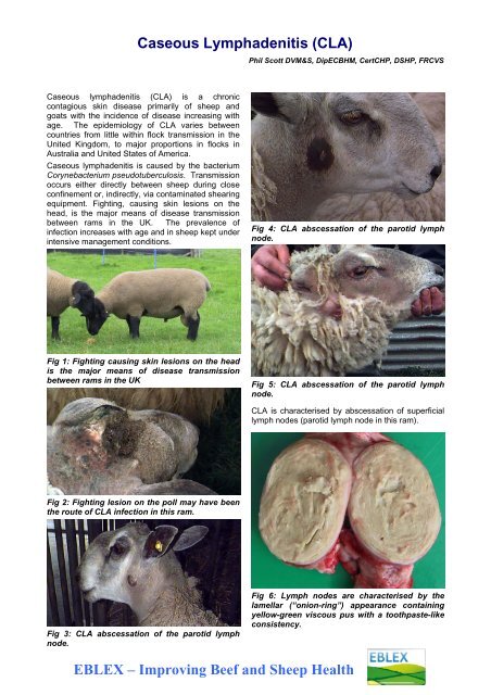
Fig 4: CLA abscessation of the parotid lymph<br />
node.<br />
Fig 1: Fighting causing skin lesions on the head<br />
is the major means of disease transmission<br />
between rams in the UK<br />
Fig 5: CLA abscessation of the parotid lymph<br />
node.<br />
CLA is characterised by abscessation of superficial<br />
lymph nodes (parotid lymph node in this ram).<br />
Fig 2: Fighting lesion on the poll may have been<br />
the route of CLA infection in this ram.<br />
Fig 3: CLA abscessation of the parotid lymph<br />
node.<br />
Fig 6: Lymph nodes are characterised by the<br />
lamellar (“onion-ring”) appearance containing<br />
yellow-green viscous pus with a toothpaste-like<br />
consistency.<br />
EBLEX – Improving Beef and <strong>Sheep</strong> Health
Clinical presentation<br />
The disease is characterised by abscessation of<br />
superficial lymph nodes particularly the parotid (base<br />
of the ear), submandibular (below the jaw), popliteal<br />
(hindleg), precrural (hindleg), and prescapular<br />
(foreleg) lymph nodes. This form of the disease is<br />
often referred to as the cutaneous or superficial form<br />
of CLA. Spread of infection to the lymph nodes<br />
within the chest and internal organs including lungs,<br />
spleen, kidneys and liver, constitutes the visceral or<br />
internal form of CLA. <strong>Sheep</strong> with the superficial<br />
form of CLA may show clinical signs of illness only<br />
when enlargement of the abscess causes<br />
compression of the airway.<br />
Fig 8: Local abscess unrelated to a lymph node<br />
(see below) – this is not CLA.<br />
Fig 7: Spread of infection to the lymph nodes<br />
within the chest – there has been no further<br />
spread and this infection did not produce clinical<br />
signs.<br />
In Australia, shearing wounds lead to infection of the<br />
prescapular and precrural lymph nodes with less<br />
than 1 per cent of lesions affecting lymph nodes of<br />
the head region. Carcass lymph nodes may be 15<br />
cm in diameter and are characterised by the lamellar<br />
(“onion-ring”) appearance of affected lymph nodes<br />
containing yellow-green viscous pus with a<br />
toothpaste-like consistency. Conversely, CLA in the<br />
United Kingdom is characterised by abscessation of<br />
the parotid and submandibular lymph nodes.<br />
In the USA, the visceral form of CLA is commonly<br />
associated with the “thin ewe syndrome” with lesions<br />
in the lungs, liver, and kidneys. Less common sites<br />
are the vertebral column, udder, and scrotum. Large<br />
lung and mediastinal lesions may result in dyspnoea<br />
and this form of the disease is common in the USA,<br />
where affected sheep are referred to as “lungers”.<br />
Spread of infection to cause significant visceral<br />
lesions is uncommon in the UK.<br />
Fig 9: Local abscess unrelated to a lymph node<br />
(see above) – this is not CLA.<br />
Differential diagnoses<br />
Definitive diagnosis of the cutaneous form of CLA<br />
should also include actinobacillosis and tuberculosis,<br />
and local abscess formation.<br />
Fig 10: Subcutaneous abscess unrelated to<br />
lymph node.
whenever possible. Alternatively, sheep should be<br />
purchased and blood testing undertaken before<br />
admission.<br />
Fig 11: Lancing and drainage of above abscess<br />
by a veterinary surgeon.<br />
Common differential diagnoses for the visceral form<br />
of CLA affecting the chest include chronic<br />
suppurative pneumonia, pleural or mediastinal<br />
abscesses, and ovine pulmonary adenocarcinoma<br />
(OPA, Jaagsiekte).<br />
In more general terms, common causes of chronic<br />
weight loss in adult sheep include restricted<br />
nutrition, poor dentition, chronic parasitism,<br />
paratuberculosis (Johne’s disease), maedi-visna<br />
virus infection, chronic suppurative processes, and<br />
tumours of the gastrointestinal tract.<br />
Diagnosis<br />
A positive blood test result indicates exposure to<br />
organism and may indicate active infection, however<br />
severely debilitated animals may yield a false<br />
negative result. The diagnosis is confirmed by<br />
culture of C. pseudotuberculosis.<br />
Treatment<br />
Despite the sensitivity of C. pseudotuberculosis to a<br />
number of antibiotics, therapy is often unsuccessful<br />
due the intracellular site of the bacteria and the<br />
fibrous capsule surrounding the lesions. Lancing<br />
lesions only results in contamination of the<br />
environment thus increasing the potential for<br />
disease spread. Abscesses frequently recur after<br />
drainage and lavage with antiseptics.<br />
Management/Prevention/Control<br />
measures<br />
Disease prevention in clean flocks can be<br />
maintained by effective biosecurity measures. In<br />
such programmes the role of shearing equipment,<br />
and other handling facilities such as mobile plunge<br />
dippers and feeders as vectors for disease, must be<br />
carefully considered. However, disease risks are<br />
highest from purchased animals which must be<br />
inspected before purchase and quarantined for at<br />
least two months. Replacement breeding stock<br />
must be purchased from disease-free flocks<br />
Fig 12: Replacement breeding stock must be<br />
purchased from disease-free flocks whenever<br />
possible.<br />
Fig 13: Purchased animals - must be inspected<br />
before purchase and quarantined for at least two<br />
months.<br />
Commercial vaccines have reduced the incidence of<br />
CLA within a flock but do not prevent all new<br />
infections nor cure sheep already infected.<br />
Commercial vaccines are used in many countries<br />
with a high CLA prevalence such as the USA and<br />
Australia but presently not in many countries within<br />
Europe.<br />
In unvaccinated flocks, all seropositive animals must<br />
be culled and testing repeated until disease is<br />
eliminated.
To test your knowledge and understanding of the control of this<br />
condition, try our instantly marked self assessments, by clicking here<br />
Health Quiz<br />
Copyright © NADIS 2011<br />
Supported by Merial Animal Health makers of Oramec<br />
NADIS seeks to ensure that the information contained within this document is accurate at the time of<br />
printing. However, subject to the operation of law NADIS accepts no liability for loss, damage or injury<br />
howsoever caused or suffered directly or indirectly in relation to information and opinions contained in or<br />
omitted from this document.<br />
To see the full range of NADIS livestock health bulletins please visit www.nadis.org.uk

