Distribution of alkaloids within cacti 270
Sacred Cacti part B preview - Largely Accurate Information Media
Sacred Cacti part B preview - Largely Accurate Information Media
- No tags were found...

Create successful ePaper yourself
Turn your PDF publications into a flip-book with our unique Google optimized e-Paper software.
Excerpted from the unpublished color edition <strong>of</strong> San Pedro & related Trichocereus species<br />
<strong>Distribution</strong> <strong>of</strong> <strong>alkaloids</strong> <strong>within</strong> <strong>cacti</strong><br />
Surprisingly there has been very little serious work published<br />
on this topic.<br />
Alkaloids in “pellote” (i.e. peyote) were reported by JANOT<br />
& BERNIER 1933 to be almost exclusively in the internal cells <strong>of</strong><br />
the cortical parenchyma at top <strong>of</strong> plant. (See TLC results by<br />
Todd elsewhere here.)<br />
In Trichocereus candicans <strong>alkaloids</strong> were found by Niedfeld<br />
to be mainly in the chlorophyllaceous cortical parenchyma.<br />
(Niedfeld used microchemical methods to determine this) RETI<br />
1950 cited NIEDFELD 1931.<br />
In T. terscheckii; <strong>alkaloids</strong> are primarily in the parenchymal<br />
tissues, 29% were found to be in the green epidermis (dry),<br />
while the central parts (dry) including cortical parenchyma<br />
contained 45% <strong>of</strong> the total alkaloid content [please note that<br />
this included the vast majority <strong>of</strong> the parenchymal tissues and<br />
the total weight <strong>of</strong> that portion <strong>of</strong> the plant is much higher than<br />
that <strong>of</strong> the green epidermis. This indicates a lower concentration<br />
for the central parts than in the green portion but potentially<br />
useful concentrations nonetheless.] RETI & CASTRILLÓN 1951<br />
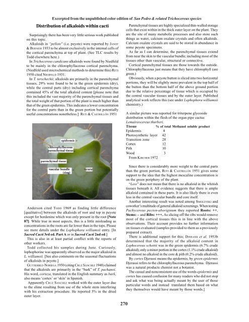
Parenchymal tissues are highly specialized thin-walled storage<br />
cells that exist <strong>within</strong> in the thick outer layer on the plant. They<br />
are the site <strong>of</strong> many metabolic processes and also store such<br />
things as water, calcium oxalate crystals and <strong>of</strong>ten <strong>alkaloids</strong>.<br />
Calcium oxalate crystals are said to be stored in abundance in<br />
some peyote specimens.<br />
As far as I can determine, the parenchymal tissues extend<br />
from near the skin to the vascular bundle; including most <strong>of</strong> the<br />
tissues other than vascular, structural or connective.<br />
Cortical parenchymal tissues are those towards the outside.<br />
Chlorophyllaceous just means that they have chlorophyll (are<br />
green.)<br />
Obviously, when a peyote button is sliced into two horizontal<br />
portions, they will be slightly more prevalent in the top half <strong>of</strong><br />
the button than the bottom half <strong>of</strong> the above ground portion<br />
due to the relative percentage <strong>of</strong> tissue which is occupied by<br />
the central vascular tissues and by the outer layer. Published<br />
analytical work reflects this (see under Lophophora williamsii<br />
chemistry.)<br />
A similar picture was reported for triterpene glycoside<br />
distribution <strong>within</strong> the flesh <strong>of</strong> the organ pipe cactus<br />
Lemaireocereus thurberi.<br />
Tissue % <strong>of</strong> total Methanol soluble product<br />
Epidermis 4<br />
Photosynthetic layer 42<br />
Transition zone 28<br />
Cortex 12<br />
Pith 10<br />
Wood 3<br />
From KIRCHER 1972<br />
Anderson cited TODD 1969 as finding little difference<br />
[qualitative] between the <strong>alkaloids</strong> <strong>of</strong> root and top in peyote<br />
except for hordenine which was only present in the root [Note<br />
87]. While true in most aspects, this is a little misleading as<br />
concentrations in the roots are far lower than in the tops. Please<br />
see more details under the Lophophora williamsii entry. [In<br />
Sacred Cacti 3rd ed. Part A or in Sacred Cacti 2nd ed.]<br />
This is also in at least partial conflict with the reports <strong>of</strong><br />
other workers.<br />
Todd collected his samples during June. Curiously,<br />
lophophorine was apparently observed as the major alkaloid in<br />
L. williamsii. [See also comments on the seasonal fluctuations<br />
<strong>of</strong> <strong>alkaloids</strong> in peyote.]<br />
GUTTIERREZ-NORIEGA 1950 (citing CRUZ SÁNCHEZ 1948) claimed<br />
that the <strong>alkaloids</strong> are primarily in the “bark” <strong>of</strong> T. pachanoi.<br />
His word, corteza, translated in the English summary as bark,<br />
also means ‘cortex’ or ‘skin’ in Spanish.<br />
Apparently CRUZ SANCHEZ worked with the outer layer due<br />
to the slime resulting from use <strong>of</strong> the whole stem interfering<br />
with his extraction procedure. He reported 5% in the dried<br />
outer layer.<br />
<strong>270</strong><br />
Since there is considerably more weight to the central parts<br />
than the green portion, RETI & CASTRILLÓN 1951 gives some<br />
support to the idea that the highest mescaline concentration is<br />
on the green periphery <strong>of</strong> the plant.<br />
“Less” does not mean that there is no alkaloid in the whitish<br />
tissues beneath it. All evidence suggests that there is ample<br />
alkaloid contained in these parts. It is also likely there is even<br />
less in the central vascular bundle and core itself.<br />
Another interesting result was noted among SMOLENSKI and<br />
coworker’s multitude <strong>of</strong> general alkaloid screenings. When testing<br />
Pachycereus pecten-aboriginum they reported Roots: ++,<br />
Stems: – and Ribs: +++. As slicing <strong>of</strong>f the ribs would remove<br />
most <strong>of</strong> the cortical tissues this is in line with the above<br />
observations. Their account provides no further information<br />
on tissues evaluated (samples provided to them as a previously<br />
prepared extract).<br />
There is additional support for this; DJERASSI et al. 1953b<br />
determined that the majority <strong>of</strong> the alkaloid content in<br />
Lophocereus schottii was in the green epidermis (6.7% crude<br />
alkaloid); only a minor portion in the cortex (1.1% crude alkaloid)<br />
and almost no alkaloid in the core & pith (0.2% crude alkaloid).<br />
By cortex Djerassi means the epidermis, by green epidermis<br />
Djerassi refers to the chlorophyllaceous parenchyma. Djerassi<br />
was a natural products chemist not a botanist.<br />
The casual and nonconsistent use <strong>of</strong> the words epidermis and<br />
cortex has caused confusion for many readers who did not stop<br />
and ask what was being actually meant by the user <strong>of</strong> those<br />
particular words and instead translated them based on what<br />
they themselves would have meant by those words.]
This area needs further work. While many <strong>alkaloids</strong> may<br />
indeed be higher towards the outside <strong>of</strong> the plant there are<br />
known exceptions. Hordenine being observed in the root rather<br />
than the top (in peyote) is a good example. Its highest<br />
concentrations being in the root was reported again in<br />
Mammillaria microcarpa by KNOX and coworkers.<br />
It is noteworthy also that all <strong>of</strong> the <strong>alkaloids</strong> measured by<br />
KNOX were much higher in the cortex itself as compared to the<br />
chlorophyll rich tubercles and several were higher in the vascular<br />
tissues than in the tubercles.<br />
We were informed by an Entheogen Review reader that they<br />
had found an unspecified amount <strong>of</strong> the cores <strong>of</strong> San Pedro to be<br />
active but they provided inadequate information for us to<br />
understand HOW they actually determined this or how much<br />
they observed.<br />
This should not be any surprise should a person ingest a large<br />
enough amount.<br />
PUMMANGURA et al. 1982 reported that mescaline did not<br />
transmigrate between grafted T. pachanoi and T. spachianus<br />
regardless <strong>of</strong> which was used as stock and scion. Their conclusion<br />
was that mescaline was locally produced and noncirculating.<br />
While it may or may not be true that transmigration <strong>of</strong> <strong>alkaloids</strong><br />
does not occur, SINISCALCO 1983 reported that the normally<br />
mescaline-free Myrtillocactus geometrizans was found to contain<br />
0.3% mescaline by dry weight after having previously been<br />
grafted with Lophophora williamsii.<br />
Many questions immediately arise.<br />
None are presently answered.<br />
San Pedro: <strong>Distribution</strong> <strong>of</strong> <strong>alkaloids</strong><br />
Trichocereus scopulicola FR991<br />
(NMCR)<br />
from Ritter’s seed<br />
It is almost unbelievable that no one has looked into the<br />
matter <strong>of</strong> alkaloid distribution <strong>within</strong> <strong>cacti</strong> more thoroughly.<br />
Analysis <strong>of</strong> only outer green layers looking at only<br />
mescaline has become the predominate analystical approach.<br />
A flowering Trichocereus peruvianus KK242<br />
Photo by Flip<br />
In an e-mail we received in 2004, Karel Knize commented<br />
“Some flowers are used (cont ca 4%) plant itself 2-3.5%”<br />
“the strongest type are 9-12 ribs or 3-4 ribs”<br />
He did not elaborate further.<br />
A friend claimed to have had good results from flower masses<br />
from peruvianoids and terscheckii but preserved no details.<br />
In more recent years, additional friends ingesting pachanoi<br />
and peruvianus flowers and ovary could discern no effects<br />
whatsoever..<br />
Clearly some analytical work seems in order to know what to<br />
believe.<br />
271<br />
Trichocereus pachanot<br />
Variant growth