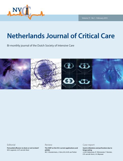
Netherlands Journal of Critical Care
Netherlands Journal of Critical Care - NJCC
Netherlands Journal of Critical Care - NJCC

Create successful ePaper yourself
Turn your PDF publications into a flip-book with our unique Google optimized e-Paper software.
Volume 17 - No 1 - February 2013<br />
<strong>Netherlands</strong> <strong>Journal</strong> <strong>of</strong> <strong>Critical</strong> <strong>Care</strong><br />
Bi-monthly journal <strong>of</strong> the Dutch Society <strong>of</strong> Intensive <strong>Care</strong><br />
Editorial<br />
Pericardial effusion: to drain or not to drain?<br />
W.K. Lagrand, J.A.P. van der Sloot<br />
Review<br />
The SSEP on the ICU: current applications and<br />
pitfalls<br />
M.C. Cloostermans, J. Horn, M.J.A.M. van Putten<br />
Case report<br />
Gastric dilatation and perforation due to<br />
binge eating<br />
J.A.M. Heijneman, R. Tahmassian, T. Karsten,<br />
E.R. van der Vorm, I.A. Meynaar
<strong>Netherlands</strong> <strong>Journal</strong> <strong>of</strong> <strong>Critical</strong> <strong>Care</strong><br />
<strong>Netherlands</strong> <strong>Journal</strong> <strong>of</strong> <strong>Critical</strong> <strong>Care</strong><br />
Executive editorial board<br />
A.B.J. Groeneveld, editor in chief<br />
J. Box, language editor<br />
COPYRIGHT<br />
<strong>Netherlands</strong> <strong>Journal</strong> <strong>of</strong> <strong>Critical</strong> <strong>Care</strong><br />
ISSN: 1569-3511<br />
NVIC p/a Domus Medica<br />
P.O. Box 2124, 3500 GC Utrecht<br />
T.: +31-(0)30-6868761<br />
© 2013 NVIC. All rights reserved. Except as outlined<br />
below, no part <strong>of</strong> this publication may be reproduced,<br />
stored in a retrieval system or transmitted in any form<br />
or by any means, electronic, mechanical, photocopying,<br />
recording or otherwise, without prior written<br />
permission <strong>of</strong> the publisher. Permission may be sought<br />
directly from NVIC.<br />
Derivative works<br />
Subscribers may reproduce tables <strong>of</strong> contents or<br />
prepare lists <strong>of</strong> articles including abstracts for internal<br />
circulation within their institutions. Permission <strong>of</strong><br />
the publisher is required for resale or distribution<br />
outside the institution. Permission <strong>of</strong> the publisher is<br />
also required for all other derivative works, including<br />
compilations and translations.<br />
Electronic storage<br />
Permission <strong>of</strong> the publisher is required to store or use<br />
electronically any material contained in this journal,<br />
including any article or part <strong>of</strong> an article.<br />
Subscriptions<br />
An annual subscription to The <strong>Netherlands</strong> <strong>Journal</strong> <strong>of</strong><br />
<strong>Critical</strong> <strong>Care</strong> consists <strong>of</strong> 6 issues. Issues within Europe<br />
are sent by standard mail and outside Europe by air<br />
delivery. Cancellations should be made, in writing, at<br />
least two months before the end <strong>of</strong> the year.<br />
The annual subscription fee for the <strong>Netherlands</strong> is 170<br />
euro, for Europe 285 euro, for the rest <strong>of</strong> the world 380<br />
euro. Subscriptions are accepted on a prepaid basis<br />
only and are entered on a calendar year basis.<br />
Please make your cheque payable to Van Zuiden<br />
Communications B.V., PO Box 2122, 2400 CC Alphen aan<br />
den Rijn, the <strong>Netherlands</strong> or you can transfer the fee to<br />
ING Bank, account number 67.87.10.872, Castellumstraat<br />
1, Alphen aan den Rijn, the <strong>Netherlands</strong>, swift-code: ING<br />
BNL 2A. Do not forget to mention the complete address<br />
for delivery <strong>of</strong> the <strong>Journal</strong>.<br />
Claims<br />
Claims for missing issues should be made within two<br />
months <strong>of</strong> the date <strong>of</strong> dispatch. Missing issues will be<br />
mailed without charge. Issues claimed beyond the twomonth<br />
limit must be prepaid at back copy rates.<br />
Advertising-exploitation/business<br />
contacts<br />
For orders, reprints and advertising, please contact<br />
Van Zuiden Communications B.V.<br />
Van Zuiden Communications B.V.<br />
PO Box 2122<br />
2400 CC Alphen aan den Rijn<br />
The <strong>Netherlands</strong><br />
Tel.: +31 (0)172-47 61 91<br />
E-mail: njcc@vanzuidencommunications.nl<br />
Internet: www.njcc.nl<br />
inhoud<br />
EDITORIAL<br />
3 Pericardial effusion: to drain or not to drain?<br />
W.K. Lagrand, J.A.P. van der Sloot<br />
REVIEWs<br />
5 The SSEP on the ICU: current applications and pitfalls<br />
M.C. Cloostermans, J. Horn, M.J.A.M. van Putten<br />
12 The pharmacologic treatment <strong>of</strong> alcohol withdrawal syndrome in the ICU<br />
D.P.F. van Nunen, D.H.T. Tjan<br />
GUIDELINE<br />
10 Recommendations for the timing and dosing <strong>of</strong> CRRT in critically ill<br />
patients with AKI<br />
H.M. Oudemans-van Straaten, C.S.C. Bouman, M. Schetz, A.B.J. Groeneveld, A.C. de Pont,<br />
A.M. van Alphen, H. de Geus, W. Boer<br />
CASE REPORTS<br />
18 Gastric dilatation and perforation due to binge eating: a case report<br />
J.A.M. Heijneman, R. Tahmassian, T. Karsten, E.R. van der Vorm, I.A. Meynaar<br />
21 Trichoderma: an unusual bystander in invasive pulmonary aspergillosis<br />
K. Ariese, L. Hulsh<strong>of</strong>f, R. Jansen, P.H.J. van der Voort<br />
25 Drug induced lung injury – a case <strong>of</strong> fatal bleomycin interstitial<br />
pneumonitis<br />
S. van der Sar-van der Brugge, H. van Ravenswaay Claasen, L. Dawson<br />
30 A rare cause <strong>of</strong> cardiac failure following transthoracic oesophagectomy<br />
D.A. Wicherts, S. Hendriks, W.L.E.M. Hesp, J.A.B. van der Hoeven, H.H. Ponssen<br />
33 Elevated liver enzymes and renal failure, with a surprising outcome.<br />
Two similar cases<br />
A.E. Boendermaker, D. Boumans, R.A.A. van Zanten, H. Idzerda, H. van de Hout, Th.F. Veneman<br />
37 Editorial board<br />
37 International advisory board<br />
39 Information for authors<br />
<strong>Netherlands</strong> <strong>Journal</strong> <strong>of</strong> <strong>Critical</strong> <strong>Care</strong> is indexed in:<br />
EMBASE EM<strong>Care</strong> Scopus<br />
Neth j crit care – volume 17 – no 1 – february 2013<br />
1
<strong>Netherlands</strong> <strong>Journal</strong> <strong>of</strong> <strong>Critical</strong> <strong>Care</strong><br />
Accepted January 2013<br />
EDITORIAL<br />
Pericardial effusion: to drain or not to drain?<br />
W.K. Lagrand 1 , J.A.P. van der Sloot<br />
1<br />
Department <strong>of</strong> Intensive <strong>Care</strong> Adults, room C3-430, Academic Medical Center, Amsterdam, The <strong>Netherlands</strong><br />
Correspondence<br />
W.K. Lagrand – e-mail: w.k.lagrand@amc.uva.nl<br />
Key words - Hepatic and renal dysfunction, pericardial effusion, cardiac tamponade<br />
In this issue <strong>of</strong> the <strong>Netherlands</strong> <strong>Journal</strong> <strong>of</strong> <strong>Critical</strong> <strong>Care</strong>,<br />
Boendermaker et al. describe two cases <strong>of</strong> hepatic and renal<br />
dysfunction caused by neoplastic pericardial effusion and resulting<br />
in cardiac tamponade 1 . The incidence <strong>of</strong> hepatic and renal<br />
dysfunction in critically ill patients is high and can have various<br />
causes. Because pericardial effusion, <strong>of</strong> oncologic origin, is not the<br />
most likely cause <strong>of</strong> these organ dysfunctions, critical care clinicians<br />
<strong>of</strong>ten omit considering cardiac tamponade as the underlying cause.<br />
In most cases <strong>of</strong> pericardial effusion, the condition is found more<br />
or less by surprise. Once found, the question arises whether the<br />
pericardial effusion is symptomatic (i.e. cardiac tamponade) or<br />
asymptomatic (i.e. an innocent bystander without therapeutic<br />
consequences). The cases described by Boendermaker et al. nicely<br />
illustrate the clinical and therapeutic considerations and place <strong>of</strong><br />
pericardial effusion and/or tamponade in the differential diagnosis<br />
<strong>of</strong> hepatic and renal dysfunction.<br />
The etiology <strong>of</strong> pericardial effusion is quite diverse: it may be caused<br />
by infection (bacterial, viral, fungal, mycobacteria, protozoal),<br />
inflammation (auto-immune diseases), myocardial infarction<br />
(Dressler syndrome), cardiac surgery, trauma or chemical e.g.<br />
uraemia. Pericardial effusion resulting from a malignant process,<br />
such as neoplastic pericardial effusion, may occur directly (from<br />
the malignant process itself) or as a consequence <strong>of</strong> therapy e.g.<br />
radiotherapy, chemotherapy, necessary to treat the tumour 2,3 .<br />
The signs and symptoms <strong>of</strong> cardiac effusion may manifest gradually,<br />
ranging from overt tamponade to no clinical signs at all. Besides<br />
this, most symptoms <strong>of</strong> cardiac tamponade are nonspecific, like<br />
anorexia, cough, hypotension, tachycardia, dyspnoea, tachypnoea<br />
and sometimes circulatory collapse. Also, like the cases presented<br />
by Boendermaker et al., patients may present with the complications<br />
<strong>of</strong> cardiac tamponade due to reduced organ perfusion resulting in<br />
hepatic and renal failure. In cases <strong>of</strong> pericardial effusion, physical<br />
examination may also reveal specific and nonspecific findings.<br />
Besides tachycardia, heart sounds may be attenuated, due to the<br />
isolating effects <strong>of</strong> the pericardial fluid. Clinically significant<br />
tamponade usually results in hypotension or shock. (i.e. Beck’s<br />
triad: hypotension, tachycardia and muffled heart tones). Jugular<br />
venous distention is usually present but may be absent in cases<br />
<strong>of</strong> hypovolemia. A key finding in cases <strong>of</strong> cardiac tamponade is<br />
pulsus paradoxus (PP) 2,4,5 . PP, however, is not pathognomonic for<br />
tamponade. It may also be present in cases <strong>of</strong> massive pulmonary<br />
embolism, pr<strong>of</strong>ound haemorrhagic shock, and obstructive<br />
pulmonary diseases. PP is defined as a decline <strong>of</strong> 10 mmHg or more<br />
in systolic arterial blood pressure after inspiration during normal<br />
breathing. It may be palpable, but sometimes arterial catheterisation<br />
is needed to identify PP.<br />
It has to be emphasized that cardiac tamponade is a clinical<br />
diagnosis. Additional investigations, however, may support the<br />
diagnosis <strong>of</strong> tamponade. Electrocardiography (ECG) may reveal<br />
signs <strong>of</strong> pericardial effusion: micro voltages, electrical alternans (as<br />
a sign <strong>of</strong> a swinging heart), PR-segment depression, ST-T segment<br />
alterations such as elevation and/or depression. These ECG findings<br />
are also not specific but may fit in with the diagnosis <strong>of</strong> pericardial<br />
effusion. Chest radiography may depict an enlarged cardiac<br />
silhouette with a tent-like shape. The cardiac silhouette, however,<br />
may also remain normal despite a large amount <strong>of</strong> pericardial<br />
effusion. Echocardiography is the principal tool for making the<br />
diagnosis pericardial effusion. Echocardiographic signs <strong>of</strong> cardiac<br />
tamponade include collapse <strong>of</strong> the right atrium and/or right<br />
ventricle. Left atrial collapse occurs in approximately 25% <strong>of</strong> patients<br />
and is highly specific for tamponade. Doppler echocardiography may<br />
show respiratory variations in transvalvular blood flow. This would<br />
be an indication that signs <strong>of</strong> PP can be demonstrated by Doppler<br />
echocardiography. By subcostal echocardiographic examination the<br />
inferior vena cava (IVC) can be visualized. In cases <strong>of</strong> tamponade,<br />
the IVC is usually dilated, with little or no collapse after inspiration<br />
during normal breathing. CT-scanning, MRI and right and left heart<br />
catheterisation may be considered but are generally not needed in<br />
the evaluation <strong>of</strong> pericardial effusion, unless it is necessary to search<br />
for underlying diseases such as malignancies 2 .<br />
So, what do we learn from the cases presented by Boendermaker et<br />
al.? First, pericardial effusion and tamponade should be considered<br />
in patients with hepatic and renal dysfunction that has been caused<br />
by diminished hemodynamics, thereby compromising the function<br />
Neth j crit care – volume 17 – no 1 – february 2013<br />
3
<strong>Netherlands</strong> <strong>Journal</strong> <strong>of</strong> <strong>Critical</strong> <strong>Care</strong><br />
<strong>of</strong> multiple organs including the kidneys and liver. Pericardial<br />
effusion in itself does not always result in cardiac tamponade: when<br />
it develops slowly, large amounts <strong>of</strong> pericardial fluid (> 2000 mL)<br />
may not result in tamponade, whereas small amounts <strong>of</strong> pericardial<br />
fluid (< 100 mL), accumulating in a very short time, may cause<br />
severe obstructive cardiogenic shock 4,5 . Neoplastic pericardial<br />
effusion usually develops slowly with gradual clinical deterioration<br />
over time due to inflow obstruction; this may eventually result in<br />
cardiac tamponade, as described in the presented cases. Second,<br />
cardiac tamponade remains a clinical diagnosis. Echocardiography<br />
is essential to assess the existence <strong>of</strong> pericardial effusion and<br />
to find clues for the severity <strong>of</strong> inflow obstruction. It is to be<br />
emphasized, however, that even when there are negative signs <strong>of</strong><br />
cardiac tamponade by echocardiography, a cardiac tamponade may<br />
be present. Clinical signs <strong>of</strong> cardiac tamponade, in the presence<br />
<strong>of</strong> pericardial effusion, are to be diagnosed as cardiac tamponade<br />
unless proven otherwise. Removal <strong>of</strong> the pericardial fluid either by<br />
pericardiocentesis or surgery, should not be postponed in cases <strong>of</strong><br />
clinically manifest tamponade with negative echocardiographic<br />
signs <strong>of</strong> inflow obstruction. In all patients who are hemodynamically<br />
unstable, pericardial drainage has to be considered when there is a<br />
pericardial effusion 6 .<br />
References<br />
1. Boendermaker AE, Boumans D, van Zanten RAA, Idzerda H, van de Hout H, Veneman<br />
ThF. Elevated liver enzymes and renal failure, with a surprising outcome. Two similar<br />
cases. Neth J Crit <strong>Care</strong> 2012;2013;17:33-6.<br />
2. LeWinter MM, Kabbani S. Pericardial diseases. In: Braunwald E, Zipes DP, Libby, eds.<br />
Heart disease: a textbook <strong>of</strong> cardiovascular medicine. 7th ed. Vol. 2. Philadelphia: W.B.<br />
Saunders, 2005:1757-80.<br />
3. Refaat MM, Katz WE. Neoplastic pericardial effusion. Clin Cardiol 2011;34:593-598<br />
4. Spodick DH. Acute cardiac tamponade. N Engl J Med 2003;349:684-90.<br />
5. Reddy PS, Curtiss EI, O’Toole JD, Shaver JA. Cardiac tamponade: hemodynamic observations<br />
in man. Circulation 1978;58:265<br />
6. Ariyarajah V, Spodick DH. Cardiac tamponade revisited: a postmortem look at cautionary<br />
case. Tex Heart Inst J 2007; 34:347-51.<br />
4 Neth j crit care – volume 17 – no 1 – february 2013
<strong>Netherlands</strong> <strong>Journal</strong> <strong>of</strong> <strong>Critical</strong> <strong>Care</strong><br />
Accepted January 2013<br />
REVIEW<br />
The SSEP on the ICU: current applications and pitfalls<br />
M.C. Cloostermans 1,2 , J. Horn 3 , M.J.A.M. van Putten 1,2<br />
1<br />
Clinical Neurophysiology, MIRA – Institute for Biomedical Technology and Technical Medicine, University <strong>of</strong> Twente, Enschede, The <strong>Netherlands</strong><br />
2<br />
Departments <strong>of</strong> Clinical Neurophysiology and Neurology, Medisch Spectrum Twente, Enschede, The <strong>Netherlands</strong><br />
3<br />
Department <strong>of</strong> Intensive <strong>Care</strong> Medicine, Academic Medical Center, University <strong>of</strong> Amsterdam, Amsterdam, The <strong>Netherlands</strong><br />
Correspondence<br />
J. Horn – e-mail: j.horn@amc.uva.nl<br />
Key words - Somatosensory evoked potentials, prognostication, post-anoxic coma, traumatic brain injury, subarachnoid haemorrhage<br />
Abstract<br />
Clinical neurological evaluation <strong>of</strong> patients in the intensive care unit<br />
(ICU) is <strong>of</strong>ten limited. Registration <strong>of</strong> the somatosensory evoked<br />
potential (SSEP) can assist in the neurological evaluation in these<br />
patients. In this paper, we discuss the principles, applications and<br />
limitations <strong>of</strong> the SSEPs in the ICU with a focus on prognostication<br />
in comatose patients. Registration <strong>of</strong> the SSEP is a very reliable and<br />
reproducible method, if it is performed and interpreted correctly. A<br />
bilateral absent cortical SSEP response is a reliable predictor for poor<br />
neurological outcome in patients with a post-anoxic coma, but not in<br />
patients with traumatic brain injury or subarachnoid haemorrhage.<br />
During SSEP recordings, great care should be taken in improving<br />
the signal to noise ratio. Since the interpreting clinician is <strong>of</strong>ten<br />
not present during the actual SSEP registration itself, the role <strong>of</strong> the<br />
lab technician is crucial in obtaining reliable SSEP results. If the<br />
noise level is too high, the peripheral responses are abnormal, or the<br />
response is not reproducible in a second set <strong>of</strong> stimuli, interpretation<br />
<strong>of</strong> the SSEP cannot be done reliably.<br />
Introduction<br />
Neurological evaluation <strong>of</strong> patients in the intensive care unit (ICU)<br />
is <strong>of</strong>ten limited, and clinical neurophysiology has provided several<br />
techniques to assist in the evaluation <strong>of</strong> the central and peripheral<br />
nervous system in these conditions. Techniques include the electroencephalogram<br />
(EEG), brainstem auditory evoked potentials (BAEP)<br />
and the somatosensory evoked potential (SSEP). In this paper, we<br />
discuss the principles, applications and limitations <strong>of</strong> the SSEP. SSEPs<br />
are used in a variety <strong>of</strong> clinical settings, including the evaluation <strong>of</strong><br />
coma, neuromonitoring in the operating theatre, and the evaluation<br />
<strong>of</strong> traumatic spinal cord injury. Here, we focus on the use <strong>of</strong> the SSEP<br />
registration in the prognostication <strong>of</strong> comatose patients in the ICU.<br />
SSEP: Principles<br />
The somatosensory evoked potential is a small (< 10-50 µV)<br />
electrical signal, that can be recorded noninvasively from the skull,<br />
after giving a set <strong>of</strong> electrical stimuli to one <strong>of</strong> the peripheral nerves.<br />
Measurement <strong>of</strong> the SSEP evaluates the complete pathway from the<br />
peripheral sensory nervous system to the sensory cortex, which<br />
runs via the dorsal column lemniscal pathway via the spinal cord,<br />
brainstem and thalamus 1,2 .<br />
The dorsal column-lemniscal pathway consists <strong>of</strong> four neuronal<br />
populations. The cell bodies <strong>of</strong> the first-order neurons are situated<br />
in the dorsal root ganglia, the trigeminal ganglion, the midbrain<br />
trigeminal nucleus, and the vagal ganglion nodosum. The<br />
second-order neuron lies in the dorsal column nuclei (the cuneate<br />
nucleus and the gracile nucleus). Axons <strong>of</strong> these second neurons<br />
cross the midline and project to the ventroposterior nuclei <strong>of</strong> the<br />
thalamus (third-order neuron). From there the pathway projects<br />
into the network <strong>of</strong> somatosensory cortex areas (fourth-order<br />
neurons), which include primary and secondary somatosensory<br />
cortex, posterior parietal cortex, posterior and mid-insula, and<br />
mid-cingulate cortex 1 . Figure 1 shows the anatomical connections<br />
evaluated by the median nerve SSEP.<br />
SSEPs are usually evoked by bipolar transcutaneous electrical<br />
stimulation applied on the skin over the selected nerve, and<br />
registered with disk-electrodes along the tract. For example, in<br />
SSEP recordings <strong>of</strong> the median nerve registration electrodes can be<br />
placed at the elbow, Erb’s point, cervical, parietal and frontal cortex.<br />
The cortical response can only be interpreted reliably, when the<br />
peripheral responses are present.<br />
In the nomenclature <strong>of</strong> SSEP waveforms, N or P followed by<br />
an integer (e.g. N20) are used to indicate the polarity and the<br />
nominal post-stimulus latency (in ms) <strong>of</strong> the recorded wave in<br />
the healthy population 1 . The earliest cortical potential is the N20,<br />
which is generated in the primary somatosensory cortex, where<br />
thalamocortical cells make synaptic connections with the superficial<br />
and deep pyramidal cell layers 3,4 . In comparison to the later cortical<br />
responses, the N20 is the most robust, and is the latest waveform to<br />
disappear during increasing levels <strong>of</strong> encephalopathy. Furthermore,<br />
the N20 is relatively independent to the level <strong>of</strong> sedation 1 . As the<br />
Neth j crit care – volume 17 – no 1 – february 2013<br />
5
<strong>Netherlands</strong> <strong>Journal</strong> <strong>of</strong> <strong>Critical</strong> <strong>Care</strong><br />
later cortical waveforms (P45, N60 and P/N100) are less reliable<br />
and more susceptible to changes by sedation, the N20 is used in all<br />
prognostic clinical routines.<br />
The N20 SSEP in Prognostication<br />
Postanoxic Coma<br />
Bilateral absence <strong>of</strong> short latency (N20) SSEP response has been<br />
identified as the most powerful predictor <strong>of</strong> poor outcome in<br />
patients who are unconscious after circulatory arrest 1 . In patients<br />
not treated with hypothermia, bilateral absence <strong>of</strong> cortical N20<br />
responses 24 hours or more after the ischemic event is a reliable<br />
predictor for a poor neurological outcome 5,6,7 . A systematic review<br />
<strong>of</strong> Robinson showed a false positive rate (FPR) <strong>of</strong> 0% 6 , while a meta<br />
analyse <strong>of</strong> Wijdicks et al. found a 0.7% false positive rate for bilateral<br />
absent N20 responses in those patients 7 .<br />
In patients treated with therapeutic hypothermia, absence <strong>of</strong> the<br />
N20 also indicates a poor prognosis. In two large prospective studies,<br />
including 228 patients, the median nerve SSEP was found to be a<br />
reliable tool to predict a poor outcome after rewarming with an FPR<br />
<strong>of</strong> 0% 8,9 . However, a retrospective study <strong>of</strong> Leithner in 122 available<br />
SSEPs revealed one patient treated with therapeutic hypothermia<br />
after cardiac arrest with bilateral absent N20 responses at day 3<br />
with good neurological outcome 10 . This SSEP was measured on day<br />
3 under sedation with midazolam and fentanyl in a patient with<br />
alcoholic polyneuropathy. Despite this single case, pooled analysis<br />
<strong>of</strong> these three recent studies 8,9,10 on cardiac arrest patients after<br />
hypothermia still give very low FPRs <strong>of</strong> 0.9%, indicating that bilateral<br />
absence <strong>of</strong> the N20 should still be viewed as a reliable predictor for<br />
poor outcome in patients treated with hypothermia.<br />
Figure 1. The anatomical connections evaluated by the median nerve SSEP<br />
Thalamus<br />
Cuneate<br />
nucleus<br />
Postcentral<br />
gyrus<br />
Recent studies show that already during the period <strong>of</strong> hypothermia<br />
the SSEP is a reliable tool to predict a poor outcome 9,11,12,13 . A pooled<br />
analysis <strong>of</strong> these four prospective studies (424 patients), shows an<br />
FPR <strong>of</strong> 1.5% with a sensitivity <strong>of</strong> 28%. The FPR <strong>of</strong> 1.5% results from<br />
three patients with good neurological outcome and bilateral absent<br />
N20 responses during hypothermia, who were reported in the study<br />
<strong>of</strong> Bouwes et al 9 . However, in a post hoc assessment <strong>of</strong> these SSEP<br />
registrations it was concluded that these three SSEP recordings<br />
were undeterminable because there was too much noise in the<br />
registration 9 . Correction <strong>of</strong> these results led to an FPR <strong>of</strong> 0%.<br />
Unfortunately, preservation <strong>of</strong> the N20 does not imply a favourable<br />
outcome in patients after cardiac arrest. In fact, only a small proportion<br />
<strong>of</strong> patients with a poor outcome after resuscitation have negative SSEP<br />
responses resulting in a low sensitivity. This low sensitivity <strong>of</strong> the SSEP<br />
is also reflected in the large variability <strong>of</strong> EEG patterns that can be<br />
observed in patients with a preserved N20, including status epilepticus<br />
or even electro-cerebral silence 4 . As pyramidal cell synaptic function<br />
is mainly reflected by the EEG, while SSEP mainly evaluates the<br />
thalamocortical synaptic function, a possible explanation is selective<br />
hypoxic damage to the cortical pyramidal cells’ synaptic function, with<br />
preserved thalamocortical synapses 4 .<br />
Traumatic Brain Injury<br />
In patients with severe traumatic brain injury (TBI), the results<br />
available on the reliability <strong>of</strong> SSEP to predict outcome have been<br />
contradictive. Sleigh et al. showed in a prospective blinded cohort<br />
study including 105 patients that the median nerve SSEP is a reliable<br />
predictor for poor neurological outcome, with a 43% sensitivity and no<br />
false positives 14 . In contrast, in several other studies TBI patients were<br />
described with initially bilateral absent N20 responses who regained<br />
consciousness and had only minor disabilities 1,15,16 . These results show<br />
that absence <strong>of</strong> cortical SSEP responses is not a reliable predictor in<br />
TBI patients. The most likely explanation is that in head trauma, a<br />
transient N20 disappearance may be consecutive to focal midbrain<br />
dysfunction due to oedema 1 . Therefore, SSEPs should never be used as<br />
a single test in TBI patients, but integrated with other neurophysiologic<br />
tools and clinical examination to improve the predictive value 1,14,15 . In<br />
TBI patients it is especially important to rule out traumatic lesions <strong>of</strong><br />
the peripheral nerves, nerve roots or spinal cord when using clinical<br />
neurophysiologic tests. Clinical examination <strong>of</strong> the peripheral nerves<br />
can be difficult in patients with a diminished consciousness.<br />
Median<br />
nerve<br />
Lower<br />
brainstem<br />
Spinal cord<br />
Subarachnoid haemorrhage<br />
In patients with subarachnoid haemorrhage, neither median or tibial<br />
nerve SSEP, flash-visual evoked potential, BAEP nor central conduction<br />
time <strong>of</strong> the median nerve SSEP can be used as a valid predictor for<br />
outcome. The patient’s initial clinical grading still provides the only<br />
satisfying predictor, independent <strong>of</strong> the patient’s clinical course 17 .<br />
Sepsis<br />
In patients with severe sepsis and septic shock, prolonged cortical<br />
SSEP peak latencies occur in 84% <strong>of</strong> the patients. These prolonged<br />
6 Neth j crit care – volume 17 – no 1 – february 2013
<strong>Netherlands</strong> <strong>Journal</strong> <strong>of</strong> <strong>Critical</strong> <strong>Care</strong><br />
The SSEP on the ICU: current applications and pitfalls<br />
latencies can be used to diagnose septic encephalopathy and its<br />
severity is associated with the severity <strong>of</strong> illness 18 . In these patients<br />
SSEP cannot be used to determine prognosis.<br />
Pitfalls and limitations <strong>of</strong> SSEPs at the ICU<br />
Noise<br />
The most severe limitation <strong>of</strong> the SSEP is the moderate interobserver<br />
agreement, which is extensively described in a study <strong>of</strong> Zandbergen<br />
et al. 19 . In their study, SSEPs <strong>of</strong> 56 consecutive patients with anoxic–<br />
ischaemic coma were interpreted independently by 5 experienced<br />
clinical neurophysiologists. The interobserver agreement for SSEPs<br />
in anoxic–ischaemic coma was only moderate (kappa 0.52, 95% CI<br />
0.20–0.65). The main source <strong>of</strong> disagreement was related to the<br />
noise levels. For recordings with a noise level <strong>of</strong> 0.25 µV or more,<br />
mean kappa was 0.34 (fair agreement); for recordings with a noise<br />
level below 0.25 µV mean kappa was 0.74, which is a substantial<br />
agreement 19 .<br />
Efforts should be made to improve the registration and diminish<br />
noise as much as possible. Zandbergen et al. recommend that the<br />
peak-to-peak amplitude <strong>of</strong> noise <strong>of</strong> the cortical and cervical leads<br />
should be lower than 0.25 µV after averaging, especially in the<br />
frequency <strong>of</strong> the SSEPs themselves (20-500 Hz) 19 . Giving muscle<br />
relaxants can <strong>of</strong>ten improve the quality <strong>of</strong> the SSEP in patients<br />
with too much muscle activity; an example is given in figure 2.<br />
Furthermore, disturbing electrical ICU equipment should be turned<br />
<strong>of</strong>f if possible. Also, giving more stimuli (up to 1000 or more) and<br />
increasing the stimulus intensity could improve the signal-to-noise<br />
ratio 19 . Since the interpreting clinician is <strong>of</strong>ten not present during<br />
the actual SSEP registration itself, the role <strong>of</strong> the lab technician is<br />
crucial in obtaining reliable SSEP results. As the quality <strong>of</strong> the SSEP<br />
recording depends on the skills <strong>of</strong> the technician, it is important that<br />
they are well trained and sufficiently familiar with SSEP registrations<br />
in the ICU. In those situations where significant artifacts appear to<br />
be present, the referring physician should be informed. Furthermore,<br />
it is always the role <strong>of</strong> the interpreting clinician to check the quality<br />
and signal-to-noise ratio <strong>of</strong> the SSEP registration, and to decide<br />
whether the SSEP registration is reliable enough for clinical decision<br />
making.<br />
Interpretation Criteria<br />
Despite the noise level, also other criteria for reliable results can be<br />
given. An N20 peak on one side can only be considered as present if<br />
it fulfils all the following criteria:<br />
• It should have an appropriate latency (i.e. at least 4.5 ms longer<br />
than the corresponding N13 peak in normal-stature adults) 19 .<br />
• It should be present on the contralateral side, and there should<br />
be a clear difference with the recording from the side ipsilateral<br />
to the stimulus 19 . Therefore it is recommended to record not<br />
only the contralateral sensory cortex after stimulation, but also<br />
co-register the ipsilateral side. This prevents misinterpretation <strong>of</strong><br />
the N18, which has its origin in the brainstem, as a N20 potential.<br />
• Any potentials found should be reproducible in a second set <strong>of</strong><br />
stimuli 1,19 .<br />
Bilateral absence <strong>of</strong> N20 peaks requires the presence <strong>of</strong> normal<br />
potentials over Erb’s point and the neck (N13) to ensure that the<br />
impulses have arrived in the central nervous system 1,19 .<br />
Disturbing factors and sedation<br />
Cortical responses are not influenced by moderate sedation<br />
or metabolic disturbances, factors that <strong>of</strong>ten hamper clinical<br />
neurological examination in the ICU. However, intoxication or<br />
metabolic disturbances and other explanations for absent SSEP<br />
potentials, for example a high cervical lesion, should be ruled out.<br />
The N20 is relatively independent to the level <strong>of</strong> sedation, and<br />
remains present even at a sedation level that is sufficient to induce<br />
an isoelectric EEG 1,19 . Prop<strong>of</strong>ol produces minimal to less than<br />
10% suppression <strong>of</strong> the SSEP amplitude 20-23 . Also midazolam and<br />
opioids have only moderate effect on the SSEP amplitude and<br />
latency 20-23 . Furthermore, remifentanyl can supress the cortical SSEP<br />
components by 20-80%, when given in a high dose (0.8 mg/kg/min)<br />
as used during neuromonitoring in the operation room 20 . On the<br />
other hand, in a small percentage <strong>of</strong> cases it may be even useful to<br />
Figure 2. Example <strong>of</strong> the effect <strong>of</strong> Esmeron on a SSEP after stimulation <strong>of</strong> the right n. medianus in a patient after resuscitation. The evoked potential is<br />
measured cortical (CP3), cervical (Cerv), at Erb’s point and at the elbow (Elb)<br />
Before administration <strong>of</strong> muscle relaxants<br />
CP3<br />
2.5 µV<br />
After administration <strong>of</strong> muscle relaxants<br />
CP3<br />
2.5 µV<br />
Cerv<br />
10 µV<br />
Cerv<br />
10 µV<br />
Erb<br />
10 µV<br />
Erb<br />
10 µV<br />
Elb<br />
10 µV<br />
Elb<br />
10 µV<br />
10 ms 10 ms<br />
Neth j crit care – volume 17 – no 1 – february 2013<br />
7
<strong>Netherlands</strong> <strong>Journal</strong> <strong>of</strong> <strong>Critical</strong> <strong>Care</strong><br />
give sedation in low dose to improve the quality <strong>of</strong> the SSEP. This is<br />
especially the case in patients with generalized periodic discharges,<br />
which in some situations can be supressed after administration <strong>of</strong><br />
prop<strong>of</strong>ol. These periodic discharges <strong>of</strong>ten have large amplitudes in<br />
comparison to the evoked potential and can disturb the cortical<br />
response. An illustration <strong>of</strong> a positive effect <strong>of</strong> prop<strong>of</strong>ol on the<br />
quality <strong>of</strong> the SSEP recording is given in figure 3.<br />
Discussion<br />
Prognostication <strong>of</strong> comatose patients in the ICU using clinical<br />
examination is <strong>of</strong>ten difficult and neurophysiological assessment<br />
may assist in clinical decision making. The SSEP is a relatively simple,<br />
inexpensive, and non-invasive method to evaluate functional damage<br />
to the complete sensory pathway from the peripheral nervous<br />
system, dorsal column <strong>of</strong> the spinal cord, lemniscal pathways in the<br />
brainstem, with eventual arrival at the somatosensory cortex.<br />
The SSEP can be used in the prediction <strong>of</strong> the neurological outcome<br />
in comatose patients with different aetiologies. However, it is<br />
only sufficiently reliable in predicting poor neurological outcome<br />
in patients with a post-anoxic coma, although the sensitivity for<br />
predicting a poor outcome is relatively low. For the prediction <strong>of</strong> a<br />
favourable neurological outcome the SSEP cannot be used. Other<br />
neurophysiologic tools, such as continuous EEG 13 and the mismatch<br />
negativity and P300 responses 24,25 may provide additional or even<br />
improved information in these cases.<br />
In TBI patients, SSEPs can assist in the prognosis, but should never<br />
be considered in isolation but integrated with other neurophysiologic<br />
tools and clinical examination. In patients with subarachnoid<br />
haemorrhage or sepsis the SSEP has no prognostic value.<br />
Since the SSEP is usually recorded for prognostication, absent<br />
cortical responses almost always lead to withdrawal <strong>of</strong> intensive<br />
care treatment, the clinician interpreting the results <strong>of</strong> the SSEP<br />
recording has to be 100% certain. Decisions to withdraw treatment<br />
are irreversible and therefore the N20 SSEPs should be considered as<br />
‘not bilaterally absent’ in cases <strong>of</strong> doubt.<br />
In conclusion, the SSEP is a very reliable and reproducible method,<br />
if it is performed and interpreted correctly. A bilateral absent N20<br />
response is a reliable predictor for poor neurological outcome in<br />
patients with a postanoxic coma. In postanoxic patients treated with<br />
hypothermia, the SSEP can reliable be measured after rewarming and<br />
probably also during the period <strong>of</strong> hypothermia. In other comatose<br />
patients, such as TBI patients or patients with a subarachnoid<br />
haemorrhage, the SSEP measurement is not reliable enough for the<br />
prognosis <strong>of</strong> poor outcome to use as a single parameter in clinical<br />
decision making. During SSEP measurements great care should be<br />
taken in improving the signal to noise ratio. If the noise level is too<br />
high, the peripheral responses are abnormal, or the response is not<br />
reproducible in a second set <strong>of</strong> stimuli, interpretation <strong>of</strong> the SSEP<br />
cannot be done reliably and the SSEP should be measured again in<br />
a later stage.<br />
References<br />
1. Cruccu G, Amin<strong>of</strong>f MJ, Curio G, et al. Recommendations for the clinical use <strong>of</strong> somatosensory-evoked<br />
potentials. Clin Neurophysiol 2008;119:1705-1719<br />
2. Morgalla MH, Bauer J, Ritz R, Tatagiba M. Coma. The prognostic value <strong>of</strong> evoked potentials<br />
in patients after traumatic brain injury. Anaesthesist 2006;55:760-768 In German.<br />
3. Allison T, McCarthy G, Wood CC, Jones SJ. Potentials evoked in human and monkey<br />
cerebral cortex by stimulation <strong>of</strong> the median nerve a review <strong>of</strong> scalp and intracranial<br />
recordings. Brain 1991;114:2465–503<br />
4. van Putten MJAM. The N20 in post-anoxic coma: Are you listening? Clin Neurophsyiol<br />
2012;123;1460-4<br />
5. Zandbergen EG, de Haan RJ, Stoutenbeek CP, et al. Systematic review <strong>of</strong> early prediction<br />
<strong>of</strong> poor outcome in anoxic-ischaemic coma. Lancet 1998;352:1808-1812<br />
6. Robinson LR, Micklesen PJ, Tirschwell DL, Lew HL. Predictive value <strong>of</strong> somatosensory<br />
evoked potentials for awakening from coma. Crit care med 2003;31:960-967<br />
7. Wijdicks EFM, Hijdra A, Young GB, Bassetti CL Wiebe A. Practice parameter: Prediction<br />
<strong>of</strong> outcome in comatose survivors after cardiopulmonary resuscitation (an evidence-based<br />
review): Report <strong>of</strong> the Quality Standards Subcommittee <strong>of</strong> the American<br />
Academy <strong>of</strong> Neurology. Neurology 2006;67;203-210<br />
8. Rossetti AO, Oddo M, Logroscino G, Kaplan PW. Prognostication after cardiac arrest and<br />
hypothermia: A prospective study. Ann Neurology 2010;67:301-307<br />
9. Bouwes A, Binnenkade JM, Kuiper MA et al. Prognosis <strong>of</strong> coma after therapeutic hypothermia:<br />
A prospective cohort study. Ann Neurology 2012;71:206-212<br />
10. Leithner C, Ploner CJ, Hasper D, Storm. Does hypothermia influence the predictive<br />
value <strong>of</strong> bilateral absent N20 after cardiac arrest? Neurology 2010;74:965-969<br />
Figure 3. Example <strong>of</strong> the effect <strong>of</strong> Esmeron and Prop<strong>of</strong>ol on a SSEP after stimulation <strong>of</strong> the right n. medianus in a patient after resuscitation. The evoked<br />
potential is measured cortical (CP3), cervical (Cerv), at Erb’s point and at the elbow (Elb). Before administration <strong>of</strong> prop<strong>of</strong>ol the EEG showed generalized<br />
periodic discharges, while after the administration <strong>of</strong> prop<strong>of</strong>ol the EEG showed diffuse delta activity<br />
Before administration <strong>of</strong> prop<strong>of</strong>ol<br />
CP3<br />
2.5 µV<br />
After administration <strong>of</strong> prop<strong>of</strong>ol<br />
CP3<br />
2.5 µV<br />
Cerv<br />
10 µV<br />
Cerv<br />
10 µV<br />
Erb<br />
10 µV<br />
Erb<br />
10 µV<br />
Elb<br />
10 µV<br />
Elb<br />
10 µV<br />
10 ms 10 ms<br />
8 Neth j crit care – volume 17 – no 1 – february 2013
<strong>Netherlands</strong> <strong>Journal</strong> <strong>of</strong> <strong>Critical</strong> <strong>Care</strong><br />
The SSEP on the ICU: current applications and pitfalls<br />
11. Tiainen M, Kovala TT, Takkunen OS, Roine RO. Somatosensory and brainstem auditory<br />
evoked potentials in cardiac arrest patients treated with hypothermia. Crit <strong>Care</strong> Med<br />
2005;33:1736-1740<br />
12. Bouwes A, Binnekade JM, Zandstra DF, et al. Somatosensory evoked potentials during<br />
mild hypothermia after cardiopulmonary resuscitation. Neurol 2009;73:1457-1461<br />
13. Cloostermans MC, van Meulen FB, Eertman CJ, Hom HW, van Putten MJAM. Continuous<br />
EEG monitoring for early prediction <strong>of</strong> neurological outcome in postanoxic patients<br />
after cardiac arrest: A prospective cohort study. Crit <strong>Care</strong> Med 2012;40;12867-2875<br />
14. Sleigh JW, Havill JH, Frith R, Kersel D, Marsh N, Ulyatt D. Somatosensory evoked potentials<br />
in severe traumatic brain injury: a blinded study. J Neurosurg 1999;91:577-580<br />
15. Lew HL, Dikmen S, Slimp J, et al. Use <strong>of</strong> somatosensory-evoked potentials and cognitive<br />
event-related potentials in predicting outcomes <strong>of</strong> patients with severe traumatic<br />
brain injury. Am J Phys Med Rehabil 2003;82:53-61<br />
16. Carter BG, Butt W. Are somatosensory evoked potentials the best predictor <strong>of</strong> outcome<br />
after severe brain injury? A systematic review. Intensive <strong>Care</strong> Med 2005;31:765-775<br />
17. Wachter D, Christophis P, Stein M, Oertel MF. Use <strong>of</strong> multimodal electrophysiological<br />
monitoring to predict outcome after subarachnoid hemorrhage? A prospective series.<br />
J Neurosurg Sci 2011;55:179-187<br />
18. Zauner C, Gendo A, Kramer L, et al. Impaired subcortical and cortical sensory evoked<br />
potential pathways in septic patients. Crit <strong>Care</strong> Med 2002;30:1136-1139<br />
19. Zandbergen EGJ, Hijdra A, de Haan RJ, et al. Interobserver variation in the interpretation<br />
<strong>of</strong> SSEPs in anoxic-ischaemic coma. Clin Neurophysiol 2006;117:1529-1535<br />
20. Asouhidou I, Katsaridis V, Vaidis G, et al. Somatosensory Evoked Potentials suppression<br />
due to remifentanil during spinal operations; a prospective clinical study. Scoliosis<br />
2010;5:8<br />
21. Langeron O, Vivien B, Paqueron X, et al. Effects <strong>of</strong> prop<strong>of</strong>ol, prop<strong>of</strong>ol–nitrous oxide and<br />
midazolam on cortical somatosensory evoked potentials during sufentanil anaesthesia<br />
for major spinal surgery. Br J Anaesth 1999;82:340-345<br />
22. Laureau E, Marciniak B, Hébrard A, Herbaux B, Guieu JD. Comparative study <strong>of</strong> prop<strong>of</strong>ol<br />
and midazolam effects on somatosensory evoked potentials during surgical treatment<br />
<strong>of</strong> scoliosis. Neurosurgery 1999;45:69-74<br />
23. Taniguchi M, Nadstawek J, Pechstein U, Schramm J. Total intravenous anesthesia for<br />
improvement <strong>of</strong> intraoperative monitoring <strong>of</strong> somatosensory evoked potentials during<br />
aneurysm surgery. Neurosurgery 1992;31:891-897<br />
24. Fischer C, Luauté J, Némoz C, Morlet D, Kirkorian G, Mauguiere F. Improved prediction<br />
<strong>of</strong> awakening or nonawakening from severe anoxic coma using tree-based classification<br />
analysis. Crit <strong>Care</strong> Med 2006; 34: 1520-1524<br />
25. Fischer C, Dailler F, Morlet D. Novelty P3 elicited by the subject’s own name in comatose<br />
patients, Clin Neurophysiology 2008; 119: 2224-2230.<br />
Neth j crit care – volume 17 – no 1 – february 2013<br />
9
<strong>Netherlands</strong> <strong>Journal</strong> <strong>of</strong> <strong>Critical</strong> <strong>Care</strong><br />
Accepted January 2013<br />
GUIDELINE<br />
Recommendations for the timing and dosing <strong>of</strong> CRRT in critically<br />
ill patients with AKI<br />
H.M. Oudemans-van Straaten 1 , C.S.C. Bouman 2 , M. Schetz 3 , A.B.J. Groeneveld 4 , A.C. de Pont 2 , A.M. van Alphen 5 , H. de Geus 6 , W. Boer 7<br />
1<br />
Department <strong>of</strong> Intensive <strong>Care</strong>, VU University Medical Center, Amsterdam, 2 Academic Medical Center, Amsterdam, 3 Universitary Hospital Leuven, Belgium,<br />
4<br />
Erasmus Medical Center, Rotterdam, 5 Department <strong>of</strong> Nephrology, Maasstad Hospital, Rotterdam, 6 Erasmus Medical Center, Rotterdam, 7 Hospital<br />
Oost Limburg, Genk, Belgium<br />
Correspondence<br />
H.M. Oudemans-van Straaten – e:-mail: h.oudemans@vumc.nl<br />
Key words Continuous renal replacement therapy, hem<strong>of</strong>iltration, hemodialysis, acute kidney injury, guideline, fluid balance<br />
Introduction<br />
This guideline is part <strong>of</strong> the guideline for renal replacement therapy<br />
(RRT) in intensive care (IC) patients and concerns recommendations<br />
for the timing and dosing <strong>of</strong> continuous renal replacement therapy<br />
(CRRT) in IC patients with acute kidney injury (AKI). Below we<br />
present a summary <strong>of</strong> the guideline. The full version and a summary<br />
<strong>of</strong> appraised studies is presented on http://www.nvic.nl/. Intermittent<br />
hemodialysis is an alternative option for stable IC patients. However, the<br />
considerations that need to be made for the choice between continuous<br />
or intermittent treatment are beyond the scope <strong>of</strong> this guideline.<br />
Considerations regarding the timing <strong>of</strong> initiation<br />
Early initiation can improve metabolic homeostasis, volume balance<br />
and body temperature and thereby contribute to the stabilization<br />
<strong>of</strong> the circulation and improve clinical outcome. In contrast, early<br />
initiation may unnecessarily expose the patient to possible adverse<br />
effects associated with the treatment should renal function recover<br />
soon. Late initiation may contribute to worsening <strong>of</strong> the patient’s<br />
condition as a result <strong>of</strong> metabolic disturbances, fluid accumulation<br />
and circulating uremic toxins.<br />
The following aspects should be considered when timing CRRT:<br />
• The etiology and short-term reversibility <strong>of</strong> the acute renal<br />
insufficiency. With persistent need <strong>of</strong> high dose vasopressors<br />
and continued exposure to other risks <strong>of</strong> AKI, renal function will<br />
likely not recover soon.<br />
• Urinary output in the context <strong>of</strong> the patient’s fluid balance and<br />
fluid needs.<br />
• The severity and consequences <strong>of</strong> the fluid overload for the<br />
individual patient (e.g. gas exchange, tissue perfusion and cellular<br />
oxygen delivery).<br />
• The severity <strong>of</strong> the metabolic disturbance and associated harm<br />
for the patient (e.g. consequences <strong>of</strong> acidosis and uremic toxicity<br />
on circulation, respiratory distress, inspiratory pressures,<br />
oxidant stress).<br />
• The trend <strong>of</strong> renal function (a decreasing-upward or downward<br />
slope <strong>of</strong> the serum creatinine curve in time indicates<br />
improvement <strong>of</strong> function).<br />
• Metabolic consequences <strong>of</strong> fluid removal. In contrast to the use<br />
<strong>of</strong> diuretics, ultrafiltration during CRRT allows the iso-osmotic<br />
removal <strong>of</strong> large amounts <strong>of</strong> fluids without inducing inevitable<br />
diuretic related disturbances <strong>of</strong> acid base and electrolyte balance.<br />
• Costs and adverse effects. CRRT is a complex and expensive<br />
extracorporeal treatment with inevitable blood activation, needing<br />
catheter insertion and anticoagulation, and there are associated<br />
risks <strong>of</strong> bleeding, thrombosis and metabolic derangements.<br />
Considerations regarding the dose<br />
The dose <strong>of</strong> CRRT corresponds to effluent flow (dialysate+filtrate<br />
volume for continuous hemodialysis or hemodiafiltration,<br />
CVVHD(F), or filtrate volume for continuous hem<strong>of</strong>iltration,<br />
CVVH), expressed per time and kilograms <strong>of</strong> body weight) (ml/kg/h).<br />
The large randomized studies used the body weight before ICU<br />
admission 1 or at randomization 2 . It should be noted that the dose<br />
<strong>of</strong> CRRT (25-45 ml/kg/h) is always less than normal renal function<br />
(120 ml/min). Dose should minimally be adequate to remove uremic<br />
toxins and metabolic acidosis 3 . The production <strong>of</strong> uremic toxins and<br />
metabolic acids is likely to be higher in hypermetabolic patients with<br />
sepsis, while the loss <strong>of</strong> beneficial substances, such as water soluble<br />
vitamins and drugs is also higher when CRRT dose is high.<br />
Furthermore, delivered dose is always lower than prescribed dose.<br />
The so-called filter down-time is due to a delay in the exchange<br />
<strong>of</strong> bags, stagnation <strong>of</strong> flow due to access or circuit clotting,<br />
discontinuation <strong>of</strong> treatment due to interventions, investigations<br />
or the circuit change. Prescribed dose should be corrected for<br />
down-time, which is <strong>of</strong>ten 20-25%. In case <strong>of</strong> predilution, dose<br />
should further be corrected for the dilution <strong>of</strong> blood in the filter.<br />
Correction factor is (blood flow +ultrafiltrate flow)/blood flow.<br />
Based on the available randomized controlled trials, there is<br />
currently no pro<strong>of</strong> that a CRRT dose <strong>of</strong> 35 ml/kg/h, as was<br />
10 Neth j crit care – volume 17 – no 1 – february 2013
<strong>Netherlands</strong> <strong>Journal</strong> <strong>of</strong> <strong>Critical</strong> <strong>Care</strong><br />
Recommendations for the timing and dosing <strong>of</strong> CRRT in critically ill patients with AKI<br />
recommended in the previous guideline, provides a better patient<br />
survival than a dose <strong>of</strong> 20-25 ml/kg/h. The benefit <strong>of</strong> a higher dose<br />
(35-45 ml/kg/h) as found in previous single center clinical trials 1,2<br />
was not confirmed in the two recent large multicenter trials 3,4 .<br />
Furthermore, previous non-randomized clinical and animal<br />
studies suggested a benefit <strong>of</strong> early high volume hem<strong>of</strong>iltration<br />
in patients with severe septic shock on stabilization <strong>of</strong> the<br />
circulation 5 . However, a recent meta-analysis including randomized<br />
controlled trials and subgroups from randomized controlled trials<br />
could not show any benefit <strong>of</strong> CRRT versus no CRRT or a higher<br />
dose <strong>of</strong> CRRT in patients with severe sepsis or septic shock on<br />
survival, hemodynamics, pulmonary gas exchange, multiple organ<br />
dysfunction syndrome or length <strong>of</strong> stay 6 . The effect <strong>of</strong> CRRT on<br />
survival was not modified by CRRT dose. Finally, preliminary results<br />
<strong>of</strong> the multicenter IVOIRE study (http://www.clinicaltrials.gov),<br />
which compared hem<strong>of</strong>iltration doses <strong>of</strong> 35 ml/kg/h to 70 ml/kg/h<br />
in patients with septic shock, AKI, and multiple organ failure, do not<br />
show a survival benefit <strong>of</strong> the higher dose (personal communication).<br />
Therefore, the best available evidence does not support the routine<br />
use <strong>of</strong> high-volume CRRT in patients with severe sepsis or septic<br />
shock. However, CRRT is recommended in patients with AKI and<br />
metabolic derangement or diuretic-resistant fluid overload, and dose<br />
should be sufficient for the control <strong>of</strong> acidosis.<br />
Appraisal <strong>of</strong> the literature and grading <strong>of</strong> the recommendations<br />
We appraised the literature according to the guidelines <strong>of</strong> the<br />
NVIC (A-D), but decided to grade the recommendations (1-2) in<br />
agreement with the KDIGO Guidelines (http://www.kdigo.org/ ),<br />
which are derived from the GRADE classification 4 , used in the sepsis<br />
guidelines. KDIGO is an acronym for Kidney Disease Improving<br />
Global Outcomes, an initiative <strong>of</strong> the National Kidney Foundation. In<br />
the KDIGO system there is room for giving a strong recommendation<br />
on clinical grounds while the level <strong>of</strong> evidence is low.<br />
We finally based the grade <strong>of</strong> recommendation on the level <strong>of</strong><br />
evidence in the literature, the physiological effects and the risks and<br />
costs <strong>of</strong> the treatment (see table 1).<br />
Table 1. Grading <strong>of</strong> guideline recommendations<br />
Grade <strong>of</strong><br />
recommendation<br />
Implications<br />
Policy<br />
Level 1<br />
‘‘We recommend’’<br />
Level 2<br />
‘‘We suggest’’<br />
No grade<br />
Most patients should receive<br />
the recommended course <strong>of</strong><br />
action.<br />
Different choices will be<br />
appropriate for different<br />
patients. Each patient needs<br />
help to arrive at a management<br />
decision consistent with his or<br />
her values and preferences.<br />
The recommendation can<br />
be evaluated as a candidate<br />
for developing a policy or a<br />
performance measure.<br />
The recommendation is<br />
likely to require substantial<br />
debate and involvement <strong>of</strong><br />
stakeholders before policy<br />
can be determined.<br />
Used, typically, to provide guidance based on common<br />
sense or where the topic does not allow adequate<br />
application <strong>of</strong> evidence.<br />
Consider starting CRRT in a patient with AKI and AKI-related<br />
metabolic derangements<br />
• before the patient is being exposed to new risk factors for AKI<br />
to improve his metabolic and fluid status and optimize his<br />
condition (no grading).<br />
Do not apply RRT if<br />
• AKI is mild (mild metabolic derangements) and probably<br />
transitory (2D);<br />
• treatment is expected to be futile (no grading).<br />
2. The dose <strong>of</strong> CRRT<br />
We recommend delivering an effluent (filtrate+dialysate) dose <strong>of</strong> at<br />
least 20-25 ml/kg/h for CRRT in AKI. (IA)<br />
We recommend compensating for a decrease <strong>of</strong> dose due to<br />
• filter down-time;<br />
• predilution.<br />
We recommend assessing the actual delivered dose and adjust<br />
prescription to reach target. (1B)<br />
CRRT dose can be increased individually to correct severe metabolic<br />
derangements more rapidly (no grading).<br />
Recommendations<br />
1. The timing <strong>of</strong> initiation <strong>of</strong> CRRT<br />
Absolute indications<br />
We recommend initiating CRRT immediately in patients with<br />
life-threatening AKI-related symptoms <strong>of</strong> fluid, electrolyte and<br />
acid-base balance (1C).<br />
Relative indications<br />
We suggest starting CRRT if, despite optimization <strong>of</strong> the circulation<br />
and other supporting interventions, the patient has AKI and<br />
• persistent AKI-related metabolic derangements and/or<br />
• severe diuretic-resistant fluid overload<br />
if and when uremic complications and organ damage are expected<br />
to develop (2C).<br />
References<br />
1. Ronco C, Belomo R, Homel P, Brendolan A, Dan M, Piccinni P, La Greca G: Effects <strong>of</strong> different<br />
doses in continuous veno-venous haem<strong>of</strong>iltration on outcomes <strong>of</strong> acute renal<br />
failure: a prospective randomised trial. EDTNA ERCA J 2002, Suppl 2: 7-12.<br />
2. Saudan P, Niederberger M, De Seigneux S, Romand J, Pugin J, Perneger T, Martin PY:<br />
Adding a dialysis dose to continuous hem<strong>of</strong>iltration increases survival in patients with<br />
acute renal failure. Kidney Int 2006, 70: 1312-1317.<br />
3. Palevsky PM, Zhang JH, O’Connor TZ, Chertow GM, Crowley ST, Choudhury D, Finkel<br />
K, Kellum JA, Paganini E, Schein RM, Smith MW, Swanson KM, Thompson BT, Vijayan<br />
A, Watnick S, Star RA, Peduzzi P: Intensity <strong>of</strong> renal support in critically ill patients with<br />
acute kidney injury. N Engl J Med 2008, 359: 7-20.<br />
4. Bellomo R, Cass A, Cole L, Finfer S, Gallagher M, Lo S, McArthur C, McGuinness S,<br />
Myburgh J, Norton R, Scheinkestel C, Su S: Intensity <strong>of</strong> continuous renal-replacement<br />
therapy in critically ill patients. N Engl J Med 2009, 361: 1627-1638.<br />
5. Bouman CS, Oudemans-van Straaten HM, Schultz MJ, Vroom MB: Hem<strong>of</strong>iltration in<br />
sepsis and systemic inflammatory response syndrome: the role <strong>of</strong> dosing and timing.<br />
J Crit <strong>Care</strong> 2007, 22: 1-12.<br />
6. Latour-Perez J, Palencia-Herrejon E, Gomez-Tellot V, Baeza-Roman A, Garcia-Garcia<br />
MA, Sanchez-Artola B: Intensity <strong>of</strong> continuous renal replacement therapies in patients<br />
with severe sepsis and septic shock: a systematic review and meta-analysis. Anaesth<br />
Intensive <strong>Care</strong> 2011, 39: 373-383.<br />
Neth j crit care – volume 17 – no 1 – february 2013<br />
11
<strong>Netherlands</strong> <strong>Journal</strong> <strong>of</strong> <strong>Critical</strong> <strong>Care</strong><br />
Accepted January 2013<br />
review<br />
The pharmacologic treatment <strong>of</strong> alcohol withdrawal<br />
syndrome in the ICU<br />
D.P.F. van Nunen, D.H.T. Tjan<br />
Department <strong>of</strong> Intensive <strong>Care</strong>, Gelderse Vallei Hospital Ede, The <strong>Netherlands</strong><br />
Correspondence<br />
DHT Tjan – e-mail: TjanD@zgv.nl<br />
Keywords - Alcohol withdrawal syndrome, delirium tremens, withdrawal seizures, benzodiazepines, anticonvulsants, gamma-hydroxybutyrate, α2-agonists, antipsychotics<br />
Abstract<br />
Alcohol withdrawal syndrome (AWS) presents a significant problem<br />
among new admissions to the intensive care unit. In patients with<br />
a history <strong>of</strong> alcohol abuse, AWS manifests itself with symptoms<br />
<strong>of</strong> autonomic hyperactivity, tremors, hallucinations, agitation,<br />
anxiety, and seizures. Progression <strong>of</strong> AWS, called delirium tremens<br />
(DT), is associated with increased mortality. Traditionally, AWS<br />
is treated with benzodiazepines which have a well-established<br />
record for reducing symptoms <strong>of</strong> withdrawal and provide adequate<br />
control <strong>of</strong> both seizures and DT. However, the side-effects <strong>of</strong><br />
benzodiazepines have prompted the introduction <strong>of</strong> alternative<br />
agents. Anticonvulsants and gamma-hydroxybutyrate do suppress<br />
symptoms <strong>of</strong> AWS, but their effectiveness in the prevention <strong>of</strong><br />
seizures and DT is doubtful. Ethanol results in less sedation<br />
than benzodiazepines, although the evidence for its role in AWS<br />
remains limited. Alpha-2 agonists are potent against symptoms<br />
<strong>of</strong> noradrenergic overdrive and are suitable as adjuvants to<br />
benzodiazepines. Antipsychotics have no demonstrable effectiveness<br />
in AWS and may even be harmful.<br />
symptoms are usually mild, 5-10% <strong>of</strong> alcohol-dependent patients 7<br />
develop a severe dysautonomic and encephalopathic state known as<br />
‘delirium tremens’ (DT) after 48-72 hours <strong>of</strong> abstinence 8,9 . In this<br />
progression <strong>of</strong> alcohol withdrawal syndrome (AWS) the autonomic<br />
disarray is further exacerbated and the patient’s cognition and level<br />
<strong>of</strong> consciousness can change within a short period <strong>of</strong> time. DT is<br />
associated with a mortality rate <strong>of</strong> 5% which is attributable to<br />
complications <strong>of</strong> its clinical symptomatology like coronary spasms,<br />
arrhythmias and myocardial infarction 8 .<br />
The natural course <strong>of</strong> AWS is a gradual lessening <strong>of</strong> symptoms<br />
72 hours after its onset 9,10 . However, given the high mortality and<br />
morbidity, early treatment <strong>of</strong> AWS is warranted. Since the first<br />
clinical descriptions <strong>of</strong> the syndrome in the nineteenth century,<br />
many pharmacologic and therapeutic treatments have been<br />
published in medical journals 11 . Reviews <strong>of</strong> this body <strong>of</strong> literature are<br />
few and inconsistent. The objective <strong>of</strong> this review is to examine the<br />
evidence supporting the popular pharmacologic treatment options<br />
for AWS. The subsequent discussion is based on a systematic search<br />
<strong>of</strong> the electronic literature database MEDLINE (PubMed). For each<br />
Introduction<br />
The incidence <strong>of</strong> alcohol dependence and associated disorders is<br />
high amongst ICU patients. Although there are no epidemiological<br />
data for the <strong>Netherlands</strong>, in the United States between 10% and 33%<br />
<strong>of</strong> patients admitted to the ICU suffer from alcohol dependence 1 .<br />
According to the fourth edition <strong>of</strong> the Diagnostic and Statistical<br />
Manual <strong>of</strong> Mental Disorders (DSM-IV) alcohol dependence is<br />
formally defined as a maladaptive pattern <strong>of</strong> alcohol use resulting<br />
in clinical impairment or stress as manifested by the development<br />
<strong>of</strong> tolerance and withdrawal, unsuccessful efforts to abstain,<br />
consumption <strong>of</strong> ever greater quantities and the involvement <strong>of</strong> a<br />
considerable amount <strong>of</strong> time that limits other activities 2 . Symptoms<br />
<strong>of</strong> alcohol withdrawal may occur in up to 91% <strong>of</strong> alcohol-dependent<br />
patients after acute abstinence 3,4 . The syndrome <strong>of</strong> alcoholic<br />
withdrawal consists <strong>of</strong> signs and symptoms (see table 1 2 ) developing<br />
in alcohol-dependent individuals within 6 to 48 hours after their<br />
last intake <strong>of</strong> alcohol or reduction in intake 5,6 . Although these<br />
Table 1. DSM-IV-TR Alcohol withdrawal – diagnostic criteria 2<br />
A. Cessation <strong>of</strong> (or reduction in) alcohol use that has been heavy and<br />
prolonged<br />
B. Two (or more) <strong>of</strong> the following, developing within several hours to a few<br />
days after Criterion A<br />
- Autonomic hyperactivity (e.g., sweating or pulse rate greater than 100)<br />
- Increased hand tremor<br />
- Insomnia<br />
- Nausea or vomiting<br />
- Transient visual, tactile, or auditory hallucinations or illusions<br />
- Psychomotor agitation<br />
- Anxiety<br />
- Grand mal seizures<br />
C. Clinically significant distress or impairment in social, occupational, or<br />
other important areas <strong>of</strong> functioning<br />
D. The symptoms are not due to a general medical condition and are not<br />
better accounted for another mental disorder<br />
12 Neth j crit care – volume 17 – no 1 – february 2013
<strong>Netherlands</strong> <strong>Journal</strong> <strong>of</strong> <strong>Critical</strong> <strong>Care</strong><br />
The pharmacologic treatment <strong>of</strong> alcohol withdrawal syndrome in the ICU<br />
pharmacologic agent a search query was composed <strong>of</strong> synonyms<br />
for the respective agent in combination with ‘alcoholwithdrawal’,<br />
‘AWS’, ‘delirium tremens’ and ‘DT’. The search results were filtered<br />
for relevant meta- analyses, trials, cohort studies and case series.<br />
Previous reviews describing a segment <strong>of</strong> the literature were also<br />
consulted.<br />
The resulting review is structured as follows. First, the pathophysiology<br />
<strong>of</strong> alcohol dependency and withdrawal is elucidated. Second,<br />
the method <strong>of</strong> diagnosing AWS is discussed. Third, the evidence<br />
supporting the popular pharmacotherapies is presented. The fourth<br />
and final section summarizes and concludes.<br />
Pathophysiology<br />
Alcohol or ethanol influences multiple stages <strong>of</strong> the neurotransmission<br />
cascade in the central nervous system. Genetic, pharmacological<br />
and electrophysiological studies have demonstrated that alcohol<br />
modifies synaptic transmission by altering neuronal excitability<br />
through an interaction with ligand and voltage-gated ion channels.<br />
The sedative effects <strong>of</strong> alcohol are principally thought to be the<br />
result <strong>of</strong> its interference with two neurotransmission systems. At<br />
low concentrations (< 100 mg/dl) alcohol enhances transmission<br />
<strong>of</strong> gamma-aminobutyric acid (GABA), by promoting chloride<br />
conductance through the GABAA-receptor. At higher concentrations<br />
(> 250 mg/dl) alcohol works directly on the GABAA-receptor and<br />
causes a prolonged opening <strong>of</strong> its chloride channel that is independent<br />
<strong>of</strong> the neurotransmitter GABA. This second mechanism makes<br />
alcohol toxic in overdose. A prolonged opening <strong>of</strong> the chloride channel<br />
causes excessive influx <strong>of</strong> chloride into neurons <strong>of</strong> the respiratory<br />
system resulting in respiratory depression 10,12 . Continued exposure to<br />
alcohol leads to tolerance with downregulation <strong>of</strong> GABAA-receptors.<br />
Besides reinforcing the inhibitory effects <strong>of</strong> GABA, alcohol<br />
tempers excitatory neurotransmission mediated by glutamate. This<br />
neurotransmitter binds N-methyl-D-aspartate (NMDA)-receptors<br />
resulting in a calcium influx depolarizing the neuron. One <strong>of</strong> the<br />
results <strong>of</strong> NMDA stimulation is an enhancement <strong>of</strong> signal transmission<br />
between neurons called long-term potentiation which underlies<br />
learning and the development <strong>of</strong> memory. Alcohol serves as a blocker<br />
<strong>of</strong> the NMDA-receptors inhibiting this process and contributing to<br />
amnesia and depression <strong>of</strong> cerebral function. Over time the brain’s<br />
reaction is to increase the number <strong>of</strong> NMDA-receptors which allow<br />
normal functioning in the presence <strong>of</strong> alcohol, the formation <strong>of</strong><br />
tolerance 10,12 .<br />
In AWS, GABA neurotransmission is decreased while glutamatergic<br />
neurotransmission is increased resulting in a state <strong>of</strong> heightened<br />
excitability. Furthermore, the increased sympathetic activity is due<br />
to an overstimulation <strong>of</strong> noradrenergic neurons following increased<br />
glutamate function and the loss <strong>of</strong> noradrenergic autoinhibition 12 .<br />
The hallucinations experienced during withdrawal are caused by<br />
an enhanced dopaminergic transmission following disinhibition<br />
<strong>of</strong> dopaminergic neurons through reduced GABAergic activity 10 .<br />
Research has shown that the increased susceptibility to seizures<br />
seen in patients is likely to have its origin in the deep layers <strong>of</strong> the<br />
superior colliculus where NMDA-receptor mediated excitation is no<br />
longer chronically suppressed by alcohol 13 .<br />
Diagnosis<br />
AWS should be considered as a diagnosis <strong>of</strong> exclusion. If the<br />
patient’s history and physical findings prompt clinical suspicion<br />
then alternative etiologies must be ruled out, such as infection<br />
(meningitis), head trauma (intracerebral hemorrhage), epilepsy,<br />
electrolyte or metabolic disturbances, hepatic failure, intoxication<br />
or withdrawal from other substances. The formal diagnostic<br />
criteria are listed in table 1. The clinical spectrum varies from<br />
uncomplicated withdrawal syndrome with patients having a clear<br />
sensorium with signs <strong>of</strong> autonomic hyperactivity and increased<br />
sympathetic stimulation. Worsening <strong>of</strong> the symptoms can result<br />
in hallucinations and progression to DT with or without seizures.<br />
When the history on alcohol consumption is unavailable or<br />
unreliable, biomarkers such as gammaglutamyl transferase (GGT)<br />
and carbohydrate-deficient transferrin (CDT) may provide clues<br />
for chronic alcohol overuse with combined sensitivities <strong>of</strong> 81-90%<br />
and specificities <strong>of</strong> 63-95% 14 . Ethanol levels on admission have no<br />
predictive value for AWS 15 .<br />
After the diagnosis <strong>of</strong> AWS hs been made, the severity <strong>of</strong> symptoms<br />
can be quantified by the Clinical Institute Withdrawal Assessment<br />
Scale for Alcohol (CIWA-Ar) 16,19 .<br />
Treatment<br />
Without therapy the symptoms <strong>of</strong> alcohol withdrawal are expected<br />
to reach their peak 72 hours after the last ingestion <strong>of</strong> alcohol<br />
and generally resolve within four days after this moment 9,12 . In<br />
most cases the symptoms are relatively mild and no pharmacotherapeutic<br />
management is required. However, in manifest AWS<br />
treatment is indicated to avoid DT or seizures. The pharmacotherapeutic<br />
management <strong>of</strong> AWS entails the substitution <strong>of</strong> a<br />
long-acting agent for alcohol and subsequently to taper its dosage<br />
over time 17 . Historically, many different classes <strong>of</strong> drugs have been<br />
tried in the management <strong>of</strong> AWS. This section provides an overview<br />
<strong>of</strong> the primary pharmacologic agents. The discussion <strong>of</strong> supportive<br />
measures is beyond the scope <strong>of</strong> this article.<br />
Benzodiazepines<br />
Benzodiazepines have been the mainstay <strong>of</strong> pharmacotherapeutic<br />
treatment <strong>of</strong> AWS and the prevention <strong>of</strong> secondary seizures since<br />
1969 18 . Benzodiazepines produce their effect by increasing the<br />
affinity <strong>of</strong> GABAA-receptors for the neurotransmitter GABA. This<br />
results in a greater influx <strong>of</strong> calcium into the neuron which inhibits<br />
neurotransmission. In this way benzodiazepines serve as a direct<br />
substitute for the GABA-modulating effects <strong>of</strong> alcohol 7 .<br />
The evidence supporting the use <strong>of</strong> benzodiazepines in AWS is<br />
relatively solid with three good quality meta-analyses independently<br />
concluding benzodiazepines to be the preferred treatment.<br />
Mayo-Smith 19 conducted a meta-analysis <strong>of</strong> six prospective, placebocontrolled<br />
trials from the 1960s to 1980s involving three<br />
Neth j crit care – volume 17 – no 1 – february 2013<br />
13
<strong>Netherlands</strong> <strong>Journal</strong> <strong>of</strong> <strong>Critical</strong> <strong>Care</strong><br />
different benzodiazepines and concluded that benzodiazepines<br />
are more effective than placebo in reducing the occurrence <strong>of</strong><br />
DT (risk reduction <strong>of</strong> 4.9 cases <strong>of</strong> delirium per 100 patients,<br />
P=0.04) and in seizure prophylaxis (risk reduction <strong>of</strong> 7.7 seizures<br />
per 100 patients treated, P
<strong>Netherlands</strong> <strong>Journal</strong> <strong>of</strong> <strong>Critical</strong> <strong>Care</strong><br />
The pharmacologic treatment <strong>of</strong> alcohol withdrawal syndrome in the ICU<br />
Ethanol<br />
The use <strong>of</strong> ethanol in the prophylaxis and treatment <strong>of</strong> AWS has<br />
mostly been limited to surgical wards and intensive care units and is<br />
controversial 45,46 . A nation-wide survey in the <strong>Netherlands</strong> published<br />
a decade ago showed that 16% <strong>of</strong> intensive care units occasionally<br />
used ethanol in the context <strong>of</strong> AWS 47 . In the surgical specialties,<br />
ethanol is perceived to possess several advantages over other agents.<br />
First, compared with benzodiazepines ethanol does not readily<br />
cause drowsiness which may hamper the evaluation <strong>of</strong> a patient, for<br />
example, in a trauma setting. The lack <strong>of</strong> drowsiness also allows for<br />
rapid mobilization <strong>of</strong> patients in the postoperative period. Second, in<br />
comparison with benzodiazepines ethanol is seen to carry less risk<br />
<strong>of</strong> respiratory depression which facilitates weaning and participation<br />
in pulmonary toilet. In non-surgical specialties ethanol is not as<br />
popular due to a short duration <strong>of</strong> action, a narrow margin <strong>of</strong> safety<br />
and possible tissue damage at the infusion site 9,48 .<br />
Research into the employment <strong>of</strong> ethanol for the treatment <strong>of</strong><br />
AWS generally consists <strong>of</strong> small case series with varying quality<br />
<strong>of</strong> methodology 46 . Two randomized controlled trials have been<br />
published. Spies et al. 49 randomized 197 alcohol-dependent surgical<br />
patients to four prophylactic regimens started on admission to<br />
the ICU: 50 patients received intravenous ethanol, 48 flunitrazepam-clonidine,<br />
49 chlormethiazole-haloperidol and 50 patients<br />
were given flunitrazepam- haloperidol. No differences were found<br />
between the groups with respect to symptoms <strong>of</strong> AWS as measured<br />
by the CIWA-Ar scale, length <strong>of</strong> stay in the intensive care and<br />
major cardiovascular and pulmonary complications. Weinberg<br />
et al. 48 randomized 49 trauma patients with a history <strong>of</strong> severe<br />
alcohol abuse into two groups on admission to the intensive care<br />
unit. 26 patients were administered ethanol (5%, max 200 ml/hour)<br />
intravenously while 24 patients received diazepam by intravenous<br />
or enteral route (max 20 mg/4 hours). The ethanol group proved<br />
to be significantly more difficult to keep in a calm and cooperative<br />
state when measured with the Riker Sedation- Agitation Scale. All<br />
patients managed to wean from therapy 96 hours after initiation with<br />
no appreciable difference between the two groups. Note that in both<br />
trials ethanol was given intravenously. The enteric administration <strong>of</strong><br />
ethanol is not recommended because <strong>of</strong> its narrow margin <strong>of</strong> safety<br />
and the dependency <strong>of</strong> its intestinal absorption on the presence<br />
and composition <strong>of</strong> gastric contents, smoking habits, medications<br />
(ranitidine, erythromycin) and inter- and intra- individual differences<br />
in the gastric emptying rate 50 . Moreover, the oral administration <strong>of</strong><br />
ethanol has been reported to expose patients to taste and behavioural<br />
clues promoting relapse into past drinking behaviour 51 .<br />
Gamma-hydroxybutyrate (GHB)<br />
Gamma-hydroxybutyrate or GHB is a metabolite <strong>of</strong> GABA, to which<br />
it is structurally similar. GHB is naturally present in the human<br />
brain and is involved in the regulation <strong>of</strong> sleep cycles, temperature<br />
regulation, cerebral glucose metabolism and blood flow, memory,<br />
and emotional control 52 . Regarding its use in the treatment <strong>of</strong> AWS,<br />
GHB has the interesting characteristic <strong>of</strong> being a weak agonist <strong>of</strong><br />
GABAB-receptors and the fact that exogenous GHB is converted to<br />
GABA which results in an indirect activation <strong>of</strong> GABAA-receptors.<br />
Consequently, GHB partly mimics the actions <strong>of</strong> alcohol in the brain<br />
and may therefore act as a substitute drug 53 . Compared with placebo,<br />
GHB is effective in the treatment <strong>of</strong> AWS as demonstrated by the<br />
results <strong>of</strong> a single published trial. Gallimberti et al. 54 randomized 23<br />
patients with AWS to placebo or GHB (50 mg/kg) and scored their<br />
symptoms on a 30- point scale during seven consecutive hours. At<br />
the end <strong>of</strong> the observation period the withdrawal symptoms in the<br />
GHB-group had virtually disappeared while in the placebo group<br />
the level <strong>of</strong> agitation had increased. In two similar trials, GHB has<br />
been compared with diazepam 51,55 . A total <strong>of</strong> 102 alcohol-dependent<br />
patients were randomized to either GHB (50 mg/kg) or diazepam<br />
(0.5-0.75 mg/kg) and for periods <strong>of</strong> up to three weeks their<br />
symptoms were measured with the CIWA-Ar scale. In both trials<br />
GHB performed at least as well as diazepam in treating AWS. In sub<br />
scores <strong>of</strong> the CIWA-Ar scale, GHB proved to be faster in suppressing<br />
symptoms <strong>of</strong> anxiety and agitation.<br />
Concerns for the use <strong>of</strong> GHB are possible side-effects and its addiction<br />
potential. In a review by the Cochrane Collaboration <strong>of</strong> 13 trials <strong>of</strong><br />
GHB for the treatment <strong>of</strong> alcohol related disorders 20% <strong>of</strong> patients<br />
developed transitory vertigo or dizziness at a dose <strong>of</strong> 50 mg/kg,<br />
while 0.6 to 2.5% reported diarrhea, headache, rhinitis or nausea 56 .<br />
No serious adverse events occurred. Craving was only seen in the<br />
treatment <strong>of</strong> alcohol dependence in up to 10% <strong>of</strong> patients.<br />
α2-agonists<br />
The symptoms <strong>of</strong> AWS are partly the product <strong>of</strong> noradrenergic<br />
overdrive. One <strong>of</strong> the prime receptors for noradrenergic transmission<br />
in the brain is the α2-receptor. Normally this receptor inhibits the<br />
firing <strong>of</strong> the presynaptic norepinephrine neuron, but during AWS<br />
its sensitivity is impaired which results in augmented noradrenergic<br />
transmission. Accordingly, an exogenous high affinity α2-agonist<br />
could potentially reinforce noradrenergic auto-inhibition and be <strong>of</strong><br />
use in the treatment <strong>of</strong> AWS 61 . Three small trials 57-59 investigated<br />
the application <strong>of</strong> the α2-agonist clonidine in the prophylaxis for<br />
withdrawal in alcohol-dependent patients. Compared with either the<br />
sedative chlormethiazole, or the benzodiazepine chlordiazepoxide,<br />
no significant difference was found in observer rated symptoms <strong>of</strong><br />
AWS. However, the groups treated with clonidine had significantly<br />
lower blood pressure and heart rate. In another trial, Robinson<br />
et al. 60 randomized 32 patients with symptoms <strong>of</strong> acute alcohol<br />
withdrawal to clonidine or chlormethiazole. In the clonidine<br />
group eight patients dropped out <strong>of</strong> treatment due to orthostatic<br />
hypotension, seizures or hallucinations. These symptoms were not<br />
observed in the chlormethiazole group. Adin<strong>of</strong>f et al. 61 examined<br />
the loading doses required to control symptoms <strong>of</strong> withdrawal in 25<br />
alcohol-dependent males for diazepam, alprazolam, diazepam and<br />
placebo. In contrast to both benzodiazepines, clonidine proved to<br />
be no more effective than placebo but did decrease systolic blood<br />
pressure significantly. Spies et al. reported that lower median doses<br />
<strong>of</strong> flunitrazepam were required to control symptoms <strong>of</strong> AWS when<br />
Neth j crit care – volume 17 – no 1 – february 2013<br />
15
<strong>Netherlands</strong> <strong>Journal</strong> <strong>of</strong> <strong>Critical</strong> <strong>Care</strong><br />
this was combined with clonidine rather than haloperidol 49 . All in<br />
all, the above-mentioned research shows that clonidine is effective<br />
in the treatment <strong>of</strong> symptoms <strong>of</strong> AWS related to noradrenergic<br />
overdrive, but does not support clonidine monotherapy for<br />
preventing delirium or seizures 62 .<br />
Dexmedetomidine, a derivative <strong>of</strong> the veterinary sedative and<br />
analgesic medetomidine, has eight times the affinity <strong>of</strong> clonidine for<br />
the α2-receptor 61 . In the context <strong>of</strong> AWS, a limited number <strong>of</strong> case<br />
reports demonstrate successful employment <strong>of</strong> dexmedetomidine<br />
for controlling symptoms <strong>of</strong> noradrenergic overdrive, mainly as an<br />
adjunct to benzodiazepines 63-67 .<br />
Antipsychotics<br />
Although antipsychotic or neuroleptic drugs, especially haloperidol,<br />
are routinely employed in the treatment <strong>of</strong> AWS 9 no randomized,<br />
placebo-controlled trials have ever been published demonstrating<br />
their effectiveness. The evidence is limited to predominantly<br />
non-randomized benzodiazepine controlled studies and only for<br />
the older category <strong>of</strong> atypical antipsychotic drugs (phenothiazines,<br />
haloperidol). In the context <strong>of</strong> AWS, no human studies have been<br />
conducted with the newer atypical antipsychotics (clozapine,<br />
olanzapine, risperidone) 68 . Mayo-Smith performed a meta-analysis 19<br />
<strong>of</strong> four prospective trials in which phenothiazines (chlorpromazine<br />
and promazine) were compared with benzodiazepines (diazepam<br />
or chlordiazepoxide) or placebo. Phenothiazines were no more<br />
effective than placebo in preventing delirium and less effective<br />
than benzodiazepines (6.6 more cases <strong>of</strong> delirium per 100 patients,<br />
P=0.002). Moreover, in comparison with benzodiazepines, treatment<br />
with phenothiazines increased the incidence <strong>of</strong> seizures (+ 11.4 cases<br />
per 100 patients, P
<strong>Netherlands</strong> <strong>Journal</strong> <strong>of</strong> <strong>Critical</strong> <strong>Care</strong><br />
The pharmacologic treatment <strong>of</strong> alcohol withdrawal syndrome in the ICU<br />
26. Wilson A, Vulcano B. A double-blind, placebo-controlled trial <strong>of</strong> magnesium sulfate<br />
in the ethanol withdrawal syndrome. Alcohol Clin Exp Res. 1984 Nov-Dec;8(6):542-5.<br />
27. Ritson B, Chick J. Comparison <strong>of</strong> two benzodiazepines in the treatment <strong>of</strong> alcohol<br />
withdrawal: effects on symptoms and cognitive recovery. Drug Alcohol Depend. 1986<br />
Dec;18(4):329-34.<br />
28. Stepanović-Petrović RM, Tomić MA, Vucković SM, Kocev N, Ugresić ND, Prostran<br />
MS, Bosković B. GABAergic mechanisms are involved in the antihyperalgesic effects<br />
<strong>of</strong> carbamazepine and oxcarbazepine in a rat model <strong>of</strong> inflammatory hyperalgesia.<br />
Pharmacology. 2008;82(1):53-8.<br />
29. Hough CJ, Irwin RP, Gao XM, Rogawski MA, Chuang DM. Carbamazepine inhibition<br />
<strong>of</strong> N-methyl- D-aspartate-evoked calcium influx in rat cerebellar granule cells. J<br />
Pharmacol Exp Ther. 1996 Jan;276(1):143-9.<br />
30. Barrons R, Roberts N. The role <strong>of</strong> carbamazepine and oxcarbazepine in alcohol withdrawal<br />
syndrome. J Clin Pharm Ther. 2010 Apr;35(2):153-67.<br />
31. Stuppaeck CH, Pycha R, Miller C, Whitworth AB, Oberbauer H, Fleischhacker WW.<br />
Carbamazepine versus oxazepam in the treatment <strong>of</strong> alcohol withdrawal: a double-blind<br />
study. Alcohol Alcohol. 1992 Mar;27(2):153-8.<br />
32. Malcolm R, Ballenger JC, Sturgis ET, Anton R. Double-blind controlled trial comparing<br />
carbamazepine to oxazepam treatment <strong>of</strong> alcohol withdrawal. Am J Psychiatry. 1989<br />
May;146(5):617-21.<br />
33. Malcolm R, Myrick H, Roberts J, Wang W, Anton RF, Ballenger JC. The effects <strong>of</strong> carbamazepine<br />
and lorazepam on single versus multiple previous alcohol withdrawals in an<br />
outpatient randomized trial. J Gen Intern Med. 2002 May;17(5):349-55.<br />
34. Minozzi S, Amato L, Vecchi S, Davoli M. Anticonvulsants for alcohol withdrawal.<br />
Cochrane Database Syst Rev. 2010 Mar 17;(3):CD005064.<br />
35. Björkqvist SE, Isohanni M, Mäkelä R, Malinen L. Ambulant treatment <strong>of</strong> alcohol withdrawal<br />
symptoms with carbamazepine: a formal multicentre double-blind comparison<br />
with placebo. Acta Psychiatr Scand. 1976 May;53(5):333-42.<br />
36. Schik G, Wedegaertner FR, Liersch J, Hoy L, Emrich HM, Schneider U. Oxcarbazepine<br />
versus carbamazepine in the treatment <strong>of</strong> alcohol withdrawal. Addict Biol. 2005<br />
Sep;10(3):283-8.<br />
37. Koethe D, Juelicher A, Nolden BM et al. Oxcarbazepine--efficacy and tolerability during<br />
treatment <strong>of</strong> alcohol withdrawal: a double-blind, randomized, placebo-controlled multicenter<br />
pilot study. Alcohol Clin Exp Res. 2007 Jul;31(7):1188-94.<br />
38. Lum E, Gorman SK, Slavik RS. Valproic acid management <strong>of</strong> acute alcohol withdrawal.<br />
Ann Pharmacother. 2006 Mar;40(3):441-8.<br />
39. Reoux JP, Saxon AJ, Malte CA, Baer JS, Sloan KL. Divalproex sodium in alcohol withdrawal:<br />
a randomized double-blind placebo-controlled clinical trial. Alcohol Clin Exp<br />
Res. 2001 Sep;25(9):1324-9.<br />
40. Longo LP, Campbell T, Hubatch S. Divalproex sodium (Depakote) for alcohol withdrawal<br />
and relapse prevention. J Addict Dis. 2002;21(2):55-64.<br />
41. Myrick H, Brady KT, Malcolm R. Divalproex in the treatment <strong>of</strong> alcohol withdrawal. Am<br />
J Drug Alcohol Abuse. 2000 Feb;26(1):155-60.<br />
42. Krupitsky EM, Rudenko AA, Burakov AM et al. Antiglutamatergic strategies for ethanol<br />
detoxification: comparison with placebo and diazepam. Alcohol Clin Exp Res. 2007<br />
Apr;31(4):604-11.<br />
43. Alldredge BK, Lowenstein DH, Simon RP. Placebo-controlled trial <strong>of</strong> intravenous diphenylhydantoin<br />
for short-term treatment <strong>of</strong> alcohol withdrawal seizures. Am J Med. 1989<br />
Dec;87(6):645-8.<br />
44. Kramp P, Rafaelsen OJ. Delirium tremens: a double-blind comparison <strong>of</strong> diazepam and<br />
barbital treatment. Acta Psychiatr Scand. 1978 Aug;58(2):174-90.<br />
45. Dissanaike S, Halldorsson A, Frezza EE, Griswold J. An ethanol protocol to prevent<br />
alcohol withdrawal syndrome. J Am Coll Surg. 2006 Aug;203(2):186-91.<br />
46. Hodges B, Mazur JE. Intravenous ethanol for the treatment <strong>of</strong> alcohol withdrawal<br />
syndrome in critically ill patients. Pharmacotherapy. 2004 Nov;24(11):1578-85.<br />
47. van Klei WA, Havenaar JM, Klijn FA, van Dijk A. [Ethanol for treatment <strong>of</strong> delirium in<br />
alcohol dependent patients on intensive care units in the <strong>Netherlands</strong>: efficacy not<br />
proven]. Ned Tijdschr Geneeskd. 2000 Apr 8;144(15):710-3.<br />
48. Weinberg JA, Magnotti LJ, Fischer PE et al. Comparison <strong>of</strong> intravenous ethanol versus<br />
diazepam for alcohol withdrawal prophylaxis in the trauma ICU: results <strong>of</strong> a randomized<br />
trial. J Trauma. 2008 Jan;64(1):99-104.<br />
49. Spies CD, Dubisz N, Funk W et al. Prophylaxis <strong>of</strong> alcohol withdrawal syndrome in<br />
alcohol- dependent patients admitted to the intensive care unit after tumour resection.<br />
Br J Anaesth. 1995 Dec;75(6):734-9.<br />
50. Norberg A, Jones AW, Hahn RG, Gabrielsson JL. Role <strong>of</strong> variability in explaining ethanol<br />
pharmacokinetics: research and forensic applications. Clin Pharmacokinet. 2003;42(1):1-31.<br />
51. DiPaula B, Tommasello A, Solounias B, McDuff D. An evaluation <strong>of</strong> intravenous ethanol<br />
in hospitalized patients. J Subst Abuse Treat. 1998 Sep-Oct;15(5):437-42.<br />
52. Addolorato G, Balducci G, Capristo E, Attilia ML, Taggi F, Gasbarrini G, Ceccanti M.<br />
Gamma- hydroxybutyric acid (GHB) in the treatment <strong>of</strong> alcohol withdrawal syndrome:<br />
a randomized comparative study versus benzodiazepine. Alcohol Clin Exp Res. 1999<br />
Oct;23(10):1596-604.<br />
53. Sewell RA, Petrakis IL. Does gamma-hydroxybutyrate (GHB) have a role in the treatment<br />
<strong>of</strong> alcoholism? Alcohol Alcohol. 2011 Jan-Feb;46(1):1-2.<br />
54. Gallimberti L, Canton G, Gentile N et al. Gamma-hydroxybutyric acid for treatment <strong>of</strong><br />
alcohol withdrawal syndrome. Lancet. 1989 Sep 30;2(8666):787-9.<br />
55. Nava F, Premi S, Manzato E, Campagnola W, Lucchini A, Gessa GL. Gammahydroxybutyrate<br />
reduces both withdrawal syndrome and hypercortisolism in<br />
severe abstinent alcoholics: an open study vs. diazepam. Am J Drug Alcohol Abuse.<br />
2007;33(3):379-92.<br />
56. Leone MA, Vigna-Taglianti F, Avanzi G, Brambilla R, Faggiano F. Gamma-hydroxybutyrate<br />
(GHB) for treatment <strong>of</strong> alcohol withdrawal and prevention <strong>of</strong> relapses. Cochrane<br />
Database Syst Rev. 2010 Feb 17;(2):CD006266.<br />
57. Manhem P, Nilsson LH, Moberg AL, Wadstein J, Hökfelt B. Alcohol withdrawal: effects <strong>of</strong><br />
clonidine treatment on sympathetic activity, the renin-aldosterone system, and clinical<br />
symptoms. Alcohol Clin Exp Res. 1985 May-Jun;9(3):238-43.<br />
58. Baumgartner GR, Rowen RC. Clonidine vs chlordiazepoxide in the management <strong>of</strong><br />
acute alcohol withdrawal syndrome. Arch Intern Med. 1987 Jul;147(7):1223-6.<br />
59. Baumgartner GR, Rowen RC. Transdermal clonidine versus chlordiazepoxide in alcohol<br />
withdrawal: a randomized, controlled clinical trial. South Med J. 1991 Mar;84(3):312-21.<br />
60. Robinson BJ, Robinson GM, Maling TJ, Johnson RH. Is clonidine useful in the treatment<br />
<strong>of</strong> alcohol withdrawal? Alcohol Clin Exp Res. 1989 Feb;13(1):95-8.<br />
61. Adin<strong>of</strong>f B. Double-blind study <strong>of</strong> alprazolam, diazepam, clonidine, and placebo in<br />
the alcohol withdrawal syndrome: preliminary findings. Alcohol Clin Exp Res. 1994<br />
Aug;18(4):873-8.<br />
62. Muzyk AJ, Fowler JA, Norwood DK, Chilipko A. Role <strong>of</strong> α2-agonists in the treatment <strong>of</strong><br />
acute alcohol withdrawal. Ann Pharmacother. 2011 May;45(5):649-57.<br />
63. Maccioli GA. Dexmedetomidine to facilitate drug withdrawal. Anesthesiology. 2003<br />
Feb;98(2):575-7.<br />
64. Rovasalo A, Tohmo H, Aantaa R, Kettunen E, Palojoki R. Dexmedetomidine as an<br />
adjuvant in the treatment <strong>of</strong> alcohol withdrawal delirium: a case report. Gen Hosp<br />
Psychiatry. 2006 Jul- Aug;28(4):362-3.<br />
65. Darrouj J, Puri N, Prince E, Lomonaco A, Spevetz A, Gerber DR. Dexmedetomidine<br />
infusion as adjunctive therapy to benzodiazepines for acute alcohol withdrawal. Ann<br />
Pharmacother. 2008 Nov;42(11):1703-5.<br />
66. DeMuro JP, Botros DG, Wirkowski E, Hanna AF. Use <strong>of</strong> dexmedetomidine for the treatment<br />
<strong>of</strong> alcohol withdrawal syndrome in critically ill patients: a retrospective case<br />
series. J Anesth. 2012 Aug;26(4):601-5.<br />
67. Muzyk AJ, Revollo JY, Rivelli SK. The use <strong>of</strong> dexmedetomidine in alcohol withdrawal. J<br />
Neuropsychiatry Clin Neurosci. 2012 Jun 1;24(3):10045-6.<br />
68. Uzbay TI. Atypical antipsychotic drugs and ethanol withdrawal syndrome: a review.<br />
Alcohol Alcohol. 2012 Jan-Feb;47(1):33-41.<br />
69. Alldredge BK. Seizure risk associated with psychotropic drugs: clinical and pharmacokinetic<br />
considerations. Neurology. 1999;53(5 Suppl 2):S68-75.<br />
70. Pisani F, Oteri G, Costa C, Di Raimondo G, Di Perri R. Effects <strong>of</strong> psychotropic drugs on<br />
seizure threshold. Drug Saf. 2002;25(2):91-110.<br />
71. Palestine ML, Alatorre E. Control <strong>of</strong> acute alcoholic withdrawal symptoms: a comparative<br />
study <strong>of</strong> haloperidol and chlordiazepoxide. Curr Ther Res Clin Exp. 1976<br />
Sep;20(3):289-99.<br />
Neth j crit care – volume 17 – no 1 – february 2013<br />
17
<strong>Netherlands</strong> <strong>Journal</strong> <strong>of</strong> <strong>Critical</strong> <strong>Care</strong><br />
Accepted January 2013<br />
CASE REPORT<br />
Gastric dilatation and perforation due to binge eating:<br />
a case report<br />
J.A.M. Heijneman 1 , R. Tahmassian 2 , T. Karsten 3 , E.R. van der Vorm 4 , I.A. Meynaar 1<br />
1<br />
Department <strong>of</strong> Intensive care, Reinier de Graaf Hospital, Delft, The <strong>Netherlands</strong><br />
2<br />
Department <strong>of</strong> Clinical Pharmacy, Reinier de Graaf Hospital, Delft, The <strong>Netherlands</strong><br />
3<br />
Department <strong>of</strong> Surgery, Reinier de Graaf Hospital, Delft, The <strong>Netherlands</strong><br />
4<br />
Department <strong>of</strong> Clinical Microbiology, Reinier de Graaf Hospital, Delft, The <strong>Netherlands</strong><br />
Correspondence<br />
J.A.M. Heijneman – e-mail: j.heijneman@rdgg.nl<br />
Keywords - Gastric dilatation, gastric ischemia, binge eating, Candida albicans, fluconazole, casp<strong>of</strong>ungin<br />
Abstract<br />
We present a case <strong>of</strong> massive gastric dilatation and necrosis in<br />
a patient with psychogenic polyphagia. The patient developed a<br />
Candida albicans sepsis due to gastric perforation and was treated<br />
with antifungal therapy and multiple surgical interventions.<br />
Case<br />
A 43-year-old female attended the emergency department with<br />
complaints <strong>of</strong> severe abdominal pain. The pain, mainly localized<br />
in the epigastric area, arose after the ingestion <strong>of</strong> a large amount<br />
<strong>of</strong> food one day prior to admission. The patient was suffering from<br />
nausea but was unable to vomit. She was afebrile and did not<br />
complain <strong>of</strong> altered bowel movements. Her medical history reported<br />
a lumbar sympathectomy, psychosis and an eating disorder (periods<br />
<strong>of</strong> polyphagia alternated with periods <strong>of</strong> extreme anorexia). An<br />
erect abdominal X-ray (figure 1) showed a remarkably distended<br />
stomach. The patient was admitted to the surgical ward with the<br />
suspected diagnosis <strong>of</strong> gastric distension, possibly caused by delayed<br />
gastric emptying provoked by her anti-psychotic drugs, namely<br />
Olanzapine and Clomipramine. She was put on ‘nil by mouth’<br />
and was given a nasogastric tube, which immediately drained 2.5<br />
litres <strong>of</strong> gastric fluid. However, in the next few hours the patient’s<br />
condition deteriorated, showing signs <strong>of</strong> shock with progressive<br />
tachypnoea, tachycardia, cold extremities and falling blood<br />
pressure. At emergency laparotomy, the ventral side <strong>of</strong> the stomach<br />
was found to be completely necrotic and had perforated from the<br />
distal oesophagus to the pylorus. Further, 3.5 litres <strong>of</strong> gastric fluid<br />
and undigested food particles had leaked into the abdominal cavity.<br />
Extensive abdominal lavage was performed, followed by resection<br />
<strong>of</strong> the necrotic ventral side with reconstruction <strong>of</strong> a “tube-like”<br />
stomach, using the vital dorsal side <strong>of</strong> the stomach.<br />
accordance with the ICU’s sepsis protocol (inotropes, cefotaxime,<br />
metronidazole and a single dose <strong>of</strong> gentamicin), continuous<br />
venovenous hem<strong>of</strong>iltration and administration <strong>of</strong> amphotericin,<br />
colistin and tobramycin in mouth and stomach (routine selective<br />
decontamination <strong>of</strong> the digestive tract). Although at this stage<br />
Figure 1. Abdominal X-ray showing a massively distended stomach<br />
After surgery, the patient was admitted to the Intensive <strong>Care</strong><br />
Unit (ICU), where she was mechanically ventilated and treated in<br />
18 Neth j crit care – volume 17 – no 1 – february 2013
<strong>Netherlands</strong> <strong>Journal</strong> <strong>of</strong> <strong>Critical</strong> <strong>Care</strong><br />
Gastric dilatation and perforation due to binge eating: a case report<br />
there was only slight clinical suspicion <strong>of</strong> candidemia, fluconazole<br />
was added as a precautionary action because <strong>of</strong> the severity <strong>of</strong> the<br />
sepsis. On the fifth day <strong>of</strong> admission, abdominal cultures taken at<br />
the laparotomy as well as a blood culture showed Candida albicans<br />
and fluconazole was replaced by casp<strong>of</strong>ungin. All intravenous<br />
and arterial lines were replaced. Fundoscopy <strong>of</strong> both eyes by an<br />
ophthalmologist did not show signs <strong>of</strong> Candida endophthalmitis.<br />
Subsequent blood cultures (9 sets) on different days were negative<br />
for C.albicans. Despite the antifungal treatment with casp<strong>of</strong>ungin,<br />
the patient’s abdominal fluid remained positive for C.albicans. After<br />
seven days <strong>of</strong> casp<strong>of</strong>ungin monotherapy, fluconazole was again<br />
added to the antifungal regimen in order to augment the treatment<br />
with higher tissue levels. C.albicans was found in abdominal samples<br />
up to 3 weeks after admission. The C.albicans found in the blood<br />
and peritoneal fluid was sensitive to fluconazole, tested in the Vitek<br />
system (Biomerieux). A total <strong>of</strong> 16 re-laparotomies were performed<br />
in the following five weeks because <strong>of</strong> recurrent gastrointestinal<br />
leakage and inadequate source control. This eventually resulted in a<br />
subtotal gastrectomy. Both the oesophagus and the leaking antrum<br />
stump were drained by Foley catheters. Also the left hemicolon was<br />
resected together with a large part <strong>of</strong> the small bowel due to ongoing<br />
leakage from various fistulas. The patient had a surgical procedure<br />
for end colostomy <strong>of</strong> the transverse colon. Eventually the patient’s<br />
Figure 2. Abdominal X-ray showing a massively distended stomach<br />
clinical condition stabilized. She was weaned from mechanical<br />
ventilation and no longer in need <strong>of</strong> inotropic agents. All antifungal<br />
therapy was stopped after 29 days and cultures remained negative<br />
thereafter.<br />
After 46 days <strong>of</strong> ICU-admission the patient was transferred to<br />
the surgical ward for further recovery. Eventually, 118 days after<br />
admission, the patient was transferred to a university hospital for<br />
home parenteral nutrition (TPN) training. After almost one year <strong>of</strong><br />
TPN, bowel continuity was restored in a ten hour surgical procedure<br />
in which, after extensive adhesiolysis, a Roux and Y oesophagojejunostomy<br />
was created with reconstruction <strong>of</strong> the abdominal wall<br />
with a Ramirez-plasty. The patient is currently recovering from this<br />
surgery.<br />
Discussion<br />
It is well known that the stomach has rich vascularisation and<br />
collateral blood supply. As a result, gastric infarction due to an<br />
insufficient perfusion is a rather rare condition and its exact<br />
pathophysiology remains unclear. The reported causes <strong>of</strong> gastric<br />
infarction are diverse and include volvulus, acute necrotizing<br />
gastritis, intrathoracic herniation, the ingestion <strong>of</strong> caustic materials,<br />
vascular compromise and acute gastric dilatation 1 . Experimental<br />
models have shown that gastric infarction will only appear when<br />
both venous and arterial occlusion is present 2 . In a massively<br />
distended stomach, the intragastric pressure can reach over 30cm<br />
H 2<br />
O exceeding the gastric venous pressure, therefore compromising<br />
venous drainage <strong>of</strong> the stomach 3 . In healthy individuals, satiety<br />
mechanisms prevent over-distension <strong>of</strong> the stomach. However, in<br />
patients suffering from eating disorders, these satiety mechanisms<br />
are <strong>of</strong>ten insufficient or absent. Geliebter et al 4 showed that patients<br />
with bulimia nervosa have a significantly larger stomach capacity,<br />
possibly due to repetitive expansion <strong>of</strong> the stomach during binge<br />
eating. Some studies have found evidence <strong>of</strong> delayed gastric emptying<br />
in bulimic patients, possibly due to smaller gastric contractions in<br />
the enlarged stomach 5,6,7 . These gastrointestinal changes in patients<br />
with eating disorders might lead to a higher risk <strong>of</strong> developing<br />
gastric infarction. The incidence <strong>of</strong> acute gastric dilatation in<br />
anorexia and psychogenic polyphagia seems to be higher in females<br />
(67%) 8 . A distended abdomen and the urge to vomit are the most<br />
common early signs <strong>of</strong> gastric distension 9 . With progression <strong>of</strong> the<br />
gastric distension, continuous abdominal pain and the inability to<br />
vomit are the predominant findings. An abdominal X-ray may show<br />
gastric dilatation (and, in rare cases <strong>of</strong> recurrent gastric distension,<br />
gastric pneumotosis caused by gas-forming pathogenic bacteria<br />
infiltrating the injured stomach wall 10 ). With clinical signs <strong>of</strong> gastric<br />
distension, prompt decompression by nasogastric suctioning should<br />
be initiated.<br />
When gastric decompression is delayed, progression <strong>of</strong> the gastric<br />
distension may lead to gastric infarction and subsequently gastric<br />
perforation. The overall mortality <strong>of</strong> gastric infarction is 73% 11 .<br />
Mortality can <strong>of</strong>ten be attributed to severe sepsis and multi-organ<br />
failure as a result <strong>of</strong> gastric perforation.<br />
Neth j crit care – volume 17 – no 1 – february 2013<br />
19
<strong>Netherlands</strong> <strong>Journal</strong> <strong>of</strong> <strong>Critical</strong> <strong>Care</strong><br />
In our case, necrosis and perforation <strong>of</strong> the stomach resulted in<br />
an ongoing peritonitis and candidemia. Candidemia requires<br />
systemic antifungal treatment. Studies show a high mortality rate<br />
associated with candidemia, which is highest in those patients<br />
who are not treated with an antifungal drug 12,13 . In a patient who is<br />
clinically suspected <strong>of</strong> candidemia (e.g. presenting with classic skin<br />
or eye lesions), preemptive antifungal therapy should be initiated<br />
while awaiting the return <strong>of</strong> blood cultures. Yet most patients<br />
have no obvious signs suggesting the presence <strong>of</strong> candidiasis. In<br />
those cases, depending upon the acuity <strong>of</strong> the patient’s condition,<br />
clinicians should consider whether it is appropriate to initiate<br />
empiric treatment with an antifungal agent. The 2009 Infectious<br />
Diseases Society America (IDSA) 14 , and the 2008 Dutch SWAB 15<br />
treatment guidelines for candidiasis recommend an echinocandin<br />
for the treatment <strong>of</strong> candidemia in patients who are considered to be<br />
‘moderately severe’ or ‘severely’ ill 14 .<br />
14. Pappas PG, Kauffman CA, Andes D, Benjamin DK Jr, Calandra TF, Edwards JE Jr, et al.<br />
Infectious Diseases Society <strong>of</strong> America. Clinical practice guidelines for the management<br />
<strong>of</strong> candidiasis: 2009 update by the Infectious Diseases Society <strong>of</strong> America. Clin<br />
Infect Dis. 2009;48:503-35.<br />
15. SWAB-richtlijnen voor de behandeling van invasieve schimmelinfecties. Stichting<br />
Werkgroep Antibioticabeleid (SWAB), September 2008. Pr<strong>of</strong>. dr. B.J. Kullberg (voorzitter)<br />
et al.<br />
16. Hernandez S, López-Ribot JL, Najvar LK, McCarthy DI, Bocanegra R, Graybill<br />
JR. Casp<strong>of</strong>ungin resistance in Candida albicans: correlating clinical outcome with laboratory<br />
susceptibility testing <strong>of</strong> three isogenic isolates serially obtained from a patient<br />
with progressive Candida esophagitis. Antimicrob Agents Chemother. 2004;48:1382-3<br />
17. Pfaller MA, Diekema DJ, Gibbs DL, Newell AV, Meis JS, Gould IM, et al. Results from the<br />
ARTEMIS DISK Global Antifungal Surveillance study, 1997 to 2005: an 8.5-year analysis<br />
<strong>of</strong> susceptibilities <strong>of</strong> Candida species and other yeast species to fluconazole and<br />
voriconazole determined by CLSI standardized disk diffusion testing. J Clin Microbiol.<br />
2007;45:1735-45<br />
Conclusion<br />
Massive gastric dilatation caused by psychogenic polyphagia can<br />
progress to gastric necrosis and perforation. Therefore, physicians<br />
should always consider the possibility <strong>of</strong> acute gastric dilatation<br />
when a patient complains <strong>of</strong> abdominal pain after the ingestion <strong>of</strong><br />
a large meal, especially in patients with psychiatric co-morbidity.<br />
Gastric dilatation can <strong>of</strong>ten be treated conservatively, but progressive<br />
gastric dilatation may result in ischemia, gastric perforation and<br />
subsequent multiple organ failure.<br />
References<br />
1. Turan M, Sen M, Canbay E, Karadayi K, Yildiz, E. Gastric necrosis and perforation caused<br />
by acute gastric dilatation: report <strong>of</strong> a case. Surg Today. 2003;33:302-4<br />
2. Babkin BP, Armour JC, Webster DR. Restoration <strong>of</strong> the Functional Capacity <strong>of</strong> the Stomach<br />
when Deprived <strong>of</strong> its Main Arterial. Blood Supply. Can Med Assoc J. 1943;48:1-10<br />
3. Edlich RF, Borner JW, Kuphal J, Wangensteen OH. Gastric blood flow. I. Its distribution<br />
during gastric distention. Am J Surg. 1970;120-35-7<br />
4. Geliebter A, Hashim SA. Gastric capacity in normal, obese, and bulimic women. Physiol<br />
Behav. 2001;74:743-6<br />
5. Kiss A, Wiesnagrotzki S, Abatzi T, Meryn S, Huabenstock A, Base W. Upper gastrointestinal<br />
endoscopy findings in patients with long-standing bulimia nervosa. Gastrointest<br />
Endosc. 1989;35:516-8<br />
6. Anderson L, Shaw JM, McCargar L. Physiological effects <strong>of</strong> bulimia nervosa on the gastrointestinal<br />
tract. Can J Gastroenterol. 1997;11:451-9<br />
7. Kamal N, Chami T, Andersen A, Rosell FA, Schuster MM, Whitehead WE. Delayed gastrointestinal<br />
transit times in anorexia nervosa and bulimia nervosa. Gastroenterology<br />
1991;101:1320-4<br />
8. Gomez MA, Blum L, Scotto B, Besson M, Roger R, Alison D. Gastric necrosis from acute<br />
dilatation without underlying psychiatric disorder. J Radiol. 2004;85:643-5<br />
9. Werner Harmse ChB, Vicci Smith DA, Aisne Stoker DMRD. Intramural gastric air – gastric<br />
pneumatosis or emphysematous gastritis? SA J Radiol. 2007;57-8<br />
10. Billitier AJ, Abrams BJ, Brunetto A. Radiographic imaging modalities for the patient in<br />
the emergency department with abdominal complaints. Emerg Med Clin North Am.<br />
1996;14:789-850<br />
11. Kerstein MD, Goldberg B, Panter B, Tilson D, Spiro H. Gastric infarction. Gastroenterology.<br />
1974;67:1238-9<br />
12. Nucci, M, Colombo, AL, Silveira, F, et al. Risk factors for death in patients with candidemia.<br />
Infect Control Hosp Epidemiol. 1998;19:846-50<br />
13. Fraser VJ, Jones M, Dunkel J, Storfer S, Med<strong>of</strong>f G. Candidemia in a tertiary care hospital:<br />
epidemiology, risk factors, and predictors <strong>of</strong> mortality. Clin Infect Dis. 1992;15:414-21<br />
20 Neth j crit care – volume 17 – no 1 – february 2013
<strong>Netherlands</strong> <strong>Journal</strong> <strong>of</strong> <strong>Critical</strong> <strong>Care</strong><br />
Accepted January 2013<br />
CASE REPORT<br />
Trichoderma: an unusual bystander in invasive pulmonary<br />
aspergillosis<br />
K. Ariese 1 , L. Hulsh<strong>of</strong>f 1 , R. Jansen 2 , P.H.J. van der Voort 3<br />
1<br />
Department <strong>of</strong> Anaesthesiology and Intensive <strong>Care</strong>, NKI-AvL, Amsterdam, The <strong>Netherlands</strong><br />
2<br />
Department <strong>of</strong> Microbiology, Onze Lieve Vrouwe Gasthuis, Amsterdam, The <strong>Netherlands</strong><br />
3<br />
Department <strong>of</strong> Intensive <strong>Care</strong>, Onze Lieve Vrouwe Gasthuis, Amsterdam, The <strong>Netherlands</strong><br />
Correspondence<br />
K. Ariese-Beldman – e-mail: k.beldman@nki.nl<br />
Keywords - Trichoderma, invasive fungal disease, immunocompromised, HIPEC<br />
Abstract<br />
In this case report we present a 64-year old female patient who was<br />
admitted to our intensive care unit (ICU) with an abdominal sepsis<br />
six days after a laparotomy and a hyperthermic intraperitoneal<br />
chemotherapy (HIPEC)-procedure. During her ICU admission she<br />
developed a pneumonia and sepsis. Bronchoalveolar lavage (BAL)<br />
was positive for Aspergillus species and an unknown fungus species.<br />
Despite maximum treatment for invasive aspergillosis, the patient<br />
died after being on the ICU for 29 days. Evaluation <strong>of</strong> the unknown<br />
species in the BAL showed Trichoderma. This case reports focuses<br />
on the clinical relevance <strong>of</strong> finding the Trichoderma species in<br />
the BAL. In addition, we provide a literature overview concerning<br />
Trichoderma species infections.<br />
Introduction<br />
Trichoderma species has always been considered as a contaminant<br />
in organ site cultures. However, in the recent literature, Trichoderma<br />
species has been increasingly reported as etiologic agents in human<br />
infections, especially in immunocompromised patients. We describe<br />
a case report and discuss recent literature in order to achieve<br />
increased awareness <strong>of</strong> this frequently fatal infection.<br />
Case<br />
A 64-year old female, with a medical history <strong>of</strong> hypertension and<br />
a right sided hemicolectomy for carcinoma three months prior to<br />
admission was admitted to our intensive care unit following a<br />
mitomycin-C hyperthermic intraperitoneal chemotherapy (HIPEC)<br />
procedure for metastatic carcinoma <strong>of</strong> the colon. The patient did not<br />
receive chemotherapy in the six months before the HIPEC procedure.<br />
Six days following the procedure, the patient developed abdominal<br />
pain and sepsis. A diagnostic laparotomy was performed which<br />
showed a perforation <strong>of</strong> the small intestine and an extensive faecal<br />
peritonitis. The perforation was surgically closed and an abdominal<br />
lavage was performed. The patient was hemodynamically unstable<br />
and mechanically ventilated at the time <strong>of</strong> admission to the ICU.<br />
Antibiotic treatment was started with amoxicillin, ceftriaxone and<br />
metronidazol. In addition, she received selective digestive tract<br />
decontamination consisting <strong>of</strong> amphotericin B, colistin/polymyxin<br />
and tobramycin in oral paste and gastric suspension. Blood cultures<br />
remained negative except for one anaerobic blood culture, dated just<br />
before the re-laparotomy, showing Bacteroides species. The patient’s<br />
condition deteriorated and a second look laparotomy was performed<br />
on day two <strong>of</strong> ICU admission. A necrotizing pancreatitis was found,<br />
most probably due to low-flow state during sepsis, and antibiotic<br />
treatment was changed to meropenem, dosed 500 mg twice daily<br />
(adjusted for renal function). On the third day <strong>of</strong> ICU admission the<br />
cultures <strong>of</strong> the abdominal fluid showed positive for Enterococcus<br />
faecium for which vancomycin was added to the meropenem.<br />
Seven days after ICU admission the patient stabilized and infection<br />
parameters improved. Antibiotic treatment was stopped after 12 and<br />
11 days <strong>of</strong> meropenem and vancomycin treatment respectively.<br />
At day 16 <strong>of</strong> ICU admission, the patient’s infection parameters<br />
deteriorated. After obtaining cultures from the wound, drain<br />
fluids, and sputum, meropenem and vancomycin were re-initiated.<br />
The cultures were negative except for the tracheal aspirate which<br />
showed less than five colonies <strong>of</strong> Aspergillus fumigatus ( figure 1).<br />
Aerosolized amphotericin B was added to the antibiotic regiment as<br />
topical treatment, as the positive tracheal aspirates were considered<br />
to reflect colonization. Because the patient could not be weaned<br />
from mechanical ventilation and continued to have fever between<br />
38 and 39° Celsius, a computer tomogram <strong>of</strong> the thorax and<br />
abdomen was performed on day 22 <strong>of</strong> ICU stay. It showed increasing<br />
pulmonary consolidations in both inferior lobes and the right<br />
superior lobe. Multiple sputum cultures grew Aspergillus fumigatus,<br />
susceptible for itraconazole (minimal inhibitory concentration [mic]<br />
after incubation for 48 hours by E-test and broth microdilution on<br />
Sabouraud dextrose agar was 0.75 mg/ml) and amphotericine B (mic<br />
0.38 mg/ml).<br />
Because <strong>of</strong> potential invasive fungal infection, casp<strong>of</strong>ungin was<br />
added on day 22 to the antibiotic regimen, initially dosed at 70 mg<br />
Neth j crit care – volume 17 – no 1 – february 2013<br />
21
<strong>Netherlands</strong> <strong>Journal</strong> <strong>of</strong> <strong>Critical</strong> <strong>Care</strong><br />
Figure 1. Aspergillus<br />
Figure 2. Trichoderma<br />
daily then reduced to 50 mg daily. Liver function abnormalities<br />
precluded the prescription <strong>of</strong> azoles. At day 27, the chest X-ray<br />
showed a cavity in the right middle lobe. The antifungal regime<br />
was intensified by adding voriconazole (6 mg/kg twice daily,<br />
followed by 4 mg/kg) to the already prescribed casp<strong>of</strong>ungin and a<br />
broncho-alveolar lavage (BAL) was performed. The galactomannan<br />
test in the BAL was strongly positive (optical density index <strong>of</strong> 9.3,<br />
cut <strong>of</strong>f 0.5 for serum). Galactomannan in serum was not performed.<br />
A CT-scan <strong>of</strong> the thorax performed on day 29 showed progression<br />
<strong>of</strong> consolidations compared with the previous one. These<br />
consolidations were highly suspicious for invasive aspergillosis<br />
because <strong>of</strong> extensive cavitation. The patient’s condition worsened<br />
and she subsequently died on day 29 in the ICU.<br />
Cultures from the tracheal aspirate taken two days before her death<br />
revealed the same Aspergillus species and another fungus which<br />
was difficult to identify. It was ultimately determined by the Dutch<br />
tertiary reference centre for fungal infections (Department <strong>of</strong> Medical<br />
Microbiology Radboud University, Nijmegen) as Trichoderma species<br />
(figure 2), which showed relative susceptibility to voriconazole<br />
and anidulafungin. The mic for Trichoderma as were (in mg/ml):<br />
itraconazole: >16, fluconazole: >64, amphotericin B: 2.0, 5-flucytosine:<br />
>64, voriconazole: 1.0, anidulafungin: 0.25 and posaconazole: >16.<br />
An autopsy was performed. The pulmonary slides showed<br />
(bilaterally) necrotizing pneumonia with clusters <strong>of</strong> mould-cords<br />
consistent with Aspergillus with vascular invasive growth.<br />
Trichoderma could not be detected in the obtained tissue samples.<br />
Discussion<br />
The diagnosis <strong>of</strong> invasive fungal disease (IFD) is challenging, since<br />
moulds are ubiquitously present in the environment and detection<br />
in a clinical sample does not necessarily represent invasive fungal<br />
infection. Criteria for IFD have been developed by the EORTC<br />
(European Organization for the Research and Treatment <strong>of</strong> Cancer)<br />
for patients with haematological malignancies to standardize<br />
clinical and epidemiological research (table 1) 1 . Despite this they<br />
are <strong>of</strong>ten applied to such patient groups 2 , as we did on day 22,<br />
facing an immunocompromised patient with increasing pulmonary<br />
consolidations on CT and moulds found in multiple sputum<br />
cultures. The autopsy results confirmed our diagnosis by showing a<br />
necrotising pneumonia with clusters <strong>of</strong> hyphae conform Aspergillus<br />
with invasive growth in vascular structures.<br />
In our patient, Trichoderma was found in combination with<br />
Aspergillus in the BAL-material. In the tissue samples obtained<br />
by autopsy, Trichoderma could not be identified. The relative<br />
contribution <strong>of</strong> Trichoderma in the clinical course is undetermined.<br />
Indeed, Trichoderma could just be an unusual bystander in invasive<br />
pulmonary Aspergillosis, emphasizing the severe immune-incompetence<br />
<strong>of</strong> our patient. However, the clinician confronted with<br />
a culture containing Trichoderma should be aware <strong>of</strong> this emerging<br />
pathogen in immunocompromised patients, as we have described,<br />
and should make a therapeutic decision.<br />
Previously, Trichoderma species were considered to be contaminants.<br />
Trichoderma infections in humans appeared to be rare, but they are<br />
increasingly reported as emergent pathogens, probably due to their<br />
opportunistic behaviour, but also because <strong>of</strong> the increasing number<br />
<strong>of</strong> immunocompromised patients nowadays 3,4 .<br />
The first case <strong>of</strong> an invasive Trichoderma infection was described<br />
by Robertson, concerning an accidental intravenous infusion <strong>of</strong><br />
contaminated fluid 5 . In the last decades <strong>of</strong> the past century, the<br />
majority <strong>of</strong> described patients suffered peritonitis as a result <strong>of</strong><br />
peritoneal dialysis. Mortality was high, with less than fifty percent<br />
survival rate. The majority <strong>of</strong> the non-peritonitis cases contain<br />
22 Neth j crit care – volume 17 – no 1 – february 2013
<strong>Netherlands</strong> <strong>Journal</strong> <strong>of</strong> <strong>Critical</strong> <strong>Care</strong><br />
Trichoderma: an unusual bystander in invasive pulmonary aspergillosis<br />
Table 2. IFD criteria by the EORTC<br />
Host criteria<br />
• Recent history <strong>of</strong> neutropenia (0.5x10⁹<br />
neutrophils/L for >10 days) temporally related to<br />
the onset <strong>of</strong> fungal disease<br />
• receipt <strong>of</strong> an allogeneic stem cell transplant<br />
• prolonged use <strong>of</strong> corticosteroids (excluding<br />
among patients with allergic bronchopulmonary<br />
aspergillosis) at a mean minimum dose <strong>of</strong> 0.3 mg/<br />
kg/day <strong>of</strong> prednisone equivalent for 13 weeks<br />
• treatment with other recognized T cell<br />
immunosuppressants, such as cyclosporine, TNF-a<br />
blockers, specific monoclonal antibodies (such as<br />
alemtuzumab), or nucleoside analogues during the<br />
past 90 days<br />
• inherited severe immunodeficiency (such as<br />
chronic granulomatous disease or severe combined<br />
immunodeficiency)<br />
Clinical criteria<br />
(<strong>of</strong> the respiratory tract)<br />
• The presence <strong>of</strong> 1 <strong>of</strong> the following 3 signs on CT:<br />
- dense, well-circumscribed lesions(s) with or<br />
without a halo sign<br />
- air-crescent sign<br />
- cavity<br />
or<br />
• tracheobronchial ulceration, nodule,<br />
pseudomembrane, plaque, or eschar seen on<br />
bronchoscopic analysis<br />
Microbiologic criteria<br />
• Direct test (cytology, direct microscopy, or culture)<br />
- mold in sputum, bronchoalveolar lavage fluid,<br />
bronchial brush, or sinus aspirate samples,<br />
indicated by 1 <strong>of</strong> the following:<br />
• presence <strong>of</strong> fungal elements indicating a mold<br />
• recovery by culture <strong>of</strong> a mold<br />
• indirect tests (detection <strong>of</strong> antigen or cell-wall<br />
constituents):<br />
- aspergillosis<br />
• galactomannan antigen detected in plasma,<br />
serum, bronchoalveolar lavage fluid, or CSF<br />
- invasive fungal disease other than cryptococcosis<br />
and zygomycoses<br />
• β-d-glucan detected in serum<br />
Proven IFD is defined as recovering a fungal species from a sterile compartment <strong>of</strong> the body (e.g. positive microscopy or culture from blood, cerebrospinal fluid or sterile tissues).<br />
Probable IFD requires the presence <strong>of</strong> a host factor, a clinical criterion and a microbiological criterion.<br />
Possible IFD is established when cases meet the criteria for a host factor and a clinical criterion but for which mycological criteria are absent.<br />
opportunistic infections complicating the course <strong>of</strong> haematological<br />
malignancies and solid organ transplantations.<br />
Little is recorded about the clinical manifestations <strong>of</strong> Trichoderma,<br />
perhaps because the clinical image seems very un-specific.<br />
Symptoms appear predominantly in immunocompromised patients<br />
as nodular pulmonary infiltrates, sometimes mimicking invasive<br />
aspergillosis 6 , localized (ulceronecrotic) cutaneous lesions or<br />
disseminated infection, including the central nervous system. In<br />
the case <strong>of</strong> peritonitis (due to infected peritoneal dialysis catheters)<br />
the symptoms range from mild (abdominal discomfort) to more<br />
severe (bowel obstruction). Sometimes fever is present. None <strong>of</strong><br />
these symptoms could suggest a fungal rather than a bacterial<br />
origin. This makes the definitive diagnosis <strong>of</strong> Trichoderma difficult.<br />
The diagnosis relies on the demonstration <strong>of</strong> hyphae in tissue<br />
sections associated with positive culture results in non-biopsy<br />
specimens, obtained from accessible sites, e.g. skin, upper- or lower<br />
airways, or urine, sputum cultures, wound swabs etc. Once hyphae<br />
have been demonstrated in tissue sections, Trichoderma infection<br />
can easily be misdiagnosed as aspergillosis or other hyalohyphomycosis,<br />
because the hyphae are morphologically quite similar.<br />
Guarro et al. emphasized the complexity <strong>of</strong> the branching pattern<br />
<strong>of</strong> Trichoderma hyphae in tissue, compared to the other hyalo hyphomycosis<br />
7 .<br />
Also the identification <strong>of</strong> Trichoderma isolates at species level may<br />
be difficult by only relying on morphology, sometimes leading to<br />
erroneous species identification. Nowadays molecular techniques<br />
are used for species identification, since Kuhls et al. identified the<br />
human pathogenic Trichoderma isolates by PCR-fingerprinting 8 .<br />
The prognosis for patients with Trichoderma infections is poor. For<br />
disseminated infections the mortality is as high as 100%. Favourable<br />
outcome <strong>of</strong> the infection is associated with immunocompetence,<br />
catheter or drain removal in cases <strong>of</strong> peritonitis and surgical<br />
debridement <strong>of</strong> the localized lesion. The major problem with<br />
Trichoderma genus is its poor susceptibility to antifungal drugs.<br />
The minimal inhibitory concentrations for fluconazole and<br />
5-fluorocytosine are far above the human toxicity level 9,10,11,12 .<br />
Possible susceptibility (i.e. fungal mic for which non-toxic blood<br />
levels are achievable) is reported for itraconazole, ketoconazole<br />
and miconazole, although for itraconazole and ketoconazole high<br />
mic levels have also been reported 7 . Apparent susceptibility to<br />
voriconazole has been described 13,14 . Recent data indicate that<br />
there may be resistance to amphotericin B 15,16,17 . There is a lack <strong>of</strong><br />
information about the efficacy <strong>of</strong> casp<strong>of</strong>ungin, although low mics<br />
have been recorded 6,18 . No clinical trials have been performed<br />
directing the treatment <strong>of</strong> Trichoderma infections. Susceptibility<br />
testing is <strong>of</strong> utmost importance, and preferably antifungal<br />
drugs with low mics should be used. There is no evidence for<br />
advising a specific antifungal agent in the empirical setting (i.e.<br />
before susceptibility testing). The most recent case report uses a<br />
combination <strong>of</strong> an echinocandine (casp<strong>of</strong>ungin) in combination<br />
with voriconazole, both more tolerable than amphotericin B, with<br />
the aim that Trichoderma will at least be susceptible to one <strong>of</strong><br />
these antifungals 18 . Since for other susceptible fungal infections<br />
duo therapy is not proven to be superior to monotherapy, we advise<br />
switching to monotherapy when results for susceptibility testing<br />
become available from the laboratory 19,20,21 .<br />
Conclusion<br />
A case <strong>of</strong> an invasive opportunistic pulmonary infection with<br />
Aspergillus and possibly Trichoderma spp. is presented. Awareness<br />
<strong>of</strong> this frequently fatal invasive Trichoderma infection is warranted<br />
for critical care physicians. Clinical backgrounds and considerations<br />
concerning the diagnosis have been discussed and suggestions for<br />
treatment are given in this article.<br />
Neth j crit care – volume 17 – no 1 – february 2013<br />
23
<strong>Netherlands</strong> <strong>Journal</strong> <strong>of</strong> <strong>Critical</strong> <strong>Care</strong><br />
References<br />
1. De Pauw B, Walsh TJ. Donelly JP et al. Revised definitions <strong>of</strong> invasive fungal disease<br />
from the European Organization for Research and Treatment <strong>of</strong> Cancer/Invasive Fungal<br />
Infections Cooperative Group and the National Institute <strong>of</strong> Allergy and Infectious<br />
Diseases Mycoses Study Group (EORTC/MSG) Consensus Group. Clin Infect Dis<br />
2008;46:1813-21<br />
2. Vandewoude KH, Blot SI, Depuydt P, Benoit D, Temmerman W, Colardyn F, Vogelaers D.<br />
Clinical relevance <strong>of</strong> Aspergillus isolation from respiratory tract samples in critically ill<br />
patients. <strong>Critical</strong> <strong>Care</strong> 2006; 10(1):R31<br />
3. De Miguel D, Gómez P, Gonzáles R, et al. Nonfatal pulmonary Trichoderma viride infection<br />
in an adult patient with acute myeloid leukemia: report <strong>of</strong> one case and review <strong>of</strong><br />
the literature. Diagn Microbiol Infect Dis 2005;53(1):33-7<br />
4. Walsh TJ, Groll A, Hiemenz J, Fleming R, Roilides E, Anaissie E. Infections due to<br />
emerging and uncommon medically important fungal pathogens. Clin Microbiol<br />
Infect 2004;10 (Suppl. 1): 48-66<br />
5. Robertson MH. Fungi in fluids- a hazard <strong>of</strong> intravenous therapy. J Med Microbiol<br />
1970;3:99-102<br />
6. Alanio A, Brethon B, Feuilhade de Chauvin M, et al. Invasive pulmonary infection due<br />
to Trichoderma longibrachiatum mimicking invasive Aspergillosis in a neutropenic<br />
patient successfully treated with voriconazole combined with casp<strong>of</strong>ungin. Clin Infect<br />
Dis 2008;46(10):e116-8<br />
7. Guarro J, Antolin-Ayala MI, Gene J, Gutierrez-Calzada J, Nieves-Diez C, Ortoneda M.<br />
Fatal case <strong>of</strong> Trichoderma harzianum infection in a renal transplant recipient. J Clin<br />
Microbiol 1999;37:3751-5<br />
8. Kuhls K, Lieckfeldt E, Borner T, Gueho E. Molecular reidentification <strong>of</strong> human pathogenic<br />
Trichoderma isolates as Trichoderma longibrachiatum and trichoderma citrinoviride.<br />
Med Mycol 1999;37:25-33<br />
9. Seguin P, Degeilh B, Grulois I, et al. Successful treatment <strong>of</strong> a brain abscess due to<br />
Trichoderma longibrachiatum after surgical resection. Eur J Clin Microbiol Infect Dis<br />
1995;14(5):445-8<br />
10. Munoz FM, Demmler GJ, Travis WR, Ogden AK, Rossmann SN, Rinaldi MG. Trichoderma<br />
longibrachiatum infection in a pediatric patient with aplastic anemia. J Clin Microbiol<br />
1997;35:499-503<br />
11. Furukawa H, Kusne S, Sutton DA, Manez R, Carrau R, Nichols L, Abu-Elmagd K, Skedros<br />
D, Todo S, Rinaldi MG. Acute invasive sinusitis due to Trichoderma longibrachiatum in a<br />
liver and small bowel transplant recipient. Clin Infect Dis 1998;26:487-9<br />
12. Richter S, Cormican MG, Pfaller MA, Lee CK, Gingrich R, Rinaldi MG, Sutton DA. Fatal<br />
disseminated Trichoderma longibrachiatum infection in an adult bone marrow transplant<br />
patient: species identification and review <strong>of</strong> the literature. J Clin Micriobiol<br />
1999;37:1154-60<br />
13. Antal Z, Kredics L, Doczi I, Manczinger L, Kevei F, Nagy E. The pysiological features <strong>of</strong><br />
opportunistic Trichoderma strains. Acta Microbiol Immunol Hung 2002;49:393<br />
14. Marco F, Pfaller MA, Messer SA, jones RN. Antifungal activity <strong>of</strong> a new triazole, voriconazole<br />
(UK-109,496), compared with three other antifungal agents tested against clinical<br />
isolates <strong>of</strong> filamentous fungi. Med Mycol 1998;36:433-6<br />
15. Ragnaud JM, Marceau C, Roche-Bezian MC, Wone C. Infection péritonéale à<br />
Trichoderma koningii sur dialyse continue ambulatoire. Med Mal Infect 1984;14:402-5<br />
16. Tanis BC, van der Pijl H, van Ogtrop ML, Kibbelaar RE, Chang PC. Fatal fungal peritonitis<br />
by Trichoderma longibrachiatum complicating peritoneal dialysis. Nephrol Dial<br />
Transplant 1995;10(1):114-6<br />
17. Campos-Herrero M, Bordes A, Perera A, Ruiz M, Fernandez A. Trichoderma koningii peritonitis<br />
in a patient undergoing peritoneal dialysis. Clin Microbiol Newslett 1996;18:150-2<br />
18. Trabelsi S, Hariga D, Khaled S. First case <strong>of</strong> Trichoderma longibrachiatum infection<br />
in a renal transplant recipiënt in Tunisia and review <strong>of</strong> the literature. Tunis Med<br />
2010;88(1):52-7<br />
19. Singh N, Limaye AP, Forrest G, Safdar N, Munoz P, Pursell K, et al. Combination<br />
<strong>of</strong> voriconazole and casp<strong>of</strong>ungin as primary therapy for invasive aspergillosis in<br />
solid organ transplant recipients: a prospective, multicenter, observational study.<br />
Transplantation 2006;81(3):320-6.<br />
20. Kontoyiannis DP, Boktour M, Hanna H, Torres HA, Hachem R, Raad, II. Itraconazole<br />
added to a lipid formulation <strong>of</strong> amphotericin B does not improve outcome <strong>of</strong> primary<br />
treatment <strong>of</strong> invasive aspergillosis. Cancer 2005;103(11):2334-7.<br />
21. Maertens J, Glasmacher A, Herbrecht R et al. Multicenter noncomparative study <strong>of</strong><br />
casp<strong>of</strong>ungin in combination with other antifungals as salvage therapy in adults with<br />
invasive aspergillosis. Cancer 2006;107: 2888-97.<br />
24 Neth j crit care – volume 17 – no 1 – february 2013
<strong>Netherlands</strong> <strong>Journal</strong> <strong>of</strong> <strong>Critical</strong> <strong>Care</strong><br />
Accepted January 2013<br />
CASE REPORT<br />
Drug induced lung injury – a case <strong>of</strong> fatal bleomycin<br />
interstitial pneumonitis<br />
S. van der Sar-van der Brugge 1 , H. van Ravenswaay Claasen 2 , L. Dawson 3<br />
1<br />
Department <strong>of</strong> Internal Medicine, Reinier de Graaf Hospital Delft, The <strong>Netherlands</strong><br />
Currently: Department <strong>of</strong> Pulmonology, Haga Hospital, Den Haag, The <strong>Netherlands</strong><br />
2<br />
Department <strong>of</strong> Pathology, Reinier de Graaf Hospital Delft, The <strong>Netherlands</strong><br />
3<br />
Department <strong>of</strong> Intensive <strong>Care</strong>, Reinier de Graaf Hospital Delft, The <strong>Netherlands</strong><br />
Correspondence<br />
S. van der Sar-van der Brugge – e-mail: svandersar@gmail.com<br />
Keywords - Drug induced lung injury, bleomycin, interstitial pneumonitis, intensive care unit<br />
Abstract<br />
Bleomycin is an antineoplastic agent that is known for its potential<br />
for fatal lung toxicity. Cell injury occurs through the formation <strong>of</strong><br />
free radicals.<br />
Timely detection <strong>of</strong> bleomycin-induced pneumonitis (BIP) can be<br />
difficult and it is vital to keep a low index <strong>of</strong> suspicion in patients<br />
receiving bleomycin. We describe a patient with Hodgkin’s lymphoma<br />
who died <strong>of</strong> bleomycin-induced lung injury in our intensive care unit.<br />
We discuss treatment options and review the literature.<br />
Introduction<br />
With the knowledge that the prognosis <strong>of</strong> patients with<br />
haematological malignancies in the ICU has been improved in the<br />
past decades 1 , dealing with life-threatening conditions in these<br />
patients forms a major challenge for the ICU clinician. Usually,<br />
respiratory failure and /or sepsis are the main reasons for ICU<br />
admission.<br />
Less <strong>of</strong>ten we are involved in the treatment <strong>of</strong> toxicity <strong>of</strong><br />
chemotherapy in patients cured <strong>of</strong> haematological malignancy. We<br />
report a patient with Hodgkin’s lymphoma who died in our ICU due<br />
to a complication <strong>of</strong> bleomycin-containing chemotherapy.<br />
Case description<br />
A 74-year old patient was transferred from the haematology-oncology<br />
department to our ICU because <strong>of</strong> respiratory failure.<br />
He had been diagnosed with M. Hodgkin stage IVB with cervical,<br />
mediastinal and retroperitineal localisations and was treated with<br />
six courses <strong>of</strong> adriamycine, bleomycine, vinblastine and dacarbazine<br />
in a 4-weekly schedule. His previous medical history included a<br />
myocardial infarction and CABG at the age <strong>of</strong> 55. For the past 54 years<br />
he had smoked one pack <strong>of</strong> cigarettes per day, and never managed to<br />
quit despite counselling. Pulmonary function tests performed before<br />
the initiation <strong>of</strong> chemotherapy showed COPD GOLD stadium II,<br />
without reversibility and without complaints (table 1).<br />
After the first two rounds <strong>of</strong> chemotherapy, however, the patient<br />
complained <strong>of</strong> rhinitis, an irritable cough and dyspnoea on exertion.<br />
CT thorax at that point showed no intrapulmonary abnormalities,<br />
and pulmonary function tests were stable (table 1). Treatment for<br />
an upper respiratory tract infection was given, and chemotherapy<br />
continued as scheduled. Chest X-ray and pulmonary function<br />
tests were repeated after the fourth month <strong>of</strong> chemotherapy, and<br />
again there were only minor changes and as his symptoms had not<br />
worsened, chemotherapy continued (table 1).<br />
After the sixth ABVD-course, the patient underwent a FDG-PET-scan,<br />
and as staff members noticed considerable dyspnoea, he was referred<br />
to the haematology-oncology department for admission afterwards.<br />
Progressive dyspnoea and cough had developed since his last round<br />
<strong>of</strong> chemotherapy three weeks earlier.<br />
On examination the patient was not acutely ill, but mildly distressed<br />
with a respiratory rate <strong>of</strong> 16 breaths per minute, reaching a PaO 2<br />
<strong>of</strong><br />
8.2 kPa and an oxygen saturation <strong>of</strong> 92%. The temperature was 37.3<br />
°C. Laboratory analysis showed: ESR 21 mm/hr, C-reactive protein<br />
29 mg/l, haemoglobin 7.5 mmol/l, LDH 544 mmol/l, creatinine 61<br />
mmol/l. The PET-CT thorax revealed diffuse ground glass opacities<br />
Table 1. Pulmonary function tests during chemotherapy<br />
VC max [L] VC [% <strong>of</strong><br />
predicted]<br />
FEV-1 [L]<br />
FEV-1 [% <strong>of</strong><br />
predicted]<br />
TLC-He [L]<br />
TLC-He [% <strong>of</strong><br />
predicted]<br />
DLCOc [mmol/<br />
min/kPa]<br />
Baseline 3.98 93 2.30 73 6.90 95 5.62<br />
Two months 3.94 92 2.38 76 6.90 94 7.12<br />
Four months 3.56 83 2.02 65 6.91 95 6.34<br />
Neth j crit care – volume 17 – no 1 – february 2013<br />
25
<strong>Netherlands</strong> <strong>Journal</strong> <strong>of</strong> <strong>Critical</strong> <strong>Care</strong><br />
and thickening <strong>of</strong> intra- and interlobular septa (figure 1), but no new<br />
localisations <strong>of</strong> Hodgkin’s lymphoma.<br />
Given the subacute onset <strong>of</strong> dyspnoea after administration <strong>of</strong><br />
bleomycin, the absence <strong>of</strong> fever and relatively low inflammation<br />
markers, the working diagnosis was bleomycin interstitial<br />
pneumonitis rather than pneumonia. However, as the patient was<br />
immunocompromised, bacterial (super)infection or an opportunistic<br />
infection (especially Pneumocystis jirovecii pneumonia) had to be<br />
considered as well. Therefore, treatment with prednisone (100 mg/<br />
day), cefuroxime and high-dose cotrimoxazole was commenced.<br />
Administration <strong>of</strong> oxygen was kept to a minimum, aiming for an<br />
oxygen saturation level <strong>of</strong> 90%. The consulting pulmonologist<br />
decided to review the patient shortly after admission in order to<br />
schedule a broncho-alveolar lavage for further diagnostic steps.<br />
During admission, however, the patient’s clinical condition<br />
deteriorated rapidly with major changes in behaviour, and<br />
progressive hypoxaemia.<br />
On day 2 <strong>of</strong> hospital admission, the ICU’s rapid response team was<br />
called to the haematology-oncology ward to assess the patient.<br />
We saw a distressed, restless and at times aggressive patient who<br />
responded to verbal stimuli. His airway was clear. With 22 breaths<br />
per minute and administration <strong>of</strong> 2 L O 2<br />
/min by nose prong, his<br />
SpO 2<br />
was 80%. On auscultation bilateral crackles were heard. His<br />
blood pressure was 135/80 mmHg, with a sinus tachycardia <strong>of</strong> 110<br />
beats per minute, and a temperature <strong>of</strong> 37.4°C. Further examination<br />
was unremarkable.<br />
Blood gas analysis at that point showed: pH 7.46; pCO 2<br />
3,7 kPa;<br />
bicarbonate 20 mmol/l; base excess -3.6; pO 2<br />
4.8; O 2<br />
saturation 65%.<br />
Further lab results were: Haemoglobin 6.5 mmol/l; leucocytes 12.4<br />
*10 9 /l, thrombocytes 132*10 9 /l, CRP 38 mg/l, LDH 570 U/l. A CXR<br />
showed bilateral consolidations ( figure 2).<br />
We transferred the patient to the ICU where high flow oxygen<br />
through nasal cannulae (Optiflow) was given, aiming for an oxygen<br />
saturation <strong>of</strong> 88-90%. Morphine and sedatives were administered<br />
for comfort.<br />
Figure 1. CT thorax: Bilateral airspace consolidation and ground glass<br />
opacities<br />
Figure 2. Chest X-ray with bibasal consolidations<br />
We considered bleomycin interstitial pneumonitis to be the<br />
most likely diagnosis, but were also concerned about possible<br />
PJP-infection. Alternative diagnoses (atypical pneumonia, acute lung<br />
injury due to a different, unknown cause) were considered much<br />
less likely. Therefore high-dose prednisolone and cotrimoxazole<br />
were continued and to include Pseudomonas aeruginosa coverage,<br />
ceftazidim was substituted for cefuroxim.<br />
We discussed intubation and mechanical ventilation <strong>of</strong> the patient<br />
in order to perform a broncho-alveolar lavage. But considering the<br />
fact that bleomycin toxicity was the most likely diagnosis, given<br />
the subacute onset and slowly progressive course coupled with a<br />
relatively low CRP, we decided against intubation. Treatment with<br />
Optiflow had the main advantage <strong>of</strong> the patient being able to<br />
communicate with his relatives. Over the next two days, the patient’s<br />
condition was poor but stable. His CRP-level dropped slightly, and<br />
consolidations on CXR barely improved. He remained strictly<br />
dependent on Optiflow (FiO 2<br />
55%, 50L/min), with oxygen saturation<br />
levels between 85-94% and had considerable respiratory distress on<br />
incidental removal <strong>of</strong> the Optiflow cannulae. Academic institutions<br />
were consulted for their opinion regarding the benefit <strong>of</strong> mechanical<br />
ventilation, e.g. extracorporeal membrane oxygenation (ECMO) or<br />
other experimental therapies. However, as lung damage was severe,<br />
and no short-term improvement was to be expected, they advised<br />
continuing the conservative treatment.<br />
We informed the patient and his relatives <strong>of</strong> the prognosis, with a<br />
very poor chance <strong>of</strong> survival in an acceptable condition. They agreed<br />
to withhold further active treatment. Patient comfort was ensured<br />
by continuous administration <strong>of</strong> morphine and midazolam. The<br />
patient died a few hours after cessation <strong>of</strong> therapy.<br />
Permission for autopsy was obtained, and findings were as follows:<br />
Both lungs were heavy and solid, with white-yellow to grey<br />
discolorations. Microscopic examination revealed areas <strong>of</strong><br />
26 Neth j crit care – volume 17 – no 1 – february 2013
<strong>Netherlands</strong> <strong>Journal</strong> <strong>of</strong> <strong>Critical</strong> <strong>Care</strong><br />
Drug induced lung injury – a case <strong>of</strong> fatal bleomycin interstitial pneumonitis<br />
congestion and haemorrhage in alveolar septa (figure 3 and 4) and<br />
fibroblast-plugging in alveoli ( figure 5). Macrophages in alveoli<br />
were noted as well as a few hyaline membranes. These findings are<br />
consistent with diffuse alveolar damage and organizing pneumonia,<br />
which are both known patterns <strong>of</strong> lung reaction to toxic drugs,<br />
like bleomycin. Additional PAS and Grocott-stain <strong>of</strong> the lungs<br />
did not reveal any micro-organisms, and culture for Aspergillus<br />
was negative. There was mediastinal lymphadenopathy but on<br />
microscopy no atypical lymphoid cells were found.<br />
Figure 5. Diffuse alveolar damage: Late phase <strong>of</strong> organizing pattern with<br />
intra-alveolar plugs, and dense connective tissue<br />
Figure 3. Diffuse alveolar damage: acute and organising pattern.<br />
Congestion <strong>of</strong> alveolar capillaries in the alveolar septal wall (left) and<br />
rupture <strong>of</strong> this wall by young organizing fibrine, fibroblasts and type II<br />
pneumocytes (right)<br />
In conclusion, there was diffuse alveolar damage and organizing<br />
pneumonia, consistent with bleomycin pulmonary toxicity, but<br />
no evidence <strong>of</strong> an opportunistic infection or recurrence <strong>of</strong> the<br />
Hodgkin’s lymphoma.<br />
Figure 4. Diffuse alveolar damage: acute and organizing patterns.<br />
Erythrocyte extravasation in alveolar spaces (upper left), congestion <strong>of</strong><br />
erythrocytes in septal capillary (right below) and fibrosis in alveolar septal<br />
wall consisting <strong>of</strong> loose organizing type <strong>of</strong> connective tissue. Prominent<br />
hyperplastic type 2 pneumocytes line the alveolar walls<br />
Discussion<br />
Drug-induced lung injury is an important cause <strong>of</strong> respiratory<br />
failure, but presentation is usually non-specific. In patients treated<br />
with antineoplastic agents, differentiation between drug toxicity<br />
and other causes <strong>of</strong> lung injury like (opportunistic) infection,<br />
cardiovascular disease or progression <strong>of</strong> primary disease can be<br />
difficult.<br />
Bleomycin is an antineoplastic agent that is known for its potential<br />
for fatal lung toxicity 2 . This toxicity can present in several distinct<br />
patterns, including eosinophilic hypersensitivity, organizing<br />
pneumonia and bronchiolitis obliterans and interstitial pneumonitis<br />
(BIP), which may eventually progress to pulmonary fibrosis 2 . The<br />
incidence <strong>of</strong> BIP increases with cumulative dose up to 18% in patients<br />
who receive a dose <strong>of</strong> over 360 units <strong>of</strong> bleomycin 3 , but it can occur<br />
even with doses < 50 units. Fatal toxicity has been reported in 0-3%<br />
<strong>of</strong> all patients receiving bleomycin-containing chemotherapy 4,5 .<br />
Patients surviving the acute phase <strong>of</strong> BIP, however, usually recover<br />
completely with total recovery <strong>of</strong> all lung function parameters after<br />
two years 6 .<br />
The main indications for bleomycin are disseminated germ cell<br />
tumours and Hodgkin’s disease. Omission <strong>of</strong> bleomycin from<br />
the standard treatment for germ cell tumours has been shown to<br />
decrease the disease free survival 4 . In patients at high risk for BIP,<br />
however, alternative chemotherapeutic regimens can be used.<br />
The antineoplastic effect <strong>of</strong> bleomycin occurs by inducing cell-death<br />
through induction <strong>of</strong> free radicals. Bleomycin binds to Fe(II), which<br />
is oxidized to Fe(III), resulting in free radicals formation. These free<br />
Neth j crit care – volume 17 – no 1 – february 2013<br />
27
<strong>Netherlands</strong> <strong>Journal</strong> <strong>of</strong> <strong>Critical</strong> <strong>Care</strong><br />
radicals cause single- and double-strand breaks in DNA (scission),<br />
leading to cell-death. The enzyme bleomycine hydrolase deactivates<br />
the drug, but as this enzyme has low activity in the skin and the<br />
lungs, these organs are most susceptible to bleomycin toxicity 2 .<br />
Bleomycin causes activation <strong>of</strong> alveolar macrophages by an unknown<br />
mechanism, resulting in release <strong>of</strong> inflammatory, pr<strong>of</strong>ibrotic<br />
cytokines 7 . Tumour necrosis factor α, transforming growth factor β<br />
and platelet-derived growth factor receptor α are believed to play a<br />
role in formation <strong>of</strong> fibrosis 8 .<br />
Bleomycin interstitial pneumonitis typically (but not exclusively)<br />
develops subacutely during treatment, up to six months afterwards 2 .<br />
Symptoms are nonspecific and include dyspnoea on exertion,<br />
dry cough, tachypnoea and fever. On examination, bibasilar fine<br />
crepitations and hypoxaemia can be noted.<br />
Pulmonary function tests (PFTs) are usually obtained before initiation<br />
<strong>of</strong> bleomycin. Subsequent PFTs are usually performed as clinically<br />
indicated. Diffusing capacity <strong>of</strong> the lung for carbon monoxide (DLCO)<br />
decreases in most patients on bleomycin, but also decreases in patients<br />
receiving non-bleomycin-containing chemotherapy 9 . Deterioration <strong>of</strong><br />
vital capacity (VC) or total lung capacity (TLC) also occurs in patients<br />
on bleomycin and is more specific for BIP 9 .<br />
Diagnosis is by exclusion. Cultures including viral and PCRs on<br />
blood, sputum or broncho-alveolar lavage (BAL) fluid can be done<br />
to rule out infection including opportunistic. Usually empiric<br />
antibiotic treatment is started pending culture results. BAL-fluid<br />
can also be checked for malignant cells.<br />
Chest radiographic findings can be normal, but typically bibasilar<br />
infiltrates, progressing to diffuse alveolar or interstitial consolidation<br />
are found. Rarely, pneumothorax and pneumomediastinum have<br />
been reported in BIP 10 . On high resolution computed tomography<br />
(HRCT) abnormalities may be detected earlier than on plain chest<br />
radiography. HRCT is useful in characterizing the pattern and<br />
distribution <strong>of</strong> abnormalities. HRCT findings vary with underlying<br />
histopathologic pattern (table 2) 11 . Rarely, organizing pneumonia<br />
due to bleomycin presents with subpleural nodules, which should<br />
not be mistaken for progression <strong>of</strong> primary disease.<br />
Lung biopsy can be obtained for histopathological examination,<br />
but findings may be nonspecific. Histopathologic patterns <strong>of</strong><br />
bleomycin-induced lung injury include diffuse alveolar damage<br />
(DAD), nonspecific interstitial pneumonia, cryptogenic organizing<br />
pneumonia (COP) and bronchiolitis obliterans and eosinophilic<br />
pneumonia 12 . DAD, which was also found in our patient, is the<br />
most common form but is nonspecific. It is also the most common<br />
histopathologic pattern seen in ARDS 13 . It is characterized by diffuse<br />
alveolar septal thickening, patchy or diffuse airspace organization<br />
and focal or diffuse hyaline membranes, in the absence <strong>of</strong> signs <strong>of</strong><br />
infection (including viral), granulomas and prominent eosinophils<br />
or neutrophils 14 .<br />
Risk factors for development <strong>of</strong> BIP have been identified (table 3) 2,15 .<br />
A previous history <strong>of</strong> pulmonary disease has not been reported<br />
as a risk factor, but the risk <strong>of</strong> dealing another blow to an already<br />
compromised organ seems obvious.<br />
Table 2. High resolution computed tomography manifestations <strong>of</strong> different<br />
histopathological types associated with bleomycin toxicity 11<br />
Diffuse alveolar damage Bilateral airspace consolidation and ground glass<br />
opacities involving dependent lung regions<br />
Alveolar haemorrhage Extensive bilateral ground glass opacities with or<br />
without superimposed interlobular linear opacities<br />
(‘crazy paving’)<br />
Hypersensitivity<br />
pneumonitis<br />
Nonspecific interstitial<br />
pneumonia<br />
Cryptogenic organizing<br />
pneumonia and<br />
bronchiolitis obliterans<br />
Eosinophilic pneumonia<br />
Bilateral ground glass opacities and/or small poorly<br />
defined centrilobular nodules. Air trapping<br />
Patchy bilateral or diffuse ground glass opacities<br />
with associated reticular opacities. Mainly lower lung<br />
zones and subpleural regions involved<br />
Ground glass opacities in bilateral random<br />
distribution. Airspace consolidation in subpleural or<br />
peribronchial distribution. Rarely solitary nodules<br />
Airspace consolidation and ground glass opacities<br />
in predominantly peripheral distribution, involving<br />
middle and upper lung zones<br />
Table 3. Risk factors for bleomycine pulmonary toxicity 2,15<br />
Age<br />
Renal insufficiency<br />
Smoking<br />
Radiation therapy<br />
High fraction <strong>of</strong> inhaled oxygen<br />
Concurrent use op granulocyte colony stimulating factor<br />
Concurrent use <strong>of</strong> cisplatin<br />
The role <strong>of</strong> high FiO2-administration is controversial. The<br />
pathogenetic mechanism (with a central role for formation <strong>of</strong> free<br />
radicals) suggests a possibly harmful effect <strong>of</strong> high FiO 2<br />
, which<br />
has been confirmed by most studies in animals. In humans, this is<br />
supported by reports <strong>of</strong> patients developing acute respiratory failure<br />
postoperatively after previous treatment with bleomycin. A major<br />
review article in 77 patients who received bleomycin, however, failed<br />
to show a correlation between FiO 2<br />
-restriction and postoperative<br />
pulmonary morbidity or survival 16 . Usual practise is to keep FiO 2<br />
as<br />
low as possible. In hypoxemic patients, oxygen is supplemented as<br />
needed to reach an oxygen saturation <strong>of</strong> 89-92%.<br />
Once BIP has been diagnosed, further bleomycin treatment should<br />
be withheld. Patients can be treated with glucocorticoids (for<br />
example, prednisone 0.75-1 mg/kg day up to 100 mg/day). There are<br />
no controlled studies <strong>of</strong> glucocorticoids, but observational studies<br />
have suggested a beneficial effect 5 . Response may vary according to<br />
histopathological pattern. Further treatment is mainly supportive.<br />
Shortly after we lost our patient, an interesting case report was<br />
published, describing a patient with life-threatening BIP, who was<br />
completely cured with imatinib-mesylate, after steroid treatment<br />
failed 17 .<br />
Our patient was at increased risk <strong>of</strong> bleomycin pulmonary toxicity<br />
due to his age, smoking status and COPD. In retrospect, he had<br />
already showed symptoms <strong>of</strong> BIP after the first two rounds <strong>of</strong><br />
chemotherapy. His symptoms were given clinical attention, but<br />
unfortunately the treating physicians were lured into a false sense <strong>of</strong><br />
28 Neth j crit care – volume 17 – no 1 – february 2013
<strong>Netherlands</strong> <strong>Journal</strong> <strong>of</strong> <strong>Critical</strong> <strong>Care</strong><br />
Drug induced lung injury – a case <strong>of</strong> fatal bleomycin interstitial pneumonitis<br />
security by the normal findings on CT-scan and pulmonary function<br />
testing and bleomycin-containing chemotherapy was continued.<br />
During ICU admission we were faced with the difficult decision<br />
regarding intubation or palliative treatment. Though the patient’s<br />
chances seemed poor, there would have been a small chance <strong>of</strong><br />
survival with aggressive management. The patient and his relatives,<br />
however, decided against this as they were afraid <strong>of</strong> prolonged<br />
suffering and a poor clinical and functional outcome.<br />
Conclusion<br />
Bleomycin treatment is <strong>of</strong>ten complicated by pulmonary toxicity,<br />
which can be severe. Screening tests (like radiographic imaging or<br />
pulmonary function tests) are unreliable, making it vital to keep<br />
a low index <strong>of</strong> suspicion especially in patients with risk factors.<br />
Withholding further bleomycin from patients with dyspnoea or a<br />
non-productive cough should be seriously considered, even in the<br />
absence <strong>of</strong> abnormalities on CT-scans or PFTs.<br />
As establishing the diagnosis is mainly by exclusion and patients<br />
are also prone to infection whether opportunistic or not, this can<br />
be problematic. Once BIP is suspected, treatment with high-dose<br />
glucocorticoids should be started. Excess administration <strong>of</strong> oxygen<br />
is to be avoided, but in patients with respiratory failure, adequate<br />
tissue oxygenation should be maintained. Initiation <strong>of</strong> mechanical<br />
ventilation requires careful consideration as the effect on BIP is<br />
unknown. Treatment with high flow oxygen allows comfort and<br />
verbal communication. By admitting this severely hypoxaemic<br />
patient to the ICU we were able to gain some valuable time for him<br />
through good palliative treatment.<br />
13. Parambil JG, Myers JL, Aubry MC, Ryu JH. Causes and prognosis <strong>of</strong> diffuse alveolar<br />
damage diagnosed on surgical lung biopsy. Chest 2007;132:50-7.<br />
14. American Thoracic Society; European Respiratory Society. American Thoracic Society/<br />
European Respiratory Society International Multidisciplinary Consensus Classification<br />
<strong>of</strong> the Idiopathic Interstitial Pneumonias. This joint statement <strong>of</strong> the American Thoracic<br />
Society (ATS), and the European Respiratory Society (ERS) was adopted by the ATS<br />
board <strong>of</strong> directors, June 2001 and by the ERS Executive Committee, June 2001. Am J<br />
Respir Crit <strong>Care</strong> Med 2002;16:277-304.<br />
15. O’Sullivan JM, Huddart RA, Norman AR, Nicholls J, Dearnaley DP, Horwich A. Predicting the<br />
risk <strong>of</strong> bleomycin lung toxicity in patients with germ cell tumours. Ann Oncol 2003;14:91-6.<br />
16. Donat SM, Levy DA. Bleomycin associated pulmonary toxicity: is perioperative oxygen<br />
restriction necessary? J Urol 1998;160:1347-52.<br />
17. Carnevale-Schianca F, Gallo S, Rota-Scalabrini D, et al. Complete resolution <strong>of</strong> life-threatening<br />
bleomycin-induced pneumonitis after treatment with imatinib mesylate in a<br />
patient with Hodgkin’s lymphoma: hope for severe chemotherapy-induced toxicity?<br />
J Clin Oncol 2011;29:e691-3.<br />
References<br />
1. Ferrà C, Marcos P, Misis M, et al. Outcome and prognostic factors in patients with hematologic<br />
malignancies admitted to the intensive care unit: a single-center experience.<br />
Int J Hematol. 2007;85:195-202.<br />
2. Sleijfer S. Bleomycin-induced pneumonitis. Chest 2001;120:617-24.<br />
3. De Wit R, Roberts JT, Wilkinson PM, et al. Equivalence <strong>of</strong> three or four cycles <strong>of</strong> bleomycin,<br />
etoposide, and cisplatin chemotherapy and <strong>of</strong> a 3- or 5-day schedule in<br />
good-prognosis germ cell cancer: a randomized study <strong>of</strong> the European Organization<br />
for Research and Treatment <strong>of</strong> Cancer Genitourinary Tract Cancer Cooperative Group<br />
and the Medical Research Council. J Clin Oncol 2001;19:1629-40.<br />
4. Simpson AB, Paul J, Graham J, Kaye SB. Fatal bleomycin pulmonary toxicity in the west <strong>of</strong><br />
Scotland 1991-95: a review <strong>of</strong> patients with germ cell tumours. Br J Cancer 1998;78:1061-6.<br />
5. White DA, Stover DE. Severe bleomycin-induced pneumonitis. Clinical features and<br />
response to corticosteroids. Chest 1984;86:723-8.<br />
6. Van Barneveld PW, Sleijfer DT, van der Mark TW, et al. Natural course <strong>of</strong> bleomycin-induced<br />
pneumonitis. A follow-up study. Am Rev Respir Dis 1987;135:48-51.<br />
7. Denholm EM, Phan SH. Bleomycin binding sites on alveolar macrophages. J Leukoc<br />
Biol 1990;48:519-23.<br />
8. Yoshida M, Sakuma J, Hayashi S, et al. A histologically distinctive interstitial pneumonia<br />
induced by overexpression <strong>of</strong> the interleukin 6, transforming growth factor beta 1, or<br />
platelet-derived growth factor B gene. Proc Natl Acad Sci U S A. 1995;92:9570-4.<br />
9. Sleijfer S, van der Mark TW, Schraffordt Koops H, Mulder NH. Decrease in pulmonary<br />
function during bleomycin-containing combination chemotherapy for testicular<br />
cancer: not only a bleomycin effect. Br J Cancer 1995;71:120-3.<br />
10. Keijzer A, Kuenen B. Fatal pulmonary toxicity in testis cancer with bleomycin-containing<br />
chemotherapy. J Clin Oncol 2007;25:3543-4.<br />
11. Silva CI, Müller NL. Drug-induced lung diseases: most common reaction patterns and corresponding<br />
high-resolution CT manifestations. Semin Ultrasound CT MR 2006, 27:111-6.<br />
12. Godoy MC, Nonaka D, Raphael BG, Vlahos I. Diffuse ground-glass opacities in a patient<br />
with Hodgkin lymphoma and progressive respiratory failure. Chest 2008;134:207-12.<br />
Neth j crit care – volume 17 – no 1 – february 2013<br />
29
<strong>Netherlands</strong> <strong>Journal</strong> <strong>of</strong> <strong>Critical</strong> <strong>Care</strong><br />
Accepted January 2013<br />
CASE REPORT<br />
A rare cause <strong>of</strong> cardiac failure following<br />
transthoracic oesophagectomy<br />
D.A. Wicherts 1 , S. Hendriks 2 , W.L.E.M. Hesp 1 , J.A.B. van der Hoeven 1 , H.H. Ponssen 2<br />
1<br />
Albert Schweitzer Hospital, Department <strong>of</strong> Surgery, Dordrecht, The <strong>Netherlands</strong><br />
2<br />
Albert Schweitzer Hospital, Department <strong>of</strong> Intensive <strong>Care</strong> Medicine, Dordrecht, The <strong>Netherlands</strong><br />
Correspondence<br />
D.A. Wicherts – e-mail: d.a.wicherts@asz.nl<br />
Keywords - Oesophagectomy, cardiac failure, chylomediastinum, chylothorax<br />
Abstract<br />
Following elective transthoracic oesophagectomy in a 75-year old<br />
female, sudden haemodynamic instability occurred on the second<br />
postoperative day, requiring re-intubation and inotropic support. A<br />
large mediastinal fluid collection with mechanical compression <strong>of</strong> the<br />
heart was found with computed tomography imaging <strong>of</strong> the thorax.<br />
Restoration <strong>of</strong> cardiac function was noted following successful surgical<br />
fluid drainage. Subsequent ligation <strong>of</strong> the thoracic duct because <strong>of</strong><br />
persisting leakage <strong>of</strong> chylous fluid resulted in final patient recovery.<br />
Chyle leakage following oesophagogastrectomy usually results in pleural<br />
effusion. However, a chylomediastinum may sometimes occur with<br />
potentially significant haemodynamic consequences. Recognition <strong>of</strong> the<br />
thoracic duct at initial surgery with or without prophylactic ligation is<br />
crucial for preventing major complications caused by chyle leakage.<br />
Introduction<br />
During oesophagogastrectomy, injury to the main thoracic duct or<br />
its branches <strong>of</strong>ten occurs. This is related to the close anatomical<br />
proximity <strong>of</strong> the thoracic duct to the oesophagus. As a consequence,<br />
lymphatic fluid may leak into the thoracic cavity, resulting in<br />
a so-called chylothorax 1 . This complication causes significant<br />
morbidity, but fortunately is relatively uncommon. The overall<br />
incidence is reported to be around 2% to 3%, depending on the type<br />
<strong>of</strong> surgical approach used 2 . Opening the thoracic cavity during a<br />
transthoracic approach logically increases the risk <strong>of</strong> a chylothorax<br />
compared to a transhiatal oesophagogastrectomy.<br />
A chylothorax usually presents as a high-volume lymphatic output<br />
from a chest tube or an undrained pleural effusion on a chest<br />
radiograph. A chylomediastinum (mediastinal chyle collection),<br />
however, may have significant haemodynamic consequences due to<br />
its close relationship with the heart and major vascular structures.<br />
This situation is illustrated in the following case report.<br />
Case report<br />
A 75-year old female was admitted to our hospital with a<br />
biopsy-proven squamous cell carcinoma <strong>of</strong> the distal oesophagus.<br />
At preoperative work-up, including endoscopic ultrasound as well<br />
as computed tomography (CT) imaging <strong>of</strong> the thorax and abdomen,<br />
it was classified as a cT 3<br />
N 1<br />
tumour. Following standard preoperative<br />
chemoradiotherapy, a transthoracic oesophagectomy was performed<br />
with a primary anastomosis located in the neck.<br />
During surgery, the oesophagus and surrounding lymph nodes were<br />
mobilized through a right thoracotomy and incisions in the upper<br />
abdomen and neck. The anastomosis was made between the cervical<br />
oesophagus and the fundus <strong>of</strong> the stomach. The initial surgery was<br />
uneventful. Drains were situated in the neck, right pleural cavity<br />
and upper abdomen. Postoperatively, the patient was admitted to<br />
the Intensive <strong>Care</strong> Unit and was extubated the following day without<br />
any problems.<br />
On postoperative day (POD) 2, the patient suddenly deteriorated<br />
haemodynamically (hypotension, tachycardia and increased central<br />
venous pressure [23 mm Hg]), requiring re-intubation and inotropic<br />
support. The chest radiograph showed bilateral pleural effusions<br />
for which an additional chest tube was placed at the left side,<br />
draining a large amount <strong>of</strong> typical chylous (milky) fluid. Laboratory<br />
analysis showed an elevated level <strong>of</strong> triglycerides (5.3 mmol/L)<br />
suggestive for chyle. Fluid cultures were negative. Additionally,<br />
conservative treatment with total parenteral nutrition (TPN) was<br />
started. However, the patient did not improve. Subsequent CT<br />
imaging <strong>of</strong> the thorax showed a large mediastinal fluid collection<br />
adjacent to the neo-oesophagus, compressing the left atrium <strong>of</strong><br />
the heart ( figure 1). Additional cardiac ultrasound confirmed low<br />
cardiac output due to external compression <strong>of</strong> the left atrium with<br />
a subsequent decrease <strong>of</strong> cardiac inflow. At re-laparotomy on POD<br />
3, the mediastinal collection was drained through a transhiatal<br />
approach using the upper abdominal incision. Immediate reduction<br />
in the patient’s need <strong>of</strong> vasopressors and inotropic support<br />
was noted as well as a marked decrease in heart rate following<br />
successful drainage. Central venous pressure decreased to 11 mm<br />
Hg. Postoperative cardiac ultrasound confirmed the presence <strong>of</strong> a<br />
normalized cardiac function. A medium-chain triglyceride (MCT)<br />
diet was started postoperatively.<br />
30 Neth j crit care – volume 17 – no 1 – february 2013
<strong>Netherlands</strong> <strong>Journal</strong> <strong>of</strong> <strong>Critical</strong> <strong>Care</strong><br />
A rare cause <strong>of</strong> cardiac failure following transthoracic oesophagectomy<br />
Figure 1. CT images <strong>of</strong> the thorax (a: transverse, b: coronal) demonstrating<br />
a large mediastinal fluid collection (c) adjacent to the neo-oesophagus and<br />
compressing the left atrium <strong>of</strong> the heart<br />
Figure 2. Marked reduction <strong>of</strong> heart rate and improvement <strong>of</strong> blood<br />
pressure during re-laparotomy. Time 0 represents the beginning <strong>of</strong><br />
surgery. Mediastinal drainage (d) occurred 2 to 3 minutes later<br />
180<br />
160<br />
140<br />
120<br />
100<br />
80<br />
60<br />
40<br />
20<br />
d<br />
0<br />
-5<br />
0<br />
HR<br />
BP syst<br />
BP dias<br />
MAP<br />
5 10 15 20<br />
Time<br />
Due to continuing production <strong>of</strong> fluid in the upper abdominal<br />
drain (5 to 6 liters <strong>of</strong> chylous fluid per day), a re-thoracotomy was<br />
performed on POD 14. During surgery, leakage <strong>of</strong> lymph from the<br />
thoracic duct was noted. The thoracic duct was subsequently ligated,<br />
resulting in a marked improvement <strong>of</strong> patient recovery.<br />
Discussion<br />
Cases and management <strong>of</strong> postoperative chylothorax following<br />
oesophagogastrectomy have been frequently presented 3,4 . However,<br />
to our knowledge, the presence <strong>of</strong> a chylomediastinum has only been<br />
reported on a few occasions 5-8 . In addition, significant secondary<br />
haemodynamic consequences were described in only one paper 8 .<br />
Postoperatively, massive chylous chest tube effusion is suggestive<br />
<strong>of</strong> thoracic duct injury. Loculated mediastinal collection <strong>of</strong> chyle<br />
is, however, rare especially following a transthoracic procedure<br />
when the pleural cavity has been opened 2 . One could assume that<br />
in these cases sufficient fluid drainage is possible through the chest<br />
tubes. Due to the high volume <strong>of</strong> physiological lymph production,<br />
insufficient mediastinal drainage may cause rapidly progressive<br />
mechanical obstruction <strong>of</strong> vital mediastinal organs. In our case,<br />
decreased left-sided cardiac inflow due to external left atrial<br />
compression resulted in a significant reduction <strong>of</strong> cardiac output.<br />
Interestingly, obvious improvement <strong>of</strong> cardiac function occurred<br />
following successful fluid drainage. Intra-operative monitoring<br />
demonstrated a marked subsequent reduction <strong>of</strong> heart rate and<br />
improvement <strong>of</strong> blood pressure (figure 2).<br />
Usually, the presence <strong>of</strong> a chylothorax can be managed conservatively<br />
by TPN or an MCT diet, occasionally resolving within a few days.<br />
Only persisting chyle leakage necessitates re-operation and ligation<br />
<strong>of</strong> the thoracic duct. However, different thresholds <strong>of</strong> continuing<br />
fluid drainage per day that require re-operation are advised 9,10 . In<br />
general, operative therapy is suggested in case <strong>of</strong> a daily output <strong>of</strong><br />
more than 2 liters after two days <strong>of</strong> optimal conservative therapy 10 .<br />
In our case, we observed an uncommonly high production <strong>of</strong> chyle <strong>of</strong><br />
5 to 6 liters per day. In cases <strong>of</strong> severe haemodynamic consequences,<br />
surgical intervention is urgently required. Some authors recommend<br />
prophylactic ligation <strong>of</strong> the thoracic duct during initial surgery to<br />
minimize the risk <strong>of</strong> postoperative morbidity and mortality 11 . Of<br />
course, mediastinal chyle collections need to be distinguished from<br />
a local abscess or anastomotic leakage. These complications require<br />
a different treatment approach. CT imaging is usually indicated to<br />
differentiate between these causes.<br />
In summary, chyle leakage following oesophagogastrectomy usually<br />
results in pleural effusion. Mediastinal collections may, however,<br />
occur with potentially significant haemodynamic consequences.<br />
Early recognition and subsequent management is essential.<br />
Neth j crit care – volume 17 – no 1 – february 2013<br />
31
<strong>Netherlands</strong> <strong>Journal</strong> <strong>of</strong> <strong>Critical</strong> <strong>Care</strong><br />
Intra-operative recognition <strong>of</strong> the thoracic duct with or without<br />
prophylactic ligation remains crucial to prevent major complications<br />
due to chyle leakage.<br />
References<br />
1. Cerfolio RJ. Chylothorax after esophagogastrectomy. Thorac Surg Clin 2006;16:49-52.<br />
2. Rindani R, Martin CJ, Cox MR. Transhiatal versus Ivor-Lewis oesophagectomy: is there a<br />
difference? Aust N Z J Surg 1999;69:187-94.<br />
3. Paul S, Altorki NK, Port JL, Stiles BM, Lee PC. Surgical management <strong>of</strong> chylothorax.<br />
Thorac Cardiovasc Surg 2009;57:226-8.<br />
4. Nair SK, Petko M, Hayward MP. Aetiology and management <strong>of</strong> chylothorax in adults. Eur<br />
J Cardiothorac Surg 2007;32:362-9.<br />
5. Lautin JL, Baran S, Dumitrescu O, Sakurai H, Halpern N, Lautin EM. Loculated mediastinal<br />
chylothorax resulting from esophagogastrectomy: a case report. J Thorac Imaging<br />
1993;8:313-5.<br />
6. Khwaja HA, Chaudhry SM. Mediastinal lymphocele following radical esophagogastrectomy.<br />
Can J Surg 2008;51:E48-9.<br />
7. Lavis RA, Barrett JA, Kinsella DC, Berrisford RG. Recurrent dysphagia after oesophagectomy<br />
caused by chylomediastinum. Interact Cardiovasc Thorac Surg 2004;3:68-70.<br />
8. Pera M, Belda J, Vidal O, Rubio M, Grande L. Mediastinal chyloma after esophageal<br />
cancer resection: an unusual complication causing left cardiac failure. J Thorac<br />
Cardiovasc Surg 2002;124:198-9.<br />
9. Cerfolio RJ, Allen MS, Deschamps C, Trastek VF, Pairolero PC. Postoperative<br />
chylothorax. J Thorac Cardiovasc Surg 1996;112:1361-5.<br />
10. Lagarde SM, Omloo JM, Jong K, Busch OR, Obertop H, van Lanschot JJ. Incidence and<br />
management <strong>of</strong> chyle leakage after esophagectomy. Ann Thorac Surg 2005;80:449-54.<br />
11. Lai FC, Chen L, Tu YR, Lin M, Li X. Prevention <strong>of</strong> chylothorax complicating extensive<br />
esophageal resection by mass ligation <strong>of</strong> thoracic duct: a random control study. Ann<br />
Thorac Surg 2011;91:1770-4.<br />
32 Neth j crit care – volume 17 – no 1 – february 2013
<strong>Netherlands</strong> <strong>Journal</strong> <strong>of</strong> <strong>Critical</strong> <strong>Care</strong><br />
Accepted January 2013<br />
CASE REPORT<br />
Elevated liver enzymes and renal failure, with a surprising<br />
outcome. Two similar cases<br />
A.E. Boendermaker 1 , D. Boumans 2 , R.A.A. van Zanten 2 , H. Idzerda 3 , H. van de Hout 4 , Th.F. Veneman 1,2<br />
1<br />
Department <strong>of</strong> Intensive <strong>Care</strong> Medicine, Ziekenhuisgroep Twente, Almelo, The <strong>Netherlands</strong><br />
2<br />
Department <strong>of</strong> Internal Medicine, Ziekenhuisgroep Twente, Almelo, The <strong>Netherlands</strong><br />
3<br />
Department <strong>of</strong> Cardiology, Ziekenhuisgroep Twente, Almelo, The <strong>Netherlands</strong><br />
4<br />
Department <strong>of</strong> Radiology, Ziekenhuisgroep Twente, Almelo, The <strong>Netherlands</strong><br />
Correspondence<br />
A.E. Boendermaker – e-mail: a.boendermaker@zgt.nl<br />
Keywords - Tamponade, shock, pericardial effusion, kidney failure<br />
Introduction<br />
The prevalence <strong>of</strong> elevated liver enzymes and acute renal failure is<br />
high and the differential diagnosis <strong>of</strong> both conditions, separately<br />
and combined, is extensive. 1-3 We present two cases <strong>of</strong> rapidly<br />
increasing liver enzymes in combination with (oliguric) renal failure<br />
with surprising outcomes. In both cases the medical condition was<br />
caused by cardiac tamponade with almost complete restoration <strong>of</strong><br />
both renal and liver function after pericardiocentesis. Pericardial<br />
effusion can be a complication <strong>of</strong> numerous medical conditions, such<br />
as malignancies, trauma, metabolic disorders and infections. 4-6 In<br />
some cases, the accumulation <strong>of</strong> pericardial fluid results in cardiac<br />
tamponade with subsequent cardiogenic shock. This life-threatening<br />
condition can then lead to multiple organ dysfunction and, unless<br />
treated promptly, even to death. 7,8 Both described cases <strong>of</strong> cardiac<br />
tamponade underline the necessity <strong>of</strong> a thorough search for the<br />
underlying cause <strong>of</strong> elevated liver enzymes and acute renal failure.<br />
Case A<br />
A previously healthy 49-year-old man (patient A) was admitted<br />
to our intensive care unit (ICU) with signs <strong>of</strong> haemodynamic<br />
impairment, elevated liver enzymes and renal failure.<br />
Several hours before admission to our ICU, the patient presented at<br />
the emergency department (ED) after an episode <strong>of</strong> transient loss<br />
<strong>of</strong> consciousness lasting a few seconds. His medical history was<br />
unremarkable. He complained <strong>of</strong> a slowly progressive cough with<br />
shortness <strong>of</strong> breath during exercise that he had had for a few months.<br />
During the few days prior to admission, he had experienced a sharp chest<br />
pain during coughing; this was accompanied by vomiting and a fever<br />
up to 38.5 o Celsius. His general practitioner had prescribed amoxicillin<br />
under the clinical suspicion <strong>of</strong> pneumonia. Furthermore, his body<br />
weight had been stable and his appetite was unchanged. In addition, he<br />
admitted nicotine abuse estimated at approximately 25 pack years.<br />
Physical examination performed by the ED resident showed a pale,<br />
slightly overweight man with a normal body temperature (37.2 o<br />
C), a blood pressure <strong>of</strong> 90/67 mmHg with a heart rate 116 bpm, a<br />
respiratory rate <strong>of</strong> 30 breaths per minute and a peripheral oxygen<br />
saturation <strong>of</strong> 99% without additional oxygen. Heart sounds were<br />
normal and no murmurs or pericardial rub were heard. An expiratory<br />
wheeze and inspiratory crackles were noticed in the lower lung fields<br />
bilaterally. Examination <strong>of</strong> the abdomen was unremarkable. There<br />
were no signs <strong>of</strong> neurological pathology. Determination <strong>of</strong> jugular<br />
vein distention (JVD), Kussmaul’s sign and pulsus paradoxus could<br />
at this point have directed towards obstructive cardiogenic shock.<br />
Unfortunately, none <strong>of</strong> these diagnostic tests were performed on<br />
admission.<br />
Laboratory investigation revealed normocytic anaemia, acute<br />
renal failure, elevated liver enzymes and markers <strong>of</strong> inflammation<br />
(table 1). The chest X-ray (figure 1) showed a small consolidation<br />
<strong>of</strong> the left posterobasal segment <strong>of</strong> the lung, cardiac enlargement<br />
(cor thorax ratio (CTR) <strong>of</strong> 0.59) and loss <strong>of</strong> the aortopulmonary<br />
window. The electrocardiogram (ECG) showed a sinus rhythm<br />
Table 1. Laboratory data from case A<br />
Case A Units At admission<br />
ED<br />
06-11-2011<br />
At admission<br />
ICU<br />
07-11-2011<br />
22 days after<br />
admission<br />
29-11-2011<br />
Haemoglobin mmol/L 7.4 6.8 7.8<br />
C-reactive protein mg/L 186 190 22<br />
Leucocyte count *10^9/L 15.9 17.5 9.5<br />
Bilirubin total μmol/l 22 19 5<br />
Alkaline phosphatase U/L 108 112 116<br />
Gamma GT U/L 91 88 40<br />
ASAT U/L 1.568 2.871 10<br />
ALAT U/L 2.496 3.968 27<br />
Lactate<br />
U/L 3.607 4.749 184<br />
dehydrogenase<br />
Creatinine μmol/l 184 248 95<br />
Urea mmol/L 20 26 8.3<br />
Estimated GFR mL/min 34 24 73<br />
Neth j crit care – volume 17 – no 1 – february 2013<br />
33
<strong>Netherlands</strong> <strong>Journal</strong> <strong>of</strong> <strong>Critical</strong> <strong>Care</strong><br />
Figure 1. Case A, Posteroanterior and lateral chest X-ray showing a small<br />
consolidation <strong>of</strong> the left posterobasal segment <strong>of</strong> the lung, cardiac enlargement<br />
and loss <strong>of</strong> the aortopulmonary window<br />
patient’s haemodynamic parameters improved and stabilized. In the<br />
following 12 hours, approximately 900 cc <strong>of</strong> sanguinolent pericardial<br />
fluid was drained. During the next few days, the liver enzymes, renal<br />
function and diuresis gradually improved (table 1).<br />
Pathologic investigation <strong>of</strong> the pericardial fluid revealed the presence<br />
<strong>of</strong> atypical cells, suspicious for metastases <strong>of</strong> adenocarinoma.<br />
The subsequent diagnostic work-up included a CT-scan <strong>of</strong> the<br />
abdomen and chest, and a bronchoscopy with lavage and biopsies.<br />
These studies confirmed the diagnosis <strong>of</strong> a cT1aN3M1a, stage IV<br />
adenocarcinoma <strong>of</strong> the lung without hepatic metastases. Treatment<br />
with palliative chemotherapy was initiated.<br />
Figure 2. ECG Case A<br />
without microvoltages or electrical alternans, and abnormal<br />
concavely elevated ST-segments in V3-V6, II, III and a VF with slight<br />
depression <strong>of</strong> the PRa-interval (figure 2).<br />
Patient A was admitted to the internal medicine ward with the<br />
preliminary diagnosis <strong>of</strong> a severe sepsis with signs <strong>of</strong> organ failure due<br />
to a community acquired pneumonia <strong>of</strong> the left lung. He was treated<br />
accordingly with fluid resuscitation and broad-spectrum antibiotics.<br />
Despite all efforts the patient’s condition deteriorated. Twelve hours<br />
after admission he was transferred to the ICU because <strong>of</strong> refractory<br />
hypotension (95/60 mmHg), signs <strong>of</strong> tissue hypoxia and progressive<br />
multiple organ dysfunction expressed by a marked increase <strong>of</strong> liver<br />
enzymes and progressive oliguric renal failure (table 1).<br />
The JVD was elevated and heart sounds were muffled. Intra-arterial<br />
blood pressure measurement showed a pulsus paradoxus.<br />
Abdominal ultrasound showed venous congestion within the portal<br />
vein, inferior vena cava and liver veins, with normal directions <strong>of</strong><br />
blood flow, and a thickened gall bladder wall. The transthoracic<br />
echocardiogram revealed a normal left ventricular ejection fraction<br />
and a tricuspid aortic valve with normal morphology and function.<br />
It showed circular pericardial effusion <strong>of</strong> apical 3.5 cm and <strong>of</strong> 4.4 cm<br />
at the right ventricle with a swinging heart. There were paradoxal<br />
septal movements and compression <strong>of</strong> the right atrium consistent<br />
with pericardial tamponade. An emergency pericardial drainage<br />
was performed. Within 15 minutes after pericardial drainage, the<br />
Case B<br />
A 61-year-old man (patient B) presented at the ED with rapidly<br />
developing shortness <strong>of</strong> breath, a non-productive cough and<br />
peripheral oedema. His medical history revealed a viral pericarditis<br />
12 years previously, a stent-graft reconstruction <strong>of</strong> the abdominal<br />
aorta 11 years previously, type 2 diabetes and chronic kidney disease<br />
stage III related to diabetic nephropathy. In addition, he admitted<br />
nicotine abuse estimated at approximately 15 pack years.<br />
Physical examination showed a dyspnoeic patient with a respiratory rate<br />
<strong>of</strong> 24 breaths per minute and peripheral oxygen saturation <strong>of</strong> 96 % while<br />
breathing room air. The patient’s blood pressure was 107/73 mmHg<br />
with a heart rate <strong>of</strong> 80 bpm and the body temperature was normal (36<br />
o<br />
C). Chest auscultation revealed normal heart sounds without a heart<br />
murmur or pericardial rub, and mild to moderate bilateral inspiratory<br />
crackles. Furthermore, pitting oedema was seen in both legs. The<br />
presence <strong>of</strong> an increased JVD or a pulsus paradoxus was not tested.<br />
Laboratory investigation at admission showed an acute on chronic<br />
renal failure, normocytic anaemia and elevated C-reactive protein<br />
(CRP) and NT-proBNP (table 2). The chest X-ray revealed a right<br />
sided retrocardial consolidation suggestive <strong>of</strong> pneumonia without<br />
significant cardiac enlargement (CTR <strong>of</strong> 0.50) (figure 3). The ECG<br />
showed a sinus rhythm with flattened ST-segments inferolateral and<br />
criteria for microvoltages were approximated but not met (figure 4).<br />
Table 2. Laboratory data from case B<br />
Case B Units At admission<br />
ED<br />
05-12-2011<br />
At admission<br />
CCU<br />
07-12-2011<br />
Haemoglobin mmol/L 7.1 6.4 6.3<br />
C-reactive protein mg/L 56 67 51<br />
Leucocyte count *10^9/L 8 8.3 7.9<br />
Bilirubin total μmol/l 9 11 16<br />
Alkaline phosphatase U/L 146 - 125<br />
Gamma GT U/L 81 117 77<br />
ASAT U/L 32 3.802 61<br />
ALAT U/L 33 2.449 396<br />
Lactate<br />
U/L 258 3.161 239<br />
dehydrogenase<br />
Creatinine μmol/l 216 353 112<br />
Urea mmol/L 13.7 25.4 7.5<br />
Estimated GFR mL/min 27 15 58<br />
NT-proBNP pmol/L 85 110 110<br />
5 days after<br />
admission<br />
12-12-2011<br />
34 Neth j crit care – volume 17 – no 1 – february 2013
<strong>Netherlands</strong> <strong>Journal</strong> <strong>of</strong> <strong>Critical</strong> <strong>Care</strong><br />
Elevated liver enzymes and renal failure, with a surprising outcome. Two similar cases<br />
Figure 3. Case B, Posteroanterior and lateral chest X-ray showing a right<br />
sided retrocardial consolidation suggestive <strong>of</strong> a pneumonia without significant<br />
cardiac enlargement<br />
Figure 4. ECG Case B<br />
The preliminary diagnosis was a community acquired pneumonia<br />
combined with right and left sided cardiac decompensation<br />
in presence <strong>of</strong> a previously unknown history <strong>of</strong> heart failure.<br />
Treatment was started accordingly with amoxicillin and intravenous<br />
administration <strong>of</strong> furosemide.<br />
The next day patient B became hypotensive and oliguric. Furosemide<br />
infusion was ceased and intravenous volume resuscitation was<br />
initiated. As a result, the blood pressure gradually normalized<br />
but despite this the patient’s condition deteriorated. Physical<br />
re-examination revealed increased bilateral lung crackles and<br />
peripheral oedema and elevated JVD, suggesting progressive heart<br />
failure for which furosemide infusion was restarted at a higher dose.<br />
During the next few hours, the patient became anuric, hypotensive<br />
and his respiratory distress progressed. Laboratory investigation<br />
showed a metabolic acidosis with respiratory compensation,<br />
dramatically increased parenchymal liver enzymes, further decrease<br />
in renal function and stable CRP (table 2). The chest X-ray (bed-side<br />
anterior posterior projection) now showed enlargement <strong>of</strong> the<br />
cardiac silhouette and the ECG remained unchanged. The patient<br />
was transferred to the Cardiac <strong>Care</strong> Unit (CCU).<br />
Emergency transthoracic echocardiography had limited visualization,<br />
but showed a normal left ventricular ejection fraction and circular<br />
pericardial effusion, apical <strong>of</strong> 3.2 cm and <strong>of</strong> 3.1 cm over the right<br />
ventricle. Paradoxal septal movement was seen in combination<br />
with compression <strong>of</strong> the right atrium consistent with pericardial<br />
tamponade. An emergency pericardial drainage was performed.<br />
After drainage <strong>of</strong> a total 800 ml serosangulent pericardial fluid, the<br />
patient stabilized haemodynamically. During the next few days, his<br />
liver enzymes and renal function and diuresis improved gradually.<br />
Pathologic investigation <strong>of</strong> the pericardial fluid revealed the presence<br />
<strong>of</strong> adenocarcinoma-cells suspicious for metastases originating in the<br />
lung. CT-scan <strong>of</strong> the thorax and abdomen revealed a small mass in the<br />
apex <strong>of</strong> the left lung and bilateral pleural effusion. Thoracocentesis<br />
was performed showing malignant cells as well. This confirmed<br />
the diagnosis <strong>of</strong> adenocarcinoma <strong>of</strong> the lung with carcinomatous<br />
pericarditis and pleuritis (stage IV disease). Due to several<br />
complications in the course <strong>of</strong> the disease, palliative chemotherapy<br />
could not be initiated and the patient died four months later.<br />
Discussion<br />
The most common disease <strong>of</strong> the pericardium is acute pericarditis. 4-6<br />
Major manifestations are a typical sharp retrosternal chest pain that<br />
is position dependent and intensifies on inspiration, a pericardial<br />
friction rub, typical ECG changes and pericardial effusion (PE).<br />
PE can also found by chance in asymptomatic patients during<br />
echocardiography. As a result <strong>of</strong> inflammation <strong>of</strong> the pericardium,<br />
PE can develop after an acute myocardial infarction, cardiac surgery,<br />
or as a consequence <strong>of</strong> autoimmune disease, trauma, metabolic<br />
disorders, infection and malignancies. Most cases are presumed to<br />
have a viral or autoimmune aetiology and follow a benign course. 4-6<br />
PE can lead to impairment <strong>of</strong> cardiac function and tamponade<br />
as a rare complication. 7-8 Cardiac tamponade with subsequent<br />
obstructive cardiogenic shock, leading to hepatocellular damage and<br />
renal dysfunction, amongst other signs <strong>of</strong> end organ dysfunction,<br />
occurs in approximately 2 out <strong>of</strong> 10,000 people per year. 5<br />
Both pericarditis and cardiac tamponade are clinical diagnoses.<br />
They can, however, be supported by the results <strong>of</strong> additional<br />
diagnostics. 4,6,8,10 As the pericardial sac is filled with excessive fluid<br />
a compressive pericardial syndrome occurs, in which especially<br />
right ventricular filling pressures are increased, diastolic filling<br />
<strong>of</strong> the heart is reduced, and the interventricular septum deviates<br />
towards the left ventricle impairing cardiac output. 7-8,10 The septum<br />
deviation causes a pulsus paradoxus, in which the physiologic<br />
decrease in systolic blood pressure and pulse wave amplitude during<br />
inspiration become abnormally large. 7-8,11 Rapid accumulation <strong>of</strong><br />
PE leads to Beck’s triad <strong>of</strong> systolic hypotension, increased JVD and<br />
muffled heart sounds. The presence <strong>of</strong> Kussmaul’s sign, which is the<br />
paradoxically increased distension <strong>of</strong> the jugular vein at inspiration,<br />
is difficult to determine and commonly only present in tamponade<br />
when a constrictive disease exists. 4-8<br />
PE can be suspected on a chest X-ray and by changes in the<br />
ECG. An enlarged cardiac silhouette, especially with loss <strong>of</strong> the<br />
aortopulmonary window supports any suspicion <strong>of</strong> PE with more<br />
than 200 mL <strong>of</strong> fluid. The ECG can show the following abnormalities<br />
divided into 4 stages based upon progression <strong>of</strong> pericarditis: diffuse<br />
concavely elevated or flattened ST-segment deviations or diffuse<br />
Neth j crit care – volume 17 – no 1 – february 2013<br />
35
<strong>Netherlands</strong> <strong>Journal</strong> <strong>of</strong> <strong>Critical</strong> <strong>Care</strong><br />
T-wave inversions, PR-depression, and microvoltages with or without<br />
electrical alternans, due to PE. 7-8 Echocardiography is a simple,<br />
reliable, non-invasive and commonly used modality in the standard<br />
work-up <strong>of</strong> PE. 10<br />
In both patients described here, cardiac tamponade due to malignant<br />
pericarditis was the first presentation <strong>of</strong> disseminated lung cancer.<br />
An estimated four to seven percent <strong>of</strong> patients with pericarditis<br />
without known malignancy are ultimately diagnosed with malignant<br />
pericarditis (MP) as first presentation. In patients with a known<br />
malignancy, pericardial involvement occurs in one to twenty percent.<br />
The incidence <strong>of</strong> MP is the highest in lung carcinoma, followed by<br />
carcinoma <strong>of</strong> the breast, oesophagus, melanoma and lymphoma. 5-6,9<br />
Multi organ dysfunction expressed by acute kidney failure<br />
and elevated parenchymal liver enzymes preceded (refractory)<br />
haemodynamic instability in both cases. Circulatory shock in<br />
definition is haemodynamic failure to provide oxygen for end<br />
organ aerobic function. Three major phenomena <strong>of</strong> shock are<br />
hypotension, tachycardia and signs <strong>of</strong> end organ dysfunction. Due<br />
to compensatory mechanisms, hypotension can be a late sign <strong>of</strong><br />
ongoing shock, as it was in our patients. 7,11<br />
The presence <strong>of</strong> acute renal failure in general is related to<br />
hypoperfusion, the so-called pre-renal kidney failure. This is generally<br />
due to hypotension or decreased cardiac output as a consequence<br />
<strong>of</strong> hypovolaemia, sepsis, cardiac failure or vasodilatation. 1,11 In both<br />
cases, the gradual improvement <strong>of</strong> kidney function after pericardiocentesis<br />
supports the hypothesis that the cause was directly related<br />
to renal hypoperfusion as a result <strong>of</strong> the cardiac tamponade. 1,7,11<br />
Pericardial effusion as the cause <strong>of</strong> acute renal failure is uncommon.<br />
The literature is limited to only several case reports. 12-14 Cardiac<br />
tamponade as a cause <strong>of</strong> the combination <strong>of</strong> acute renal failure and<br />
elevated liver enzymes, as in our patients, is also a rare finding. 14<br />
Increased liver enzymes can be caused by viral, toxic, or ischemic<br />
hepatitis. Hypoperfusion <strong>of</strong> the liver results in ischemia with<br />
hepatocellular damage which can be detected by a rapid rise in<br />
serum aminotransferase levels associated with an early massive rise<br />
in lactate dehydrogenase (LDH). Generally, the serum bilirubin level<br />
and phosphatase levels rise far less and hepatic synthetic function<br />
usually remains normal or is only mildly impaired. 2-3 Without<br />
ongoing haemodynamic instability, the biochemical markers<br />
usually return to normal. In addition to hypoperfusion <strong>of</strong> the liver,<br />
congestion <strong>of</strong> blood (congestive hepatopathy) due to heart failure or<br />
obstruction <strong>of</strong> heart function, can play a role in the aetiology <strong>of</strong> the<br />
elevated liver enzymes. 2-3,14<br />
Different causes <strong>of</strong> the elevated liver enzymes, hepatic ischemia or<br />
congestive hepatopathy, could possibly explain the difference in<br />
elevation <strong>of</strong> serum aminotransferase levels and ASAT/ALAT ratio<br />
between patients A and B. Also, a combination <strong>of</strong> both conditions,<br />
with a different contribution <strong>of</strong> each cause, is possible in these<br />
patients. However, laboratory findings in both patients are highly<br />
suggestive <strong>of</strong> an ischemic cause since congestive hepatopathy is most<br />
commonly characterized by marked elevation <strong>of</strong> cholestatic liver<br />
enzymes and much lower levels <strong>of</strong> aminotransferase. 2-3<br />
When PE has been confirmed, a subsequent diagnostic and<br />
therapeutic pericardiocentesis can be performed either blinded or by<br />
means <strong>of</strong> ECG, echocardiographic, CT or fluoroscopic guidance. 4-7,10<br />
In cases <strong>of</strong> cardiac tamponade, pericardiocentesis is instantly<br />
required to prevent further life-threatening complications and even<br />
death. 4-7 In patients with suspected malignancy, tuberculosis or<br />
purulent pericarditis, a pericardiocentesis should be performed as<br />
diagnostic measure, due to the necessity <strong>of</strong> specific therapy. 4-7<br />
In the cases described here, the initial evaluation and additional<br />
investigation either did not point directly to, or were not recognized<br />
as signs <strong>of</strong> the presence <strong>of</strong> pericardial effusion with cardiac<br />
tamponade. Both patients were initially considered to be suffering<br />
from hypoperfusion <strong>of</strong> the liver and kidneys due to severe pulmonary<br />
sepsis. However, in retrospect, misinterpretation <strong>of</strong> physical signs and<br />
additional diagnostics (ECG, chest X-ray and laboratory investigation)<br />
lead to the incorrect diagnosis on admission and eventually delayed<br />
the diagnosis <strong>of</strong> cardiac tamponade. This underlines the need for<br />
more awareness <strong>of</strong> PE as a cause <strong>of</strong> haemodynamic instability.<br />
In conclusion, we have described two rare cases in which the presence<br />
<strong>of</strong> acute renal failure and elevated liver enzymes are the result <strong>of</strong> PE<br />
with cardiac tamponade as a consequence <strong>of</strong> underlying malignant<br />
disease. Simultaneous development <strong>of</strong> kidney and liver failure should<br />
increase the suspicion <strong>of</strong> the presence <strong>of</strong> shock. 1-3,11 Moreover, more<br />
rare forms <strong>of</strong> shock should be considered and sought for. Awareness<br />
amongst clinicians that signs <strong>of</strong> end organ dysfunction can precede<br />
haemodynamic shock, due to compensatory mechanisms, is<br />
necessary for prompt treatment <strong>of</strong> the underlying cause.<br />
References<br />
1. Lameire N, Van Biesen W, Vanholder R. Acute renal failure. Lancet. 2005;365(9457):417-30.<br />
2. Birrer R, Takuda Y, Takara T. Hypoxic hepatopathy: pathophysiology and prognosis.<br />
Intern Med. 2007;46(14):1063.<br />
3. Henrion J, Schapira M, Luwaert R, Colin L, Delannoy A, Heller FR, Hypoxic hepatitis:<br />
Clinical and hemodynamic study in 142 consecutive cases. Medicine 2003; 82(6):392-406<br />
4. Sagristà-Sauleda J, Sarrias Mercé A, Soler-Soler J. Diagnosis and management <strong>of</strong> pericardial<br />
effusion. World J Cardiol 2011; 3(5): 35-143<br />
5. Jacob R, Grimm RA. Pericardial disease. Carvey WD ed. Cleveland Clinic: Current clinical<br />
medicine. 1 st ed. Philadelphia, Saunders Elsevier, 2008, chap 2<br />
6. Imazio M, Trinchero R. Triage and management <strong>of</strong> acute pericarditis. Int J Cardiol 2007;<br />
118 (3): 286-94.<br />
7. Spodick DH, Acute cardiac tamponade, N Engl J Med 2003; 349:684-90<br />
8. Roy CL, Minor MA, Brookhart MA, Choudhry NK, Does this patient with a pericardial<br />
effusion have cardiac tamponade? JAMA, 2007;297 (16): 1810-1818<br />
9. Lestuzzi C. Neoplastic pericardial disease: Old and current strategies for diagnosis and<br />
management. World J Cardiol 2010; 2(9): 270-279<br />
10. Merce J, Sagrista-Sauleda J, Permanyer-Miralda G, Evangelista A, Soler-Soler J.<br />
Correlation between clinical and doppler echocardiographic findings in patients with<br />
moderate and large pericardial effusion: implications for the diagnosis <strong>of</strong> cardiac tamponade.<br />
Am Heart J 1999;138: 759-764.<br />
11. Abboud, FM. Pathophysiology <strong>of</strong> hypotension and shock. In: Hurst, JW (Ed), The heart,<br />
New York, McGraw-Hill, 1982. p. 452<br />
12. Saklayen M, Anne VV, Lapuz M. Pericardial effusion leading to acute renal failure: two<br />
case reports and discussion <strong>of</strong> pathophysiology. Am J Kidney Dis 2002;40: 837-841<br />
13. Gluck N, Fried M, Porat R, Acute renal failure as the presenting symptom <strong>of</strong> pericardial<br />
effusion, Intern Med 2011; 50:719-721<br />
14. Khan R, Gessert C, Bockhold S, Pericardial effusion presenting with anuric acute renal<br />
failure and hepatocellular damage, J Hosp Med 2009;4:68-70<br />
36 Neth j crit care – volume 17 – no 1 – february 2013
<strong>Netherlands</strong> <strong>Journal</strong> <strong>of</strong> <strong>Critical</strong> <strong>Care</strong><br />
Editorial Board <strong>of</strong> the <strong>Netherlands</strong> <strong>Journal</strong> <strong>of</strong> <strong>Critical</strong> <strong>Care</strong><br />
A.B. Johan Groeneveld, Editor in<br />
Chief<br />
Dept. <strong>of</strong> Intensive <strong>Care</strong> Medicine<br />
Erasmus Medical Center<br />
Rotterdam<br />
PO Box 2040<br />
3000 CA Rotterdam<br />
Jan Bakker, Section Editor<br />
Hemodynamics<br />
Dept. <strong>of</strong> Intensive <strong>Care</strong> Medicine<br />
Erasmus Medical Center<br />
Rotterdam<br />
PO Box 2040<br />
3000 CA Rotterdam<br />
Alexander Bindels, Section Editor<br />
Endocrinology<br />
Dept. <strong>of</strong> Internal Medicine<br />
Catharina Hospital<br />
Michelangelolaan 2<br />
5623 EJ Eindhoven<br />
Bert Bos, Section Editor<br />
Pediatrics<br />
Department <strong>of</strong> Pediatrics<br />
Academic Medical Center<br />
University <strong>of</strong> Amsterdam<br />
Meibergdreef 9<br />
1105 AZ Amsterdam<br />
Frank Bosch, Section Editor<br />
Imaging<br />
Dept. <strong>of</strong> Internal Medicine<br />
Rijnstate Hospital<br />
PO Box 9555<br />
6800 TA Arnhem<br />
Wolfgang Buhre, Section Editor<br />
Anesthesiology<br />
Dept. <strong>of</strong> Anesthesiology<br />
University Medical Center Utrecht<br />
PO Box 85500<br />
3508 GA Utrecht<br />
Hans van der Hoeven, Section Editor<br />
Mechanical Ventilation<br />
Dept. <strong>of</strong> Intensive <strong>Care</strong> Medicine<br />
Radboud University Nijmegen<br />
Medical Centre<br />
PO Box 9101<br />
6500 HB Nijmegen<br />
Can Ince, Section Editor<br />
Physiology<br />
Dept. <strong>of</strong> Physiology<br />
Academic Medical Center<br />
University <strong>of</strong> Amsterdam<br />
Meibergdreef 9<br />
1105 AZ Amsterdam<br />
Evert de Jonge, Section Editor<br />
Scoring and quality assessment<br />
Dept. <strong>of</strong> Intensive <strong>Care</strong> Medicine<br />
Leiden University Medical Center<br />
P.O. Box 9600<br />
2300 RC Leiden<br />
Nicole Juffermans<br />
Section Editor<br />
Hemostasis and Thrombosis<br />
Dept. <strong>of</strong> Intensive <strong>Care</strong><br />
Academic Medical Center<br />
University <strong>of</strong> Amsterdam<br />
Meibergdreef 9<br />
1105 AZ Amsterdam<br />
Heleen Oudemans-van Straaten,<br />
Section Editor Nephrology<br />
Dept. <strong>of</strong> Intensive <strong>Care</strong> Medicine<br />
VU University Medical Center<br />
PO Box 7057<br />
1007 MB Amsterdam<br />
Peter Pickkers, Section Editor<br />
Sepsis and inflammation<br />
Dept. <strong>of</strong> Intensive <strong>Care</strong> Medicine<br />
Radboud University Nijmegen<br />
Medical Centre<br />
PO Box 9101<br />
6500 HB Nijmegen<br />
Arjen Slooter, Section Editor<br />
General<br />
Dept. <strong>of</strong> Intensive <strong>Care</strong><br />
University Medical Center Utrecht<br />
PO Box 85500<br />
3508 GA Utrecht<br />
Peter Spronk, Section Editor<br />
General<br />
Dept. <strong>of</strong> Intensive <strong>Care</strong> Medicine<br />
Gelre Hospital, location Lukas<br />
PO Box 9014<br />
7300 DS Apeldoorn<br />
Jaap Tulleken, Section Editor<br />
General<br />
Dept. <strong>of</strong> Intensive <strong>Care</strong> Medicine<br />
University Medical Center<br />
Groningen<br />
PO Box 30001<br />
9700 RB Groningen<br />
Anton van Kaam, Section Editor<br />
Neonatology<br />
Dept. <strong>of</strong> Neonatal Intensive <strong>Care</strong><br />
Emma Children’s Hospital,<br />
Academic Medical Center<br />
University <strong>of</strong> Amsterdam<br />
Meibergdreef 9<br />
1105 AZ Amsterdam<br />
Jozef Kesecioglu, Section Editor<br />
Pulmonology<br />
Dept. Of Intensive <strong>Care</strong> Medicine<br />
University Medical Center Utrecht<br />
PO Box 85500<br />
3508 GA Utrecht<br />
Michael Kuiper, Section Editor<br />
Neurology<br />
Dept. <strong>of</strong> Intensive <strong>Care</strong> Medicine<br />
Medical Center Leeuwarden<br />
PO Box 888<br />
8901 BR Leeuwarden<br />
Maarten Nijsten, Section Editor<br />
Surgery<br />
Dept. <strong>of</strong> Intensive <strong>Care</strong> Medicine<br />
University Medical Center<br />
Groningen<br />
PO Box 30 001<br />
9700 RB Groningen<br />
International Advisory Board<br />
Charles Gomersall<br />
Dept. <strong>of</strong> Anaesthesia and Intensive<br />
<strong>Care</strong><br />
The Chinese University <strong>of</strong> Hong<br />
Kong, Prince <strong>of</strong> Wales Hospital<br />
Hong Kong, China<br />
Frank van Haren<br />
A/ Pr<strong>of</strong>essor, Australian National<br />
University Medical School<br />
Department <strong>of</strong> Intensive <strong>Care</strong><br />
Medicine<br />
The Canberra Hospital<br />
PO Box 11, Woden, ACT 2606<br />
Canberra, Australia<br />
Charles Hinds<br />
Pr<strong>of</strong>essor <strong>of</strong> Intensive <strong>Care</strong> Medicine<br />
St. Bartholomew’s Hospital<br />
West Smithfield, London, UK<br />
Patrick Honoré<br />
Heads <strong>of</strong> Clinics<br />
Director <strong>of</strong> <strong>Critical</strong> <strong>Care</strong><br />
Nephrology Platform<br />
ICU department<br />
Universitair Ziekenhuis Brussel,<br />
VUB University<br />
Brussels, Belgium<br />
Alun Hughes<br />
Pr<strong>of</strong>essor <strong>of</strong> Clinical Pharmacology<br />
Imperial College London<br />
South Kensington Campus<br />
London, UK<br />
Manu Malbrain<br />
Dept. <strong>of</strong> Intensive <strong>Care</strong> Unit<br />
Hospital Netwerk Antwerp<br />
Campus Stuivenberg<br />
Antwerp, Belgium<br />
Paul Marik<br />
Associate Pr<strong>of</strong>essor<br />
Dept. <strong>of</strong> Medicine and Medical<br />
Intensive <strong>Care</strong> Unit<br />
University <strong>of</strong> Massachusetts<br />
St. Vincent’s Hospital, USA<br />
Greg Martin<br />
Dept. <strong>of</strong> Medicine<br />
Division <strong>of</strong> Pulmonary, Allergy and<br />
<strong>Critical</strong> <strong>Care</strong><br />
Emory University School <strong>of</strong> Medicine<br />
Atlanta, USA<br />
Ravindra Mehta<br />
Pr<strong>of</strong>essor <strong>of</strong> Clinical Medicine<br />
Associate Chair for Clinical Research<br />
Department <strong>of</strong> Medicine<br />
UCSD Medical Centre<br />
8342, 200 W Arbor Drive<br />
San Diego, USA<br />
Xavier Monnet<br />
Service de réanimation médicale<br />
Centre Hospitalier Universitaire<br />
de Bicêtre<br />
France<br />
Jean-Charles Preiser<br />
Dept. Intensive <strong>Care</strong><br />
CHU Liege – Domaine Universitaire<br />
Liege, Belgium<br />
Yasser Sakr<br />
Dept. <strong>of</strong> Anaesthesiology and<br />
Intensive <strong>Care</strong><br />
Friedrich-Schiller University Hospital<br />
Jena, Germany<br />
Hannah Wunsch<br />
Dept. <strong>of</strong> Anaesthesia<br />
New York Presbyterian Columbia<br />
New York, USA<br />
Neth j crit care – volume 17 – no 1 – february 2013<br />
37
<strong>Netherlands</strong> <strong>Journal</strong> <strong>of</strong> <strong>Critical</strong> <strong>Care</strong><br />
Information for authors<br />
The <strong>Netherlands</strong> <strong>Journal</strong> <strong>of</strong> <strong>Critical</strong> <strong>Care</strong> (Neth J Crit <strong>Care</strong>) is the <strong>of</strong>ficial<br />
journal <strong>of</strong> the <strong>Netherlands</strong> Society <strong>of</strong> Intensive <strong>Care</strong> (Nederlandse Vereniging<br />
voor Intensive <strong>Care</strong>-NVIC). The journal has a circulation <strong>of</strong> about 1,750 copies<br />
bimonthly in the <strong>Netherlands</strong> and Belgium.<br />
High quality reports <strong>of</strong> research related to any aspect <strong>of</strong> intensive care<br />
medicine, whether laboratory, clinical, or epidemiological, will be considered<br />
for publication in the Neth J Crit <strong>Care</strong>. This includes original articles, reviews,<br />
case reports, clinical images, book review, structured abstracts <strong>of</strong> papers<br />
from the literature, notes, correspondence etc. All manuscripts pass through<br />
an independent review process managed by the editorial board.<br />
The journal is indexed by Embase, Emcare and Scopus. A Medline annotation<br />
is in prepara tion. The following manuscript types apply.<br />
Structured abstracts<br />
All manuscripts should be submitted with structured abstracts as described<br />
below. No information should be reported in the abstract that does not<br />
appear in the text <strong>of</strong> the manuscript.<br />
Manuscripts should include an abstracts <strong>of</strong> no more than 300 words using the<br />
following headings: Background and objectives, Design, Methods and results<br />
and Conclusions. For the sake <strong>of</strong> brevity, parts <strong>of</strong> the abstract may be written<br />
as phrases rather than complete sentences.<br />
Background and objectives<br />
State the precise primary objective <strong>of</strong> the review. Indicate whether the review<br />
emphasizes factors such as cause, diagnosis, prognosis, therapy, or prevention<br />
and include information about the specific population, intervention, exposure,<br />
and tests or outcomes that are being reviewed.<br />
Design<br />
Describe the design <strong>of</strong> the study indicating, as appropriate, use <strong>of</strong> randomization,<br />
blinding, gold standards for diagnosis test and temporal direction (retrospective<br />
or prospective).<br />
Methods and results<br />
Summarize here accurately, although concisely, summarize how you will<br />
proceed in learning the answer to the objective. Also provide the main<br />
outcomes <strong>of</strong> the study.<br />
Conclusions<br />
The conclusions and their applications (clinical or otherwise) should be<br />
clearly stated, limiting interpretation to the domain <strong>of</strong> the review.<br />
Original articles/reviews<br />
Articles should describe original investigations that have been brought to an<br />
acceptable degree <strong>of</strong> completion. Articles should not exceed 3000 words. The<br />
editorial board also welcomes review papers which should also not exceed<br />
3000 words.<br />
The manuscript should be clear in outline (with subheadings) for maximum<br />
clarity. Only a limited number <strong>of</strong> figures (coloured figures are encouraged<br />
without extra charge) and tables may be included; double presentations in<br />
the form <strong>of</strong> figures and tables should be avoided. The text should follow<br />
the IMRAD format and contain an abstract, introduction, materials and<br />
methods, results, discussion section and references. The abstract should<br />
not exceed 250 words and should be structured. Authors should provide<br />
a minimum <strong>of</strong> three keywords, a running title, and list not more than 30<br />
references for original articles and 70 references for review articles.<br />
Case Reports<br />
The text <strong>of</strong> a case reports should also include an abstract, introduction, case<br />
report/case history, discussion section, legends for figures and references. The<br />
abstract should not exceed 250 words and may be unstructured. The journal<br />
kindly requests authors to provide a minimum <strong>of</strong> three keywords and to list<br />
not more than 30 references.<br />
Clinical Images<br />
A clinical image should contain one or two pictures and a short case history,<br />
and should preferably not be referenced. The legend to the image should<br />
succinctly present relevant clinical information, including a short description<br />
<strong>of</strong> the patient’s history, relevant physical and laboratory findings, clinical<br />
course, response to treatment (if any), and condition at last follow-up. The<br />
journal kindly requests authors to provide a minimum <strong>of</strong> three keywords.<br />
General information<br />
The original manuscript and two copies (or electronic file) are to be submitted<br />
to the editor in chief at the NVIC <strong>of</strong>fice by e-mail (see below). The manuscript<br />
must be accompanied by a cover letter stating the following: the complete<br />
mailing address, e mail address, telephone number and fax number <strong>of</strong> the<br />
corresponding author, and if it is a re submission, the previous Neth J Crit<br />
<strong>Care</strong> number and year. Receipt <strong>of</strong> the manuscript will be acknowledged<br />
in writing within 14 days. If this is not the case, authors are requested to<br />
check. The language <strong>of</strong> the journal is British English. Authors who are unsure<br />
<strong>of</strong> proper English usage will have their manuscript checked by someone<br />
pr<strong>of</strong>icient in the English language.<br />
Layout<br />
Paragraphs starting immediately under headings and subheadings should<br />
begin at the left margin. Subsequent paragraphs should be indented. All text<br />
should be double spaced, on one side <strong>of</strong> the paper and with a wide margin.<br />
The manuscript pages, including references and legends, must be sequentially<br />
numbered throughout.<br />
Tables<br />
Tables are to be numbered independently <strong>of</strong> the figures with Arabic numbers,<br />
with headings and kept separate from the text.<br />
Figures<br />
Figures must also be numbered with Arabic numbers and kept separate from<br />
the text. Legends must be given on a separate sheet. Schematic line drawings<br />
are preferred. Figures already published elsewhere cannot usually be included,<br />
except in survey articles. Colour figures can be published. Short, clear legends<br />
make additional description in the text unnecessary. The desired placement<br />
<strong>of</strong> figures and tables can be marked in the margins <strong>of</strong> the manuscript sheets.<br />
Figures should be provided in electronic format TIFF or better.<br />
References<br />
Only articles cited in the text are to be listed. They are to be arranged in order<br />
<strong>of</strong> appearance in the text …. and numbered consecutively. Only the reference<br />
number should appear in the text. Include all author names (unless there are seven<br />
or more, in which case abbreviate to three and, add ‘et al.’), and page numbers.<br />
Article in journals: Calandra T, Cometta A. Anti biotic therapy for gram negative<br />
bacteremia. Infect Dis Clin North Am 1991;5:817-34<br />
Books (-sections): Thijs LG. Fluid therapy in septic shock. In: Sibbald WJ, Vincent<br />
J L (eds). Clinical trials for the treatment <strong>of</strong> sepsis. (Update in intensive care and<br />
emergency medicine, volume 19). Berlin Heidelberg New York, Springer, 1995,<br />
pp 167-190. Conference Meetings: Rijneveld AW, Lauw FN, te Velde AA, et al.<br />
The role <strong>of</strong> interferon gamma in murine pneumococcal pneumonia.<br />
38th Interscience Conference on Antimicrobial Agents and Chemotherapy<br />
(ICAAC). San Diego, Ca., 1998, pp 290<br />
Copyright<br />
Copyright ownership is to be transferred in a written statement, which must<br />
accompany all manuscript submissions and must be signed by all authors.<br />
The agreement should state, “The undersigned authors transfer all copyright<br />
ownership <strong>of</strong> the manuscript (title <strong>of</strong> article) to the <strong>Netherlands</strong> <strong>Journal</strong> <strong>of</strong><br />
<strong>Critical</strong> <strong>Care</strong>. Authors must disclose any potential financial or ethical conflicts <strong>of</strong><br />
interest regarding the contents <strong>of</strong> the submission. Any relevant papers that may<br />
be considered as duplicating in part the current submission should be reported.<br />
How to submit<br />
Submit manuscript directly to: Editorial <strong>of</strong>fice e-mail: nethjcritcare@nvic.nl<br />
Neth j crit care – volume 17 – no 1 – february 2013<br />
39
<strong>Netherlands</strong> <strong>Journal</strong> <strong>of</strong> <strong>Critical</strong> <strong>Care</strong><br />
Pro<strong>of</strong>s<br />
The corresponding author will receive pro<strong>of</strong>s by e-mail. Corrected pro<strong>of</strong>s<br />
must be returned within 48 hours <strong>of</strong> receipt.<br />
Production process<br />
Decisions <strong>of</strong> the editors are final. All material accepted for publication is<br />
subject to copyediting. The original manuscript will be discarded one month<br />
after publication unless the publisher is requested to return the originals to<br />
the author Neth J Crit <strong>Care</strong> reserves the right to edit for house style, clarity,<br />
precision <strong>of</strong> expression, and grammar. Authors review these changes at the<br />
pro<strong>of</strong> stage but must limit their alterations in pro<strong>of</strong> to correcting errors and<br />
to clarifying misleading statements.<br />
For guidelines on the NJCC’s house style see website<br />
General guidelines on house style<br />
• The title <strong>of</strong> manuscript should be in typeface Times New Roman, size 20.<br />
With the exception <strong>of</strong> the first word and proper nouns, initial capitals are<br />
not used in the title.<br />
• The names <strong>of</strong> departments should be in typeface Times New Roman, size 12.<br />
• The names <strong>of</strong> hospitals should be written in English.<br />
• Generally, abbreviations should not be used in the title (see Table <strong>of</strong><br />
standard abbreviations) for exceptions).<br />
• The corresponding author need only provide their e-mail address on the<br />
title page.<br />
• Please provide a minimum <strong>of</strong> three keywords and a running title.<br />
• In addresses write The <strong>Netherlands</strong>. In running text, the <strong>Netherlands</strong>.<br />
• The abstract should be written in the structured format (with the exception<br />
<strong>of</strong> case reports).<br />
• Unstructured abstracts should take the form <strong>of</strong> a single paragraph.<br />
• The abstract should be bold typeface Times New Roman, size 12.<br />
• Headings must be in bold.<br />
• Non-standard abbreviations (see Table <strong>of</strong> standard abbreviations) should<br />
always be explained and their use kept to a minimum.<br />
• Please use British English spelling, except in titles <strong>of</strong> institutions that have<br />
chosen to use US spelling, e.g. Academic Medical Center, Amsterdam.<br />
• The journal uses British English spelling, e.g. aetiology, oestradiol, anaemia,<br />
haemorrhage, oesophagus, practice (noun), practise (verb), fetus. This<br />
should be used consistently. Use z-spellings, e.g. minimize, organization<br />
(Oxford spelling).<br />
• Do not use exclamation marks except in direct quotes from other sources.<br />
• No full stops in initials,abbreviations and academic titles.<br />
• Reference numbers go after commas and full stops, before semicolons and<br />
colons.<br />
• Quotation marks – please use double, not single, inverted commas for<br />
reported speech Full stops go inside quotation marks.<br />
• Genus names should be in italics, e.g. Staphylo coccus aureus, S. aureus.<br />
• Numbers under 10 are spelled out except for measurements with a unit (10<br />
mmol/l) or age (4 weeks old), or when in a list with other numbers (5 mice,<br />
6 rats, 12 gerbils).<br />
• When referring to tables or figures in the text use a capital letter, e.g. see<br />
Table 2.<br />
Guidelines on writing style for Dutch-speaking authors<br />
• Following English language convention pr<strong>of</strong>. dr. should be written as<br />
Pr<strong>of</strong>essor.<br />
• The gender <strong>of</strong> an author is not specifically reported. Do not use Ms or Mrs<br />
in front <strong>of</strong> Pr<strong>of</strong>essor or Doctor.<br />
• Spell check your article before submission using UK English (references<br />
keep original spelling).<br />
• Abbreviating names. Use initials only J Smit not Joh Smit.<br />
• Avoid “he” as a general pronoun. Make nouns and pronouns plural, use<br />
“they”. If this is not possible then use “he or she”.<br />
• Drugs should be referred to by their English language non-proprietary<br />
names, e.g. not fosfomycin but phosphomycin.<br />
• Brackets. In English, information in brackets is not crucial to the meaning<br />
<strong>of</strong> the sentence and may be omitted without detracting from its meaning.<br />
The Dutch convention <strong>of</strong> using brackets to contain information crucial<br />
to the sentence should not be applied, e.g. (immuno) histology should be<br />
written as immunohistology and histology, (un) sterile gloves as sterile or<br />
unsterile gloves.<br />
• Apostrophe. In English the apostrophe is used to indicate possession or<br />
omission, e.g. the patient’s notes, not to form a plural, e.g. ECG’s should be<br />
ECGs.<br />
• “False friends.” Please be aware that although some words sound like<br />
they have the same meaning they do not, e.g. adequaat is not always<br />
synonymous with adequate (adequate = toereikend), e.g. “Bij 98% werd<br />
technisch adequate wervelmorfometrie verricht” becomes “In 98% spinal<br />
morphometry was technically successful.” “Klachten” may not universally<br />
be translated as “complaints”; please use “signs and/or symptoms” where<br />
appropriate.<br />
• ± is a mathematical symbol and should not be used in a non-mathematical<br />
context to mean approximately or about.<br />
• Generally, organizations and groups <strong>of</strong> people take single verbs, e.g. the<br />
team has researched.<br />
Table <strong>of</strong> abbreviations<br />
AIDS<br />
acquired immunodeficiency syndrome<br />
ALI<br />
acute lung injury<br />
ARDS<br />
adult respiratory distress syndrome<br />
APACHE acute phyisology and chronic health evaluation<br />
BIPAP<br />
biphasic positive airways pressure<br />
CCU<br />
coronary care unit<br />
COPD<br />
chronic obstructive pulmonary disease<br />
CPAP<br />
continuous positive airway pressure<br />
CT<br />
computerized or computed tomography<br />
ECG<br />
electrocardiogram<br />
ECMO<br />
extracorporeal membrane oxygenation<br />
EEG<br />
electroencephalogram<br />
ELISA<br />
enzyme-linked immunosorbent assay<br />
ETCO2<br />
end-tidal carbon dioxide<br />
HDU<br />
high dependency unit<br />
HIV<br />
human immunodeficiency virus<br />
IC<br />
intensive care<br />
ICU<br />
intensive care unit<br />
IM<br />
intramuscular<br />
INR<br />
international normalized ratio<br />
IPPV<br />
intermittent positive pressure ventilation<br />
IV<br />
intravenous<br />
MAP<br />
mean arterial pressure<br />
MODS<br />
multiorgan dysfunction syndrome<br />
MRI<br />
magnetic resonance imaging<br />
PACU<br />
post anaesthesia care unit<br />
PEEP<br />
postive end expiratory pressure<br />
PET<br />
positron emission tomography<br />
SARS<br />
severe adult respiratory syndrome<br />
SIRS<br />
systemic inflammatory response syndrome<br />
SOFA<br />
sequential or gan failure assessment<br />
SPECT<br />
single-photon emission ct<br />
TIA<br />
transient ischemic attack<br />
TRALI<br />
transfusion-related acute lung injury<br />
40 Neth j crit care – volume 17 – no 1 – february 2013