The Cerebral Cortex
The Cerebral Cortex
The Cerebral Cortex
- No tags were found...

You also want an ePaper? Increase the reach of your titles
YUMPU automatically turns print PDFs into web optimized ePapers that Google loves.
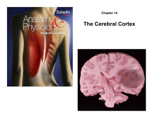
Chapter 14<br />
<strong>The</strong> <strong>Cerebral</strong> <strong>Cortex</strong>
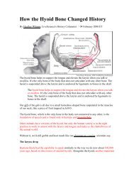
Cerebrum - Gross Anatomy<br />
<strong>Cerebral</strong><br />
hemispheres<br />
Rostral<br />
Caudal<br />
Central sulcus<br />
Frontal lobe<br />
Gyri<br />
Lateral sulcus<br />
Cerebrum<br />
Central sulcus<br />
Temporal lobe<br />
Cerebellum<br />
Parietal lobe<br />
Brainstem<br />
Occipital lobe<br />
Spinal cord<br />
Longitudinal fissure<br />
(b) Lateral view<br />
(a) Superior view<br />
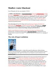
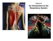
• two cerebral hemispheres divided by longitudinal fissure<br />
– connected by white fibrous tract the corpus callosum<br />
– gyri and sulci – increases amount of cortex in the cranial cavity<br />
– gyri increases surface area for information processing capability<br />
– some sulci divide each hemisphere into five lobes named for the<br />
cranial bones that overly them
Functions of Cerebrum - Lobes<br />
• frontal lobe<br />
– voluntary motor functions<br />
– motivation, foresight, planning,<br />
memory, mood, emotion, social<br />
judgment, and aggression<br />
Precentral<br />
gyrus<br />
Frontal lobe<br />
Rostral<br />
Caudal<br />
Parietal lobe<br />
• parietal lobe<br />
– receives and integrates general<br />
sensory information, taste and<br />
some visual processing<br />
Central<br />
sulcus<br />
Insula<br />
Postcentral gyrus<br />
Occipital lobe<br />
• occipital lobe<br />
– primary visual center of brain<br />
Lateral sulcus<br />
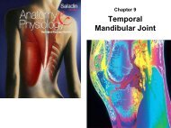
• temporal lobe<br />
– areas for hearing, smell, learning,<br />
memory, and some aspects of<br />
vision and emotion<br />
Temporal lobe<br />
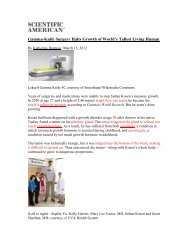
• insula (hidden by other regions)<br />
– understanding spoken language,<br />
taste and sensory information from<br />
visceral receptors
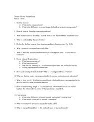
Insula of Dissected Brain<br />
Precentral<br />
gyrus<br />
Central sulcus<br />
leaves<br />
Postcentral<br />
gyrus<br />
Frontal lobe<br />
Parietal lobe<br />
Insula<br />
Arachnoid<br />
mater<br />
Temporal lobe<br />
Occipital lobe<br />
Cerebellum<br />
Blood vessels<br />
Medulla<br />
oblongata<br />
(c) Lateral view<br />
© <strong>The</strong> McGraw-Hill Companies, Inc./Rebecca Gray, photographer/Don Kincaid, dissections
<strong>The</strong> <strong>Cerebral</strong> White Matter<br />
• most of the volume of cerebrum is white matter<br />
– glia and myelinated nerve fibers transmitting signals from one<br />
region of the cerebrum to another and between cerebrum and<br />
lower brain centers<br />
• three types of tracts<br />
– projection tracts<br />
• extends vertically between higher and lower brain and spinal cord<br />
centers<br />
• Only tracts to carry information between cerebrum and rest of the<br />
body<br />
– commissural tracts<br />
• cross from one cerebral hemisphere to other side through bridges<br />
called commissures<br />
– most pass through corpus callosum<br />
– anterior and posterior commissures<br />
– enables the two sides of the cerebrum to communicate with each other<br />
– association tracts<br />
• connect different regions within the same cerebral hemisphere<br />
• long association fibers – connect different lobes of a hemisphere<br />
to each other<br />
• short association fibers – connect different gyri within a single lobe
<strong>Cerebral</strong> White Matter<br />
Association tracts<br />
Projection tracts<br />
Frontal lobe<br />
Corpus callosum<br />
Parietal lobe<br />
Temporal lobe<br />
Occipital lobe<br />
(a) Sagittal section<br />
Longitudinal fissure<br />
Corpus callosum<br />
Commissuralta tracts<br />
Lateral ventricle<br />
Basal nuclei<br />
<strong>Cerebral</strong> peduncle<br />
Thalamus<br />
Third ventricle<br />
Mammillary body<br />
Projection tracts<br />
Decussation in pyramids<br />
pons<br />
Pyramid<br />
Medulla oblongata<br />
(b) Frontal section
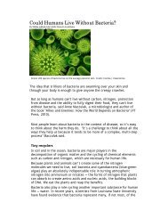
<strong>Cerebral</strong> <strong>Cortex</strong><br />
• neural integration is carried<br />
out in the gray matter of the<br />
cerebrum<br />
Copyright © <strong>The</strong> McGraw-Hill Companies, Inc. Permission required for reproduction or display.<br />
• cerebral gray matter found in<br />
three places<br />
– cerebral cortex<br />
– basal nuclei<br />
– limbic system<br />
I<br />
II<br />
Cortical surface<br />
Small pyramidal<br />
cells<br />
• cerebral cortex – layer<br />
covering the surface of the<br />
hemispheres<br />
– only 2 – 3 mm thick<br />
– cortex constitutes about 40%<br />
of the mass of the brain<br />
– contains 14 – 16 billion<br />
neurons<br />
III<br />
IV<br />
V<br />
VI<br />
Stellate cells<br />
Large pyramidal<br />
cells<br />
White<br />
matter
<strong>Cerebral</strong> <strong>Cortex</strong><br />
– contains two principal types of<br />
neurons<br />
• stellate cells<br />
– have spheroid somas<br />
with dendrites<br />
projecting in all<br />
directions<br />
– receive sensory input<br />
and process information<br />
on a local level<br />
• pyramidal cells<br />
– tall, and conical, with<br />
apex toward the brain<br />
surface<br />
– a thick dendrite with<br />
many branches with<br />
small, knobby dendritic<br />
spines<br />
– include the output<br />
neurons of the<br />
cerebrum<br />
– only neurons that leave<br />
the cortex and connect<br />
with other parts of the<br />
CNS<br />
• neocortex – six layered tissue that<br />
constitutes about 90% of the<br />
human cerebral cortex<br />
– relatively recent in evolutionary origin<br />
Copyright © <strong>The</strong> McGraw-Hill Companies, Inc. Permission required for reproduction or display.<br />
I<br />
II<br />
III<br />
IV<br />
V<br />
VI<br />
White<br />
matter<br />
Cortical surface<br />
Small pyramidal<br />
cells<br />
Stellate cells<br />
Large pyramidal<br />
cells
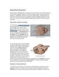
<strong>The</strong> Basal Nuclei<br />
Copyright © <strong>The</strong> McGraw-Hill Companies, Inc. Permission required for reproduction or display.<br />
Cerebrum<br />
Corpus callosum<br />
Lateral ventricle<br />
Thalamus<br />
Internal capsule<br />
Insula<br />
Third ventricle<br />
Hypothalamus<br />
Pituitary gland<br />
Caudate nucleus<br />
Putamen<br />
Globus pallidus<br />
Subthalamic nucleus<br />
Optic tract<br />
Lentiform<br />
nucleus<br />
Corpus<br />
striatum<br />
• Masses of cerebral gray matter buried deep in the white matter, lateral to the<br />
thalamus<br />
– receives input from the substantia nigra of the midbrain and the motor areas of the cortex<br />
– send signals back to both these locations<br />
– involved in motor control<br />
• at least three brain centers form basal nuclei<br />
– caudate nucleus<br />
– putamen<br />
– globus pallidus<br />
• lentiform nucleus – putamen and globus pallidus collectively<br />
• corpus striatum – putamen and caudate nucleus collectively
• important center of emotion and<br />
learning<br />
• most anatomically prominent<br />
components are:<br />
– cingulate gyrus – arches over<br />
the top of the corpus callosum<br />
in the frontal and parietal lobes<br />
– hippocampus – in the medial<br />
temporal lobe – memory<br />
(formed here but not stored<br />
here)<br />
– amygdala – immediately<br />
rostral to the hippocampus -<br />
emotion<br />
• limbic system components are<br />
connected through a complex<br />
loop of fiber tracts allowing for<br />
somewhat circular patterns of<br />
feedback<br />
• limbic system structures have<br />
centers for both gratification<br />
and aversion<br />
– gratification – sensations of<br />
pleasure or reward<br />
– aversion –sensations of fear or<br />
sorrow<br />
Limbic System<br />
Medial<br />
prefrontal<br />
cortex<br />
Corpus<br />
callosum<br />
Cingulate<br />
gyrus<br />
Orbitofrontal<br />
cortex<br />
Basal nuclei<br />
Amygdala<br />
Temporal lobe<br />
Copyright © <strong>The</strong> McGraw-Hill Companies, Inc. Permission required for reproduction or display.<br />
Fornix<br />
Thalamic<br />
nuclei<br />
Mammillary<br />
body<br />
Hippocampus
Note: Review Video “Brain Stem Model”