Urinalysis
Urinalysis: Bayer's Multistix-10 SG Reagent Strips for Urinalysis
Urinalysis: Bayer's Multistix-10 SG Reagent Strips for Urinalysis
- No tags were found...

You also want an ePaper? Increase the reach of your titles
YUMPU automatically turns print PDFs into web optimized ePapers that Google loves.
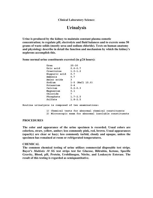
Clinical Laboratory Science:<br />
<strong>Urinalysis</strong><br />
Urine is produced by the kidney to maintain constant plasma osmotic<br />
concentration; to regulate pH, electrolyte and fluid balances and to excrete some 50<br />
grams of waste solids (mostly urea and sodium chloride). Texts on human anatomy<br />
and physiology describe in detail the function and mechanism by which the kidney's<br />
nephrons accomplish this.<br />
Some normal urine constituents excreted (in g/24 hours):<br />
Urea 25-30<br />
Uric acid 0.6-0.7<br />
Creatinine 1.0-1.2<br />
Hippuric acid 0.7<br />
Ammonia 0.7<br />
Amino acids 3<br />
Sodium 1-5 (NaCl 15.0)<br />
Potassium 2-4<br />
Calcium 0.2-0.3<br />
Magnesium 0.1<br />
Chloride 7<br />
Phosphate 1.7-2.5<br />
Sulfate 1.8-2.5<br />
Routine urinalysis is composed of two examinations:<br />
PROCEDURES<br />
1) Chemical tests for abnormal chemical constituents<br />
2) Microscopic exam for abnormal insoluble constituents<br />
The color and appearance of the urine specimen is recorded. Usual colors are<br />
colorless, straw, yellow, amber; less commonly pink, red, brown. Usual appearances<br />
(opacity) are clear or hazy; less commonly turbid, cloudy and opaque, unless the<br />
specimen has remained at room or refrigerated temperatures.<br />
CHEMICAL<br />
The common chemical testing of urine utilizes commercial disposable test strips.<br />
Bayer's Multistix 10 SG test strips test for Glucose, Bilirubin, Ketone, Specific<br />
Gravity, Blood, pH, Protein, Urobilinogen, Nitrite, and Leukocyte Esterase. The<br />
result of this testing is regarded as semiquantitative.
A fresh urine specimen is collected in a clean, dry container. A Multistix strip is<br />
briefly immersed in the urine specimen, covering all reagent areas.<br />
The edge of the Multistix strip is run against the rim of the urine container to<br />
remove excess urine. The strip is held in a horizontal<br />
METHODOLOGIES AND INTERPRETATIONS<br />
Glucose:<br />
This test is based on a double sequential enzyme reaction. One enzyme, glucose<br />
oxidase, catalyzes the formation of gluconic acid and hydrogen peroxide from the<br />
oxidation of glucose. A second enzyme, peroxidase, catalyzes the reaction of<br />
hydrogen peroxide with a potassium iodide chromogen to oxidize the chromogen to<br />
colors ranging from green to brown.<br />
In general the presence of glucose indicates that the filtered load of glucose exceeds<br />
the maximal tubular reabsorptive capacity for glucose. In diabetes mellitus, urine<br />
testing for glucose is often substituted for blood glucose monitoring.
Bilirubin:<br />
This test is based on the coupling of bilirubin with diazotized dichloroanaline in a<br />
strongly acid medium. The color ranges through various shades of tan.<br />
Bilirubin in the urine indicates the presence of liver disease or biliary obstruction.<br />
Very low amounts of bilirubin can be detected in the urine, even when serum levels<br />
are below the clinical detection of jaundice.<br />
Ketone:<br />
This test is based on the development of colors ranging from buff-pink, for a<br />
negative reading, to purple when acetoacetic acid reacts with nitroprusside.<br />
Urine testing only detects acetoacetic acid, not the other ketones, acetone or betahydroxybuteric<br />
acid. In ketoacidosis (insulin deficiency or starvation), it can be<br />
present in large amounts in the urine before any elevation in plasma levels.<br />
Specific Gravity:<br />
This test is based on the apparent pKa change of certain pretreated polyelectrolytes,<br />
poly(methyl-vinyl-ether/maleic anhydride), in relation to ionic concentration. In the<br />
presence of bromthymol blue, colors range deep blue-green in urine of low ionic<br />
concentration through green and yellow-green in urines of increasing ionic<br />
concentration.<br />
The specific gravity is a convenient index of urine concentration. It measures<br />
density and is only an approximate guide to true concentration. A specific gravity of<br />
1.025, in the<br />
absence of protein, glucose and other large molecular weight substances such as<br />
contrast media, usually indicates normal renal concentration and makes chronic<br />
renal insufficiency unlikely.
Blood:<br />
This test is based on the peroxidase-like activity of hemoglobin, which catalyzes the<br />
reaction of diisopropylbenzene dihydroperoxide and 3,3',5,5'-tetramethylbenzidine.<br />
The resulting color ranges from orange through green; very high levels of blood<br />
may cause the color development to continue to blue.<br />
The presence of large numbers of RBCs in the urine sediment establishes the<br />
diagnosis of hematuria. If the dipstick is more strongly positive than would be<br />
expected from the number of RBCs, then the possibility of hemoglobinuria or<br />
myoglobinuria should be considered.<br />
pH:<br />
The test is based on the double indicator (methyl red/bromthymol blue) principle<br />
that gives a broad range of colors covering the entire urinary pH range. Colors<br />
range from orange through yellow and green to blue.<br />
The urine pH should be recorded, although it is seldom of diagnostic value.<br />
Phosphates will precipitate in an alkaline urine, and uric acid will precipitate in an<br />
acidic urine.<br />
Protein:<br />
This test is based on the protein-error-of-indicators (tetrabromphenol blue)<br />
principle. At a constant pH, the development of any green color is due to the<br />
presence of protein. Colors range from yellow for negative through yellow-green<br />
and green to green-blue for positive reactions.<br />
Heavy proteinuria usually represents an abnormality in the glomerular filtration<br />
barrier. The test is more sensitive for albumin than for globulins or hemoglobin.<br />
Urobilinogen:<br />
This test is based on the modified Ehrlich reaction, in which paradiethylaminobenzaldehyde<br />
in conjunction with a color enhancer reacts with<br />
urobilinogen in a strongly acid medium to produce a pink-red color.
Urine urobilinogen is increased in any condition that causes an increase in<br />
production or retention of bilirubin.<br />
Nitrite:<br />
This test depends upon the conversion of nitrate (derived from the diet) to nitrite by<br />
the action of Gram negative bacteria in the urine. At the acid pH of the reagent<br />
area, nitrite in the urine reacts with para-arsanilic acid to form a diazonium<br />
compound. This diazonium compound in turn couples with 1,2,3,4-<br />
tetrahydrobenzo(h)quinoline-3-ol to produce a pink color.<br />
Bacteriuria caused by some Gram negative bacteria which produce the nitrate<br />
reductase enzyme give a positive test.<br />
Leukocytes:<br />
Granulocytic leukocytes contain esterases that catalize the hydrolysis of the<br />
derivatized pyrrole amino acid ester to liberate 3-hydroxy-5-phenyl pyrrole. This<br />
pyrrole then reacts with a diazonium salt to produce a purple product.<br />
A positive leukocyte esterase test provides indirect evidence for the presence of<br />
bacteriuria.