The Meninges and The Cerebral Spinal Fluid
The Meninges and The Cerebral Spinal Fluid
The Meninges and The Cerebral Spinal Fluid
- No tags were found...

Create successful ePaper yourself
Turn your PDF publications into a flip-book with our unique Google optimized e-Paper software.
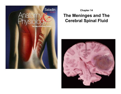
Chapter 14<br />
<strong>The</strong> <strong>Meninges</strong> <strong>and</strong> <strong>The</strong><br />
<strong>Cerebral</strong> <strong>Spinal</strong> <strong>Fluid</strong>
<strong>Meninges</strong> of the Brain<br />
Skull<br />
Brain:<br />
Blood vessel<br />
Pia mater<br />
Gray matter<br />
White matter<br />
Dura mater:<br />
Periosteal layer<br />
Meningeal layer<br />
Arachnoid villus<br />
Arachnoid mater<br />
Subdural space<br />
Subarachnoid<br />
space<br />
Superior sagittal<br />
sinus<br />
Falx cerebri<br />
(in longitudinal<br />
fissure only)
<strong>Meninges</strong><br />
• meninges – three connective tissue membranes that<br />
envelop the brain<br />
– lies between the nervous tissue <strong>and</strong> bone<br />
– as in spinal cord, they are the dura mater, arachnoid mater,<br />
<strong>and</strong> the pia mater<br />
– protect the brain <strong>and</strong> provide structural framework for its arteries<br />
<strong>and</strong> veins<br />
• dura mater<br />
– in cranial cavity - 2 layers<br />
• outer periosteal – equivalent to periosteum of cranial bones<br />
• inner meningeal – continues into vertebral canal <strong>and</strong> forms dural sac<br />
around spinal cord<br />
– cranial dura mater is pressed closely against cranial<br />
bones<br />
• no epidural space<br />
• not attached to bone except: around foramen magnum, sella<br />
turcica, the crista galli, <strong>and</strong> sutures of the skull<br />
• layers separated by dural sinuses – collect blood circulating through<br />
brain<br />
– folds inward to extend between parts of the brain<br />
• falx cerebri separates the two cerebral hemispheres<br />
• tentorium cerebelli separates cerebrum from cerebellum<br />
• falx cerebelli separates the right <strong>and</strong> left halves of cerebellum
<strong>Meninges</strong><br />
• arachnoid mater <strong>and</strong> pia mater are similar<br />
to those in the spinal cord<br />
• arachnoid mater<br />
– transparent membrane over brain surface<br />
– subarachnoid space separates it from pia<br />
mater below<br />
– subdural space separates it from dura mater<br />
above in some places<br />
• pia mater<br />
– very thin membrane that follows contours of<br />
brain, even dipping into sulci<br />
– not usually visible without a microscope
<strong>Meninges</strong> of the Brain<br />
Skull<br />
Brain:<br />
Blood vessel<br />
Pia mater<br />
Gray matter<br />
White matter<br />
Dura mater:<br />
Periosteal layer<br />
Meningeal layer<br />
Arachnoid villus<br />
Arachnoid mater<br />
Subdural space<br />
Subarachnoid<br />
space<br />
Superior sagittal<br />
sinus<br />
Falx cerebri<br />
(in longitudinal<br />
fissure only)
Meningitis<br />
• meningitis - inflammation of the meninges<br />
– serious disease of infancy & childhood<br />
– especially between 3 months <strong>and</strong> 2 years of age<br />
• caused by bacterial <strong>and</strong> virus invasion of the CNS by<br />
way of the nose <strong>and</strong> throat<br />
• pia mater <strong>and</strong> arachnoid are most often affected<br />
• bacterial meningitis can cause swelling the brain,<br />
enlarging the ventricles, <strong>and</strong> hemorrhage<br />
• signs include high fever, stiff neck, drowsiness, <strong>and</strong><br />
intense headache <strong>and</strong> may progress to coma – death<br />
within hours of onset<br />
• diagnosed by examining the CSF for bacteria<br />
– lumbar puncture (spinal tap) draws fluid from subarachnoid<br />
space between two lumbar vertebrae
Brain Ventricles<br />
Copyright © <strong>The</strong> McGraw-Hill Companies, Inc. Permission required for reproduction or display.<br />
Caudal<br />
Rostral<br />
Lateral ventricles<br />
Interventricular<br />
foramen<br />
Third ventricle<br />
<strong>Cerebral</strong><br />
aqueduct<br />
Fourth ventricle<br />
Lateral aperture<br />
Median aperture<br />
Central canal<br />
Cerebrum<br />
Lateral ventricle<br />
Interventricular<br />
foramen<br />
Third ventricle<br />
<strong>Cerebral</strong><br />
aqueduct<br />
Fourth ventricle<br />
Lateral aperture<br />
Median aperture<br />
(a) Lateral view<br />
(b) Anterior view
Ventricles of the Brain<br />
Copyright © <strong>The</strong> McGraw-Hill Companies, Inc. Permission required for reproduction or display.<br />
Rostral (anterior)<br />
Longitudinal<br />
fissure<br />
Frontal lobe<br />
Gray matter<br />
(cortex)<br />
White matter<br />
Lateral ventricle<br />
Temporal lobe<br />
Third ventricle<br />
Lateral sulcus<br />
Insula<br />
Lateral ventricle<br />
Corpus callosum<br />
(anterior part)<br />
Caudate nucleus<br />
Septum<br />
pellucidum<br />
Sulcus<br />
Gyrus<br />
Thalamus<br />
Choroid plexus<br />
Occipital lobe<br />
Corpus callosum<br />
(posterior part)<br />
Longitudinal<br />
fissure<br />
(c)<br />
Caudal (posterior)
Ventricles <strong>and</strong> Cerebrospinal <strong>Fluid</strong><br />
• ventricles – four internal chambers within the brain<br />
– two lateral ventricles – one in each cerebral hemisphere<br />
• interventricular foramen - a tiny pore that connects to third<br />
ventricle<br />
– third ventricle - single narrow medial space beneath<br />
corpus callosum<br />
• cerebral aqueduct runs through midbrain <strong>and</strong> connects third<br />
to fourth ventricle<br />
– fourth ventricle – small triangular chamber between pons<br />
<strong>and</strong> cerebellum<br />
• connects to central canal runs down through spinal cord<br />
• choroid plexus – spongy mass of blood capillaries on<br />
the ceiling of each ventricle<br />
• ependyma – neuroglia that lines the ventricles <strong>and</strong><br />
covers the choroid plexus<br />
– produces 30% of the cerebrospinal fluid
Cerebrospinal <strong>Fluid</strong> (CSF)<br />
• cerebrospinal fluid (CSF) – clear, colorless liquid<br />
that fills the ventricles <strong>and</strong> canals of CNS<br />
– bathes its external surface<br />
• brain produces <strong>and</strong> absorbs 500 mL/day<br />
– 100 – 160 mL normally present at one time<br />
– 40% formed in subarachnoid space external to brain<br />
– 30% by the general ependymal lining of the brain<br />
ventricles<br />
– 30% by the choroid plexuses<br />
• production begins with the filtration of blood<br />
plasma through the capillaries of the brain<br />
– ependymal cells modify the filtrate, so CSF has<br />
more sodium <strong>and</strong> chloride than plasma, but less<br />
potassium, calcium, glucose, <strong>and</strong> very little protein
Cerebrospinal <strong>Fluid</strong> (CSF) Circulation<br />
• CSF continually flows through <strong>and</strong> around the CNS<br />
– driven by its own pressure, beating of ependymal cilia, <strong>and</strong><br />
pulsations of the brain produced by each heartbeat<br />
• CSF secreted in lateral ventricles flows through<br />
intervertebral foramina into third ventricle<br />
• then down the cerebral aqueduct into the fourth<br />
ventricle<br />
• third <strong>and</strong> fourth ventricles add more CSF along the way<br />
• small amount of CSF fills the central canal of the spinal<br />
cord<br />
– all escapes through three pores<br />
• median aperture <strong>and</strong> two lateral apertures<br />
• leads into subarachnoid space of brain <strong>and</strong> spinal cord surface<br />
• CSF is reabsorbed by arachnoid villi<br />
– cauliflower-shaped extension of the arachnoid meninx<br />
– protrudes through dura mater<br />
– into superior sagittal sinus<br />
– CSF penetrates the walls of the villi <strong>and</strong> mixes with the blood<br />
in the sinus
Functions of CSF<br />
• buoyancy<br />
– allows brain to attain considerable size without being<br />
impaired by its own weight<br />
– if it rested heavily on floor of cranium, the pressure<br />
would kill the nervous tissue<br />
• protection<br />
– protects the brain from striking the cranium when the<br />
head is jolted<br />
– shaken child syndrome <strong>and</strong> concussions do occur<br />
from severe jolting<br />
• chemical stability<br />
– flow of CSF rinses away metabolic wastes from<br />
nervous tissue <strong>and</strong> homeostatically regulates its<br />
chemical environment
Flow of Cerebrospinal <strong>Fluid</strong><br />
Copyright © <strong>The</strong> McGraw-Hill Companies, Inc. Permission required for reproduction or display.<br />
8<br />
Arachnoid villus<br />
Superior<br />
sagittal<br />
sinus<br />
Arachnoid mater<br />
1<br />
2<br />
CSF is secreted by<br />
choroid plexus in<br />
each lateral ventricle.<br />
CSF flows through<br />
Interventricular foramina<br />
into third ventricle.<br />
2<br />
1<br />
Subarachnoid<br />
space<br />
Dura mater<br />
Choroid plexus<br />
3<br />
Choroid plexus in third<br />
ventricle adds more CSF.<br />
3<br />
Third ventricle<br />
4<br />
CSF flows down cerebral<br />
aqueduct to fourth ventricle.<br />
4<br />
7<br />
<strong>Cerebral</strong><br />
aqueduct<br />
Lateralaper ture<br />
5<br />
Choroid plexus in fourth<br />
ventricle adds more CSF.<br />
Fourth ventricle<br />
6<br />
CSF flows out two lateral apertures<br />
<strong>and</strong> one median aperture.<br />
6<br />
5<br />
7<br />
CSF fills subarachnoid space <strong>and</strong><br />
bathes external surfaces of brain<br />
<strong>and</strong> spinal cord.<br />
7<br />
Median aperture<br />
8<br />
At arachnoid villi, CSF is reabsorbed<br />
into venous blood of dural<br />
venous sinuses.<br />
Centralcanal<br />
of spinal cord<br />
Subarachnoid<br />
space of<br />
spinal cord
Blood Supply to the Brain<br />
• brain is only 2% of the adult body weight, <strong>and</strong><br />
receives 15% of the blood<br />
– 750 mL/min<br />
• neurons have a high dem<strong>and</strong> for ATP, <strong>and</strong><br />
therefore, oxygen <strong>and</strong> glucose, so a constant<br />
supply of blood is critical to the nervous system<br />
– 10 second interruption of blood flow may cause loss of<br />
consciousness<br />
– 1 – 2 minute interruption can cause significant<br />
impairment of neural function<br />
– 4 minutes with out blood causes irreversible brain<br />
damage
Brain Barrier System<br />
• blood is also a source of antibodies,<br />
macrophages, bacterial toxins, <strong>and</strong> other<br />
harmful agents<br />
• brain barrier system – strictly regulates<br />
what substances can get from the<br />
bloodstream into the tissue fluid of the brain<br />
• two points of entry must be guarded:<br />
– blood capillaries throughout the brain tissue<br />
– capillaries of the choroid plexus
Brain Barrier System<br />
• blood-brain barrier - protects blood capillaries throughout brain<br />
tissue<br />
– consists of tight junctions between endothelial cells that form<br />
the capillary walls<br />
– astrocytes reach out <strong>and</strong> contact capillaries with their<br />
perivascular feet<br />
– induce the endothelial cells to form tight junctions that<br />
completely seal off gaps between them<br />
– anything leaving the blood must pass through the cells, <strong>and</strong><br />
not between them<br />
– endothelial cells can exclude harmful substances from<br />
passing to the brain tissue while allowing necessary ones to<br />
pass<br />
• blood-CSF barrier - protects the brain at the choroid plexus<br />
– form tight junctions between the ependymal cells<br />
– tight junctions are absent from ependymal cells elsewhere<br />
• important to allow exchange between brain tissue <strong>and</strong><br />
CSF
Brain Barrier System<br />
• blood barrier system is highly permeable to water, glucose,<br />
<strong>and</strong> lipid-soluble substances such as oxygen, carbon<br />
dioxide, alcohol, caffeine, nicotine, <strong>and</strong> anesthetics<br />
• slightly permeable to sodium, potassium, chloride, <strong>and</strong> the<br />
waste products urea <strong>and</strong> creatinine<br />
• obstacle for delivering medications such as antibiotics <strong>and</strong><br />
cancer drugs<br />
• trauma <strong>and</strong> inflammation can damage BBS <strong>and</strong> allow<br />
pathogens to enter brain tissue<br />
• Circumventricular organs (CVOs) – places in the third <strong>and</strong><br />
fourth ventricles where the barrier is absent<br />
• blood has direct access to the brain<br />
• enables the brain to monitor <strong>and</strong> respond to fluctuations<br />
in blood glucose, pH, osmolarity, <strong>and</strong> other variables<br />
• CVOs afford a route for invasion by the human<br />
immunodeficiency virus (HIV)