Hydronephrosis: Prenatal Diagnosis - UCSF Department of Urology
Hydronephrosis: Prenatal Diagnosis - UCSF Department of Urology
Hydronephrosis: Prenatal Diagnosis - UCSF Department of Urology
- No tags were found...

You also want an ePaper? Increase the reach of your titles
YUMPU automatically turns print PDFs into web optimized ePapers that Google loves.
<strong>UCSF</strong> Pediatric <strong>Urology</strong><br />
Child and Family Information Material<br />
------------------------------------------------------------------------<br />
<strong>Hydronephrosis</strong>: <strong>Prenatal</strong> <strong>Diagnosis</strong><br />
What is hydronephrosis?<br />
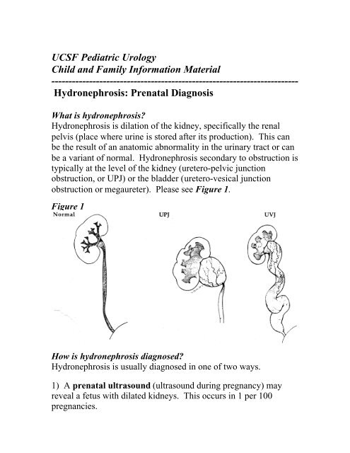
<strong>Hydronephrosis</strong> is dilation <strong>of</strong> the kidney, specifically the renal<br />
pelvis (place where urine is stored after its production). This can<br />
be the result <strong>of</strong> an anatomic abnormality in the urinary tract or can<br />
be a variant <strong>of</strong> normal. <strong>Hydronephrosis</strong> secondary to obstruction is<br />
typically at the level <strong>of</strong> the kidney (uretero-pelvic junction<br />
obstruction, or UPJ) or the bladder (uretero-vesical junction<br />
obstruction or megaureter). Please see Figure 1.<br />
Figure 1<br />
How is hydronephrosis diagnosed?<br />
<strong>Hydronephrosis</strong> is usually diagnosed in one <strong>of</strong> two ways.<br />
1) A prenatal ultrasound (ultrasound during pregnancy) may<br />
reveal a fetus with dilated kidneys. This occurs in 1 per 100<br />
pregnancies.
2) An ultrasound done as a routine evaluation for another<br />
medical problem, such as a urinary tract infection or incontinence,<br />
may also reveal hydronephrosis.<br />
Once hydronephrosis is noted, whether is it during pregnancy or<br />
later, additional tests are <strong>of</strong>ten required in order to find out the<br />
significance <strong>of</strong> the hydronephrosis. These tests are important<br />
because children with hydronephrosis may have an anatomic<br />
abnormality or urinary tract blockage. Early diagnosis and<br />
treatment <strong>of</strong> a potential urologic abnormality can prevent urinary<br />
tract infections and permanent kidney damage or scarring.<br />
What, if any, other test should be done?<br />
• VCUG (voiding cystourethrogram): This study gives us<br />
important information regarding the shape and size <strong>of</strong> the<br />
bladder, the bladder neck (or opening) and the tubes that drain<br />
the urine from the kidneys into the bladder, called ureters. It<br />
allows us to diagnose reflux (the abnormal back-flow <strong>of</strong> urine<br />
from the bladder into the ureter and up to the kidney). It also<br />
gives us additional anatomic information about the urethra<br />
(urine tube which takes urine from the bladdder outside the<br />
body) to make sure no blockage is present (posterior urethral<br />
valves).<br />
• Kidney (Renal) Scan: This test may be done depending on the<br />
history <strong>of</strong> urinary tract infection(s), result <strong>of</strong> VCUG, and/or the<br />
severity <strong>of</strong> the hydronephrosis. It is used to better demonstrate<br />
the actual function and/or drainage <strong>of</strong> the kidneys. A kidney<br />
scan can also show if there is kidney damage and/or scarring<br />
that may have resulted from a previous urinary tract infection or<br />
long-standing hydronephrosis. Two types <strong>of</strong> renal scans are<br />
typically performed depending on the diagnosis.
1. Lasix Renogram or MAG-III diuretic renogram to test for<br />
significant blockage in the urinary tract, OR<br />
2. DMSA renal scan to test for scarring or damage to the renal<br />
tissue (more comon in patients with vesico-ureteral reflux).<br />
When should these tests be performed if a prenatal ultrasound<br />
showed hydronephrosis?<br />
If your newborn child had hydronephrosis (kidney, ureter or<br />
bladder dilation) noted on a screening pre-natal ultrasound, a<br />
repeat ultrasound should be conducted after the first 1 to 3 days <strong>of</strong><br />
life. It is normal for a newborn to be dehydrated, and make less<br />
urine, on the first day <strong>of</strong> life, so it may falsely appear that<br />
hydronephosis has gone. A VCUG will be performed next, within<br />
the next several weeks <strong>of</strong> life. Certain conditions seen on the<br />
ultrasound may warrant a more expeditious work-up and we will<br />
let you know if this is necessary (for example, in the event <strong>of</strong><br />
severe hydronephrosis in both kidneys or a dilated bladder).<br />
How is hydronephrosis graded and why is this important?<br />
<strong>Hydronephrosis</strong> is graded on a scale from zero to four, with one<br />
being the mildest form and four being severe. Please see Figure<br />
2. The degree <strong>of</strong> hydronephrosis is used to assist in decision<br />
making with regard to treating the underlying cause <strong>of</strong> the<br />
hydronephrosis and the ultimate prognosis <strong>of</strong> patients. More<br />
severe grades <strong>of</strong> hydronephrosis are associated with closer<br />
pediatric urology follow-up. For example, grade III and IV<br />
hydronephrosis (not due to vesicoureteral reflux) typically require<br />
a renal scan.
Figure 2<br />
(SFU Grading <strong>of</strong> <strong>Hydronephrosis</strong>)
Why does hydronephrosis occur?<br />
There are numerous reasons why hydronephrosis occurs. Please<br />
see the list <strong>of</strong> potential diagnoses below:<br />
1) Vesicoureteral reflux<br />
2) Non-obstructive hydronephrosis<br />
3) Ureteropelvic junction (UPJ) obstruction<br />
4) Ureterocele<br />
5) Posterior urethral valves<br />
6) Ureterovesical junction (UVJ) obstruction<br />
7) Megaureter<br />
8) Multicystic Dysplastic Kidney<br />
9) Ectopic ureter<br />
10) Neurogenic/nonneurogenic bladder<br />
This list is quite extensive, but most <strong>of</strong>ten the cause <strong>of</strong> the<br />
hydronephrosis is from one <strong>of</strong> the first three (in bold) diagnoses.<br />
The special x-ray tests mentioned previously will help us to find<br />
the cuase <strong>of</strong> the hydronephrosis.<br />
Will my child require any medication to assist in treating the<br />
hydronephrosis?<br />
If your child is not presently on antibiotics, he or she will receive a<br />
prescription at the time <strong>of</strong> your visit to our <strong>of</strong>fice. Your child will<br />
receive antiobiotics in a low dose and on a daily basis. The types<br />
<strong>of</strong> antibiotics are very specific for the urinary tract and have very<br />
few, if any, side effects. The specific type <strong>of</strong> antiobitics will<br />
depend upon your child's age, weight and allergies. The goal <strong>of</strong><br />
antibiotics is to prevent kidney infections that may occur as a result<br />
<strong>of</strong> the hydronephrosis. Once the special x-ray tests have been<br />
completed, we will be able to estimate the total time <strong>of</strong> antibiotic<br />
treatment.
Will the hydronephrosis go away or will my child require<br />
surgery?<br />
Typically, non-obstructive hydronephrosis (ie, hydronephrosis<br />
secondary to dilation at the ureterovesical junction, the place where<br />
the ureter meets the bladder; please see Figure 3) and grade 1 to 3<br />
hydronephrosis secondary to uretero-pelvic junction type<br />
hydronephrosis do not need surgical intervention and resolve over<br />
time. The timing <strong>of</strong> resolution depends on the severity <strong>of</strong> the<br />
hydronephrosis and is different for each child. Children diagnosed<br />
with dilation from uretervesical junction abnormalities called<br />
megaureters rarely if ever need surgical repair. Patients with grade<br />
IV hydronephrosis (severe) are the most likely to require surgery to<br />
prevent renal damage and recurrent infection.<br />
Figure 3