COMBINATION MODIFIED ACID-FAST MODIFIED TRICHROME STAIN (Coccidia Microsporidia)
Combination Modified Acid Fast, Modified Trichrome - Medical ...
Combination Modified Acid Fast, Modified Trichrome - Medical ...
- No tags were found...

You also want an ePaper? Increase the reach of your titles
YUMPU automatically turns print PDFs into web optimized ePapers that Google loves.
<strong>COMBINATION</strong> <strong>MODIFIED</strong> <strong>ACID</strong>-<strong>FAST</strong>,<br />
<strong>MODIFIED</strong> <strong>TRICHROME</strong> <strong>STAIN</strong><br />
(<strong>Coccidia</strong>, <strong>Microsporidia</strong>)<br />
Preanalytical Considerations<br />
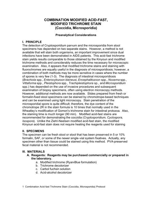
I. PRINCIPLE<br />
The detection of Cryptosporidium parvum and the microsporidia from stool<br />
specimens has depended on two separate stains. However, a method is not<br />
available that will stain both organisms, an important improvement since dual<br />
infections have been demonstrated in AIDS patients. This acid-fast trichrome<br />
stain yields results comparable to those obtained by the Kinyoun and modified<br />
trichrome methods and considerably reduces the time necessary for microscopic<br />
examination. Also, it appears that modified trichrome stains and staining with<br />
fluorochromes are equally useful in the diagnosis of microsporidiosis; however, a<br />
combination of both methods may be more sensitive in cases where the number<br />
of spores is very few (1-3). The diagnosis of intestinal microsporidiosis<br />
(Brachiola spp., Enterocytozoon bieneusi, Encephalitozoon spp., Nosema spp.,<br />
Vittaforma spp., Pleistophora spp., Trachipleistophora sp., and Microsporidium<br />
spp.) has depended on the use of invasive procedures and subsequent<br />
examination of biopsy specimens, often using electron microscopy methods.<br />
However, additional methods are now available. Slides prepared from fresh or<br />
formalin-fixed stool specimens can be stained by chromotrope-based techniques<br />
and can be examined using light microscopy. Stain penetration of the<br />
microsporidial spore is quite difficult; therefore, the dye content of the<br />
chromotrope 2R in the stain formula is 10 times that normally used in the<br />
Wheatley’s modification of Gomori’s trichrome stain for intestinal protozoa. Also,<br />
the staining time is much longer (90 min). Modified acid-fast stains are<br />
recommended for demonstrating the coccidia (Cryptosporidium, Cyclospora,<br />
Isospora). Unlike the Ziehl-Neelsen modified acid-fast stain, the modified<br />
Kinyoun acid-fast stain does not require heating the reagents used for staining<br />
II. SPECIMENS<br />
The specimen can be fresh stool or stool that has been preserved in 5 or 10%<br />
formalin, SAF, or some of the newer single-vial system fixatives. Actually, any<br />
specimen other than tissue could be stained using this method. PVA-preserved<br />
fecal material is not recommended.<br />
III. MATERIALS<br />
A. Reagents: Reagents may be purchased commercially or prepared in<br />
the laboratory.<br />
a. Modified trichrome (Ryan-Blue formulation)<br />
b. Trichrome decolorizer<br />
c. Carbol fuchsin solution<br />
d. Acid-alcohol decolorizer<br />
1 Combination Acid-fast Trichrome Stain (<strong>Coccidia</strong>, <strong>Microsporidia</strong>) Protocol
B. Supplies<br />
a. Glass slides (25 by 75 mm), frosted ends desirable<br />
b. Coverslips (22 by 22 mm; no. 1)<br />
c. Glass or plastic centrifuge tubes<br />
d. Pasteur pipettes<br />
e. Coplin jars or other suitable staining containers<br />
f. Immersion oil<br />
C. Equipment: Optional materials, depending on specimen source of<br />
laboratory protocol<br />
a. Binocular microscope with 10X, 40X, and 100X objectives (or the<br />
approximate magnifications for low power, high dry power, and oil<br />
immersion examination).<br />
b. Oculars should be 10X. Some workers prefer 5X; however, overall<br />
smaller magnification may make final organism identifications more<br />
difficult.<br />
c. Tabletop centrifuge<br />
Analytical Considerations<br />
IV. QUALITY CONTROL<br />
A. Unfortunately, the only way to perform acceptable QC procedures for<br />
this method is to use actual microsporidial spores and coccidian<br />
oocysts as the control organisms. Obtaining these positive controls<br />
may be somewhat difficult. It is particularly important to use the actual<br />
organisms because the spores are difficult to stain and the size is very<br />
small (spores: 1 to 1.5 µm)(oocysts: 4 to 10 µm).<br />
B. A QC slide should be included with each run of stained slides,<br />
particularly if the staining setup is used infrequently.<br />
C. All staining dishes should be covered to prevent evaporation of<br />
reagents (screw-cap Coplin jars or glass lids).<br />
D. Depending on the volume of slides stained, staining solutions will have<br />
to be changed on an as-needed basis.<br />
E. When the smear is thoroughly fixed and the stain is performed<br />
correctly, the spores will be ovoid and refractile, with the spore wall<br />
being bright pinkish red. Occasionally, the polar tube can be seen<br />
either as a stripe or as a diagonal line across the spore. The majority of<br />
the bacteria and other debris will tend to stain blue. However, there will<br />
still be some bacteria and debris that will stain red. The coccidia<br />
(Cryptosporidium, Cyclospora, Isospora) will stain as with any modified<br />
acid-fast stain: from pink to violet (some Cyclospora may not take the<br />
stain – acid-fast variable).<br />
F. The specimen is also checked for adherence to the slide<br />
(macroscopically).<br />
G. The microscope should be calibrated, and the objectives and oculars<br />
used for the calibration procedure should be used for all<br />
2 Combination Acid-fast Trichrome Stain (<strong>Coccidia</strong>, <strong>Microsporidia</strong>) Protocol
measurements on the microscope. The calibration factors for all<br />
objectives should be posted on the microscope for easy access<br />
(multiplication factors can be pasted on the body of the microscope).<br />
Although recalibration every 12 months may not be necessary, this will<br />
vary from laboratory to laboratory, depending on equipment care and<br />
use. Although there is not universal agreement, the microscope<br />
should probably be recalibrated once each year. This<br />
recommendation should be considered with heavy use or if the<br />
microscope has been bumped or moved multiple times. If the<br />
microscope does not receive heavy use, then recalibration is not<br />
recommended on a yearly basis.<br />
H. Known positive microscope slides, Kodachrome 2 x 2 projection<br />
slides, and photographs (reference books) should be available at the<br />
work station.<br />
I. Record all QC results; the laboratory should also have an action plan<br />
for ``out of control'' results.<br />
V. PROCEDURE<br />
A. Using a 10-µl aliquot of concentrated (formalin ethyl-acetate<br />
sedimentation concentration; 500 X g for 10 min centrifugation),<br />
preserved liquid stool (5 or 10% formalin or SAF), prepare the smear<br />
by spreading the material over an area of 45 by 25 mm.<br />
B. Allow the smear to air dry.<br />
C. Place the smear in absolute methanol for 5 or 10 min.<br />
D. Allow the smear to air dry.<br />
E. Place in carbol-fuchsin solution for 10 min (no heat required).<br />
F. Briefly rinse with tap water.<br />
G. Decolorize with 0.5% acid-alcohol.<br />
H. Briefly rinse with tap water.<br />
I. Place in trichrome stain for 30 min at 37°C.<br />
J. Rinse in acid-alcohol for no more than 10 s (1 to 3 s).<br />
K. Briefly rinse; dip slides several times in 95% alcohol. Use this step as a<br />
rinse (no more than 10 s).<br />
L. Place in 95% alcohol for 30 s.<br />
M. Allow slides to air dry.<br />
N. Examine smears under oil immersion (1,000 x) and read at least 100<br />
fields; the examination time will probably be at least 10 min per slide.<br />
VI. RESULTS<br />
A. <strong>Microsporidia</strong> spores might be seen. The spore wall should stain<br />
pinkish to red, with the interior of the spore being clear or perhaps<br />
showing a horizontal or diagonal stripe that represents the polar tube.<br />
The background will appear blue (Ryan Stain). A vacuole may also be<br />
visible in some spores. The coccidian oocysts will stain bright pink or<br />
3 Combination Acid-fast Trichrome Stain (<strong>Coccidia</strong>, <strong>Microsporidia</strong>) Protocol
violet. The background will appear green. If Cyclospora oocysts are<br />
present (uncommon), they tend to be approximately 10 µm, they<br />
resemble C. parvum but are larger, and they have no definite internal<br />
morphology; the acid-fast staining will tend to be more variable than<br />
that seen with Cryptosporidium or Isospora spp. Modified acid-fast<br />
stains stain the Cyclospora oocysts from light pink to deep red, some<br />
of which will contain granules or have a bubbly appearance, often<br />
being described as looking like “wrinkled cellophane.”<br />
B. Other bacteria, some yeast cells, and some debris will stain pink to<br />
red; the shapes and sizes of the various components may be helpful in<br />
differentiating the spores from other structures.<br />
C. The results from this staining procedure should be reported only if the<br />
positive control smears are acceptable. The production of<br />
immunoassay reagents should provide a more specific and sensitive<br />
approach to the identification of the microsporidia in fecal specimens.<br />
Postanalytical Considerations<br />
VII. REPORTING RESULTS<br />
A. Report the organism and stage (do not use abbreviations<br />
Examples (Stool Specimens): <strong>Microsporidia</strong> spores present<br />
Enterocytozoon bieneusi or Encephalitozoon<br />
(Septata) intestinalis present (if from fecal<br />
specimen); the two organisms cannot be<br />
differentiated on the basis of size or<br />
morphology.<br />
Example from urine: Encephalitozoon (Septata) intestinalis present<br />
(identification to species highly likely);<br />
generally this organism is involved in<br />
disseminated cases from GI tract to kidneys<br />
and will be found in urine.<br />
Example from stool: Cryptosporidium parvum oocysts present.<br />
(Dual Infection) <strong>Microsporidia</strong> spores present<br />
Enterocytozoon bieneusi or Encephalitozoon<br />
(Septata) intestinalis present (if from fecal<br />
specimen); the two organisms cannot be<br />
differentiated on the basis of size or<br />
morphology.<br />
B. Quantitate the number of spores and oocysts seen (rare, few,<br />
moderate, many).<br />
VIII. PROCEDURE NOTES<br />
A. It is mandatory that positive control smears be stained and examined<br />
each time patient specimens are stained and examined.<br />
4 Combination Acid-fast Trichrome Stain (<strong>Coccidia</strong>, <strong>Microsporidia</strong>) Protocol
B. Because of the difficulty in getting stain penetration through the spore<br />
wall, prepare thin smears and do not reduce the staining time in<br />
trichrome. Also, make sure the slides are not left too long in the<br />
decolorizing agent (acid-alcohol). If the control organisms are too light,<br />
leave them in the trichrome longer and shorten the time to two dips in<br />
the acid-alcohol solution. Also, remember that the 95% alcohol rinse<br />
after the acid-alcohol should be performed quickly to prevent<br />
additional destaining from the acid alcohol reagent.<br />
C. In the final stages of dehydration, the 100% ethanol and the xylenes<br />
(or xylene substitutes) should be kept as free from water as possible.<br />
Coplin jars must have tight-fitting caps to prevent both evaporation of<br />
reagents and absorption of moisture. If the xylene becomes cloudy<br />
after addition of slides from 100% alcohol, return the slides to 100%<br />
alcohol and replace the xylene with fresh stock.<br />
D. Polyvinyl alcohol-preserved specimens are not acceptable for staining<br />
with the modified acid-fast stain. However, specimens preserved in<br />
SAF are perfectly acceptable.<br />
E. Avoid the use of wet gauze filtration (an old, standardized method of<br />
filtering stool prior to centrifugation) with too many layers of gauze that<br />
may trap organisms and allow them to flow into the fluid to be<br />
concentrated. It is recommended that no more than two layers of<br />
woven (not pressed) gauze be used; another option is to use the<br />
commercially available concentrators that use no gauze but instead<br />
use plastic or metal screens.<br />
F. Other organisms, such as acid-fast bacteria and some Nocardia spp.,<br />
stain positive.<br />
G. It is very important that smears not be too thick. Thick smears may<br />
not adequately destain.<br />
H. Concentration of the specimen is essential for demonstrating<br />
organisms. The number of organisms seen in the specimens may<br />
vary from numerous to very few.<br />
I. Because of their mucoid consistency, some specimens require<br />
treatment with 10% KOH. Add 10 drops of 10% KOH to the sediment,<br />
and vortex until homogeneous. Rinse with 10% formalin, and<br />
centrifuge (500 x g for 10 min). Without decanting supernatant, take 1<br />
drop of the sediment and smear it thinly on a slide.<br />
J. Commercial concentrators and reagents are available<br />
K. Weak concentrations of sulfuric or hydrochloric acid (1.0 to 3.0%) are<br />
normally used. Stronger concentrations will remove too much stain.<br />
L. There is some debate about whether organisms lose their abilities to<br />
take up the acid-fast stain after long-term storage in 10% formalin.<br />
Use of the hot modified acid-fast method might eliminate this problem.<br />
M. Centrifuge specimens in capped tubes, and wear gloves during all<br />
phases of specimen processing.<br />
5 Combination Acid-fast Trichrome Stain (<strong>Coccidia</strong>, <strong>Microsporidia</strong>) Protocol
IX. LIMITATIONS OF THE PROCEDURE<br />
A. Although this staining method will stain the microsporidia, the range of<br />
stain intensity and the small size of the spores will cause some<br />
difficulty in identifying these organisms. Since this procedure will result<br />
in many other organisms or objects staining in stool specimens,<br />
differentiation of the microsporidia from surrounding material will still be<br />
very difficult. There also tends to be some slight size variation among<br />
the spores.<br />
B. If the patient has severe watery diarrhea, there will be less artifact<br />
material in the stool to confuse with the microsporidial spores;<br />
however, if the stool is semiformed or formed, the amount of artifact<br />
material will be much greater; thus, the spores will be much harder to<br />
detect and identify. Also, remember that the number of spores will vary<br />
according to the stool consistency (the more diarrhetic, the more<br />
spores that will be present).<br />
C. Those who developed some of these procedures feel that<br />
concentration procedures result in an actual loss of microsporidial<br />
spores; thus there is a strong recommendation to use unconcentrated,<br />
formalinized stool. However, there are no data indicating what<br />
centrifugation speeds, etc., were used in the study.<br />
D. In the UCLA Clinical Microbiology Laboratory, we have generated data<br />
(unpublished) to indicate that centrifugation at 500 X g for 10 min<br />
increases dramatically the number of microsporidial spores available<br />
for staining (from the concentrate sediment). This is the same protocol<br />
we use for centrifugation of all stool specimens, regardless of the<br />
suspected organism.<br />
E. Avoid the use of wet gauze filtration (an old, standardized method of<br />
filtering stool prior to centrifugation) with too many layers of gauze that<br />
may trap organisms and allow them to flow into the fluid to be<br />
concentrated. It is recommended that no more than two layers of<br />
gauze be used. Another option is to use the commercially available<br />
concentration systems that use metal or plastic screens for filtration.<br />
F. Light infections (low number of oocysts) may be missed.<br />
Immunoassay methods for Cryptosporidium parvum are more<br />
sensitive.<br />
G. Multiple specimens must be examined, since the numbers of oocysts<br />
in the stool will vary from day to day. A series of three specimens<br />
submitted on alternate days is recommended.<br />
REFERENCES<br />
1. Garcia, L.S. 2001. Diagnostic Medical Parasitology, ed 4., ASM<br />
Press, Washington, D.C.<br />
2. Ignatius, R., M. Lehmann, K. Miksits, T. Regnath, M. Arvand,<br />
E. Engelmann, U. Futh, H. Hahn, and J. Wagner. 1997. A<br />
new acid-fast trichrome stain for simultaneous detection of<br />
6 Combination Acid-fast Trichrome Stain (<strong>Coccidia</strong>, <strong>Microsporidia</strong>) Protocol
Cryptosporidium parvum and microsporidial species in stool<br />
specimens. J. Clin. Microbiol. 35:446-449.<br />
3. Ryan, N.J., G. Sutherland, K. Coughlan, M. Globan, J.<br />
Doultree, J.Marshall, R.W. Baird, J. Pedersen, and B. Dwyer.<br />
1993. A New Trichrome-Blue Stain for Detection of<br />
<strong>Microsporidia</strong>l Species in Urine, Stool, and Nasopharyngeal<br />
Specimens. J. Clin. Microbiol. 31:3264–3269.<br />
7 Combination Acid-fast Trichrome Stain (<strong>Coccidia</strong>, <strong>Microsporidia</strong>) Protocol
APPENDIX<br />
Acid-fast trichrome stain reagents available from Medical Chemical Corporation<br />
are as follows:<br />
REAGENT<br />
CATALOG<br />
NUMBER<br />
SIZE AND CATALOG<br />
NUMBER<br />
Ryan modification of Trichrome-Blue 601A 601A 16 oz<br />
Kinyoun Carbol Fuchsin 483A 483A -8oz 8 oz<br />
483A -1gl 1 gallon<br />
Acid Alcohol Decolorizer 311A 311A -8oz 8 oz<br />
311A -1gl 1 gallon<br />
Methylene Blue 1% 675A 675A -8oz 8 oz<br />
675A -1gl 1 gallon<br />
Brilliant Green 1% 460B 460A -8oz 8oz<br />
460A -1gl 1 gallon<br />
SED-CONNECT (Closed Concentration<br />
System)<br />
PARA-SED (Closed Concentration<br />
System)<br />
MICRO-SED (Open Concentration<br />
System)<br />
693A 693A 15 ml conc kit<br />
693A -E With Ethyl-acetate<br />
50 kits/cs<br />
695A 695A 50 ml conc kit<br />
50 kits/cs<br />
694A 694A 15 ml conc kit<br />
694A -E With Ethyl-acetate<br />
50 kits/cs<br />
SAF Vials 574-05 574-05 10 vials/pk<br />
100 vials/cs<br />
Formalin 5% Vials 5753-05 5753-05 10 vials/pk<br />
100 vials/cs<br />
Formalin 10% Vials 575-05 575-05 10 vials/pk<br />
100 vials/cs<br />
Ethyl Acetate 4992 4992-16oz 16 oz<br />
4992-1gl 1 gallon<br />
95% Reagent Alcohol 3719A 3719A 1 gal<br />
Reagent Alcohol<br />
90% ethyl alcohol<br />
5% methyl alcohol<br />
5% isopropyl alcohol<br />
374B 374B -16 oz 16 oz<br />
374B -1gal 1 gal<br />
Trichrome Decolorizer 3720A 3720A-32 oz 32 oz<br />
3720-A1 gal 1 gal<br />
Xylene 134B 134B -16 oz 16 oz<br />
134B -1 gal 1 gal<br />
Xylene Substitute (d-limonene) 930E 930E 1 gal<br />
8 Combination Acid-fast Trichrome Stain (<strong>Coccidia</strong>, <strong>Microsporidia</strong>) Protocol
9 Combination Acid-fast Trichrome Stain (<strong>Coccidia</strong>, <strong>Microsporidia</strong>) Protocol