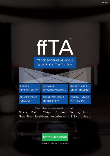
ffTA a Workstation for Trace Evidence Analysis
The ffTA is a powerful and flexible multi-functional system that provides the crime laboratory with a range of analytical facilities on a single microscope operated through a single PC.
The ffTA is a powerful and flexible multi-functional system that provides the
crime laboratory with a range of analytical facilities on a single microscope
operated through a single PC.

You also want an ePaper? Increase the reach of your titles
YUMPU automatically turns print PDFs into web optimized ePapers that Google loves.
<strong>ffTA</strong><br />
TM<br />
TRACE EVIDENCE ANALYSIS<br />
W O R K S T A T I O N<br />
RAMAN<br />
SPECTROSCOPY<br />
UV-VIS-IR<br />
MICROSPECTROMETRY<br />
GRIM GLASS RI<br />
MEASUREMENT<br />
FLUORESCENT<br />
IMAGING<br />
POLARIZED LIGHT<br />
MICROSCOPY<br />
DIGITAL IMAGE<br />
PROCESSING<br />
For the examination of:<br />
Glass, Paint Chips, Fibres, Drugs, Inks,<br />
Gun Shot Residues, Accelerants & Explosives.<br />
foster+freeman<br />
Forensic Science Equipment
<strong>ffTA</strong><br />
TM<br />
A WORKSTATION FOR TRACE EVIDENCE ANALYSIS<br />
The <strong>ffTA</strong> TM is a powerful and flexible multi-functional system that provides the<br />
crime laboratory with a range of analytical facilities on a single microscope<br />
operated through a single PC.<br />
By switching from module to module the operator is able to per<strong>for</strong>m a wide<br />
range of analytical tasks to extract the maximum amount of <strong>for</strong>ensic evidence in<br />
the shortest possible time.<br />
Available modules<br />
RAMAN<br />
SPECTROSCOPY<br />
For the study of materials<br />
including paint chips, fibres,<br />
inks, & drugs<br />
FLUORESCENT<br />
IMAGING<br />
Identification of biological<br />
samples, chemicals, &<br />
illegal substances<br />
UV-VIS-IR<br />
MICROSPECTROMETRY<br />
Non-destructive analysis of<br />
organic compounds including<br />
paint & fibres<br />
POLARIZED LIGHT<br />
MICROSCOPY<br />
For the examination and<br />
identification of natural and<br />
synthetic fibres<br />
GRIM GLASS RI<br />
MEASUREMENT<br />
<strong>Analysis</strong> and grouping of<br />
glass fragments<br />
DIGITAL IMAGE<br />
PROCESSING<br />
Enhancement & comparison<br />
of images captured using a<br />
5MP digital camera
<strong>ffTA</strong> TM Modular Design<br />
Built around the Leica DM2700M laboratory microscope with high power LED illumination,<br />
the <strong>ffTA</strong> ’ s unique modular design enables the user to add functions to meet specific<br />
laboratory requirements. Using an optical multiplexer the operator simply switches the<br />
image from one module to another.<br />
1<br />
2<br />
3<br />
4<br />
5<br />
6<br />
7<br />
Optical Multiplexer<br />
5MP CCD Camera<br />
GRIM3 Video Camera<br />
Foram X3 Raman Spectrometer<br />
Fluorescent Imaging Light Source<br />
360 o Rotating Stage<br />
GRIM3 Hot Stage<br />
3 2<br />
1<br />
Incident Illumination Package<br />
DM2700 Microscope<br />
DM2700 Objectives<br />
GRIM3 Processor<br />
Microspectrometer Module<br />
8<br />
9<br />
10<br />
11<br />
12<br />
4<br />
11 12<br />
8<br />
9<br />
5<br />
6<br />
10<br />
7<br />
System Specification<br />
The modular design of Foster+Freeman <strong>ffTA</strong> workstations offers the trace examiner increased efficiency and flexibility.<br />
The core <strong>ffTA</strong> workstation includes a DM2700 microscope connected to a high specification Windows PC. This provides a basic microscopy<br />
and image processing system.<br />
At the time of purchase, any number of <strong>ffTA</strong> modules may be selected alongside the core system to provide the user with further<br />
functionality. Post-installation, new modules may be seemlessly retro-fitted to the <strong>ffTA</strong> at any time.<br />
To configure a <strong>ffTA</strong> system that meets your laboratory’s requirements please<br />
contact your local Foster + Freeman sales representative or send an email to<br />
sales@fosterfreeman.com
RAMAN SPECTROSCOPY <strong>for</strong> the comparison and identification of materials<br />
<strong>ffTA</strong> TM FORAM ® X3<br />
Triple Raman Spectrometer Module<br />
The FORAM ® X3 module can be equipped with a choice<br />
of one or three laser wavelengths:<br />
785nm (invisible), 638nm and 532nm.<br />
High levels of sensitivity can be achieved with the 532nm<br />
laser, while the 785nm infrared laser is better able to suppress<br />
fluorescence. A balance between power and sensitivity can<br />
be achieved through the use of the 638nm laser.<br />
A Powerful Analytical Tool<br />
Raman spectroscopy is widely used in <strong>for</strong>ensic science<br />
<strong>for</strong> the study of a variety of organic and inorganic<br />
materials including paint chips, fibres, inks, and<br />
controlled substances, as well as residues from<br />
explosives, flammables and accelerants.<br />
Through the analysis of Raman spectra, specific to<br />
molecular structure, this powerful module is able to<br />
provide valuable "fingerprints" <strong>for</strong> comparing,<br />
differentiating and identifying materials.<br />
Raman Spectroscopy features include:<br />
• Non-contact, non-destructive analysis<br />
• <strong>Analysis</strong> of materials in solid or liquid <strong>for</strong>m<br />
• Rapid examination with minimal preparation<br />
Explosives<br />
Match Spectrum: Recrystallised RDX<br />
Percentage Match: 93.5%<br />
Search Spectrum: Unknown explosive material<br />
500 600 700 800 900 1000 1100 1200 1300 1400 1500 1600<br />
Raman Shift (cm o1 )<br />
Illicit drugs<br />
Match Spectrum: Methamphetamine<br />
Percentage Match: 93.7%<br />
Search Spectrum: Suspect Methamphetamine<br />
500 600 700 800 900 1000 1100 1200 1300 1400 1500 1600<br />
Raman Shift (cm o1 )<br />
<strong>ffTA</strong> TM FORAM ® Module includes<br />
Select one or choose all three laser wavelengths<br />
• Diode pumped SSL 532nm 8mW laser<br />
- Spectrometer range 250 - 2700 cm-1<br />
• Stabilised 638nm 9mW laser<br />
- Spectrometer range 300 - 3700 cm-1<br />
• Stabilised 785nm 80mW laser<br />
- Spectrometer range 200 - 2500 cm-1<br />
Detector<br />
- Low etalation CCD detector with Peltier cooling.<br />
- Peak Quantum efficiency of greater than 40%.<br />
Software<br />
- Automated and manual Wavelength/Wavenumber<br />
and photometric response calibration routines.<br />
- Comprehensive casework management system.<br />
NIST Correction Standards<br />
Relative intensity correction standard <strong>for</strong> Raman<br />
spectroscopy: 785nm & 532nm<br />
Optional Reference Databases<br />
Further enhance the capability of the<br />
Foram X3 through the addition of<br />
Raman databases <strong>for</strong> the identification<br />
of unknown materials and compounds.<br />
• Materials of Forensic Interest<br />
• Hazardous Chemicals<br />
• Pharmaceuticals, Drugs & Antibiotics<br />
Minerals & Inorganic Materials
GRIM ® GLASS RI MEASUREMENT <strong>for</strong> the analysis and grouping of glass samples<br />
<strong>ffTA</strong> TM GRIM ® 3<br />
Glass Refractive Index Measurement Module<br />
The <strong>ffTA</strong> TM GRIM ® 3 module can be used to identify and<br />
group fragments of glass through the measurement of<br />
their refractive indices using the oil immersion/<br />
temperature variation technique.<br />
The system can monitor up to four glass fragment edges<br />
per operation, speeding the casework examination process,<br />
giving improved statistical accuracy and reducing<br />
calibration time.<br />
Glass as <strong>Evidence</strong><br />
As different types of glass have different refractive indices, it is possible to use<br />
this in<strong>for</strong>mation to group fragments of glass together and to<br />
establish whether or not they may have originally been part of the same pane of<br />
glass.<br />
The Foster + Freeman <strong>ffTA</strong> TM GRIM ® 3 module determines the refractive indices of<br />
glass using the widely accepted laboratory standard method of oil immersion /<br />
temperature variation.<br />
Multi-edge processing of fragments improves efficiency<br />
<strong>ffTA</strong> TM GRIM ® 3 Module includes<br />
GRIM 3 glass RI Processor Module<br />
• Monochrome firewire video camera<br />
• GRIM ® 3 GLASS system operating software<br />
• FRAG Analyser statistical analysis software<br />
Hotstage<br />
• Mettler FP82HT hotstage<br />
• Includes hotstage fixing bracket<br />
Reference Oils and Glasses<br />
• Set of 3 purified silicon oils and 19 glass<br />
• standards to cover the range of refractive<br />
indices.<br />
Phase ring x10 objective and<br />
focusing telescope<br />
Interference filters<br />
• 488, 589, 656nm<br />
GLASS: Updated system software<br />
The GRIM3 operating software, GLASS,<br />
has been updated in 2015 to provide<br />
improved casework management with<br />
the following features:<br />
• Searchable case data<br />
• Customisable graphical user interface<br />
• Automated system per<strong>for</strong>mance<br />
monitoring<br />
• Improved flexible reporting system
POLARISED LIGHT MICROSCOPY <strong>for</strong> the analysis and identification of fibres<br />
<strong>ffTA</strong> TM PLM<br />
Light Polarisation Module<br />
Possibly the most widely used application <strong>for</strong> microscopy<br />
in <strong>for</strong>ensic science, Polarised Light Microscopy (PLM) is a<br />
valuable tool <strong>for</strong> the detection and analysis of small<br />
unknown particles and <strong>for</strong> the identification of fibres.<br />
A contrast-enhancing technique, PLM uses the birefringent<br />
properties of materials to improve the quality of the images<br />
obtained with the microscope.<br />
The addition of a 360-degree circular rotating specimen<br />
stage enables the user to make accurate measurements of<br />
a sample’s birefringence and to quickly identify fibres as<br />
acetate, acrylic, nylon, or polyester.<br />
<strong>Evidence</strong> identified by its birefringence<br />
Birefringence is the decomposition of a single ray of light into two separate rays<br />
as it passes through birefingent materials. By taking a measurement of the<br />
samples diameter and the colour retardation of the decomposed light, unknown<br />
materials can be identified against the interference colour chart.<br />
The <strong>ffTA</strong> TM Light Polarisation Module consists of a linear polariser mounted on<br />
the condenser lens, a second polariser (the analyser) placed in the optical<br />
pathway be<strong>for</strong>e the observation tubes and camera, and a retardation plate to<br />
enhance optical path differences between the cross-polarisers.<br />
Specimens are placed on a 360-degree circular rotating specimen stage<br />
equipped with two Vernier scales so that rotation angle can be measured to an<br />
accuracy of 0.1 degree.<br />
Cross-polar examination of a fibre<br />
<strong>ffTA</strong> TM PLM Module includes<br />
Interference Colour Chart<br />
360 o rotary polarisation stage<br />
• Ball bearing stage with locking clamp<br />
• Stage bracket and condenser holder.<br />
• Stage diameter 178mm<br />
• XY object guide<br />
Microscope Objective<br />
• Hi Plan x63 Polarisation Objective<br />
Polariser<br />
• 52mm mounted linear polariser<br />
• mounted analyser<br />
• 1 x waveplate<br />
• 1 x quarter waveplate<br />
Interference Colour Chart<br />
• Michel-Lévy table of birefringence<br />
Each <strong>ffTA</strong> TM<br />
PLM Module is supplied<br />
with a copy of the Michel-Lévy table of<br />
birefringence.<br />
In use <strong>for</strong> more than 100 years, the<br />
chart enables PLM examiners to<br />
identify fibres and minerals by<br />
measurement of a samples thickness<br />
and colour retardation.
FLUORESCENCE IMAGING <strong>for</strong> multi-spectral visual comparison of evidence<br />
<strong>ffTA</strong> TM Fluorescence Imaging<br />
Real-time Fluorescence Excitation Module<br />
A widely used technique <strong>for</strong> the identification and comparison<br />
of trace materials including paint chips, fibres and biological<br />
materials.<br />
Equipped with four excitation bandwidths: UV, Violet, Blue and<br />
Green, this module provides versatile high resolution<br />
fluorescence imaging and spectra when used in conjunction<br />
with the <strong>ffTA</strong> TM Micro-spectrometer module.<br />
Wide Ranging Applications<br />
In the <strong>for</strong>ensic laboratory, fluorescence imaging is an ideal<br />
technique <strong>for</strong> the inspection of biological samples, examination<br />
of accelerants (petrol, diesel, kerosene etc.) and <strong>for</strong> the<br />
characterisation and identification of illegal substances.<br />
In addition to visual inspection, fluorescence microspectrometry<br />
is a powerful technique <strong>for</strong> the discrimination of textile fibres<br />
and <strong>for</strong> the comparison of paint evidence.<br />
Absorbance - Visible Absorbance - Full Range Transmission - Full Range<br />
<strong>ffTA</strong> TM Fluorescence Modules includes<br />
Fluorescence Module A<br />
• External Light Source<br />
• 100W Mercury Vapour Lamp<br />
Or...<br />
Fluorescence Module B<br />
• External Light Source<br />
• 120W Metal Halide Light Source<br />
Filtercubes<br />
• Filtercube A 340-380nm<br />
• Filtercube D 355-425nm<br />
• Filtercube l3 450-490nm<br />
• Filtercube N2.1 515-560nm<br />
Light source selection<br />
Foster+Freeman offer a choice of high<br />
intensity discharge light sources.<br />
The Metal Halide lamp is an energy<br />
efficient light source offering excellent<br />
colour retention over a long service life.<br />
An older technology, the Mercury<br />
Vapour light source offers greater lamp<br />
life but is less energy efficient.
UV-VIS-IR MICROSPECTROMETER <strong>for</strong> the analysis of organic compounds<br />
<strong>ffTA</strong> TM Microspectrometer<br />
Available in a choice of 4 Wavelength Ranges<br />
Micro-spectrometry is a powerful analytical tool that is<br />
widely used in <strong>for</strong>ensic science <strong>for</strong> the study and<br />
comparison of trace materials including paint chips, fibres<br />
and inks.<br />
Spectra in the visible region provide the user with objective<br />
measurements of colour, and through the examination of<br />
ultra-violet and infra-red spectra, users are able to make<br />
comparisons between two materials that may be<br />
indistinguishable to the naked eye.<br />
Spectral analysis<br />
Depending on the wavelength range selected, the Microspectrometer will record<br />
absorbtion, reflection and transmission spectra in the UV, Visible or Infrared<br />
wavebands.<br />
When used in conjunction with the <strong>ffTA</strong> TM Fluorescence Imaging module it is also<br />
capable of recording fluorescence spectra.<br />
Microspectrometry is highly effective in the discrimination of fibres that may<br />
have identical physical properties.<br />
Spectral response of a suspect fibre<br />
<strong>ffTA</strong> TM Microspectrometer Modules include<br />
<strong>Trace</strong>able Standards<br />
A choice of four fibre-coupled spectrometers<br />
240-830nm FWHM resolution better than 1.98nm<br />
Or...<br />
240-1000nm FWHM resolution better than 2.5nm<br />
Or...<br />
350-1000nm FWHM resolution better than 2.54nm<br />
Or...<br />
400-1000nm FWHM resolution better than 3nm<br />
Incident Illumination Package<br />
5MP FireWire Camera<br />
• High-res 2/3” 5MP CCD camera<br />
• 400nm to 700nm spectral response<br />
Leica DM2700M objectives<br />
• x5, x10, x20, x40, x50<br />
Optical Multiplexer<br />
• 1x input, 3x selectable outputs<br />
To guarantee the accuracy of results,<br />
each <strong>ffTA</strong> Microspectrometer module is<br />
supplied with relevant calibration<br />
standards including:<br />
• NIST traceable filters<br />
• NIST wavelength calibration standard<br />
• Labsphere colour measurement<br />
• calibration standard
IMAGE PROCESSING enhancement and comparison of high-resolution images<br />
<strong>ffTA</strong> TM Image Processing<br />
High-Resolution Camera and Software Module<br />
A comprehensive module <strong>for</strong> processing images from a<br />
scientific grade 5 million pixel CCD colour camera.<br />
High quality imaging enables the examiner to zoom,<br />
orientate and position and item of evidence on screen <strong>for</strong><br />
critical visual examination.<br />
Live and stored images may be compared side-by-side,<br />
subjected to digital enhancement (contrast stretch, Fast<br />
Fourier Trans<strong>for</strong>m, Sharpen etc.), or measured and<br />
annotated.<br />
Side-by-side comparison of a fibre: Live image Vs Stored image<br />
Visual Examination of <strong>Evidence</strong><br />
<strong>Analysis</strong> of almost any item of trace evidence begins with a visual examination,<br />
to assess a samples physical characteristics, followed by macro-photography<br />
and then microscopic analysis.<br />
The <strong>ffTA</strong> TM Image Processing Module provides the examiner with a selection of<br />
facilities to ‘digitally enhance’ the visible image of a sample.<br />
Facilities include:<br />
• Image Enhancement<br />
- Contrast stretch, HSL, RGB, Filters, Equalization, FFT, Gamma Correction<br />
Visual analysis of a paint chip<br />
• Image <strong>Analysis</strong><br />
- Measurement facilities <strong>for</strong> distance, angle and area using calibrated grids<br />
- Measurements can be added to image.<br />
• Image Annotation<br />
- Editable image annotation facilities (text and shapes including arrows, lines,<br />
boxes and circles)<br />
• Image Comparison & Trans<strong>for</strong>mations<br />
- Side-by-side comparison of live and stored images<br />
- User adjustable split position<br />
- Superimposition and subtraction of live and stored image (option of<br />
red/green rendered images <strong>for</strong> extra clarity)<br />
- Live and stored images can be rotated through 90 o<br />
<strong>ffTA</strong> TM Image Processing Module includes<br />
Incident Illumination Package<br />
5MP FireWire Camera<br />
• High-res 2/3” 5MP CCD camera<br />
• 400nm to 700nm spectral response<br />
• on-chip image integration from 8mms to 22s<br />
Leica DM2700M objectives<br />
• x5, x10, x20, x40, x50
Head Office, UK Sales Office<br />
Vale Park | Evesham | WR11 1TD | United Kingdom<br />
Tel: +44 (0)1386 768 050 | sales@fosterfreeman.com<br />
USA Sales Office<br />
46030 Manekin Plaza | Suite 170 | Sterling | VA 20166 | USA<br />
Tel: 888 445 5048 | usoffice@fosterfreeman.com<br />
foster+freeman<br />
fosterfreeman. com