new secondary metabolites of phenylbutyrate in humans
new secondary metabolites of phenylbutyrate in humans
new secondary metabolites of phenylbutyrate in humans

Create successful ePaper yourself
Turn your PDF publications into a flip-book with our unique Google optimized e-Paper software.
0090-9556/04/3201-10–19$20.00<br />
DRUG METABOLISM AND DISPOSITION Vol. 32, No. 1<br />
Copyright © 2004 by The American Society for Pharmacology and Experimental Therapeutics 1181/1111187<br />
DMD 32:10–19, 2004 Pr<strong>in</strong>ted <strong>in</strong> U.S.A.<br />
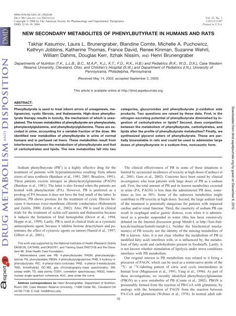
NEW SECONDARY METABOLITES OF PHENYLBUTYRATE IN HUMANS AND RATS<br />
Takhar Kasumov, Laura L. Brunengraber, Bland<strong>in</strong>e Comte, Michelle A. Puchowicz,<br />
Kathryn Jobb<strong>in</strong>s, Kather<strong>in</strong>e Thomas, France David, Renee K<strong>in</strong>man, Suzanne Wehrli,<br />
William Dahms, Douglas Kerr, Itzhak Nissim, AND Henri Brunengraber<br />
Departments <strong>of</strong> Nutrition (T.K., L.L.B., B.C., M.A.P., K.J., K.T., F.D., R.K., H.B.) and Pediatrics (R.K., W.D., D.K.), Case Western<br />
Reserve University, Cleveland, Ohio; and Children’s Hospital (S.W.) and Department <strong>of</strong> Pediatrics (I.N.), University <strong>of</strong><br />
Pennsylvania, Philadelphia, Pennsylvania<br />
ABSTRACT:<br />
Phenylbutyrate is used to treat <strong>in</strong>born errors <strong>of</strong> ureagenesis, malignancies,<br />
cystic fibrosis, and thalassemia. High-dose <strong>phenylbutyrate</strong><br />
therapy results <strong>in</strong> toxicity, the mechanism <strong>of</strong> which is unexpla<strong>in</strong>ed.<br />
The known <strong>metabolites</strong> <strong>of</strong> <strong>phenylbutyrate</strong> are phenylacetate,<br />
phenylacetylglutam<strong>in</strong>e, and phenylbutyrylglutam<strong>in</strong>e. These are excreted<br />
<strong>in</strong> ur<strong>in</strong>e, account<strong>in</strong>g for a variable fraction <strong>of</strong> the dose. We<br />
identified <strong>new</strong> <strong>metabolites</strong> <strong>of</strong> <strong>phenylbutyrate</strong> <strong>in</strong> ur<strong>in</strong>e <strong>of</strong> normal<br />
<strong>humans</strong> and <strong>in</strong> perfused rat livers. These <strong>metabolites</strong> result from<br />
<strong>in</strong>terference between the metabolism <strong>of</strong> <strong>phenylbutyrate</strong> and that<br />
<strong>of</strong> carbohydrates and lipids. The <strong>new</strong> <strong>metabolites</strong> fall <strong>in</strong>to two<br />
Sodium <strong>phenylbutyrate</strong> (PB 1 ) is a highly effective drug for the<br />
treatment <strong>of</strong> patients with hyperammonemia result<strong>in</strong>g from <strong>in</strong>born<br />
errors <strong>of</strong> urea synthesis (Batshaw et al., 1981, 2001; Brusilow, 1991).<br />
These patients excrete nitrogen as phenylacetylglutam<strong>in</strong>e (PAGN)<br />
(Batshaw et al., 1981). The latter is also formed when the patients are<br />
treated with phenylacetate (PA). However, PB is preferred as a<br />
prodrug <strong>of</strong> PA because it does not have the foul smell <strong>of</strong> the latter. In<br />
addition, PB shows promise for the treatment <strong>of</strong> cystic fibrosis because<br />
it <strong>in</strong>creases trans-membrane chloride conductance (Rubenste<strong>in</strong><br />
and Zeitl<strong>in</strong>, 2000; Zeitl<strong>in</strong> et al., 2002). Also, PB is used <strong>in</strong> cl<strong>in</strong>ical<br />
trials for the treatment <strong>of</strong> sickle-cell anemia and thalassemia because<br />
it <strong>in</strong>duces the formation <strong>of</strong> fetal hemoglob<strong>in</strong> (Dover et al., 1994;<br />
Hoppe et al., 1999). Lastly, PB is used <strong>in</strong> cl<strong>in</strong>ical trials as a cytostatic<br />
ant<strong>in</strong>eoplastic agent, because it <strong>in</strong>hibits histone deacetylases and potentiates<br />
the effect <strong>of</strong> cytotoxic agents on tumors (Samid et al., 1997;<br />
Gilbert et al., 2001).<br />
This work was supported by the National Institutes <strong>of</strong> Health (Research Grants<br />
DK58126, CA79495, and DK53761, and Tra<strong>in</strong><strong>in</strong>g Grant DK07319) and the Cleveland<br />
Mt. S<strong>in</strong>ai Health Care Foundation.<br />
1 Abbreviations used are: PB, 4-<strong>phenylbutyrate</strong>; PAGN, phenylacetylglutam<strong>in</strong>e;<br />
PA, phenylacetate; PBGN, 4-phenylbutyrylglutam<strong>in</strong>e; PHB, 3-hydroxy-4<strong>phenylbutyrate</strong>;<br />
PtC, 4-phenyl-trans-crotonate; PKB, 4-phenyl-3-ketobutyrate;<br />
TMS, trimethylsilyl; GC-MS, gas chromatography-mass spectrometry; SW,<br />
sweep width; TD, data po<strong>in</strong>ts; COSY, correlation spectroscopy; HSQC, heteronuclear<br />
s<strong>in</strong>gle-quantum coherence; AUC, area under the curve.<br />
Address correspondence to: Henri Brunengraber, Department <strong>of</strong> Nutrition,<br />
Room 280, Case Western Reserve University, 11000 Cedar Rd., Cleveland OH<br />
44106-7139. E-mail: hxb8@cwru.edu<br />
(Received May 14, 2003; accepted September 2, 2003)<br />
This article is available onl<strong>in</strong>e at http://dmd.aspetjournals.org<br />
10<br />
categories, glucuronides and <strong>phenylbutyrate</strong> �-oxidation side<br />
products. Two questions are raised by these data. First, is the<br />
nitrogen-excret<strong>in</strong>g potential <strong>of</strong> <strong>phenylbutyrate</strong> dim<strong>in</strong>ished by <strong>in</strong>gestion<br />
<strong>of</strong> carbohydrates or lipids? Second, does competition<br />
between the metabolism <strong>of</strong> <strong>phenylbutyrate</strong>, carbohydrates, and<br />
lipids alter the pr<strong>of</strong>ile <strong>of</strong> <strong>phenylbutyrate</strong> <strong>metabolites</strong>? F<strong>in</strong>ally, we<br />
synthesized glycerol esters <strong>of</strong> <strong>phenylbutyrate</strong>. These are partially<br />
bioavailable <strong>in</strong> rats and could be used to adm<strong>in</strong>ister large<br />
doses <strong>of</strong> <strong>phenylbutyrate</strong> <strong>in</strong> a sodium-free, noncaustic form.<br />
The cl<strong>in</strong>ical effectiveness <strong>of</strong> PB <strong>in</strong> some <strong>of</strong> these situations is<br />
limited by occasional <strong>in</strong>cidences <strong>of</strong> toxicity at high doses (Carducci et<br />
al., 2001; Gore et al., 2002). Concerns have been raised by cl<strong>in</strong>ical<br />
<strong>in</strong>vestigators who treat patients with large doses <strong>of</strong> PB as a sodium<br />
salt. First, the total amount <strong>of</strong> PB and its known <strong>metabolites</strong> excreted<br />
<strong>in</strong> ur<strong>in</strong>e (PA, PAGN) is less than the adm<strong>in</strong>istered PB dose, sometimes<br />
as low as 50%. Some <strong>of</strong> the unknown <strong>metabolites</strong> might<br />
contribute to PB toxicity at high doses. Second, the large sodium load<br />
<strong>of</strong> the treatment is potentially dangerous for patients with impaired<br />
cardiac and/or renal function. Third, the causticity <strong>of</strong> sodium PB can<br />
result <strong>in</strong> esophagal and/or gastric distress, even when it is adm<strong>in</strong>istered<br />
as a powder suspended <strong>in</strong> water (this has been extensively<br />
debated on the Internet discussion group Metab-L at http://lists.franken.de/mailman/list<strong>in</strong>fo/metab-L).<br />
Neither the biochemical mechanism(s)<br />
<strong>of</strong> PB toxicity nor the identity <strong>of</strong> the miss<strong>in</strong>g <strong>metabolites</strong> <strong>of</strong><br />
PB is known. Also, it is not clear whether the metabolism <strong>of</strong> PB (a<br />
modified fatty acid) <strong>in</strong>terferes with, or is <strong>in</strong>fluenced by, the metabolism<br />
<strong>of</strong> fatty acids and carbohydrates present <strong>in</strong> foodstuffs. Lastly, it<br />
is not known whether stimulation <strong>of</strong> lipolysis under stress conditions<br />
<strong>in</strong>terferes with PB metabolism.<br />
Our orig<strong>in</strong>al <strong>in</strong>terest <strong>in</strong> PB metabolism was related to it be<strong>in</strong>g a<br />
precursor <strong>of</strong> PAGN, which can be used as a non<strong>in</strong>vasive probe <strong>of</strong> the<br />
14 C- or 13 C-label<strong>in</strong>g pattern <strong>of</strong> citric acid cycle <strong>in</strong>termediates <strong>in</strong><br />
human liver (Magnusson et al., 1991; Yang et al., 1996). As part <strong>of</strong><br />
these <strong>in</strong>vestigations, we recently identified phenylbutyrylglutam<strong>in</strong>e<br />
(PBGN) as a <strong>new</strong> metabolite <strong>of</strong> PB (Comte et al., 2002). PBGN is<br />
presumably formed from the reaction <strong>of</strong> PB-CoA with glutam<strong>in</strong>e, by<br />
analogy with the formation <strong>of</strong> PAGN from the reaction between<br />
PA-CoA and glutam<strong>in</strong>e (Webster et al., 1976). In normal adult sub-<br />
Downloaded from<br />
dmd.aspetjournals.org by guest on December 6, 2012
jects who <strong>in</strong>gested a fairly low dose <strong>of</strong> PB (5 g/75 kg), we found that<br />
the total excretion <strong>of</strong> PB � PA � PAGN � PBGN accounted for only<br />
half the <strong>in</strong>gested PB dose (Comte et al., 2002). The miss<strong>in</strong>g fraction<br />
<strong>of</strong> the dose may be disposed <strong>of</strong> either <strong>in</strong> ur<strong>in</strong>e as unknown <strong>metabolites</strong>,<br />
or <strong>in</strong> feces as unabsorbed PB and/or PB <strong>metabolites</strong>.<br />
In the present study, we report the identification <strong>of</strong> additional<br />
<strong>metabolites</strong> <strong>of</strong> PB <strong>in</strong> <strong>humans</strong>, i.e., R- and S-3-hydroxy-4-<strong>phenylbutyrate</strong><br />
(PHB), phenylacetone, and 1-phenyl-2-propanol, as well as PA<br />
and PB glucuronides. We studied the mechanism <strong>of</strong> PHB formation<br />
from PB <strong>in</strong> perfused rat livers and identified two additional <strong>metabolites</strong>,<br />
4-phenyl-trans-crotonate (PtC) and 4-phenyl-3-ketobutyrate<br />
(PKB). Lastly, we prepared sodium-free esters <strong>of</strong> PB and <strong>in</strong>vestigated<br />
their bioavailability <strong>in</strong> rats. Glycerol-PB esters appear promis<strong>in</strong>g for<br />
the adm<strong>in</strong>istration <strong>of</strong> large amounts <strong>of</strong> PB without the correspond<strong>in</strong>g<br />
sodium load.<br />
Materials and Methods<br />
Materials. All chemicals used <strong>in</strong> syntheses, general chemicals, and solvents<br />
were obta<strong>in</strong>ed from Sigma-Aldrich (St. Louis, MO). All organic solvents were<br />
dried and distilled immediately before use. [ 2 H7]Phenylacetic acid (99%) and<br />
[ 2 H6]benzene were purchased from Isotec Inc. (Miamisburg, OH). The derivatization<br />
agent N-methyl-N-(trimethylsilyl)trifluoroacetamide was supplied by Regis<br />
Technologies, Inc. (Morton Grove, IL). All aqueous solutions were made with<br />
water purified with the Milli-Q system (Millipore Corporation, Bedford, MA).<br />
Preparation <strong>of</strong> Unlabeled and Deuterated Standards. S-2-Phenylbutyryl<br />
chloride was prepared by react<strong>in</strong>g S-2-phenylbutyric acid with SOCl2, and was<br />
vacuum-distilled and stored at 4°C as a 0.5 M solution <strong>in</strong> benzene. [ 2 H5]PB was prepared by alum<strong>in</strong>um chloride-catalyzed condensation <strong>of</strong> �-butyrolactone<br />
with [ 2 H6]benzene as previously described (Comte et al., 2002). �-Phenyl-trans-crotonic<br />
acid was prepared by a Fridel-Crafts reaction <strong>of</strong> benzene<br />
with ethyl �-bromo-trans-crotonate, followed by acid hydrolysis <strong>of</strong> ethyl<br />
�-phenyl-trans-crotonate (L<strong>of</strong>fler et al., 1970). The trans configuration <strong>of</strong> the<br />
product was confirmed by 1 H NMR. [ 2 H7]Phenylacetylglyc<strong>in</strong>e was synthesized<br />
by react<strong>in</strong>g [ 2 H7]phenylacetyl chloride with glyc<strong>in</strong>e, as described previ-<br />
NEW PHENYLBUTYRATE METABOLITES<br />
FIG. 1.Scheme for the synthesis <strong>of</strong> unlabeled and deuterated standards.<br />
ously for the unlabeled analog (Ramsdell and Tanaka, 1977).<br />
[ 2 H 7]Phenylacetyl chloride was prepared by activation <strong>of</strong> [ 2 H 7]phenylacetic<br />
acid with freshly distilled dichloromethyl methyl ether and used immediately<br />
after evaporation <strong>of</strong> excess dichloromethyl methyl ether and <strong>of</strong> the methyl<br />
chlor<strong>of</strong>ormate byproduct. The synthetic protocol for preparation <strong>of</strong> PHB, PKB,<br />
[ 2 H 5]PHB, phenylacetone, and 1-phenyl-2-[2- 2 H]propanol is outl<strong>in</strong>ed <strong>in</strong> Fig. 1.<br />
Ethyl 4-Phenyl-3-ketobutyrate (2; Fig. 1) was synthesized by a method<br />
adapted from Capozzi et al. (1993). The commercial isopropylidene malonate<br />
(2,2-dimethyl-4,6-diketo-1,3-dioxane, also called Meldrum’s acid; Aldrich<br />
Chemical Co., Milwaukee, WI) was reacted with phenylacetyl chloride <strong>in</strong> the<br />
presence <strong>of</strong> dry pyrid<strong>in</strong>e <strong>in</strong> anhydrous methylene chloride. The crude phenylacetylated<br />
Meldrum’s acid (compound 1, Fig. 1) was refluxed <strong>in</strong> absolute<br />
ethanol until evolution <strong>of</strong> CO 2 ceased (about 3 h). After evaporation <strong>of</strong> the<br />
solvent, ethyl 4-phenyl-3-ketobutyrate was purified on a silica gel column.<br />
4-Phenyl-3-ketobutyric acid (PKB). A 10% molar excess <strong>of</strong> 1 N NaOH<br />
solution was added slowly to ice-cooled ethyl 4-phenyl-3-ketobutyrate and<br />
stirred at room temperature until the organic phase disappeared (approximately<br />
12 h). The solution was acidified with 1 N HCl to pH 2 and extracted three<br />
times with 3 volumes <strong>of</strong> ethyl ether. After dry<strong>in</strong>g over Na 2SO 4, the solvent was<br />
evaporated to give the white solid product (yield 94%, m.p. 70°C). 1 H NMR<br />
(300 MHz, �, CDCl 3): keto, 3.33 (s, 2H, CH 2COO), 3.78 (s, 2H, CH 2Ph),<br />
7.17–7.33 (m, 5H, Ph); enol, 4.82 (s, 1H, CH), 12.13 (s, 1H, OH); keto/enol �<br />
7.8:1. 13 C NMR (75 MHz, �, CDCl 3): 48.36 (CH 2COO), 49.87 (CH 2Ph),<br />
127.41 (C-4 Ph), 128.73 (C-3, C-5 Ph), 129.50 (C-2, C-6 Ph), 133.14 (C-ipso,<br />
Ph), 169.26 (COO), 201.57 (CO).<br />
The identity <strong>of</strong> the product was also confirmed by 1) reduction <strong>of</strong> 4-phenyl-<br />
3-ketobutyrate with NaBH 4, 2) extraction <strong>of</strong> R,S-PHB with ethyl acetate, and<br />
3) TMS derivatization and NH 3-positive chemical ionization GC-MS. The<br />
mass spectrum <strong>of</strong> the derivative was identical to that <strong>of</strong> PHB synthesized as<br />
described below. The 4-phenyl-3-ketobutyric acid was stored at �80°C to<br />
prevent decomposition. Just before use <strong>in</strong> liver perfusion experiments, the acid<br />
was dissolved <strong>in</strong> water and titrated to pH � 7.40.<br />
R,S-3-Hydroxy-4-phenylbutyric acid (PHB). Ethyl 4-phenyl-3-ketobutyrate<br />
(2.06 g, 10 mmol) was mixed with 10 ml <strong>of</strong> H 2O and a calculated amount <strong>of</strong><br />
NaOH/H 2O was added dropwise with cool<strong>in</strong>g to give 0.5M solution <strong>of</strong> [OH - ].<br />
11<br />
Downloaded from<br />
dmd.aspetjournals.org by guest on December 6, 2012
12 KASUMOV ET AL.<br />
NaBH 4 (0.37g, 10 mmol) was added and the mixture stirred for 24 h. After the<br />
reaction mixture was cooled to 0°C, the pH was brought to 7.0 with HCl (6N)<br />
and more than half <strong>of</strong> water removed by lyophilizer. The pH was brought to 2<br />
by HCl (6 N) and the solution extracted 3 times with 3 volumes <strong>of</strong> ethyl ether.<br />
After dry<strong>in</strong>g the comb<strong>in</strong>ed ether extract, the ether was evaporated giv<strong>in</strong>g a<br />
white solid product. Yield 79%. Purity <strong>of</strong> product was assayed by GC-MS after<br />
derivatization with TMS.<br />
R-3-Hydroxy-4-<strong>phenylbutyrate</strong> (R-PHB) was prepared by reduc<strong>in</strong>g the correspond<strong>in</strong>g<br />
4-phenyl-3-ketobutyric acid with B-chlorodiisop<strong>in</strong>ocampheylbo<br />
rane [(�)-DIP-Cl] (Wang et al., 1999). The yield was 89% from PKB;<br />
enantiomeric excess was 97% [GC-MS <strong>of</strong> the methyl S-2-phenylbutyryl derivative<br />
(see below), and purity was confirmed by NMR].<br />
R,S-3-Hydroxy-4-phenyl[2,2,3,4,4- 2 H 5]butyrate (R,S-[ 2 H 5]PHB). 4-Phenyl-3-ketobutyric<br />
acid (0.267 g, 1.5 mmol) was suspended <strong>in</strong> 3 ml <strong>of</strong> 2 H 2O<br />
(99.9%) to which 0.36 ml <strong>of</strong> 40% <strong>of</strong> NaO 2 H (3.6 mmol) <strong>in</strong> 2 H 2O was slowly<br />
added at 0–5°C. This procedure exchanges 1 H for 2 H atoms on the methylene<br />
groups adjacent to the carbonyl. The solution was stirred at room temperature<br />
overnight and then lyophilized. The residue was dissolved <strong>in</strong> 3 ml <strong>of</strong> 2 H 2O and<br />
stirred for another 5hatroom temperature. The solution was cooled on ice,<br />
treated with NaB 2 H 4 (63 mg, 1.5 mmol), and stirred overnight at room<br />
temperature (Des Rosiers et al., 1988). After acidification to pH 1 to 2 with<br />
HCl (6 N), the solution was saturated with NaCl and extracted three times with<br />
3 volumes <strong>of</strong> diethyl ether. Solvent evaporation yielded R,S-[ 2 H 5]PHB (yield<br />
74%, purity 99% by NMR, M5 isotopic enrichment 95% by GC-MS <strong>of</strong> the<br />
TMS derivative). The free acid was titrated to pH 8 with NaOH and stored<br />
frozen as a 0.5 M solution until use.<br />
Phenylacetone was prepared by decarboxylat<strong>in</strong>g phenylketobutyrate <strong>in</strong> acid<br />
at 100°C. It was reduced to 1-phenyl-2-[2- 2 H]propanol with NaB 2 H 4.<br />
Esters <strong>of</strong> Phenylbutyrate. Dihydroxyacetone-di-PB, glycerol-tri-PB, ribose-tetra-PB,<br />
glucose-penta-PB, and sorbitol-hexa-PB were prepared by react<strong>in</strong>g<br />
the polyol/sugar with excess phenylbutyryl chloride <strong>in</strong> the presence <strong>of</strong><br />
pyrid<strong>in</strong>e and catalytic amounts <strong>of</strong> N,N-dimethylam<strong>in</strong>opyrid<strong>in</strong>e. Products were<br />
purified by flash column chromatography on silica. To prepare glycerol-mono-<br />
PB, isopropylidene glycerol was reacted with phenylbutyryl chloride as above,<br />
and the isopropylidene group was removed by mild acidic hydrolysis <strong>in</strong> water.<br />
The structure and purity <strong>of</strong> all products were confirmed by 1 H and 13 C NMR.<br />
The structure <strong>of</strong> glycerol-mono-PB was confirmed by acetylation with acetic<br />
anhydride, followed by GC-MS analysis <strong>of</strong> the derivative.<br />
Sample Preparation. For the determ<strong>in</strong>ation <strong>of</strong> the free acids concentration<br />
(PA, PHB, PKB, PtC, and PB) <strong>in</strong> perfusate, samples (0.1 ml) were spiked with<br />
0.17 �mol <strong>of</strong> [ 2 H 7]PA, [ 2 H 5]PB, and R,S-[ 2 H 5]PHB before deprote<strong>in</strong>ization<br />
with 20 �l <strong>of</strong> saturated sulfosalicylic acid. The slurries were saturated with<br />
NaCl, acidified with one drop <strong>of</strong> 6 M HCl, and extracted three times with 5 ml<br />
<strong>of</strong> diethyl ether. For the assay <strong>of</strong> conjugates <strong>of</strong> PB and PA, 0.1-ml aliquots <strong>of</strong><br />
f<strong>in</strong>al liver perfusate or <strong>of</strong> human ur<strong>in</strong>e were spiked with <strong>in</strong>ternal standards and<br />
treated with 1.0 ml HCl (6 N) at 90°C overnight to hydrolyze the conjugates.<br />
Glucuronides <strong>of</strong> PB and PA were identified by the amount <strong>of</strong> these compounds<br />
released after <strong>in</strong>cubation <strong>of</strong> perfusate and ur<strong>in</strong>e samples with �-glucuronidase<br />
<strong>in</strong> 0.2 M ammonium acetate buffer, pH 5.0, overnight at 37°C.<br />
Bile samples were analyzed for free and total (conjugated � free) PB and<br />
its <strong>metabolites</strong>. In the first series <strong>of</strong> assays, 0.05-ml samples <strong>of</strong> bile were spiked<br />
with <strong>in</strong>ternal standards, acidified to pH 2 to 2.5, and extracted three times with<br />
diethyl ether. In the second series <strong>of</strong> assays, samples spiked with <strong>in</strong>ternal<br />
standards were hydrolyzed with 0.3 ml <strong>of</strong> NaOH (6 N) at 90°C for 3 h before<br />
acidification and extraction.<br />
Phenylacetylglyc<strong>in</strong>e was extracted <strong>in</strong> acid and derivatized with methanol/<br />
HCl. For the assay <strong>of</strong> phenylacetone and 1-phenyl-2-propanol, ur<strong>in</strong>e and<br />
perfusate samples were spiked with the structural analog 1-phenyl-<br />
[ 2 H 5]ethanol and then treated with NaB 2 H 4 to reduce phenylacetone to monodeuterated<br />
1-pheny-2-propanol. The labeled and unlabeled 1-phenyl-2propanol<br />
were assayed as TMS derivatives.<br />
For the assays <strong>in</strong> ur<strong>in</strong>e, 0.1-ml samples were spiked with 0.15 �mol<br />
[ 2 H 5]PHB, acidified to pH 1 to 2 with HCl, saturated with NaCl, and extracted<br />
three times with 3 ml <strong>of</strong> diethyl ether. The comb<strong>in</strong>ed extracts were dried with<br />
Na 2SO 4 and evaporated before react<strong>in</strong>g the residues with 70 �l <strong>of</strong> TMS at<br />
60°C for 20 m<strong>in</strong>.<br />
For the chiral assay <strong>of</strong> PHB enantiomers (Powers et al., 1994), 0.1-ml<br />
samples were spiked with R,S-[ 2 H 5]PHB, and either deprote<strong>in</strong>ized with 50 �l<br />
<strong>of</strong> saturated sulfosalicylic acid (if conta<strong>in</strong><strong>in</strong>g prote<strong>in</strong>s) or acidified to pH 1 to<br />
2 with HCl (for ur<strong>in</strong>e). Then, the slurries or solutions were saturated with NaCl<br />
and extracted three times with 3 ml <strong>of</strong> diethyl ether. The comb<strong>in</strong>ed extracts<br />
were dried with Na 2SO 4 and evaporated before react<strong>in</strong>g the residues with 0.15<br />
ml <strong>of</strong> methanol/HCl for 1hat65°C, to derivatize the carboxyl groups <strong>of</strong> the<br />
PHB enantiomers. After cool<strong>in</strong>g, 1 ml <strong>of</strong> water was added to the mixture and<br />
the hydroxyacid methyl ester was extracted with diethyl ether (three times <strong>in</strong><br />
3 ml). After complete evaporation <strong>of</strong> the comb<strong>in</strong>ed ether extract, S-2phenylbutyryl<br />
chloride benzene solution (0.1 ml, 0.5 M) and 0.05 ml <strong>of</strong><br />
aqueous 12 N NaOH were added. After vortex<strong>in</strong>g, the mixture was <strong>in</strong>cubated<br />
for1honaslow shaker at room temperature. The derivatives were extracted<br />
with ether (three times <strong>in</strong> 3 ml) and 1 ml <strong>of</strong> water. The comb<strong>in</strong>ed ether phase<br />
was dried with Na 2SO 4 and evaporated completely. The residue was dissolved<br />
<strong>in</strong> 0.1 ml <strong>of</strong> ethyl acetate, and 1 �l was <strong>in</strong>jected <strong>in</strong>to the GC-MS.<br />
GC-MS Methods. All <strong>of</strong> the <strong>metabolites</strong>, except phenylacetylglyc<strong>in</strong>e, were<br />
analyzed as their TMS derivatives on a Hewlett-Packard 5890 gas chromatograph<br />
equipped with a ZB-5 capillary column (60 m � 0.25 mm i.d., 0.5 mm<br />
film thickness; Hewlett Packard, Palo Alto, CA) and coupled to a 5989A mass<br />
selective detector. Samples (0.2–1 �l) were <strong>in</strong>jected with a split ratio 20 to<br />
50:1. The carrier gas was helium (1 ml/m<strong>in</strong>) and nom<strong>in</strong>al <strong>in</strong>itial pressure was<br />
20.61 psi. The <strong>in</strong>jector port temperature was at 270°C, the transfer l<strong>in</strong>e at<br />
305°C, the source temperature at 200°C and quadrupole at 150°C. The column<br />
temperature program was: start at 100°C, hold for 1 m<strong>in</strong>, <strong>in</strong>crease by 8°C/m<strong>in</strong><br />
to 236°C, <strong>in</strong>crease by 35°C/m<strong>in</strong> to 310°C, 6 m<strong>in</strong> at 310°C. After automatic<br />
calibration, the mass spectrometer was operated under ammonia-positive ionization<br />
mode. Appropriate ion sets were monitored with a dwell time <strong>of</strong> 25 to<br />
35 ms/ion, at m/z 226/233 (PA/[ 2 H 7]PA), 254/259 (PB/[ 2 H 5]PB), 325/330<br />
(PHB/[ 2 H 5]PHB), and 252 (PtC). Note that PKB and phenylacetone had been<br />
reduced with NaB 2 H 4 to monodeuterated R,S-3-hydroxy-4-<strong>phenylbutyrate</strong><br />
(monitored at m/z 326/330) and 1-phenyl-2-propanol (monitored at m/z 200/<br />
210 and/or 217/227). Also, s<strong>in</strong>ce 1-phenyl-2-propanol was assayed with a<br />
standard <strong>of</strong> 1-phenyl[ 2 H 5]ethanol, ions monitored for this assay were 200/209<br />
or 217/226.<br />
For the analysis <strong>of</strong> chiral PHB derivatives, the GC <strong>in</strong>jector temperature was<br />
set at 280°C. The column (DB-5, 60 m � 0.25 mm i.d., 0.5 mm film thickness;<br />
Hewlett Packard) program was modified to the <strong>in</strong>itial 150°C for 2 m<strong>in</strong>,<br />
<strong>in</strong>creased by 15°C/m<strong>in</strong> to 230°C, 25 m<strong>in</strong> at 230°C, <strong>in</strong>creased by 35°C to<br />
290°C, and held 10 m<strong>in</strong>. Ions monitored were 1) 358 (M � 18, i.e., M �<br />
NH 4 � ) for analytes and 2) 363 (M � 18 � 5, i.e., M � 5�NH4 � ) for<br />
[ 2 H 5]PHB. The mass spectrometer was operated under ammonia- positive<br />
chemical ionization and was tuned automatically.<br />
Phenylacetylglyc<strong>in</strong>e was analyzed as its methyl ester derivative us<strong>in</strong>g an<br />
OV-225 column (29 m � 0.32 mm i.d., 1 �m film thickness; Quadrex<br />
Corporation, Woodbridge, CT). This column yielded better resolution <strong>of</strong><br />
N-phenylacetylglyc<strong>in</strong>e methyl ester with no peak tail<strong>in</strong>g. Samples (0.2–1 �l)<br />
were <strong>in</strong>jected with a split ratio 20:1. The carrier gas was helium (constant flow:<br />
1.2 ml/m<strong>in</strong>). The <strong>in</strong>jector port temperature was at 220°C, the transfer l<strong>in</strong>e at<br />
240°C, the source temperature at 200°C, and quadrupole at 106°C. The column<br />
temperature program was: start at 90°C, hold for 1 m<strong>in</strong>, <strong>in</strong>crease by 10°C/m<strong>in</strong><br />
to 240°C, 15 m<strong>in</strong> at 240°C. After automatic calibration, the mass spectrometer<br />
was operated under ammonia-positive ionization mode (pressure adjusted to<br />
optimize peak areas). Ions monitored were 1) 208 (M � 1, i.e., M � H �) and<br />
225 (M � 18, i.e., M � NH 4 � ) for the analyte and 2) 215 (M � 7 � 1, M �<br />
7 � H � ) and 232 (M � 7 � 18, M � 7 � NH 4 � ) for N-[ 2 H7]PA-glyc<strong>in</strong>e with<br />
a dwell time <strong>of</strong> 25 ms/ion.<br />
Areas under each chromatogram were determ<strong>in</strong>ed by <strong>in</strong>teractive computer<br />
<strong>in</strong>tegration, and corrected for naturally occurr<strong>in</strong>g heavy isotopes and light<br />
isotopic impurities <strong>in</strong> the synthesized labeled <strong>in</strong>ternal standards.<br />
NMR Spectroscopy. Proton NMR spectroscopy was performed at 400<br />
MHz on a Bruker Avance (Bruker, Newark, DE) DMX 400 wide-bore spectrometer<br />
us<strong>in</strong>g a 5-mm <strong>in</strong>verse probe. Full-strength ur<strong>in</strong>e samples were obta<strong>in</strong>ed<br />
by lyophiliz<strong>in</strong>g 5 ml <strong>of</strong> ur<strong>in</strong>e to dryness. The residue was dissolved <strong>in</strong><br />
0.5 ml <strong>of</strong> 2 H 2O, and the solution was <strong>in</strong>troduced <strong>in</strong>to a 5-mm NMR tube. An<br />
external standard made <strong>of</strong> a sealed capillary conta<strong>in</strong><strong>in</strong>g a solution <strong>of</strong> trimethylsilylpropionic<br />
acid <strong>in</strong> 2 H 2O was <strong>in</strong>troduced <strong>in</strong>to the NMR tube and used as<br />
chemical shift reference. Standard acquisition conditions were as follows for<br />
one-dimensional spectra: 45° pulse, 8-s repetition time, water saturation dur<strong>in</strong>g<br />
the relaxation delay, sweep width (SW) 6775 Hz, 64K data po<strong>in</strong>ts (TD), and<br />
Downloaded from<br />
dmd.aspetjournals.org by guest on December 6, 2012
32 scans <strong>of</strong> data collection. Two-dimensional correlation spectroscopy<br />
(COSY) spectra were obta<strong>in</strong>ed with the follow<strong>in</strong>g conditions for the second<br />
dimension: SW 3500 Hz, TD 2K, 16 scans, and for the first dimension, 512<br />
<strong>in</strong>crements <strong>of</strong> 278 �s zer<strong>of</strong>illed to 1K. A nonshifted s<strong>in</strong>ebell w<strong>in</strong>dow was<br />
applied <strong>in</strong> both dimensions, and magnitude spectra were calculated. Twodimensional<br />
1 H/ 13 C correlations via double <strong>in</strong>sensitive nuclei enhanced by<br />
polarization transfer (HSQC) were performed <strong>in</strong> the phase-sensitive mode<br />
(TPPI; time-proportional receiver phase <strong>in</strong>crementation) us<strong>in</strong>g gradients for<br />
coherence selection and carbon decoupl<strong>in</strong>g dur<strong>in</strong>g acquisition. The follow<strong>in</strong>g<br />
conditions were used <strong>in</strong> the second dimension: SW 3200 Hz, TD 2K, 128<br />
scans, and <strong>in</strong> the first dimension: SW 12 kHz, 256 <strong>in</strong>crements <strong>of</strong> 20.7 �s<br />
zer<strong>of</strong>illed to 512. A shifted s<strong>in</strong>ebell w<strong>in</strong>dow was applied <strong>in</strong> both dimensions.<br />
Proton-decoupled carbon spectra <strong>of</strong> the concentrated ur<strong>in</strong>e samples were<br />
obta<strong>in</strong>ed at 100.62 MHz <strong>in</strong> a 5-mm dual probe. Acquisition conditions were as<br />
follows: 20° pulse, repetition time 1.3 s, SW 25 kHz, TD 64K, 40,000 scans.<br />
The free <strong>in</strong>duction decays were zer<strong>of</strong>illed to 128K, and a Lorentz to Gauss<br />
transformation (LB ��1 Hz, GB � 0.1) was applied before Fourier transformation.<br />
Cl<strong>in</strong>ical Investigation. The protocol was reviewed and approved by the<br />
Institutional Review Board <strong>of</strong> University Hospitals <strong>of</strong> Cleveland. All subjects<br />
were free <strong>of</strong> any chronic or acute illness. Women had a negative pregnancy test<br />
and were not breastfeed<strong>in</strong>g. Seven subjects (three men, four women; 31.7 �<br />
5.0 years; 171.3 � 3.4 cm; 79.5 � 5.9 kg) received detailed <strong>in</strong>formation on the<br />
purpose <strong>of</strong> the <strong>in</strong>vestigation and signed an <strong>in</strong>formed consent form. After an<br />
overnight fast, the subjects were admitted to the Cl<strong>in</strong>ical Research Center at<br />
7:30 AM. They rema<strong>in</strong>ed fast<strong>in</strong>g until the completion <strong>of</strong> the study. An<br />
<strong>in</strong>travenous l<strong>in</strong>e was <strong>in</strong>stalled <strong>in</strong> the forearm with a sal<strong>in</strong>e <strong>in</strong>fusion (20 ml/h)<br />
and a short blood sampl<strong>in</strong>g catheter was <strong>in</strong>serted <strong>in</strong>to a superficial ve<strong>in</strong> <strong>of</strong> the<br />
contralateral hand. The hand was placed <strong>in</strong> a heat<strong>in</strong>g box at 60°C for sampl<strong>in</strong>g<br />
<strong>of</strong> arterialized venous blood. At 8:00 AM, after basel<strong>in</strong>e blood and ur<strong>in</strong>e<br />
sampl<strong>in</strong>g, each subject <strong>in</strong>gested 0.36 mmol/kg (5 g/75 kg) Na-PB. This dose<br />
corresponds to 11 to 17% <strong>of</strong> the doses commonly used <strong>in</strong> the treatment <strong>of</strong><br />
patients with <strong>in</strong>born errors <strong>of</strong> urea synthesis (0.4–0.6 g � kg �1 � day �1 ). Water<br />
<strong>in</strong>take was adjusted to <strong>in</strong>duce a diuresis <strong>of</strong> at least 100 ml/30 m<strong>in</strong>. Ur<strong>in</strong>e<br />
samples were collected at 30-m<strong>in</strong> <strong>in</strong>tervals for the first 3 h after PB <strong>in</strong>gestion,<br />
and then every hour until 8 h. Ur<strong>in</strong>e samples were quickly frozen and stored<br />
at �80°C until analysis.<br />
Organ Perfusion Experiments. Livers from fed male Sprague-Dawley rats<br />
kept on standard rat chow (200–230 g) were perfused (Brunengraber et al.,<br />
1975) with recirculat<strong>in</strong>g Krebs-R<strong>in</strong>ger-bicarbonate buffer conta<strong>in</strong><strong>in</strong>g 4% bov<strong>in</strong>e<br />
serum album<strong>in</strong> (fraction V, fatty acid poor; Intergen, Purchase, NY) and<br />
10 mM glucose. The bile duct was cannulated with PE 10 tub<strong>in</strong>g (BD<br />
Biosciences, San Jose, CA) for bile collection. Throughout the 2-h experiment,<br />
sodium taurocholate (38 �mol/h) was <strong>in</strong>fused <strong>in</strong>to the perfusion reservoir to<br />
stimulate bile flow (Rob<strong>in</strong>s and Brunengraber, 1982). After 30 m<strong>in</strong> <strong>of</strong> equilibration,<br />
a calculated amount <strong>of</strong> either PB, R,S-PHB, or PKB was added to the<br />
perfusate to set an <strong>in</strong>itial concentration <strong>of</strong> 5 mM. The perfusion cont<strong>in</strong>ued until<br />
120 m<strong>in</strong>. The pH <strong>of</strong> the perfusate was monitored and kept at 7.3 to 7.4 by<br />
add<strong>in</strong>g 0.3 M NaOH. Samples <strong>of</strong> bile and perfusate were collected at regular<br />
<strong>in</strong>tervals. For the assay <strong>of</strong> PKB, perfusate samples (2 ml) were treated immediately<br />
with 0.2 ml <strong>of</strong> 0.1 M NaB 2 H 4 <strong>in</strong> 0.1 mM NaOH to convert unstable<br />
PKB to stable monodeuterated R,S-PHB. Bile samples were collected every 30<br />
m<strong>in</strong>. At the end <strong>of</strong> the experiment, the livers were quick-frozen with alum<strong>in</strong>um<br />
tongs precooled <strong>in</strong> liquid nitrogen.<br />
Rat <strong>in</strong> Vivo Experiments. We tested the bioavailability <strong>of</strong> two PB esters as<br />
a means to deliver large amounts <strong>of</strong> PB without the correspond<strong>in</strong>g sodium<br />
load. Overnight-fasted rats (330–400 g) were divided <strong>in</strong>to six groups (5–7 rats<br />
per group) for the test<strong>in</strong>g <strong>of</strong> three different PB preparations: Na-PB, glycerolmono-PB,<br />
glycerol-tri-PB, ribose-tetra-PB, glucose-penta-PB, and sorbitolhexa-PB.<br />
Each rat received one stomach gavage <strong>of</strong> the sodium salt or ester <strong>in</strong><br />
an amount that delivered 2.15 mmol PB/kg. The weighed dose for each rat was<br />
mixed with 3 ml <strong>of</strong> Tween and adm<strong>in</strong>istered to the rats through a stomach<br />
gavage needle.<br />
Whole blood samples (100–200 �l) were taken at �5, 15, 30, 60, 150, 240,<br />
330, 420, and 480 m<strong>in</strong> from a small <strong>in</strong>cision <strong>in</strong> a tail ve<strong>in</strong>. Blood was collected<br />
<strong>in</strong> hepar<strong>in</strong>ized microcapillary tubes and centrifuged. The plasma (50–100 �l)<br />
was transferred to an Eppendorf tube and quick frozen.<br />
NEW PHENYLBUTYRATE METABOLITES<br />
Results<br />
Human Study. In our previous study, we had identified PBGN <strong>in</strong><br />
the plasma and ur<strong>in</strong>e <strong>of</strong> normal adults who had <strong>in</strong>gested a small dose<br />
<strong>of</strong> PB. We now report the data <strong>of</strong> additional analyses conducted on the<br />
same samples <strong>of</strong> human ur<strong>in</strong>e. First, we subjected to NMR analysis<br />
two samples <strong>of</strong> ur<strong>in</strong>e produced by each subject before and 2 h after<br />
<strong>in</strong>gestion <strong>of</strong> PB. The NMR spectrum <strong>of</strong> the second sample, but not <strong>of</strong><br />
the first, was highly suggestive <strong>of</strong> the presence <strong>of</strong> a product <strong>of</strong><br />
hydroxylation <strong>of</strong> the side cha<strong>in</strong> <strong>of</strong> PB. In the COSY spectrum <strong>of</strong> the<br />
lyophilized ur<strong>in</strong>e dissolved <strong>in</strong> D2O (after PB <strong>in</strong>gestion), we identified<br />
a proton at 4.25 ppm coupled with two CH2’s. The first CH2 has<br />
protons at 2.87 and 2.70 ppm; the second has nearly identical protons<br />
at 2.4 ppm. A chemical shift at 4.25 ppm is likely correspond<strong>in</strong>g to a<br />
proton coupled to the OH group. Therefore, the COSY spectrum<br />
revealed the presence <strong>of</strong> a metabolite hav<strong>in</strong>g –CH2–CHOH–CH2– moiety. In the HSQC spectrum these proton signals correlated with<br />
the follow<strong>in</strong>g carbons: CH at 70 ppm overlapp<strong>in</strong>g with other CH<br />
carbohydrate carbons, CH2 at 44.6 ppm and CH2 at 42.5 ppm. Lastly,<br />
<strong>in</strong> the aromatic region, signals at 129.6, 128.7, and 126.6 ppm corresponded<br />
to a monosubstituted phenyl group hav<strong>in</strong>g the same <strong>in</strong>tensity<br />
per carbon as the signals <strong>of</strong> the –CH2–CHOH–CH2– group. We<br />
therefore concluded that �-hydroxy-PB (PHB) was present <strong>in</strong> the<br />
ur<strong>in</strong>e <strong>of</strong> patients treated with PB. PHB would be a very likely<br />
metabolite s<strong>in</strong>ce it would be formed via partial �-oxidation <strong>of</strong> PB to<br />
PHB-CoA (presumably the S-enantiomer), which would be hydrolyzed<br />
to free PHB.<br />
To further confirm the identity <strong>of</strong> the ur<strong>in</strong>ary metabolite detected by<br />
NMR, we synthesized unlabeled and R,S[ 2 H5]PHB. The di-TMS<br />
derivative <strong>of</strong> synthetic R,S-PHB was analyzed by GC-MS <strong>in</strong> parallel<br />
with an extract <strong>of</strong> human ur<strong>in</strong>e (after PB <strong>in</strong>gestion) that had been<br />
reacted with TMS. In the sample derived from ur<strong>in</strong>e, we found a peak<br />
at the same retention time and with the same mass spectra (electron<br />
ionization and NH3-positive chemical ionization) as the standard <strong>of</strong><br />
R,S-PHB. In addition, the NMR spectrum <strong>of</strong> synthetic PHB had the<br />
same chemical shifts as the material identified <strong>in</strong> human ur<strong>in</strong>e. This<br />
confirmed the identity <strong>of</strong> PHB <strong>in</strong> human ur<strong>in</strong>e but did not yield<br />
<strong>in</strong>formation about its chirality. The chirality <strong>of</strong> excreted PHB yields<br />
<strong>in</strong>formation on the mechanism <strong>of</strong> its formation (see below).<br />
For the chromatographic separation <strong>of</strong> PHB enantiomers from the<br />
synthetic racemate, we tried various chiral hydroxyl derivatization<br />
reagents before select<strong>in</strong>g the comb<strong>in</strong>ation <strong>of</strong> 1) methylation <strong>of</strong> the<br />
carboxyl group, and 2) reaction <strong>of</strong> the hydroxyl group with S-2phenylbutyryl<br />
chloride (Powers et al.,1994). The expected derivatives<br />
<strong>of</strong> R- and S-PHB were well separated, and their order <strong>of</strong> elution was<br />
confirmed us<strong>in</strong>g a sample <strong>of</strong> R-PHB that we had synthesized. R-PHB<br />
elutes ahead <strong>of</strong> the S-derivative (Fig. 2).<br />
Chiral GC-MS analysis <strong>of</strong> the human ur<strong>in</strong>e samples (after PB<br />
<strong>in</strong>gestion) revealed that PHB is present as an enantiomeric mixture<br />
with 10% R-PHB and 90% S-PHB (Fig. 2). Figure 3 shows the time<br />
pr<strong>of</strong>ile <strong>of</strong> (R�S)-PHB excretion <strong>in</strong> ur<strong>in</strong>e after an oral bolus <strong>of</strong> Na-PB.<br />
The excretion <strong>of</strong> (R�S)-PHB peaked at 120 to 240 m<strong>in</strong>. Eight hours<br />
after <strong>in</strong>gestion <strong>of</strong> PB, the cumulative excretion <strong>of</strong> (R�S)-PHB<br />
(1.35 � 0.13 mmol) amounted to 4.4 � 0.56% <strong>of</strong> the PB dose.<br />
Small amounts <strong>of</strong> phenylacetone and 1-phenyl-2-propanol were<br />
identified <strong>in</strong> the ur<strong>in</strong>e samples (Table 1). Treatment <strong>of</strong> ur<strong>in</strong>e samples<br />
with �-glucuronidase <strong>in</strong>creased their PB � PA content. The total<br />
amount <strong>of</strong> PB � PA released by �-glucuronidase amounted to 2.4 �<br />
0.3% <strong>of</strong> the PB dose.<br />
Perfused Rat Liver Study. The metabolism <strong>of</strong> PB was studied <strong>in</strong><br />
perfused rat livers by the addition <strong>of</strong> 5 mM PB to the recirculat<strong>in</strong>g<br />
perfusate. The time pr<strong>of</strong>ile <strong>of</strong> the PB concentration was curvil<strong>in</strong>ear<br />
13<br />
Downloaded from<br />
dmd.aspetjournals.org by guest on December 6, 2012
14 KASUMOV ET AL.<br />
FIG. 2.Chiral GC-MS assay <strong>of</strong> R- and S-PHB excreted <strong>in</strong> one sample <strong>of</strong> human<br />
ur<strong>in</strong>e after oral <strong>in</strong>gestion <strong>of</strong> 0.36 mmol/kg Na-PB (bold trace).<br />
The sample was spiked with R,S-[ 2 H5]PHB, the enantiomer pr<strong>of</strong>ile <strong>of</strong> which is<br />
shown by the th<strong>in</strong> trace.<br />
FIG. 3.Time course <strong>of</strong> (R�S)-PHB ur<strong>in</strong>ary excretion <strong>in</strong> normal <strong>humans</strong> after<br />
oral <strong>in</strong>gestion <strong>of</strong> 0.36 mmol/kg Na-PB (mean � S.E.M.; n � 7).<br />
(Fig. 4A). Plott<strong>in</strong>g <strong>of</strong> the data under semilogarithmic coord<strong>in</strong>ates<br />
yielded a l<strong>in</strong>ear relationship compatible with a first order k<strong>in</strong>etic<br />
process for PB uptake. The total uptake <strong>of</strong> PB amounted to 310 � 16<br />
�mol � (90 m<strong>in</strong> �1 ) � liver �1 .<br />
Figure 4B shows the time accumulation <strong>of</strong> <strong>metabolites</strong> derived<br />
from PB and released <strong>in</strong>to the perfusate. Phenylacetylglyc<strong>in</strong>e is a<br />
TABLE 1<br />
Recovery <strong>of</strong> PB and its <strong>metabolites</strong> <strong>in</strong> human ur<strong>in</strong>e (n � 7) after the oral<br />
<strong>in</strong>gestion <strong>of</strong> 0.36 mmol/kg Na-PB and production <strong>of</strong> PB <strong>metabolites</strong> by rat liver<br />
(n � 5) perfused with 5 mM PB<br />
Metabolite<br />
% <strong>of</strong> PB Uptake<br />
Humans Rats<br />
Free PB 0.97 � 0.23<br />
PB-�-glucuronide 1.29 � 0.33 21.52 � 4.32<br />
Free PA 0.26 � 0.06 7.64 � 0.87<br />
PA-glyc<strong>in</strong>e 7.68 � 0.84<br />
PA-�-glucuronide 1.11 � 0.19 5.25 � 0.88<br />
PAGN a 32.6 � 1.9<br />
PBGN a 21.5 � 2.4<br />
PHB 4.4 � 0.56 15.71 � 1.63<br />
PKB 4.54 � 0.29<br />
PtC 5.15 � 0.59<br />
Phenylacetone 0.11 � 0.007 3.67 � 0.23<br />
1-Phenyl-2-propanol 0.01 � 0.001 Trace amounts<br />
Total bile met 2.97 � 0.61<br />
Total 62.4 � 2.1 74.12 � 5.58<br />
a Previously identified <strong>metabolites</strong> (Comte et al., 2002).<br />
FIG. 4.Metabolism <strong>of</strong> PB <strong>in</strong> perfused rat livers.<br />
A, uptake <strong>of</strong> PB from the recirculat<strong>in</strong>g perfusate. B, accumulation <strong>of</strong> PB<br />
<strong>metabolites</strong> <strong>in</strong> the perfusate (n � 5 for all compounds except for PKB, where n �<br />
4).<br />
known metabolite <strong>of</strong> PA <strong>in</strong> rats and dogs (Knoop, 1904; Ambrose and<br />
Sherw<strong>in</strong>, 1933; James et al., 1972). About 16% <strong>of</strong> the uptake <strong>of</strong> PB<br />
was accounted for by the production <strong>of</strong> R- and S-PHB, <strong>of</strong> which about<br />
90% is the S-enantiomer. We also identified PtC, PKB, phenylacetone,<br />
and 1-phenyl-2-propanol, which, to our best review <strong>of</strong> the<br />
literature, have not been previously described as <strong>metabolites</strong> <strong>of</strong> PB.<br />
The identity <strong>of</strong> these <strong>metabolites</strong> was confirmed by GC-MS us<strong>in</strong>g<br />
standards we synthesized. Incubation <strong>of</strong> liver perfusate samples with<br />
Downloaded from<br />
dmd.aspetjournals.org by guest on December 6, 2012
FIG. 5.Biliary excretion <strong>of</strong> PB and its <strong>metabolites</strong> (R�S)-PHB and PA by<br />
perfused rat livers.<br />
The white bars show the biliary concentrations <strong>of</strong> the free <strong>metabolites</strong>. The dark<br />
bars show the total concentrations <strong>of</strong> <strong>metabolites</strong> after alkal<strong>in</strong>e hydrolysis.<br />
�-glucuronidase revealed the formation <strong>of</strong> PB and PA glucuronides,<br />
which accounted for 22 and 5% <strong>of</strong> the PB uptake, respectively (Fig.<br />
4B; Table 1).<br />
Figure 5 shows the time pr<strong>of</strong>ile <strong>of</strong> biliary excretion <strong>of</strong> PB and its<br />
<strong>metabolites</strong>. At the end <strong>of</strong> the experiments, the concentrations <strong>of</strong> free<br />
PB, PA, and PHB <strong>in</strong> bile (2.14, 0.08, and 0.18 mM, respectively, Fig.<br />
5) were 50 to 70% <strong>of</strong> the correspond<strong>in</strong>g concentrations <strong>in</strong> the perfusate<br />
(3.11, 0.16, and 0.28 mM, respectively, Fig. 4). Alkal<strong>in</strong>e treatment<br />
<strong>of</strong> the bile significantly <strong>in</strong>creased the amounts <strong>of</strong> PB, PA, and PHB,<br />
presumably via hydrolysis <strong>of</strong> glucuronides. The total amount <strong>of</strong> free<br />
� alkali-released PB, PA, and PHB (Fig. 5, dark bars) amounted to<br />
3.0 � 0.6% <strong>of</strong> the amount <strong>of</strong> PB taken up by the liver from the<br />
perfusate.<br />
To compare the metabolism <strong>of</strong> the PHB enantiomers, one rat liver<br />
was perfused with 5 mM R,S-PHB added at 30 m<strong>in</strong>. Figure 6 shows<br />
the GC-MS chromatograms <strong>of</strong> the assays <strong>of</strong> R- and S-PHB <strong>in</strong> the<br />
perfusate at 32 and 120 m<strong>in</strong>. At 32 m<strong>in</strong>, the perfusate conta<strong>in</strong>ed equal<br />
concentrations <strong>of</strong> R- and S-PHB, which was expected s<strong>in</strong>ce a racemic<br />
mixture <strong>of</strong> PHB had been used (Fig. 6A). However, at 120 m<strong>in</strong>, the<br />
concentration <strong>of</strong> S-PHB had decreased much more than that <strong>of</strong> R-PHB<br />
(Fig. 6B). Us<strong>in</strong>g the <strong>in</strong>ternal standards <strong>of</strong> R,S-[ 2 H 5]PHB, we found<br />
that the uptake <strong>of</strong> R-PHB was immeasurable. In contrast, the uptake <strong>of</strong><br />
S-PHB was 134 �mol. A very small amount <strong>of</strong> PKB (3 �mol) was<br />
found, derived presumably from the oxidation <strong>of</strong> R-PHB by R-�hydroxybutyrate<br />
dehydrogenase. At 120 m<strong>in</strong>, the perfusate conta<strong>in</strong>ed<br />
55 �mol <strong>of</strong> PA result<strong>in</strong>g from the �-oxidation <strong>of</strong> S-PHB.<br />
To test for the <strong>in</strong>terconversion <strong>of</strong> R-PHB and PKB, another rat liver<br />
was perfused with 5 mM PKB added at 30 m<strong>in</strong>. At 120 m<strong>in</strong>, 358 �mol<br />
<strong>of</strong> PKB had disappeared from the perfusate, and 90% <strong>of</strong> this had<br />
NEW PHENYLBUTYRATE METABOLITES<br />
FIG. 6.Metabolism <strong>of</strong> R,S-PHB <strong>in</strong> a perfused rat liver.<br />
A, chiral assay <strong>of</strong> the PHB enantiomers <strong>in</strong> a sample <strong>of</strong> perfusate taken 2 m<strong>in</strong> after<br />
add<strong>in</strong>g 5 mM R,S-PHB to the recirculat<strong>in</strong>g perfusate. The th<strong>in</strong> trace corresponds to<br />
the R,S-[ 2 H5]PHB <strong>in</strong>ternal standard added dur<strong>in</strong>g sample analysis. B, similar analysis<br />
on a sample <strong>of</strong> perfusate taken after 90 m<strong>in</strong> <strong>of</strong> recirculat<strong>in</strong>g perfusion.<br />
appeared as phenylacetone, the product <strong>of</strong> PKB decarboxylation. Only<br />
traces <strong>of</strong> 1-phenyl-2-propanol were detected. Figure 7 shows the<br />
GC-MS chromatogram <strong>of</strong> the assays <strong>of</strong> R- and S-PHB <strong>in</strong> the perfusate<br />
at 120 m<strong>in</strong>. Small amounts <strong>of</strong> R-PHB (2.3 �mol) and S-PHB (about<br />
0.2 �mol) were detected. Table 1 presents a balance <strong>of</strong> the conversion<br />
<strong>of</strong> PB to <strong>metabolites</strong> <strong>in</strong> <strong>humans</strong> (ur<strong>in</strong>ary <strong>metabolites</strong>) and <strong>in</strong> perfused<br />
rat livers (<strong>metabolites</strong> released <strong>in</strong> the perfusate and bile).<br />
Bioavailability <strong>of</strong> PB Esters. Rats were gavaged with equivalent<br />
doses <strong>of</strong> Na-PB, glycerol-mono-PB, or glycerol-tri-PB suspended <strong>in</strong><br />
Tween. The plasma concentrations <strong>of</strong> PB and PA were then followed<br />
for 8 h (Fig. 8). Table 2 shows the comparison <strong>of</strong> the areas under the<br />
curves (AUC; determ<strong>in</strong>ed us<strong>in</strong>g the l<strong>in</strong>ear trapezoidal rule) for each <strong>of</strong><br />
the compounds tested. Based on the AUCs for PB, the bioavailability<br />
<strong>of</strong> the glycerol monoester or triester are about 63 and 42%, respectively,<br />
<strong>of</strong> that <strong>of</strong> the sodium salt. However, the bioavailability <strong>of</strong> the<br />
glycerol-mono-PB ester is not significantly different from that <strong>of</strong> the<br />
sodium salt. The bioavailability <strong>of</strong> the glycerol-tri-PB ester is significantly<br />
lower than that <strong>of</strong> the sodium salt. In all cases, the concentrations<br />
<strong>of</strong> PB and PA had decreased to very low levels after 8h<strong>of</strong><br />
gavage. In fact, Na-PB and glycerol-mono-PB esters have the same<br />
T max at 30 m<strong>in</strong>, with a similar C max for PB concentration <strong>in</strong> plasma,<br />
namely, 0.32 mM and 0.22 mM. However, for the glycerol-tri-PB, the<br />
C max is much lower, reach<strong>in</strong>g a concentration <strong>of</strong> only 0.16 mM with<br />
its T max at 15 m<strong>in</strong>. For all three preparations, the T max values for the<br />
PA metabolite are identical at 150 m<strong>in</strong>, whereas their respective C max<br />
values are different, namely, 0.25 mM, 0.23 mM, and 0.14 mM for<br />
Na-PB, glycerol-mono-PB, and glycerol-tri-PB, respectively. Other<br />
rats were gavaged with equivalent doses <strong>of</strong> ribose tetra-PB, glucose<br />
penta-PB, or sorbitol-hexa-PB. Over the next 6 h, no PB or PA was<br />
detected <strong>in</strong> their plasma (not shown), suggest<strong>in</strong>g that these compounds<br />
were not bioavailable.<br />
Discussion<br />
Phenylbutyrate (PB) was one <strong>of</strong> the �-phenyl fatty acids used<br />
orig<strong>in</strong>ally by Knoop as model compounds to elucidate the mechanism<br />
<strong>of</strong> fatty acid �-oxidation (Knoop, 1904). He showed that after oral<br />
adm<strong>in</strong>istration <strong>of</strong> PB to dogs, phenylacetylglyc<strong>in</strong>e (phenaceturic acid)<br />
is excreted <strong>in</strong> ur<strong>in</strong>e. This suggested that PB underwent one cycle <strong>of</strong><br />
cha<strong>in</strong> shorten<strong>in</strong>g, form<strong>in</strong>g PA, which was then coupled with glyc<strong>in</strong>e.<br />
The metabolism <strong>of</strong> PB is different <strong>in</strong> <strong>humans</strong>, <strong>in</strong> whom PA conjugates<br />
with glutam<strong>in</strong>e to form PAGN, rather than phenylacetylglyc<strong>in</strong>e (Ambrose<br />
and Sherw<strong>in</strong>, 1933). The mechanism <strong>of</strong> this conjugation was<br />
studied by Webster et al. (1976), who showed that it proceeds via<br />
phenylacetyl-CoA and is catalyzed by an acyl-CoA: L-glutam<strong>in</strong>e<br />
N-acyltransferase.<br />
15<br />
Downloaded from<br />
dmd.aspetjournals.org by guest on December 6, 2012
16 KASUMOV ET AL.<br />
FIG. 7.Production <strong>of</strong> the R- and S- enantiomers <strong>of</strong> PHB from PKB <strong>in</strong> a perfused<br />
rat liver.<br />
The liver was perfused for 90 m<strong>in</strong> with recirculat<strong>in</strong>g perfusate <strong>in</strong>itially conta<strong>in</strong><strong>in</strong>g<br />
5 mM PKB. The f<strong>in</strong>al perfusate sample was analyzed for PHB enantiomers (thick<br />
trace). The sample was spiked with R,S-[ 2 H5]PHB, the enantiomer pr<strong>of</strong>ile <strong>of</strong> which<br />
is shown by the th<strong>in</strong> trace.<br />
In the 1980s, Brusilow and collaborators started us<strong>in</strong>g PA for the<br />
treatment <strong>of</strong> patients with <strong>in</strong>born errors <strong>of</strong> the urea cycle (Batshaw et<br />
al., 1981). Later, PB was used as a precursor <strong>of</strong> PA because it did not<br />
have the foul smell <strong>of</strong> PA, and thus was more acceptable to patients<br />
(Brusilow, 1991; Batshaw et al., 2001). Most recently, PB has been<br />
used <strong>in</strong> various cl<strong>in</strong>ical trials for the treatment <strong>of</strong> malignancies, cystic<br />
fibrosis, sickle cell anemia, and thalassemia (Dover et al., 1994;<br />
Hoppe et al., 1999; Rubenste<strong>in</strong> and Zeitl<strong>in</strong>, 2000; Gilbert et al., 2001).<br />
The use <strong>of</strong> large doses <strong>of</strong> Na-PB for therapeutic purposes has raised<br />
several issues. First, the recovery <strong>of</strong> known ur<strong>in</strong>ary <strong>metabolites</strong> <strong>of</strong> PB<br />
(PAGN � traces <strong>of</strong> PB and PA) was very variable, rang<strong>in</strong>g from 50<br />
to 90% <strong>of</strong> the <strong>in</strong>gested dose (Piscitelli et al., 1995, Comte et al., 2002).<br />
S<strong>in</strong>ce PB is known to have toxic effects at high doses, some <strong>of</strong> the<br />
unknown <strong>metabolites</strong> might contribute to its toxicity. It is also possible<br />
that part <strong>of</strong> the <strong>in</strong>gested PB was not absorbed <strong>in</strong> the gut and was<br />
excreted as such (or as <strong>secondary</strong> <strong>metabolites</strong> formed <strong>in</strong> the colon) <strong>in</strong><br />
feces. Second, <strong>in</strong> cancer patients (and presumably other patients as<br />
well), the high sodium load associated with the treatment (0.4–0.6<br />
g/kg) results <strong>in</strong> an expansion <strong>of</strong> the extracellular fluid, which could be<br />
dangerous <strong>in</strong> patients with cardiac or renal dysfunction.<br />
When we learned <strong>of</strong> the under-recovery <strong>of</strong> the PB dose, we hypothesized<br />
that part <strong>of</strong> PB could be excreted <strong>in</strong> the form <strong>of</strong> PBGN, an<br />
analog <strong>of</strong> PAGN. Indeed, we previously identified PBGN <strong>in</strong> the ur<strong>in</strong>e<br />
<strong>of</strong> normal adult <strong>humans</strong> who had <strong>in</strong>gested small doses <strong>of</strong> PB (5 g)<br />
(Comte et al., 2002). Still, the total recovery <strong>of</strong> the PB dose as ur<strong>in</strong>ary<br />
PBGN � PAGN � PB � PA accounted for only about half <strong>of</strong> the<br />
dose. We thus looked for other <strong>metabolites</strong> <strong>of</strong> PB <strong>in</strong> the ur<strong>in</strong>e <strong>of</strong> the<br />
orig<strong>in</strong>al subjects, concentrat<strong>in</strong>g on 1) compounds related to <strong>in</strong>termediates<br />
<strong>of</strong> fatty acid �-oxidation, and 2) glucuronide conjugates. The<br />
compounds identified are shown <strong>in</strong> the scheme <strong>of</strong> PB metabolism<br />
presented <strong>in</strong> Fig. 9.<br />
Logical <strong>in</strong>termediates <strong>in</strong> the �-oxidation <strong>of</strong> PB are 4-phenyl-transcrotonyl-CoA,<br />
S-3-hydroxy-4-phenylbutyryl-CoA, 4-phenyl-3-keto-<br />
FIG. 8.Bioavailability <strong>of</strong> <strong>phenylbutyrate</strong> adm<strong>in</strong>istered as sodium salt (A),<br />
monoglycerol ester (B), and triglycerol ester (C).<br />
The bioavailability is reflected by the plasma concentrations <strong>of</strong> PB and PA<br />
follow<strong>in</strong>g gavage <strong>of</strong> the rats with 2.15 mmol PB equivalents/kg.<br />
TABLE 2<br />
Comparison <strong>of</strong> AUCs for PA and PB concentrations<br />
Rats were gavaged with the <strong>in</strong>dicated compounds at a dose <strong>of</strong> 2.15 mmol PB<br />
equivalents/kg. The AUCs (mM � m<strong>in</strong>) were calculated from the plasma PA and PB<br />
concentrations. Data are presented as mean � S.E.M., n � 7 for each group.<br />
Compound<br />
Adm<strong>in</strong>istered<br />
AUC PB AUC PA<br />
Ratio <strong>of</strong> the Means<br />
[(AUC PB)/AUC PA]<br />
Na-PB 83.1 � 14.7 74.9 � 21 1.1<br />
Glycerol mono-PB 51.7 � 7.1 71.5 � 24 1.4<br />
Glycerol tri-PB 34.6 � 5.0* 26.6 � 8.0 1.3<br />
* Significantly different from Na-PB controls (p � 0.01).<br />
butyryl-CoA, and phenylacetyl-CoA. The latter is known to undergo<br />
partial hydrolysis to free PA, which accumulates transiently <strong>in</strong> body<br />
fluids after the <strong>in</strong>gestion <strong>of</strong> PB. We hypothesized that the other CoA<br />
<strong>in</strong>termediates could also undergo partial hydrolysis to free acids,<br />
which would then be excreted <strong>in</strong> ur<strong>in</strong>e. Although we identified S-3hydroxy-4-<strong>phenylbutyrate</strong><br />
(S-PHB), we were unable to f<strong>in</strong>d PKB or<br />
4-phenyl-trans-crotonate <strong>in</strong> the ur<strong>in</strong>e <strong>of</strong> the human subjects who had<br />
Downloaded from<br />
dmd.aspetjournals.org by guest on December 6, 2012
FIG. 9.Scheme <strong>of</strong> PB metabolism <strong>in</strong> <strong>humans</strong> and rats.<br />
The <strong>new</strong>ly identified <strong>metabolites</strong> are identified by asterisks. Enzymes: (1), acyl-CoA synthetase; (2), acyl-CoA dehydrogenase; (3), enoyl-CoA hydratase; (4),<br />
S-3-hydroxyacyl-CoA dehydrogenase; (5), 3-ketoacyl-CoA thiolase; (6), acyl-CoA-L-glutam<strong>in</strong>e N-acyltransferase; (7), UDP-glucuronyl transferase; (8), acyl-CoA<br />
hydrolase; (9), S-3-hydroxyacyl-CoA synthetase; (10), R-�-hydroxybutyrate dehydrogenase; (11), glyc<strong>in</strong>e N-acylase; (12), spontaneous phenylketobutyrate decarboxylation;<br />
(13), alcohol dehydrogenase.<br />
<strong>in</strong>gested a small dose <strong>of</strong> PB (5 g). It is possible that these compounds<br />
are excreted after the <strong>in</strong>gestion <strong>of</strong> therapeutic doses <strong>of</strong> PB (0.4–0.6<br />
g/kg).<br />
The chiral GC-MS assay we used to identify S-PHB (Powers et al.,<br />
1994) revealed that the R-enantiomer accounted for only about 10% <strong>of</strong><br />
the total PHB excreted by <strong>humans</strong>. The probable orig<strong>in</strong> <strong>of</strong> this<br />
compound would <strong>in</strong>volve hydrolysis <strong>of</strong> PKB-CoA to PKB, which<br />
would then be reduced to R-PHB by R-�-hydroxybutyrate dehydrogenase<br />
(Fig. 9). We did not identify PKB <strong>in</strong> the human ur<strong>in</strong>e, possibly<br />
because it undergoes spontaneous decarboxylation similar to acetoacetate<br />
(Pedersen, 1929). Indeed, <strong>in</strong> the subjects’ ur<strong>in</strong>e, we found<br />
phenylacetone, which derives from the presumably spontaneous decarboxylation<br />
<strong>of</strong> PKB. In addition, we found measurable amounts <strong>of</strong><br />
1-phenyl-2-propanol, which is also formed from the reduction <strong>of</strong><br />
phenylacetone <strong>in</strong> microsomes (Coutts et al., 1981). The absence <strong>of</strong><br />
1-phenyl-2-propanol <strong>in</strong> the ur<strong>in</strong>e before PB <strong>in</strong>gestion confirms its<br />
orig<strong>in</strong> from PB. Note that phenylacetone and 1-phenyl-2-propanol are<br />
also products <strong>of</strong> amphetam<strong>in</strong>e catabolism (Shiiyama et al., 1997; Lee<br />
et al., 1999). It is not known whether phenylacetone and 1-phenyl-2propanol<br />
contribute to the toxic side effects <strong>of</strong> amphetam<strong>in</strong>e.<br />
In search<strong>in</strong>g for <strong>metabolites</strong> <strong>of</strong> PB, we looked for glucuronides<br />
s<strong>in</strong>ce it was known that benzoate, which is also used for the treatment<br />
<strong>of</strong> urea cycle disorders, is elim<strong>in</strong>ated <strong>in</strong> part as a glucuronide (Riddick,<br />
1998). Small amounts <strong>of</strong> PB and PA glucuronides were <strong>in</strong>directly<br />
identified <strong>in</strong> human ur<strong>in</strong>e by the <strong>in</strong>creases <strong>in</strong> PB and PA<br />
concentrations <strong>in</strong>duced by <strong>in</strong>cubat<strong>in</strong>g the ur<strong>in</strong>e with �-glucuronidase.<br />
The total recovery <strong>of</strong> PB and its <strong>metabolites</strong> <strong>in</strong> human ur<strong>in</strong>e<br />
accounted for only 62% <strong>of</strong> the <strong>in</strong>gested dose. Part <strong>of</strong> the miss<strong>in</strong>g<br />
fraction could still be disposed <strong>of</strong> as unknown ur<strong>in</strong>ary <strong>metabolites</strong>.<br />
Also, one cannot exclude the possibility that some PB and/or PB<br />
<strong>metabolites</strong> are excreted <strong>in</strong> feces. We do not know whether PB<br />
NEW PHENYLBUTYRATE METABOLITES<br />
glucuronide excreted <strong>in</strong> bile is entirely hydrolyzed <strong>in</strong> the gut with<br />
complete reabsorption <strong>of</strong> the PB moiety <strong>of</strong> the glucuronide.<br />
To ga<strong>in</strong> more <strong>in</strong>sight <strong>in</strong>to the metabolism <strong>of</strong> PB that does not<br />
<strong>in</strong>volve glutam<strong>in</strong>e conjugation (which is specific to <strong>humans</strong> and<br />
primates), we perfused rat livers with recirculat<strong>in</strong>g perfusate conta<strong>in</strong><strong>in</strong>g<br />
an <strong>in</strong>itial PB concentration <strong>of</strong> 5 mM. To ensure that PKB (which<br />
could possibly be formed <strong>in</strong> these experiments) would not undergo<br />
spontaneous decarboxylation, we treated some <strong>of</strong> the samples with<br />
NaB 2 H 4. This converts PKB to R,S-[ 2 H]PHB, which can then be<br />
differentiated from the orig<strong>in</strong>al PHB by GC-MS. This technique has<br />
been used previously to protect acetoacetate and �-ketopentanoate<br />
prior to analysis (Des Rosiers et al., 1988; Leclerc et al., 1995).<br />
The uptake <strong>of</strong> PB by rat liver [310 � 16 �mol � (90 m<strong>in</strong> �1 ) � liver �1<br />
or roughly 0.34 �mol � m<strong>in</strong> �1 � g �1 ] is <strong>of</strong> the same magnitude as the<br />
uptake <strong>of</strong> long-cha<strong>in</strong> fatty acids such as oleate (Brunengraber et al.,<br />
1978). The apparent first order character <strong>of</strong> PB uptake shows that it<br />
was not saturated at 5 mM substrate (Fig. 4A). Perfusion <strong>of</strong> rat livers<br />
with PB resulted <strong>in</strong> the production <strong>of</strong> PKB and PtC. Although these<br />
compounds have not been found <strong>in</strong> human ur<strong>in</strong>e after <strong>in</strong>gestion <strong>of</strong> a<br />
small dose <strong>of</strong> PB, the presence <strong>of</strong> phenylacetone and R-PHB is<br />
<strong>in</strong>direct evidence for the formation <strong>of</strong> PKB (Fig. 9). It is quite possible<br />
that, after <strong>in</strong>gestion <strong>of</strong> large therapeutic doses <strong>of</strong> PB, <strong>humans</strong> would<br />
excrete measurable amounts <strong>of</strong> PKB and PtC.<br />
Sixteen percent <strong>of</strong> the uptake <strong>of</strong> PB by the perfused rat liver was<br />
released <strong>in</strong> the form <strong>of</strong> PHB, 90% <strong>of</strong> which was the S-enantiomer,<br />
similar to what we found <strong>in</strong> <strong>humans</strong>. To probe the orig<strong>in</strong> <strong>of</strong> the R-PHB<br />
enantiomer, one liver was perfused with 5 mM PKB, and the perfusate<br />
was analyzed for the presence <strong>of</strong> PHB enantiomers. As expected, most<br />
<strong>of</strong> the PHB generated from PKB was <strong>in</strong> the R form (Fig. 7), which<br />
most likely derived from the action <strong>of</strong> R-�-hydroxybutyrate dehydrogenase<br />
on PKB (Fig. 9). Presumably, the production <strong>of</strong> small amounts<br />
<strong>of</strong> R-PHB <strong>in</strong> <strong>humans</strong> who <strong>in</strong>gested PB can also be attributed to the<br />
17<br />
Downloaded from<br />
dmd.aspetjournals.org by guest on December 6, 2012
18 KASUMOV ET AL.<br />
reduction <strong>of</strong> PKB. Note that the bacterial R-�-hydroxybutyrate dehydrogenase<br />
used <strong>in</strong> the enzymatic assay <strong>of</strong> acetoacetate and R-�hydroxybutyrate<br />
does not use PKB or R,S-PHB as a substrate.<br />
Rat livers perfused with 5 mM PB produced large amounts <strong>of</strong> PB<br />
and PA glucuronides, correspond<strong>in</strong>g to 22% and 5% <strong>of</strong> the PB uptake,<br />
respectively. These glucuronides were formed presumably from free<br />
PB and PA, s<strong>in</strong>ce glucuronidation <strong>of</strong> carboxylates does not require<br />
activation to a CoA ester. The difference <strong>in</strong> the fractions <strong>of</strong> the PB<br />
uptake recovered as PB versus PA glucuronide results probably from<br />
the low concentration <strong>of</strong> PA derived from PB (Fig. 4).<br />
Figure 9 summarizes the proposed metabolism <strong>of</strong> PB, <strong>in</strong>clud<strong>in</strong>g the<br />
<strong>new</strong> compounds identified <strong>in</strong> this report. The proportion <strong>of</strong> PB and PA<br />
glucuronides is much smaller <strong>in</strong> <strong>humans</strong> compared with perfused rat<br />
livers (Table 1). Note that the <strong>humans</strong> were overnight-fasted and,<br />
therefore, had presumably low portal ve<strong>in</strong> glucose concentrations, and<br />
fairly low glycogen concentrations <strong>in</strong> the liver. In contrast, the livers<br />
from rats fed ad libitum were perfused with a high glucose concentration<br />
(10 mM), a concentration likely to occur <strong>in</strong> the portal ve<strong>in</strong><br />
dur<strong>in</strong>g the absorption <strong>of</strong> digested food. Liver glucok<strong>in</strong>ase has a high<br />
K m for glucose (10 mM); thus, the uptake <strong>of</strong> glucose is practically<br />
proportional to the portal glucose concentration <strong>in</strong> the physiological<br />
range. S<strong>in</strong>ce the production <strong>of</strong> glucuronides uses glucose 1-phosphate<br />
derived from glucose phosphorylation and from glycogenolysis, the<br />
rate <strong>of</strong> glucuronidation may be partially dependent on the glucose<br />
concentration <strong>in</strong> the portal ve<strong>in</strong>. We could not f<strong>in</strong>d <strong>in</strong> the literature<br />
reports on studies on the <strong>in</strong>fluence <strong>of</strong> portal ve<strong>in</strong> glucose concentration<br />
on rates <strong>of</strong> xenobiotic glucuronidation <strong>in</strong> <strong>in</strong>tact livers. We found<br />
one report on the stimulation <strong>of</strong> acetam<strong>in</strong>ophen glucuronidation <strong>in</strong><br />
<strong>humans</strong> fed a high-carbohydrate diet compared with a low-carbohydrate<br />
diet (Pantuck et al., 1991). Although the stimulation <strong>of</strong> glucuronidation<br />
can result from a high glucok<strong>in</strong>ase flux or a high rate <strong>of</strong><br />
glycogen cycl<strong>in</strong>g, both mechanisms would operate after the <strong>in</strong>gestion<br />
<strong>of</strong> a high-carbohydrate diet.<br />
Our data raise several cl<strong>in</strong>ically relevant questions. First, if patients<br />
with urea cycle defects <strong>in</strong>gest their PB dose with a high-carbohydrate<br />
meal, or if decompensated patients are <strong>in</strong>fused with <strong>in</strong>travenous<br />
glucose and PA, would a large fraction <strong>of</strong> the PB/PA dose be excreted<br />
as glucuronides, i.e., compounds that do not carry nitrogen out <strong>of</strong> the<br />
organism? Second, if patients <strong>in</strong>gest their PB dose with a high-fat<br />
meal, would competition between PB and dietary long-cha<strong>in</strong> fatty<br />
acids for �-oxidation result <strong>in</strong> excretion <strong>of</strong> side products (PHB, PKB,<br />
PtC), which are also non-nitrogenous? Either <strong>of</strong> these scenarios would<br />
decrease the cl<strong>in</strong>ical effectiveness <strong>of</strong> PB therapy. Third, <strong>in</strong>dependent<br />
<strong>of</strong> the composition <strong>of</strong> the diet, could saturation <strong>of</strong> �-oxidation by PB<br />
itself lead to the excretion <strong>of</strong> these same side products? Fourth, could<br />
the accumulation <strong>of</strong> �-oxidation side products such as PtC expla<strong>in</strong><br />
episodes <strong>of</strong> toxicity encountered occasionally dur<strong>in</strong>g treatment with<br />
PB (Hoppe et al., 1999; Carducci et al., 2001; Gilbert et al., 2001;<br />
Gore et al., 2002)? Although we were unable to f<strong>in</strong>d reports <strong>of</strong><br />
phenylcrotonate toxicity, this compound is present <strong>in</strong> toxic pesticides<br />
(D<strong>in</strong>ocap). Other side products <strong>of</strong> PB metabolism, phenylacetone and<br />
1-phenyl-2-propanol, are also formed dur<strong>in</strong>g amphetam<strong>in</strong>e <strong>in</strong>toxication<br />
(Shiiyama et al., 1997; Lee et al., 1999), but it is not clear whether<br />
they contribute to the toxicity <strong>of</strong> amphetam<strong>in</strong>e or <strong>phenylbutyrate</strong>. The<br />
production <strong>of</strong> phenylacetone and 1-phenyl-2-propanol may be <strong>in</strong>creased<br />
under conditions associated with stress and stimulation <strong>of</strong><br />
lipolysis. It is well known that stimulation <strong>of</strong> lipolysis raises the<br />
concentration <strong>of</strong> plasma free fatty acids, thus <strong>in</strong>creas<strong>in</strong>g the competition<br />
for �-oxidation.<br />
In conclusion, by present<strong>in</strong>g our prelim<strong>in</strong>ary data, we hope to draw<br />
the attention <strong>of</strong> cl<strong>in</strong>icians to the potential <strong>in</strong>terference between the<br />
metabolism <strong>of</strong> PB and the metabolism <strong>of</strong> dietary carbohydrates and<br />
fatty acids. It might be advisable to adm<strong>in</strong>ister PB <strong>in</strong> multiple doses<br />
between meals, rather than with meals. Also, the potential toxicity <strong>of</strong><br />
side products <strong>of</strong> PB metabolism needs to be thoroughly <strong>in</strong>vestigated.<br />
From the metabolic balances <strong>of</strong> Table 1, it is clear that a substantial<br />
fraction <strong>of</strong> the <strong>metabolites</strong> <strong>of</strong> PB has not yet been identified (38% <strong>in</strong><br />
<strong>humans</strong> and 26% <strong>in</strong> our perfused rat liver experiments). Carnit<strong>in</strong>e<br />
derivatives <strong>of</strong> PB and PA, as well as compounds result<strong>in</strong>g from<br />
hydroxylation <strong>of</strong> the benzene r<strong>in</strong>g <strong>of</strong> PB, might be formed, and we are<br />
presently search<strong>in</strong>g for them.<br />
Lastly, the sodium-free glycerol-mono-PB ester (Fig. 8; Table 2)<br />
could be considered for patients who might be harmed by a large<br />
sodium load and/or by the causticity <strong>of</strong> sodium PB. S<strong>in</strong>ce this ester is<br />
a tasteless solid compound, it could be <strong>in</strong>corporated <strong>in</strong> processed<br />
foods and <strong>in</strong>gested as snacks <strong>in</strong> between meals. The above questions<br />
need to be <strong>in</strong>vestigated <strong>in</strong> detailed cl<strong>in</strong>ical and animal studies.<br />
References<br />
Ambrose AM and Sherw<strong>in</strong> CP (1933) Further studies on the detoxication <strong>of</strong> phenylacetic acid.<br />
J Biol Chem 101:669–675.<br />
Batshaw ML, MacArthur RB, and Tuchman M (2001) Alternative pathway therapy for urea cycle<br />
disorders: twenty years later. J Pediatr 138:S46–S54.<br />
Batshaw ML, Thomas GH, and Brusilow SW (1981) New approaches to the diagnosis and<br />
treatment <strong>of</strong> <strong>in</strong>born errors <strong>of</strong> urea synthesis. Pediatrics 68:290–297.<br />
Brunengraber H, Boutry M, Daikuhara Y, Kopelovich L, and Lowenste<strong>in</strong> JM (1975) Use <strong>of</strong> the<br />
perfused liver for the study <strong>of</strong> lipogenesis. Methods Enzymol 35:597–607.<br />
Brunengraber H, Boutry M, and Lowenste<strong>in</strong> JM (1978) Fatty acid, 3-�-hydroxysterol and ketone<br />
synthesis <strong>in</strong> the perfused rat liver. Effects <strong>of</strong> (�)-hydroxycitrate and oleate. Eur J Biochem<br />
82:373–384.<br />
Brusilow SW (1991) Phenylacetylglutam<strong>in</strong>e may replace urea as a vehicle for waste nitrogen<br />
excretion. Pediatr Res 29:147–150.<br />
Capozzi G, Roelens S, and Talami S (1993) A protocol for the efficient synthesis <strong>of</strong> enantiopure<br />
beta-substituted beta-lactones. J Org Chem 58:7932–7936.<br />
Carducci MA, Gilbert J, Bowl<strong>in</strong>g MK, Noe D, Eisenberger MA, S<strong>in</strong>ibaldi V, Zabel<strong>in</strong>a Y, Chen<br />
TL, Grochow LB, and Donehower RC (2001) A Phase I cl<strong>in</strong>ical and pharmacological<br />
evaluation <strong>of</strong> sodium <strong>phenylbutyrate</strong> on a 120-h <strong>in</strong>fusion schedule. Cl<strong>in</strong> Cancer Res 7:3047–<br />
3055.<br />
Comte B, Kasumov T, Pierce BA, Puchowicz MA, Scott ME, Dahms W, Kerr D, Nissim I, and<br />
Brunengraber H (2002) Identification <strong>of</strong> phenylbutyrylglutam<strong>in</strong>e, a <strong>new</strong> metabolite <strong>of</strong> <strong>phenylbutyrate</strong><br />
metabolism <strong>in</strong> <strong>humans</strong>. J Mass Spectrom 37:581–590.<br />
Coutts RT, Prelusky DB, and Jones GR (1981) The effects <strong>of</strong> c<strong>of</strong>actor and species differences on<br />
the <strong>in</strong> vitro metabolism <strong>of</strong> propiophenone and phenylacetone. Can J Physiol Pharmacol<br />
59:195–201.<br />
Des Rosiers C, Montgomery JA, Desrochers S, Garneau M, David F, Mamer OA, and Brunengraber<br />
H (1988) Interference <strong>of</strong> 3-hydroxyisobutyrate with measurements <strong>of</strong> ketone body<br />
concentration and isotopic enrichment by gas chromatography-mass spectrometry. Anal Biochem<br />
173:96–105.<br />
Dover GJ, Brusilow S, and Charache S (1994) Induction <strong>of</strong> fetal hemoglob<strong>in</strong> production <strong>in</strong><br />
subjects with sickle cell anemia by oral sodium <strong>phenylbutyrate</strong>. Blood 84:339–343.<br />
Gilbert J, Baker SD, Bowl<strong>in</strong>g MK, Grochow L, Figg WD, Zabel<strong>in</strong>a Y, Donehower RC, and<br />
Carducci MA (2001) A phase I dose escalation and bioavailability study <strong>of</strong> oral sodium<br />
<strong>phenylbutyrate</strong> <strong>in</strong> patients with refractory solid tumor malignancies. Cl<strong>in</strong> Cancer Res 7:2292–<br />
2300.<br />
Gore SD, Weng LJ, Figg WD, Zhai S, Donehower RC, Dover G, Grever MR, Griff<strong>in</strong> C,<br />
Grochow LB, Hawk<strong>in</strong>s A, et al. (2002) Impact <strong>of</strong> prolonged <strong>in</strong>fusions <strong>of</strong> the putative<br />
differentiat<strong>in</strong>g agent sodium <strong>phenylbutyrate</strong> on myelodysplastic syndromes and acute myeloid<br />
leukemia. Cl<strong>in</strong> Cancer Res 8:963–970.<br />
James MO, Smith RL, Williams RT, and Reidenberg M (1972) The conjugation <strong>of</strong> phenylacetic<br />
acid <strong>in</strong> man, sub-human primates and some non-primate species. Proc R Soc Lond B Biol Sci<br />
182:25–35.<br />
Knoop F (1904) Der Abbau aromatischer Fettsauren im Tierkörper. Ernst Kuttruff, Freiburg,<br />
Germany.<br />
Leclerc J, Des Rosiers C, Montgomery JA, Brunet J, Ste-Marie L, Reider MW, Fernandez CA,<br />
Powers L, David F, and Brunengraber H (1995) Metabolism <strong>of</strong> R-beta-hydroxypentanoate and<br />
<strong>of</strong> beta-ketopentanoate <strong>in</strong> conscious dogs. Am J Physiol 268:E446–E452.<br />
Lee MR, Jeng J, Hsiang WS, and Hwang BH (1999) Determ<strong>in</strong>ation <strong>of</strong> pyrolysis products <strong>of</strong><br />
smoked methamphetam<strong>in</strong>e mixed with tobacco by tandem mass spectrometry. J Anal Toxicol<br />
23:41–45.<br />
L<strong>of</strong>fler A, Norris F, Taub W, Swanholt KL, and Dreid<strong>in</strong>g AS (1970) Thermische Lactonisierung<br />
von Estern gamma-Brom-alpha, beta-ungesattigter Carbonsauren zy Delta alpha Butenolieden.<br />
Helv Chim Acta 53:403–417.<br />
Magnusson I, Schumann WC, Bartsch GE, Chandramouli V, Kumaran K, Wahren J, and Landau<br />
BR (1991) Non<strong>in</strong>vasive trac<strong>in</strong>g <strong>of</strong> Krebs cycle metabolism <strong>in</strong> liver. J Biol Chem 266:6975–<br />
6984.<br />
Pantuck EJ, Pantuck CB, Kappas A, Conney AH, and Anderson KE (1991) Effects <strong>of</strong> prote<strong>in</strong> and<br />
carbohydrate content <strong>of</strong> diet on drug conjugation. Cl<strong>in</strong> Pharmacol Ther 50:254–258.<br />
Pedersen KJ (1929) The ketonic decomposition <strong>of</strong> beta-keto carboxylic acids. J Am Chem Soc<br />
51:2098–2107.<br />
Piscitelli SC, Thibault A, Figg WD, Tompk<strong>in</strong>s A, Headlee D, Lieberman R, Samid D, and Myers<br />
CE (1995) Disposition <strong>of</strong> <strong>phenylbutyrate</strong> and its <strong>metabolites</strong>, phenylacetate and phenylacetylglutam<strong>in</strong>e.<br />
J Cl<strong>in</strong> Pharmacol 35:368–373.<br />
Powers L, Ciraolo ST, Agarwal KC, Kumar A, Bomont C, Soloviev MV, David F, Desrochers<br />
S, and Brunengraber H (1994) Assay <strong>of</strong> the enantiomers <strong>of</strong> 1,2-propanediol, 1,3-butanediol,<br />
Downloaded from<br />
dmd.aspetjournals.org by guest on December 6, 2012
1,3-pentanediol and the correspond<strong>in</strong>g hydroxyacids by gas chromatography-mass spectrometry.<br />
Anal Biochem 221:323–328.<br />
Ramsdell HS and Tanaka K (1977) Gas chromatographic studies <strong>of</strong> twenty metabolically<br />
important acylglyc<strong>in</strong>es. Cl<strong>in</strong> Chim Acta 74:109–114.<br />
Riddick DS (1998) Drug biotransformation, <strong>in</strong> Pr<strong>in</strong>ciples <strong>of</strong> Medical Pharmacology (Kalant H<br />
and Roschlau WHE eds) pp 38–54, Oxford University Press, New York.<br />
Rob<strong>in</strong>s SJ and Brunengraber H (1982) Orig<strong>in</strong> <strong>of</strong> biliary cholesterol and lecith<strong>in</strong> <strong>in</strong> the rat:<br />
contribution <strong>of</strong> <strong>new</strong> synthesis and preformed hepatic stores. J Lipid Res 23:604–608.<br />
Rubenste<strong>in</strong> RC and Zeitl<strong>in</strong> PL (2000) Sodium 4-<strong>phenylbutyrate</strong> downregulates Hsc70: implications<br />
for <strong>in</strong>tracellular traffick<strong>in</strong>g <strong>of</strong> DeltaF508-CFTR. Am J Physiol 278:C259–C267.<br />
Samid D, Hudg<strong>in</strong>s WR, Shack S, Liu L, Prasanna P, and Myers CE (1997) Phenylacetate and<br />
<strong>phenylbutyrate</strong> as novel, nontoxic differentiation <strong>in</strong>ducers. Adv Exp Med Biol 400A:501–505.<br />
Shiiyama S, Soejima-Ohkuma T, Honda S, Kumagai Y, Cho AK, Yamada H, Oguri K, and<br />
Yoshimura H (1997) Major role <strong>of</strong> the CYP2C isozymes <strong>in</strong> deam<strong>in</strong>ation <strong>of</strong> amphetam<strong>in</strong>e and<br />
NEW PHENYLBUTYRATE METABOLITES<br />
benzphetam<strong>in</strong>e: evidence for the qu<strong>in</strong>id<strong>in</strong>e-specific <strong>in</strong>hibition <strong>of</strong> the reactions catalysed by<br />
rabbit enzyme. Xenobiotica 27:379–387.<br />
Wang Z, Zhao C, Pierce ME, and Fortunak JM (1999) Enantioselective synthesis <strong>of</strong> beta-hydroxy<br />
carboxylic acids: direct conversion <strong>of</strong> beta-oxocarboxylic acids to enantiomerically enriched betahydroxy<br />
carboxylic acids via neighbor<strong>in</strong>g control group. Tetrahedron Asymmetry 10:225–228.<br />
Webster LT, Siddiqui UA, Lucas SV, Strong JM, and Mieyal JJ (1976) Identification <strong>of</strong> separate<br />
acyl-CoA:glyc<strong>in</strong>e and acyl-CoA:L-glutam<strong>in</strong>e N-acyltransferase activities <strong>in</strong> mitochondrial<br />
fractions from liver <strong>of</strong> rhesus monkey and man. J Biol Chem 251:3352–3358.<br />
Yang D, Previs SF, Fernandez CA, Dugelay S, Soloviev MV, Hazey JW, Agarwal KC, Lev<strong>in</strong>e<br />
WC, David F, R<strong>in</strong>aldo P, et al. (1996) Non<strong>in</strong>vasive prob<strong>in</strong>g <strong>of</strong> citric acid cycle <strong>in</strong>termediates<br />
<strong>in</strong> primate liver with phenylacetylglutam<strong>in</strong>e. Am J Physiol 270:E882–E889.<br />
Zeitl<strong>in</strong> PL, Diener-West M, Rubenste<strong>in</strong> RC, Boyle MP, Lee CK, and Brass-Ernst L (2002)<br />
Evidence <strong>of</strong> CFTR function <strong>in</strong> cystic fibrosis after systemic adm<strong>in</strong>istration <strong>of</strong> 4-<strong>phenylbutyrate</strong>.<br />
Mol Ther 6:119–126.<br />
19<br />
Downloaded from<br />
dmd.aspetjournals.org by guest on December 6, 2012