Infield Gehoer 16-S GB - infield-safety
Infield Gehoer 16-S GB - infield-safety
Infield Gehoer 16-S GB - infield-safety

Create successful ePaper yourself
Turn your PDF publications into a flip-book with our unique Google optimized e-Paper software.
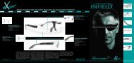
Anatomy of the human ear<br />
Diagram of the ear<br />
1. Outer ear<br />
2. Ear canal<br />
3. Eardrum<br />
4. Hammer<br />
5. Anvil<br />
6. Stirrup<br />
7. Oval window<br />
8. Semicircular canals<br />
9. Auditory nerve<br />
10. Cochlea<br />
11. Eustachian tube<br />
12. Round window<br />
13. Tympanic cavity<br />
Outer ear<br />
The outer ear is made up of the visible portion called the pinna and the ear<br />
canal. The human pinna’s functions are of no great importance, whilst<br />
animals can move and turn their ears to locate sounds.<br />
Middle ear<br />
The middle ear consists of eardrum, tympanic cavity and Eustachian tube.<br />
Inner ear<br />
The inner ear is made up of the organ of Corti and the cochlea.<br />
Ear canal<br />
The ear canal links the outer ear and the eardrum. It is approximately 3-4cm<br />
in depth and 0.5-1cm in diameter. It is completely covered with skin. The<br />
outermost part of the ear canal near the pinna is supported by cartilage, the<br />
inner part by bone. In the section made up of cartilage sebaceous glands<br />
secrete the yellow cerumen which is expelled by tiny hairs.<br />
Eardrum<br />
Between the ear canal and the tympanic cavity lies the eardrum. This membrane<br />
is approximately 1cm in diameter and 0.1mm thick. On the inside of<br />
the eardrum is a mucous membrane; from the outside it looks grey and shiny.<br />
Tympanic cavity<br />
In the tympanic cavity there are the three ear bones or ossicles: hammer,<br />
anvil and stirrup as well as the semicircular canals of the vestibular<br />
apparatus.<br />
Hammer, anvil and stirrup<br />
The hammer, anvil and stirrup are three tiny bones that are connected to<br />
each other. They directly couple and amplify sound energy from the eardrum<br />
to the inner ear.<br />
Oval window<br />
Sound waves travel via this membrane from the ossicles to the inner ear. The<br />
inner ear is located in the cochlea, well protected by the petrous portion of<br />
the temporal bone (‘pyramid’).<br />
Round Window<br />
The round window is positioned adjacent to the oval window and is the end<br />
point of the scala tympani.<br />
Eustachian tube<br />
The Eustachian tube links the tympanic cavity with the pharynx. Its main<br />
function is pressure equalisation in the ear.<br />
Cochlea<br />
The cochlea has three ducts called scalae: the scala vestibuli and the scala<br />
tympani, which both contain perilymph. Between these two is the scala<br />
media filled with endolymph which contains the organ of Corti. The word<br />
cochlea is the latin word for snail and comes from the shape of the organ.<br />
Organ of Corti<br />
The organ of Corti is the essential part of our hearing. It is located in the<br />
cochlea and contains 25,000 hair cells, each equipped with 100 hairs<br />
called stereocilia. These hairs are the sensory receptors of the auditory<br />
system and pass on information from sound waves via the auditory nerve to<br />
the brain.<br />
4 5<br />
To be precise, the human ear doesn’t process sounds, but sound waves that<br />
resonate with the eardrum. The resonance is amplified by the ossicles in the<br />
tympanic cavity and reaches the inner ear through the oval window, where<br />
the actual hearing process begins.<br />
A very important factor in the hearing process is the frequency of sound. The<br />
unit of frequency is Hertz (Hz) and is a measure of the number of sound<br />
waves per second.<br />
The amplification of sounds by the ossicles depends on frequency. The best<br />
amplification of sounds is achieved in the range of the human voice, i.e. between<br />
1000 and 2000 Hz.<br />
When a sound wave reaches the oval window via the previously mentioned<br />
pathway, it is passed on via the perilymph inside the scala vestibuli to<br />
trigger the basilar membrane. Depending on the frequency, sensory hairs in<br />
different locations on the organ of Corti are triggered. An analogy would be<br />
waves breaking at specific points on the beach, depending on their size.<br />
This way we can distinguish between high and low pitches. From the<br />
stereocilia the sound wave travels on towards and resonates with Reissner’s<br />
membrane. This resonance then triggers receptor cells that send an electric<br />
impulse along the auditory nerve to the brain. Here the sound is recognised<br />
and defined e.g. as a note or a word. The ear responds 7 times faster than<br />
the eye. If we could see as fast as we can hear, we would see every single<br />
frame in TV broadcasts instead of fluid movements.<br />
A sound is not only defined by its frequency but also by its volume. This is<br />
measured in decibel (dB). The table below lists typical everyday sounds and<br />
the noise level they cause.<br />
The human pain barrier for sounds is about 120dB, i.e. at volumes above<br />
120dB we perceive the sound as a physical pain in our ears.<br />
If we have to raise our voice in a conversation at a distance of 1m, the<br />
background noise level is about 80dB or more. It is not advisable to expose<br />
the ears to such noise levels for prolonged periods without hearing<br />
protection.<br />
Hearing – how does it work?<br />
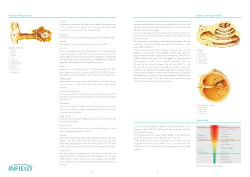
Cochlea<br />
1. Stirrup<br />
2. Oval window<br />
3. Helicotrema<br />
4. Scala vestibule<br />
5. Organ of Corti<br />
6. Scala tympani<br />
7. Round window<br />
Diagram of the cochlea<br />
1. Scala vestibule<br />
2. Organ of Corti<br />
3. Scala tympani<br />
4. Auditory nerve<br />
What is loud?<br />
Examples of noise levels in Decibel